Plasmonic Optical Biosensors for Detecting C-Reactive Protein: A Review - MDPI
←
→
Page content transcription
If your browser does not render page correctly, please read the page content below
micromachines

Review
Plasmonic Optical Biosensors for Detecting
C-Reactive Protein: A Review
Joo Seon Seok 1,2 and Heongkyu Ju 1,2, *
 1   Department of Physics, Gachon University, Seongnam-si, Gyeonggi-do 13120, Korea; wntjs0807@gmail.com
 2   Gachon Bionano Research Institute, Gachon University, Seongnam-si, Gyeonggi-do 13120, Korea
 *   Correspondence: batu@gachon.ac.kr; Tel.: +82-31-750-8552

 Received: 26 August 2020; Accepted: 25 September 2020; Published: 27 September 2020

 Abstract: C-reactive protein (CRP), a potent acute-phase reactant that increases rapidly in response
 to inflammation, tissue damage or infections, is also considered an indicator of the risk of
 cardiovascular diseases and neurological disorders. Recent advances in nanofabrication and
 nanophotonic technologies have prompted the optical plasmonic phenomena to be tailored for
 specific detection of human serum CRP into label-free devices. We review the CRP-specific detection
 platforms with high sensitivity, which feature the thin metal films for surface plasmon resonance,
 nano-enhancers of zero dimensional nanostructures, and metal nanoparticles for localized surface
 plasmon resonance. The protocols used for various types of assay reported in literature are also outlines
 with surface chemical pretreatment required for specific detection of CRPs on a plasmonic surface.
 Properties including sensitivity and detection range are described for each sensor device reviewed,
 while challenges faced by plasmonic CRP sensors are discussed in the conclusion, with future
 directions towards which research efforts need to be made.

 Keywords: C-reactive protein (CRP); optical biosensor; surface plasmon; nanoparticle

1. Introduction
      In 1930, Tillett and Francis discovered the protein that responded to the encapsulated
(c)-polysaccharide of pneumococcus in patients with acute pneumococcal infections [1], later named
the C-reactive protein (CRP). In blood sera of healthy individuals, CRP is known to be present
usually in a small amount. CRP production in the circulation elevates its level in serum in response
to various inflammations, infection, stress, trauma, surgery, tissue damage, cardiovascular disease,
and neurological degeneration [2–13]. During an acute phase response, the CRP level in serum
increases rapidly (by more than 100-fold) and reaches a peak within 48 h with its half-life of about
19 h [9,14–21].
      The CRP levels in blood plasma are considered to be lower than about 5 µg/mL for individuals
with no inflammation. However, the small variation of the level below it may depend on onset of
other ailments such as cardiovascular diseases (CVD). It is seen that the advent of a high-sensitivity
immunoassay of CRP allows the use of the risk predictive characteristic of its levels for evaluating
CVD such as acute coronary syndromes [2,14–17,22–25]. The American Heart Association (AHA) and
the US Centers for Disease Control and Prevention (CDC) defined the CVD risk groups using CRP
levels as follows [26,27]: low risk, CRP concentration 3.0 µg/mL.
      CRP mainly produced from hepatocytes in the liver, can also be produced in smooth muscle
cells, macrophages, endothelial cells, lymphocytes, and adipocytes. CRP has a pentameric structure of
the five identical monomers combined, moves through the blood to the region where inflammatory
reaction occurs, and then irreversibly dissociates into the monomers that play a direct role in the

Micromachines 2020, 11, 0895; doi:10.3390/mi11100895                   www.mdpi.com/journal/micromachines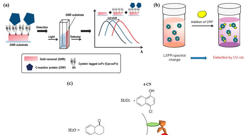
Micromachines 2020, 11, 0895                                                                         2 of 16

inflammatory response [14–17,28]. The mechanisms behind the CRP variants’ generation needs to be
clarified to deal with various abnormalities such as acute coronary syndromes. This also addresses the
requirement of developing ultra-high sensitivity biosensors for CRP detection [29].
      Highly sensitive CRP detection performed mostly in vitro exploits the immunoreaction-based
methodologies such as the turbidoimmunometric assays [30–32], immunonephelometric
assays [33–36], the enzyme-linked immunosorbent assay (ELISA) [37,38], the fluorescence assay
with immunoreaction [39–41], and the optical plasmonic immunoassays [19,29,42–51].
      Amid the methodologies mentioned above, optical plasmonic sensors have attracted much
attention for detecting biomarker molecules without labels such as fluorescent tags, due to a number
of advantages [52–59]. They include the high sensitivity without contaminating target biomolecules,
the capability of multiplexed assay, a small amount of an analyte sample required, the relative immunity
to external factors (ambient temperature variation/external electric disturbance), and the observability
of time-dependent kinetic behavior of bio-molecular interactions [3,60].
      In particular, the high sensitivity in optical plasmonic sensors arises from locally enhanced density
of photonic modes near where plasmons are excited [56,61–66]. When bringing target biomolecules
of non-negligible molecular weight such as CRP molecules (molecular weight of 118–144 kDa [67])
into close proximity with a plasmonic metal surface, their strong interaction with the plasmonic local
fields modifies the optical resonance condition via the effective refractive index change. This thus
produces the drastically altered value of the optical output coupled into the medium that allows
photon momentum conservation [68–72]. This kind of a detection platform enables only the target
molecules near the metal surface to be detected, forming the basis of plasmonic biosensors of any kind
which can excite either localized surface plasmon resonance (LSPR) or propagating surface plasmon
resonance (SPR).
      This paper reviews the CRP-detecting immunoassays relying on optical plasmonic resonances.
Section 2 presents the SPR-based CRP immunoassays, while Section 3 the LSPR-aided CRP
immunoassays. Various protocols on the basis of immune reaction for specific detection of CRP
such as direct assays, sandwich assays and sandwich assays with nano-enhancers, are described with
the relevant surface chemistry accessible to the plasmonic surface. Characteristics of each CRP assay
are also outlined for comparison, including the detection limit and range associated with the given
plasmonic transducers.

2. Surface Plasmon Resonance-Based C-Reactive Protein (CRP) Detection
     SPR biosensors detect changes in an effective refractive index of dielectric media on the sensing
surface, which is caused by adsorbing biomolecules on it. The analyte biomolecules need to be
adsorbed within the penetration range of plasmonic evanescent fields from the surface, the interface
between the nanometer thickness metallic film and the dielectric media [61,73]. SPR can occur at
the interface between metal and dielectric media that include target biomolecules (e.g., CRPs), via a
coherent coupling of surface plasmons with external photons. The surface parallel component of
photon momenta offered by a high index prism [19,29,42–47,72,74] or a fiber core [48,55,75–79], can
match those of surface plasmons, generating a coherently coupled state, i.e., the surface plasmon
polaritons. The momenta required for such matching are subject, with great sensitivity, to a change
in an effective refractive index of dielectric media near a metal film, due to the Debye screening
effects, thus overriding the resonance conditions [61,80,81]. This serves as the basis for detecting, with
high sensitivity, biomolecules adsorbed on the metal surface. SPR biosensors typically interrogate a
wavelength or an angle of incident light to pick up the reflectance dip, i.e., the so-called “plasmonic
absorption”. The absorption peak shifts in a wavelength or in an angle, as a function of effective
refractive index, its sensitivity being dominated by a complex dielectric constant of the metal film
used. Meanwhile, the peak broadening caused by metallic ohmic loss and Landau damping of surface
plasmons determines the sensor resolution [54,61,73,82–84].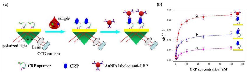
Micromachines 2020, 11, x                                                                             3 of 16
Micromachines 2020, 11, 0895                                                                          3 of 16
reaction. The three protocols all need bio-recognition receptors to capture CRPs selectively, even
amid the presence of many other kinds of biomolecule. The receptor immobilization requires the
      Figure 1 illustrates the three protocols used to build the SPR immunoassay chip structures for
prerequisite surface treatment such as via surface functionalization into a self-assembled monolayer
CRP detection [19,29,42,45]. Gold (Au), the chemically stable metal, is widely used for a plasmonic
(SAM) [42,43,48], through biotin-streptavidin/extravidin interaction [19,44,46] and via the G proteins
film due to the great availability for biochemical surface treatment required prior to target-specific
[29], via the protein adhesive films [45], and using thiol-modified receptors [47]. Upon the receptor
reaction. The three protocols all need bio-recognition receptors to capture CRPs selectively, even amid
immobilization, injection of CRPs onto the sensing surface leads to selective capture as seen in
the presence of many other kinds of biomolecule. The receptor immobilization requires the prerequisite
Figure 1(a), and in turn induces change in a SPR signal, being referred to as a direct immunoassay in
surface treatment such as via surface functionalization into a self-assembled monolayer (SAM) [42,43,48],
SPR sensors [29,42,44,45,48]. Subsequent injection of detection elements such as the detection
through biotin-streptavidin/extravidin interaction [19,44,46] and via the G proteins [29], via the protein
antibodies (dAbs) or aptamers, as seen in Figure 1(b), amplifies the SPR signal together with
adhesive films [45], and using thiol-modified receptors [47]. Upon the receptor immobilization, injection
escalating selectivity in detecting CRP, as referred to as a sandwich immunoassay in SPR sensors
of CRPs onto the sensing surface leads to selective capture as seen in Figure 1a, and in turn induces
[19]. Further enhancement of sensitivity in CRP detection can be realized via replacing the detection
change in a SPR signal, being referred to as a direct immunoassay in SPR sensors [29,42,44,45,48].
elements by nano-enhancer-conjugated detection elements such as Au nanoparticles conjugated
Subsequent injection of detection elements such as the detection antibodies (dAbs) or aptamers, as seen
antibodies [47] or quantum dots (QDs) conjugated aptamers, as seen in Figure 1(c) [46]. It is noted
in Figure 1b, amplifies the SPR signal together with escalating selectivity in detecting CRP, as referred to
that, all substances injected consecutively in a sandwich assay, which are supposed to induce the
as a sandwich immunoassay in SPR sensors [19]. Further enhancement of sensitivity in CRP detection
effective index change near metal surface, need to fall within the penetration range of plasmonic
can be realized via replacing the detection elements by nano-enhancer-conjugated detection elements
evanescent fields (~200 at visible wavelengths).
such as Au nanoparticles conjugated antibodies [47] or quantum dots (QDs) conjugated aptamers,
      We organize Section 2 into three sections according to the immunoassay types used in the
as seen in Figure 1c [46]. It is noted that, all substances injected consecutively in a sandwich assay,
SPR-based plasmonic sensing platforms, as follows: Section 2.1 covers the direct assay, Section 2.2
which are supposed to induce the effective index change near metal surface, need to fall within the
the sandwich assays and Section 2.3 the nano-enhancers in a sandwich assay.
penetration range of plasmonic evanescent fields (∼ 200 at visible wavelengths).

               Schematic of
     Figure 1. Schematic of three
                             three strategies
                                    strategies for
                                               for the surface plasmon resonance (SPR)-based C-reactive
     protein (CRP) sensors. (a):
                            (a): aa direct
                                    direct immunoassay, (b): aa sandwich
                                                                 sandwich immunoassay, (c): a sandwich
     immunoassay with nano-enhancers such as nanoparticles, and quantum dots (QDs).

      We organize
2.1. Direct          Section 2 into three sections according to the immunoassay types used in the
            Immunoassay
SPR-based plasmonic sensing platforms, as follows: Section 2.1 covers the direct assay, Section 2.2 the
     Casa et al. [42] used the chemical bonding via 4,4′-dithiodibutyric acid (DDA) to immobilize
sandwich assays and Section 2.3 the nano-enhancers in a sandwich assay.
CRP antibodies on the Au surface. Through its disulfide bond, DDA attached to the surface, with a
consequence    of the carboxyl group modified Au surface. The amino group of the CRP antibody, in
2.1. Direct Immunoassay
turn,       bonded       with     it    through      1-ethyl-3-(3-dimethylaminopropyl)carbodiimide
      Casa et al. [42] used the chemical bonding via 4,40 -dithiodibutyric acid (DDA) to immobilize
hydrochloride-N-hydroxysuccinimide (EDC-NHS) crosslinking. The biosensor chip was tested with
CRP antibodies on the Au surface. Through its disulfide bond, DDA attached to the surface,
various concentrations of CRP (10         10 μg/mL) to obtain the SPR angle shifts. The angle shift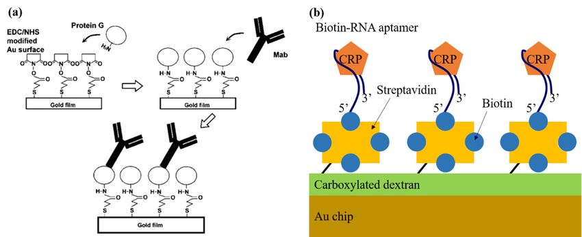
Micromachines 2020, 11, 0895                                                                                     4 of 16

with a consequence of the carboxyl group modified Au surface. The amino group of the
CRP antibody, in turn, bonded with it through 1-ethyl-3-(3-dimethylaminopropyl)carbodiimide
hydrochloride-N-hydroxysuccinimide (EDC-NHS) crosslinking. The biosensor chip was tested with
various concentrations of CRP (10−1 − 102 µg/mL) to obtain the SPR angle shifts. The angle shift
began at the CRP concentration of 1 µg/mL and plateaued at concentrations higher than 10 µg/mL.
The SPR biosensor had a detection range of 1–10 µg/mL and a limit of detection (LOD) of 1 µg/mL.
Comparison was also made between outcomes from using different flow cell shapes in the SPR chip.
The flow cell type A had a circular shape, whereas type B was rectangular, and both types had the
same depth of 10 µm. It was found that the rectangular flow cell (type B) resulted in a higher signal
shift. An SPR analyzer (NTT Advanced Technology Corp., Kanagawa, Japan) was used for their SPR
system. The sensor chips were made by sputtering Au on the glass plate and polydimethylsioxane
(PDMS) was used to make the flow cell by a photolithography method.
      The CRP biosensor developed by Hu et al. [29] also relied on the direct immunoassay with an
SPR-based signal transducer. Figure 2a illustrates the schematic of the antibody immobilizing model
using the G proteins. The G protein has a Fc-binding domain that can interact with the Fc portion
of IgG and thereby hold onto the antibody. After the Au surface of the chip had been functionalized
with 16-mercaptohexadecanoic acid, the carboxyl group of the Au surface and the amine group of
the G protein were conjugated through EDC–NHS crosslinking. Then, the CRP IgG antibody was
immobilized by bonding with G protein. In this study, three different monoclonal mouse antibodies
were used to differentiate between pCRP and mCRP; namely, antibody C8 to detect both pCRP and
mCRP, antibody 8D8 to detect only pCRP, and antibody 9C9 to detect only mCRP. Each process
was monitored in real time by measuring the SPR angle shifts. The SPR angle shifts were also
observed when introducing pCRP of 0.226 µM (26 µg/mL) and mCRP of 0.437 µM (10 µg/mL) upon
the three different antibodies used. With the antibody C8, the sensor could detect both types of CRPs.
However, while the SPR angle shift by pCRP remained unchanged after surface washing, while those
by mCRP decreased noticeably. Meanwhile, for 8D8 or 9C9 antibodies, only one of both types of CRPs
produced a significantly detectable signal, although its sensitivity substantially lower than that for C8.
This encouraged the use of the C8–pCRP immunoreaction pair for detecting the CRP level ranging
from 1 µg/mL to 26 µg/mL with a LOD of 1 µg/mL [29]. This detection range appeared to meet the
AHAMicromachines
       and CDC2020,
                 criteria
                     11, x for clinical diagnosis of CRP.                                      5 of 16

           Figure 2. (a) Schematic of immobilizing antibody on protein G layer. Reprinted from [29], Copyright
      Figure 2. (a) Schematic of immobilizing antibody on protein G layer. Reprinted from [29],
          2006, with permission from Elsevier. (b) Schematic of the sensing surface of the RNA-aptasensor for
      Copyright 2006, with permission from Elsevier. (b) Schematic of the sensing surface of the
          specific detection of CRP.
      RNA-aptasensor for specific detection of CRP.
            Optical fibers were introduced as the detection platform for SPR-based CRP assays by Aray et
      Bini et al. used aptamers as the bio-recognition receptors that were believed to be more cost-effective
      al. [48]. The fiber used as a sensor head comprised a 980 μm diameter core made of poly(methyl
and more     stable than
      methacrylate)   and antibodies  for a SPR-based
                           20 μm fluorinated            CRP biosensor
                                             polymer cladding.            [44].
                                                                   The gold filmThe
                                                                                 for schematic
                                                                                     creation of of
                                                                                                 thetheir
                                                                                                     SPR system
                                                                                                          was     is
illustrated   in Figure  2b.  The biotinylated  RNA    aptamers    were  immobilized    on  the Au
      formed on a flat surface into which the fiber core was polished along a 10 mm length. Unlike a surface of the
      typical prism based SPR sensor, the fiber SPR sensor does not need any precision device that can
      tune an incident angle of light for SPR excitation [85,86], but may be suitable for application where a
      miniaturized detection platform is required without compensating for the sensitivity at low cost. In a
      way similar to what was described above, the gold surface was first carboxylated using
      11-mercaptoundecanoic acid, and the antibody was then immobilized to the carboxylated gold via
      the EDC–NHS reaction. The SPR signal was obtained by measuring the absorption peak shift of the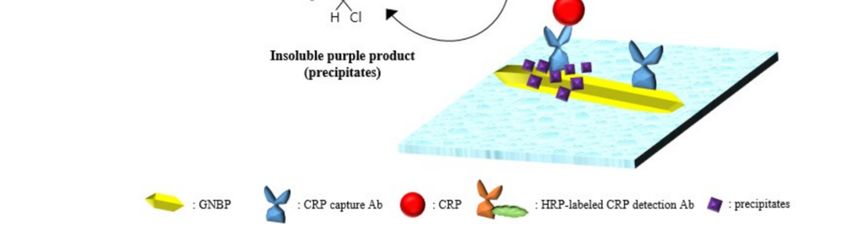
Micromachines 2020, 11, 0895                                                                         5 of 16

sensor chip, which was pretreated with layers of dextran, and streptavidin. The RNA aptamer sequence
was selected through 10 Systematic Evolution of Ligands by Exponential enrichment (SELEX) rounds.
Two types of aptamers, both biotinylated at the 50 end but having different spacers (i.e., one with a 20mer
polyT tail and the other with a triethylene glycol (TEG) tail), were tested. CRP binding curves for both
cases exhibited the LOD of 5 ng/mL and the detection range of 5 ng/mL to 100 ng/mL. The reusability
of both aptamers using HCl and NaOH were also demonstrated without losing sensitivity.
     Choi et al. [45] immobilized antibodies using an oxygen plasma-treated parylene N film,
and enhanced the sensitivity of the SPR-based sensor. The parylene N films treated with various
oxygen plasma conditions were compared with a polystyrene substrate in terms of their ability to attach
proteins such as bovine serum albumin (BSA). Various oxygen plasma conditions such as power levels
and processing time for the parylene N film were tested. This enabled the optimized condition to be
found for the most amplified SPR signal, i.e., 100 W for 1 min. Following CRP antibody immobilization
onto the film, CRP concentrations of 1, 10, 100 ng/mL and 1 µg/mL were tested with this biosensor.
The CRP concentrations and SPR signals appeared to be in a linear relationship. The biosensor with
the plasma-treated parylene N film had a significantly better sensitivity than those with the bare gold
surface of the chip or with the non-plasma treated film.
     Optical fibers were introduced as the detection platform for SPR-based CRP assays by
Aray et al. [48]. The fiber used as a sensor head comprised a 980 µm diameter core made of
poly(methyl methacrylate) and 20 µm fluorinated polymer cladding. The gold film for creation of
the SPR was formed on a flat surface into which the fiber core was polished along a 10 mm length.
Unlike a typical prism based SPR sensor, the fiber SPR sensor does not need any precision device
that can tune an incident angle of light for SPR excitation [85,86], but may be suitable for application
where a miniaturized detection platform is required without compensating for the sensitivity at low
cost. In a way similar to what was described above, the gold surface was first carboxylated using
11-mercaptoundecanoic acid, and the antibody was then immobilized to the carboxylated gold via
the EDC–NHS reaction. The SPR signal was obtained by measuring the absorption peak shift of the
spectrum of light transmitted through the fiber. CRP concentrations ranging from 0.01 µg/mL to
500 µg/mL were tested. The red shift of the absorption peak wavelength occurred with increasing the
concentration, producing the LOD of 9 ng/mL.

2.2. Sandwich Immunoassay
      The first SPR-based immunosensor for CRP detection was reported in 2006 [19]. The SPR sensor
system relied on a typical SPR spectroscope. As shown in Figure 3a, the gold surface of the sensor
chip was treated with (3-aminopropyl)triethoxysilane to generate amino groups, and the biotin–NHS
was subsequently used to create a biotin layer. The biotin layer then reacted with the streptavidin
added prior to injection of biotin-labeled CRP capture antibody (cAb). The biotin-labeled cAb reacted
with streptavidin, being fixed onto a chip surface. Injection of CRP caused the signal change due to
its immunoreaction with cAb. Another signal change occurred due to the detection antibody (dAb)
that was additionally injected to bind with CRP via another immunoreaction, ultimately forming a
sandwich bonding. Figure 3b shows the continuously monitored signal changes during all procedures
of biochemical molecules injection mentioned above. The SPR signal change from the dAb (C2) was
greater than that caused by the CRP. This indicates that use of dAb allowed the enhanced sensitivity
together with enhancing selectivity. Of the CRP concentrations (1–5 µg/mL) tested, the range of
2–5 µg/mL showed the nearly linear relationship of CRP concentration with the SPR signal with LOD
of 1 µg/mL.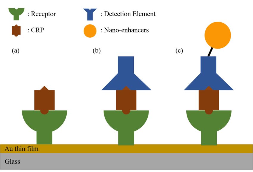
Micromachines 2020, 11, 0895 6 of 16
Micromachines 2020, 11, x 6 of 16
Figure3.3.(a)
Figure (a)Schematic
Schematic of SPR chip
of SPR chip(B,
(B,biotin;
biotin;C6C6 , biotinylated
, biotinylated
amino-b
amino-b antiCRP
antiCRP C6;antiCRP
C6; C2, C2, antiCRP
C2) (b)C2)
(b)Detection
Detectionprinciple-SPR
principle-SPRsensorgram
sensorgramof of the CRP
CRP sandwich
sandwichassay.assay.Reprinted
Reprintedfrom [19],
from Copyright
[19], 2006,
Copyright
with permission
2006, from Elsevier.
with permission from Elsevier.
2.3.
2.3.Nano-Enhancers
Nano-Enhancersfor
forSensitivity
Sensitivity Enhancement
Enhancement
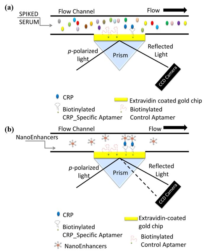
Vance
VanceandandSandros
Sandros[46] [46]designed
designed the aptamer-coatedQDs
the aptamer-coated QDsfor foruse
useasasdetection
detection elements
elements in ain a
sandwich
sandwichassay
assaytotodetect
detectCRP.
CRP.QDsQDs conjugated withCRP-specific
conjugated with CRP-specificaptamers
aptamersenhanced
enhanced thethe
SPRSPR signal
signal
change
changesignificantly,
significantly,thusthusQDs
QDsare are referred
referred to as nano-enhancers
nano-enhancersininthis thisassay.
assay.The
The schematic
schematic of the
of the
sensing
sensingsystem
systemisisillustrated
illustratedinin Figure
Figure 4. The
The immobilization
immobilizationofofthe theaptamers
aptamers that
that were
were biotinylated
biotinylated
required
requiredthe
thegold
goldsurface
surface on aa prism
prismtotobebecoated
coated with
with cystamine,
cystamine, glutaraldehyde,
glutaraldehyde, and extravidin
and extravidin in
insequence.
sequence.Then,Then,biotin-extravidin
biotin-extravidin interaction was was
interaction used used
to bind the biotinylated
to bind aptamers
the biotinylated to the to
aptamers
surface
the surfaceof of
thethe
SPRSPRchip, permitting
chip, permitting CRPCRPto betobound
be boundselectively to thetoaptamers.
selectively Meanwhile,
the aptamers. the
Meanwhile,
QDs (enhancers) surrounded by a streptavidin shell were prepared and
the QDs (enhancers) surrounded by a streptavidin shell were prepared and bound to biotinylatedbound to biotinylated
aptamers,forming
aptamers, formingthe the QDs
QDs conjugated
conjugated with with secondary
secondaryaptamers
aptamers(the(theso-called nano-enhancers).
so-called nano-enhancers).
Injection of these nano-enhancers consisting of QDs conjugated with
Injection of these nano-enhancers consisting of QDs conjugated with the secondary the secondary aptamers
aptamers thatthat
were CRP-specific would bind to the CRP selectively, thus enhancing both the SPR
were CRP-specific would bind to the CRP selectively, thus enhancing both the SPR signal sensitivity signal sensitivity
and the selectivity. The SPR signal (the difference in reflectance, i.e., ∆R measured by the SPR system)Micromachines 2020, 11, 0895 7 of 16
Micromachines 2020, 11, x 7 of 16
and the selectivity. The SPR signal (the difference in reflectance, i.e., ∆R measured by the SPR
changes by introducing such nano-enhancers at the various CRP concentrations (ranging from 5 fg/mL
system) changes by introducing such nano-enhancers at the various CRP concentrations (ranging
to 500 ng/mL).
from Injection
5 fg/mL of the nano-enhancers
to 500 ng/mL). caused the SPRcaused
Injection of the nano-enhancers signal the
(∆R) to increase
SPR bytoa increase
signal (∆R) larger degree
at a higher concentration
by a larger with respect
degree at a higher to the with
concentration control signal.
respect The
to the approximately
control linear relationship
signal. The approximately
between
linearthe concentration
relationship and the
between the concentration
SPR signal was andobtained
the SPR in the range
signal of 5 fg/mL–5
was obtained in thepg/mL,
range ofwith
5 the
LODfg/mL–5
of 5 fg/mL.
pg/mL, with the LOD of 5 fg/mL.
Figure
Figure 4. A4.schematic
A schematicillustration
illustration of
of the
thesandwich
sandwich protocol implemented
protocol for the
implemented detection
for of CRP in
the detection of CRP
biological fluid.
in biological fluid. The
The gold-coated
gold-coatedprism prismis isprefunctionalized
prefunctionalized withwith
aptamers specific
aptamers to CRP
specific and and
to CRP
control aptamers followed by the (a) direct detection of CRP (fg/mL) spiked in human serum and the
control aptamers followed by the (a) direct detection of CRP (fg/mL) spiked in human serum and
(b) sandwich based assay using CRP-specific aptamer-coated QDs for SPRi signal amplification.
the (b) sandwich based assay using CRP-specific aptamer-coated QDs for SPRi signal amplification.
Direct detection of CRP (fg/mL) does not generate a quantifiable sensor response as depicted with no
Direct detection of CRP (fg/mL) does not generate a quantifiable sensor response as depicted with no
change in the angle of reflectivity, however, with sandwich assay the nano-enhancers induce a
change in the
change angle
in the of reflectivity,
reflectivity. however,
Reproduced withwith sandwich
permission fromassay
[46]. the nano-enhancers induce a change
in the reflectivity. Reproduced with permission from [46].
A hybrid sandwich assay using an aptamer and an antibody was performed for detecting CRP,
with its schematic illustrated in Figure 5a [47]. First, the aptamer was immobilized onto Au surfaceMicromachines 2020, 11, x 8 of 16
Micromachines 2020, 11, 0895 8 of 16
A hybrid sandwich assay using an aptamer and an antibody was performed for detecting CRP,
with its schematic illustrated in Figure 5(a) [47]. First, the aptamer was immobilized onto Au surface
of of
a SPR chip via the –SH functional group. CRP was then injected to bind to the aptamer on
a SPR chip via the –SH functional group. CRP was then injected to bind to the aptamer on the Au
thesurface.
Au surface.
The SPRThe SPRwhich
signal signal
was which was shift
the angle the angle shift ofspot
of the darkest the on
darkest
a CCDspot
imageonsensor,
a CCDcouldimage
sensor, couldshifted
be further be further shifted by
by injecting injectingasantibodies
antibodies as detection
detection elements thatelements
would bindthattowould bind to CRP,
CRP, eventually
eventually
forming a hybrid sandwich assay. In addition, replacing the detection antibody by by
forming a hybrid sandwich assay. In addition, replacing the detection antibody an anAuAu
nanoparticle
nanoparticle (AuNP)-conjugated antibody as another kind of detection element further enhances thethe
(AuNP)-conjugated antibody as another kind of detection element further enhances
SPRSPRsignal.
signal.This
Thiswas
wasillustrated
illustrated in Figure
Figure 5(b)
5b where
where the
the SPR signals
signalswere
werecompared
comparedamongamong three
three
types
typesofofassays,
assays,the
the aptamer-CRP (directassay),
aptamer-CRP (direct assay),thethe aptamer-CRP-dAb
aptamer-CRP-dAb (hybrid
(hybrid sandwich
sandwich assay),assay),
the
theaptamer-CRP-AuNP
aptamer-CRP-AuNPconjugated conjugateddAbdAb(another
(another kind
kind ofof hybrid
hybrid sandwich
sandwich assay).
assay). TheThe CRP
CRP detection
detection
range
range was was 1.15
1.15 ng/mL–11.5
ng/mL–11.5 μg/mL
µg/mL with
with thethe
LODLOD of of 1.15
1.15 ng/mL.
ng/mL. WeWe summarize
summarize thethe features
features of of
thethe
SPR
SPRCRP
based based CRP sensors
sensors in the in the Table
Table 1. 1.
Figure
Figure 5. 5.(a)
(a)Schematic
Schematic illustration
illustration of
ofthe
theAuAunanoparticle
nanoparticle(AuNP)-enhanced
(AuNP)-enhancedSPRSPR
biosensor with an
biosensor with
an aptamer-antibody sandwich assay. (b) The relationship between ∆θ and CRP concentrations by
aptamer-antibody sandwich assay. (b) The relationship between Δθ and CRP concentrations by
using
using different
different sensing
sensing strategies.Direct
strategies. Directmeasurement
measurementa,a,aptamer-antibody
aptamer-antibody sandwich
sandwich measurement
measurement b,
b, AuNPs enhanced aptamer-antibody sandwich measurement c. Republished with
AuNPs enhanced aptamer-antibody sandwich measurement c. Republished with permission of Royal permission of
RoyalofSociety
Society of Chemistry,
Chemistry, from [47],from
2016[47], 2016 copyright;
copyright; permissionpermission
conveyedconveyed through Copyright
through Copyright Clearance
Clearance
Center, Inc. Center, Inc.
Table1.1.The
Table Thecharacteristics
characteristics of
of SPR-based CRP sensors
SPR-based CRP sensorsreviewed.
reviewed.
Detection Detection Detection
Detection
Reference
Reference AssayAssay
Type Type ReceptorReceptor
Detection Element LOD LOD Detection Range Remark
Remark
Element Range Time[s]
Time[s]
[42] direct assay antibody CRP 1 µg/mL 1 1~10 µg/mL 1200
[42] direct assay antibody CRP 1~10 μg/mL 1200
[29] direct assay antibody CRP - μg/mL 1~26 µg/mL 2400
[44][29] direct direct
assay assay aptamer antibodyCRP CRP 5 ng/mL - 5~100
1~26ng/mL
μg/mL 1400
2400 reusable
parylene
[45] direct assay CRP - 5 1 ng/mL~1 µg/mL 5000
[44] direct assay N film aptamer CRP 5~100 ng/mL 1400 reusable
[48] direct assay antibody CRP 9 ng/mL ng/mL 9 ng/mL~70 µg/mL 1200 Fiber used
[19] sandwich assay antibody parylene
antibody 1 µg/mL 1 ng/mL~1
2~5 µg/mL 8000
[45] direct assay CRP - 5000
nano-enhancer-sandwich N film μg/mL
[46] aptamer QD-antobody 5 fg/mL 5 fg/mL~5 pg/mL 11,000
assay 9 9 ng/mL~70 Fiber
[48] nano-enhancer-sandwich
direct assay antibody CRP 1200
[47] aptamer AuNP-antibody 1.15 ng/mLng/mL μg/mLµg/mL
1.15 ng/mL~11.5 - used
assay
1 μ
[19] sandwich assay antibody antibody 2~5 μg/mL 8000
g/mL
3. Localized Surface Plasmon Resonance for
nano-enhancer-sandwich Detecting CRP 5
5 fg/mL~5
[46] aptamer QD-antobody 11,000
assay fg/mL
pg/mL
Localized surface plasmon resonance (LSPR) occurs as a results of coherent
1.15 interaction between
nano-enhancer-sandwich 1.15
incident
[47] light and surface electrons aptamer
assay
of metal nanoparticles
AuNP-antibody at visible ng/mL~11.5
ng/mL
and near infrared
- wavelengths.
μg/mL
For LSPR, their diameters need to be smaller than the incident wavelength such that the electric field
of the light across the nanoparticle is nearly constant, giving rise to electron density oscillation at the
incident light frequency [87]. This generates the subsequent attraction between background ions and
surface electrons, and the repulsion between electrons of enhanced density. These two effects would
lead to restoring force. This determines the inherent resonance frequency of the oscillation, such as in
a driven harmonic oscillator model. It is noted that the nanoparticle geometry that look like a zero
dimensional dot can provide the momenta required for exciting the plasmonic resonances.
The oscillating plasmons would re-radiate optical energy of light in the form of elastic scattering
with its cross section much larger than the physical cross section of a nanoparticle [88]. In addition,Micromachines 2020, 11, 0895 9 of 16
Ohmic loss occurs due to electron-electron scattering into heat. These radiative and non-radiative
damping effects lead to optical extinction of light that propagates through an ensemble of nanoparticles.
The peak of the extinction spectrum with its broadening properties would be determined by the
type/size/shape of metal nanoparticles, and the molecules adjacent to them. Molecules in close
proximity to nanoparticles can be polarized into additional electric dipole moments, weakening the
restoring force (dielectric screening effects) [87]. This accounts for red shift of the extinction spectrum
peak, enabling the metal nanoparticles to lend themselves to use for sensing bio-interaction and
bonding. This working principle of sensing is strongly supported by the plasmonic enhancement of
electric field strength around nanoparticles, which would be much higher than that of the incident
field despite its locality in space within 10 nm from the particle surface [89,90].
The CRP detection was demonstrated in a direct immunoassay by exploiting the peak shifts of
extinction spectra of light transmitting through an ensemble of AuNPs). This detection scheme used
the single chain variable fragment (scFv) tagged with cysteine as the CRP receptors immobilized on a
gold nanorod (GNR) substrate as shown in Figure 6a [49]. The reduced size of scFv compared to that
of a full-length antibody would permit CRP to more strongly interact with the LSPR evanescent field
that had limited
Micromachines penetration
2020, 11, x depth, accounting for the detection limit as low as 1 ng/mL. 10 of 16
6. (a)
Figure 6.
Figure (a)Schematic
Schematicillustration
illustrationof of
label-free detection
label-free of CRP
detection usingusing
of CRP gold nanorods as localized
gold nanorods surface
as localized
surface plasmon resonance (LSPR) sensor. Republished with permission of Royal Society[49],
plasmon resonance (LSPR) sensor. Republished with permission of Royal Society of Chemistry, from of
2013 of copyright;
Chemistry, permission
from [49], 2013 ofconveyed
copyright;through Copyright
permission Clearance
conveyed Center,
through Inc. (b)Clearance
Copyright CRP nanosensing
Center,
based
Inc. (b)on
CRP LSPR property based
nanosensing of PMPC-g-AuNPs
on LSPR property by ultraviolet–visible
of PMPC-g-AuNPs(UV–Vis) spectrophotometer.
by ultraviolet–visible (UV–
Reprinted with permission from [50] Copyright 2014 American Chemical Society.
Vis) spectrophotometer. Reprinted with permission from [50] Copyright 2014 American (c) Illustration of the
Chemical
LSPR immunosensor
Society. forof
(c) Illustration CRP
thedetection using 4-CN precipitation
LSPR immunosensor on GNBP
for CRP detection substrate.
using 4-CN Reproduced
precipitationwith
on
permission from [51]; published by SPIE, 2017.
GNBP substrate. Reproduced with permission from [51]; published by SPIE, 2017.
Table 2. The characteristics of LSPR-based CRP sensors reviewed.
Sensing Receptor Detection
Reference LOD Detection Range Remark
Strategy Type MediaMicromachines 2020, 11, 0895 10 of 16
Use of a polymer based artificial receptor for CRP capture was demonstrated in the LSPR based
detection [50]. The poly(2-methacryloyloxyethyl phosphorylcholine)-grafted AuNPs (PMPC-g-AuNPs)
were synthesized as the substrate that underwent LSPR changes as a result of CRP attachment as
illustrated in Figure 6b. The extinction spectra of light through the PMPC-g-AuNPs were analyzed
using the peak-area ratio-metric sensing [91–93]. In this analysis, the measured spectral absorbance
was numerically integrated over two different ranges of wavelengths, e.g., 490–540 nm (dispersed
state), and 550–700 nm (aggregated state), into the two values A and D, respectively. The ratio A/D
represents the characteristic of the extinction spectrum. Thus, injection of CRP could change the
extinction spectra, thereby modifying the ratio A/D. In contrast, injection of human serum albumin
(HSA) as the protein references that contained proteins that would bind to the receptor non-specifically,
caused little change in the spectra. Eventually the PMPC-g-AuNPs based sensing system enabled CRP
to be detected specifically with the detection limit of 50 ng/mL (detection range of 10 ng/mL–3 µg/mL).
The sandwich immunoassay with horseradish peroxide (HRP) found in the ELISA could be
utilized to detect CRP in a LSPR-based sensing system as shown in Figure 6c, gold nano-bipyramid
(GNBP) was used as a substrate on which CRP capture antibody was immobilized by physisorption.
CRP was injected to bind the capture antibody [51]. The HRP-labeled detection antibody was then
injected to bind CRP, followed by reduction of 4-chloro-1-naphthol (4-CN) by the HRP catalyst with
hydrogen peroxide. The 4-CN precipitated near GNBP, thus changing the extinction spectrum of light
through them. The increase in CRP concentration induced the redshift, producing the detection range
of 100 pg/mL–100 ng/mL with the detection limit of 87 pg/mL. We summarize the characteristics of the
LSPR-based CRP detection sensors reviewed in Table 2.
Table 2. The characteristics of LSPR-based CRP sensors reviewed.
Sensing Receptor Detection
Reference LOD Detection Range Remark
Strategy Type Media
[49] direct assay scFv CRP - 1 ng/mL~10 µg/mL
[50] direct assay PMPC CRP 50 ng/mL 10 ng/mL~3 µg/mL
sandwich
[51] antibody precipitates 87 pg/mL 0~100 ng/mL
assay
4. Discussion
Plasmonic optical biosensors are reviewed with a focus on those developed for highly sensitive
and specific detection of CRPs, the important biomarkers not only for general inflammatory responses,
but also for cardiovascular diseases and neurological disorders. This review categorizes the plasmonic
biosensors into two kinds, i.e., those on SPR occurring between a flat metal film and dielectric analyte
media, and those using LSPR occurring on metal nanostructures with their size much smaller than
incident light wavelength.
CRP immunoassays that exploited the antibodies or aptamers as recognition receptors were applied
on the sensing surface of the plasmonic devices. We could divide types of the CRP immunoassay used
in the plasmonic device into three kinds, i.e., the direct assay, the sandwich assay, and the sandwich
assay modified with metal nanoparticles or catalyst for enhancing sensitivity. It is inferred that the
limited range of penetration depth of plasmonic evanescent fields needs to be circumvented [57] to
enhance sensitivity in the CRP plasmonic immunoassay without narrowing the range of detection for
a quantitative assay, such as by exploiting the extended penetration depth of plasmonic evanescent
fields [57,94–100].
It was revealed that the SPR-based sensors were more effective in detecting the CRPs at low
concentration levels than the LSPR-based ones. This is due to the fact that the LSPR relied on the more
limited range over which the plasmonic evanescent field extended to interact with molecules of the
detection elements. However, it turned out that the LSPR-based device did not require the optical
alignment as sophisticated as the SPR-based ones. Meanwhile, regeneration of the sensor surfaceMicromachines 2020, 11, 0895 11 of 16
may be more likely to be possible in the SPR-based ones than the LSPR-based ones due largely to the
extraordinarily limited range of evanescent field penetration depth from the surface in the LSPR cases.
Further work may place an additional emphasis on how to enhance the coefficient of variation
in CRP immunoassay for reliable detection. These can include the homogeneous distribution of
immunoassay biomolecules on the plasmonic surface, minimization of the intervention of manual
expertise required to implement such label-free plasmonic sensing, and the development of highly
stable recognition receptors with uniform binding affinity. This optimization can possibly lead to use
of the plasmonic immunoassay for small-sized label-free devices required where time-dependent CRP
concentration needs to be monitored.
Author Contributions: J.S.S. conducted the search of the relevant literature materials, summarized the search,
analyzed the results, and write the first draft for this review paper. H.J. planned this review paper, determined the
review scope, supervised the analysis and completed the paper writing. All authors have read and agreed to the
published version of the manuscript
Funding: This work was supported by the National Research Foundation of Korea (NRF) grant funded by the
Korean government (MSIT) (No. 2020R1F1A1050885), and also supported by the Gachon University Research
Fund of 2018 (GCU-2018-0674).
Conflicts of Interest: The authors declare no conflict of interest.
References
1. Tillett, W.S.; Francis, T. Serological Reactions in Pneumonia with a Non-Protein Somatic Fraction of
Pneumococcus. J. Exp. Med. 1930, 52, 561–571. [CrossRef] [PubMed]
2. Sproston, N.R.; Ashworth, J.J. Role of C-Reactive Protein at Sites of Inflammation and Infection. Front. Immunol.
2018, 9, 754. [CrossRef] [PubMed]
3. Salvo, P.; Dini, V.; Kirchhain, A.; Janowska, A.; Oranges, T.; Chiricozzi, A.; Lomonaco, T.; Di Francesco, F.;
Romanelli, M. Sensors and Biosensors for C-Reactive Protein, Temperature and pH, and Their Applications
for Monitoring Wound Healing: A Review. Sensors 2017, 17, 2952. [CrossRef] [PubMed]
4. Hall, W.A.; Rossi, P.J.; Cooper, S.; Master, V.A.; Jani, A.B. C-Reactive Protein (CRP): Initial Exploratory Study
of an Inflammatory Biomarker for Prostate Cancer Radiation Therapy. Int. J. Radiat. Oncol. Biol. Phys. 2012,
84, S710–S711. [CrossRef]
5. Guzel, E.C.; Fidan, C.; Guzel, S.; Paketci, C. C-reactive protein (CRP)/mean platelet volume (MPV) ratio
as a new biomarker for community-acquired pneumonia in children. Cukurova Med. J. 2017, 42, 451–458.
[CrossRef]
6. Silva, O.D.; Ohlsson, A.; Kenyon, C. Accuracy of leukocyte indices and C-reactive protein for diagnosis of
neonatal sepsis. Pediatric Infect. Dis. J. 1995, 14, 362–366. [CrossRef]
7. Bansal, T.; Pandey, A.; Deepa, D.; Asthana, A.K. C-Reactive Protein (CRP) and its Association with Periodontal
Disease: A Brief Review. J. Clin. Diagn Res. 2014, 8, ZE21–ZE24. [CrossRef]
8. Pepys, M.B.; Hirschfield, G.M. C-reactive protein: A critical update. J. Clin. Investig. 2003, 111, 1805–1812.
[CrossRef]
9. Aziz, N.; Fahey, J.L.; Detels, R.; Butch, A.W. Analytical performance of a highly sensitive C-reactive
protein-based immunoassay and the effects of laboratory variables on levels of protein in blood. Clin. Diagn.
Lab. Immunol. 2003, 10, 652–657. [CrossRef]
10. Ridker, P.M. Clinical Application of C-Reactive Protein for Cardiovascular Disease Detection and Prevention.
Circulation 2003, 107, 363–369. [CrossRef]
11. Hergenroeder, G.; Redell, J.B.; Moore, A.N.; Dubinsky, W.P.; Funk, R.T.; Crommett, J.; Clifton, G.L.; Levine, R.;
Valadka, A.; Dash, P.K. Identification of serum biomarkers in brain-injured adults: Potential for predicting
elevated intracranial pressure. J. Neurotrauma 2008, 25, 79–93. [CrossRef] [PubMed]
12. Genest, J. C-reactive protein: Risk factor, biomarker and/or therapeutic target? Can. J. Cardiol. 2010, 26,
41A–44A. [CrossRef]
13. Saito, K.; Kihara, K. C-reactive protein as a biomarker for urological cancers. Nat. Rev. Urol. 2011, 8, 659–666.
[CrossRef] [PubMed]
14. Young, B.; Gleeson, M.; Cripps, A.W. C-reactive protein: A critical review. Pathology 1991, 23, 118–124.
[CrossRef] [PubMed]Micromachines 2020, 11, 0895 12 of 16
15. Pepys, M.B. C-reactive protein fifty years on. Lancet 1981, 1, 653–657. [CrossRef]
16. Du Clos, T.W. Function of C-reactive protein. Ann. Med. 2000, 32, 274–278. [CrossRef] [PubMed]
17. Casas, J.P.; Shah, T.; Hingorani, A.D.; Danesh, J.; Pepys, M.B. C-reactive protein and coronary heart disease:
A critical review. J. Intern. Med. 2008, 264, 295–314. [CrossRef] [PubMed]
18. Vigushin, D.M.; Pepys, M.B.; Hawkins, P.N. Metabolic and scintigraphic studies of radioiodinated human
C-reactive protein in health and disease. J. Clin. Investig. 1993, 91, 1351–1357. [CrossRef]
19. Meyer, M.H.F.; Hartmann, M.; Keusgen, M. SPR-based immunosensor for the CRP detection—A new method
to detect a well known protein. Biosens. Bioelectron. 2006, 21, 1987–1990. [CrossRef] [PubMed]
20. Clyne, B.; Olshaker, J.S. The C-reactive protein. J. Emerg. Med. 1999, 17, 1019–1025. [CrossRef]
21. Valkanova, V.; Ebmeier, K.P.; Allan, C.L. CRP, IL-6 and depression: A systematic review and meta-analysis of
longitudinal studies. J. Affect. Disord. 2013, 150, 736–744. [CrossRef] [PubMed]
22. Kostner, A.H.; Kersten, C.; Lowenmark, T.; Ydsten, K.A.; Peltonen, R.; Isoniemi, H.; Haglund, C.;
Gunnarsson, U.; Isaksson, B. The prognostic role of systemic inflammation in patients undergoing resection of
colorectal liver metastases: C-reactive protein (CRP) is a strong negative prognostic biomarker. J. Surg. Oncol.
2016, 114, 895–899. [CrossRef] [PubMed]
23. Li, Y.; Zhong, X.; Cheng, G.; Zhao, C.; Zhang, L.; Hong, Y.; Wan, Q.; He, R.; Wang, Z. Hs-CRP and all-cause,
cardiovascular, and cancer mortality risk: A meta-analysis. Atherosclerosis 2017, 259, 75–82. [CrossRef]
24. Zhou, Y.; Han, W.; Gong, D.; Man, C.; Fan, Y. Hs-CRP in stroke: A meta-analysis. Clin. Chim. Acta 2016, 453,
21–27. [CrossRef] [PubMed]
25. Speidl, W.S.; Graf, S.; Hornykewycz, S.; Nikfardjam, M.; Niessner, A.; Zorn, G.; Wojta, J.; Huber, K.
High-sensitivity C-reactive protein in the prediction of coronary events in patients with premature coronary
artery disease. Am. Heart J. 2002, 144, 449–455. [CrossRef] [PubMed]
26. Ridker, P.M. C-reactive protein-A simple test to help predict risk of heart attack and stroke. Circulation 2003,
108, E81–E85. [CrossRef]
27. Pearson, T.A.; Mensah, G.A.; Alexander, R.W.; Anderson, J.L.; Cannon, R.O., III; Criqui, M.; Fadl, Y.Y.;
Fortmann, S.P.; Hong, Y.; Myers, G.L.; et al. Markers of inflammation and cardiovascular disease:
Application to clinical and public health practice: A statement for healthcare professionals from the Centers
for Disease Control and Prevention and the American Heart Association. Circulation 2003, 107, 499–511.
[CrossRef]
28. Marnell, L.; Mold, C.; Du Clos, T.W. C-reactive protein: Ligands, receptors and role in inflammation.
Clin. Immunol. 2005, 117, 104–111. [CrossRef]
29. Hu, W.P.; Hsu, H.Y.; Chiou, A.; Tseng, K.Y.; Lin, H.Y.; Chang, G.L.; Chen, S.J. Immunodetection of pentamer
and modified C-reactive protein using surface plasmon resonance biosensing. Biosens. Bioelectron. 2006,
21 1631–1637. [CrossRef]
30. Eda, S.; Kaufmann, J.; Roos, W.; Pohl, S. Development of a new microparticle-enhanced turbidimetric assay
for C-reactive protein with superior features in analytical sensitivity and dynamic range. J. Clin. Lab. Anal.
1998, 12, 137–144. [CrossRef]
31. Hamwi, A.; Vukovich, T.; Wagner, O.; Rumpold, H.; Spies, R.; Stich, M.; Langecker, C. Evaluation of
Turbidimetric High-Sensitivity C-Reactive Protein Assays for Cardiovascular Risk Estimation. Clin. Chem.
2001, 47, 2044–2046. [CrossRef] [PubMed]
32. Zhou, T.; Zhou, S.H.; Qi, S.S.; Shen, X.Q.; Zeng, G.F.; Zhou, H.N. The effect of atorvastatin on serum
myeloperoxidase and CRP levels in patients with acute coronary syndrome. Clin. Chim. Acta 2006, 368,
168–172. [CrossRef] [PubMed]
33. Correia, L.C.L.; Lima, J.C.; Gerstenblith, G.; Magalhães, L.P.; Moreira, A.; Barbosa, O., Jr.; Dumet, J.;
Passos, L.C.S.; D’Oliveira Júnior, A.; Esteves, J.P. Correlation between turbidimetric and nephelometric
methods of measuring C-reactive protein in patients with unstable angina or non-ST elevation acute
myocardial infarction. Arq. Bras. Cardiol. 2003, 81, 133–136. [CrossRef] [PubMed]
34. Thuillier, F.; Demarquilly, C.; Szymanowicz, A.; Gaillard, C.; Boniface, M.; Braidy, C.; Daunizeau, A.;
Gascht, D.; Gruson, A.; Lagabrielle, J.F.; et al. Nephelometry or turbidimetry for the determination of
albumin, ApoA, CRP, haptoglobin, IgM and transthyretin: Which choice? Ann. Biol. Clin. 2008, 66, 63–78.
[CrossRef]Micromachines 2020, 11, 0895 13 of 16
35. Connors, L.H.; Gertz, M.A.; Skinner, M.; Cohen, A.S. Nephelometric measurement of human serum
prealbumin and correlation with acute-phase proteins CRP and SAA: Results in familial amyloid
polyneuropathy. J. Lab. Clin. Med. 1984, 104, 538–545.
36. Montagne, P.; Laroche, P.; Cuilliere, M.L.; Varcin, P.; Pau, B.; Duheille, J. Microparticle-enhanced nephelometric
immunoassay for human C-reactive protein. J. Clin. Lab. Anal. 1992, 6, 24–29. [CrossRef]
37. Wu, T.L.; Tsao, K.C.; Chang, C.P.; Li, C.N.; Sun, C.F.; Wu, J.T. Development of ELISA on microplate for serum
C-reactive protein and establishment of age-dependent normal reference range. Clin. Chim. Acta 2002, 322,
163–168. [CrossRef]
38. Kjelgaard-Hansen, M.; Kristensen, A.T.; Jensen, A.L. Evaluation of a Commercially Available Enzyme-Linked
Immunosorbent Assay (ELISA) for the Determination of C-Reactive Protein in Canine Serum. J. Vet. Med.
Ser. A 2003, 50, 164–168. [CrossRef]
39. Oh, S.W.; Moon, J.D.; Park, S.Y.; Jang, H.J.; Kim, J.H.; Nahm, K.B.; Choi, E.Y. Evaluation of fluorescence
hs-CRP immunoassay for point-of-care testing. Clin. Chim. Acta 2005, 356, 172–177. [CrossRef]
40. Manolov, D.E.; Ro Cker, C.; Hombach, V.; Nienhaus, G.U.; Torzewski, J. Ultrasensitive Confocal Fluorescence
Microscopy of C-Reactive Protein Interacting With FcγRIIa. Arterioscler. Thromb. Vasc. Biol. 2004, 24,
2372–2377. [CrossRef]
41. Zhang, Y.; Keegan, G.L.; Stranik, O.; Brennan-Fournet, M.E.; McDonagh, C. Highly sensitive C-reactive
protein (CRP) assay using metal-enhanced fluorescence (MEF). J. Nanopart. Res. 2015, 17. [CrossRef]
42. Casa, E.; Kurosawa, C.; Kurosawa, S.; Aizawa, H.; Park, J.W.; Suzuki, H. Immunosensor using surface
plasmon resonance for C-reactive protein detection. Electrochemistry 2006, 74, 153–155. [CrossRef]
43. Jung, S.H.; Jung, J.W.; Suh, I.B.; Yuk, J.S.; Kim, W.J.; Choi, E.Y.; Kim, Y.M.; Ha, K.S. Analysis of C-reactive
protein on amide-linked N-hydroxysuccinimide-Dextran arrays with a spectral surface plasmon resonance
biosensor for serodiagnosis. Anal. Chem. 2007, 79, 5703–5710. [CrossRef]
44. Bini, A.; Centi, S.; Tombelli, S.; Minunni, M.; Mascini, M. Development of an optical RNA-based aptasensor
for C-reactive protein. Anal. Bioanal. Chem. 2008, 390, 1077–1086. [CrossRef]
45. Choi, Y.H.; Ko, H.; Lee, G.Y.; Chang, S.Y.; Chang, Y.W.; Kang, M.J.; Pyun, J.C. Development of a sensitive SPR
biosensor for C-reactive protein (CRP) using plasma-treated parylene-N film. Sens. Actuator B-Chem. 2015,
207, 133–138. [CrossRef]
46. Vance, S.A.; Sandros, M.G. Zeptomole Detection of C-Reactive Protein in Serum by a Nanoparticle Amplified
Surface Plasmon Resonance Imaging Aptasensor. Sci. Rep. 2014, 4, 7. [CrossRef] [PubMed]
47. Wu, B.; Jiang, R.; Wang, Q.; Huang, J.; Yang, X.H.; Wang, K.M.; Li, W.S.; Chen, N.D.; Li, Q. Detection of
C-reactive protein using nanoparticle- enhanced surface plasmon resonance using an aptamer-antibody
sandwich assay. Chem. Commun. 2016, 52, 3568–3571. [CrossRef]
48. Aray, A.; Chiavaioli, F.; Arjmand, M.; Trono, C.; Tombelli, S.; Giannetti, A.; Cennamo, N.; Soltanolkotabi, M.;
Zeni, L.; Baldini, F. SPR-based plastic optical fibre biosensor for the detection of C-reactive protein in serum.
J. Biophotonics 2016, 9, 1077–1084. [CrossRef]
49. Byun, J.Y.; Shin, Y.B.; Li, T.; Park, J.H.; Kim, D.M.; Choi, D.H.; Kim, M.G. The use of an engineered single
chain variable fragment in a localized surface plasmon resonance method for analysis of the C-reactive
protein. Chem. Commun. 2013, 49, 9497–9499. [CrossRef]
50. Kitayama, Y.; Takeuchi, T. Localized Surface Plasmon Resonance Nanosensing of C-Reactive Protein with
Poly(2-methacryloyloxyethyl phosphorylcholine)-Grafted Gold Nanoparticles Prepared by Surface-Initiated
Atom Transfer Radical Polymerization. Anal. Chem. 2014, 86, 5587–5594. [CrossRef]
51. Ha, S.-J.; Park, J.-H.; Byun, J.-Y.; Ahn, Y.-D.; Kim, M.-G. A localized surface plasmon resonance (LSPR)
immunosensor for CRP detection using 4-chloro-1-naphtol (4-CN) precipitation. In Proceedings of the
International Conference on Nano-Bio Sensing, Imaging, and Spectroscopy, Jeju, Korea, 22–24 February 2017.
52. Fan, X.D.; White, I.M.; Shopova, S.I.; Zhu, H.Y.; Suter, J.D.; Sun, Y.Z. Sensitive optical biosensors for unlabeled
targets: A review. Anal. Chim. Acta 2008, 620, 8–26. [CrossRef] [PubMed]
53. Damborský, P.; Švitel, J.; Katrlík, J. Optical biosensors. Essays Biochem. 2016, 60, 91–100. [CrossRef] [PubMed]
54. Spackova, B.; Wrobel, P.; Bockova, M.; Homola, J. Optical Biosensors Based on Plasmonic Nanostructures:
A Review. Proc. IEEE 2016, 104, 2380–2408. [CrossRef]
55. Tran, V.T.; Yoon, W.J.; Lee, J.H.; Ju, H. DNA sequence-induced modulation of bimetallic surface plasmons in
optical fibers for sub-ppq (parts-per-quadrillion) detection of mercury ions in water. J. Mater. Chem. A 2018,
6, 23894–23902. [CrossRef]Micromachines 2020, 11, 0895 14 of 16
56. Tran, N.H.T.; Trinh, K.T.L.; Lee, J.H.; Yoon, W.J.; Ju, H. Reproducible Enhancement of Fluorescence by
Bimetal Mediated Surface Plasmon Coupled Emission for Highly Sensitive Quantitative Diagnosis of
Double-Stranded DNA. Small 2018, 14, e1801385. [CrossRef] [PubMed]
57. Tran, N.H.T.; Phan, B.T.; Yoon, W.J.; Khym, S.; Ju, H. Dielectric Metal-Based Multilayers for Surface Plasmon
Resonance with Enhanced Quality Factor of the Plasmonic Waves. J. Electron. Mater. 2017, 46, 3654–3659.
[CrossRef]
58. Nu, T.T.V.; Tran, N.H.T.; Nam, E.; Nguyen, T.T.; Yoon, W.J.; Cho, S.; Kim, J.; Chang, K.A.; Ju, H. Blood-based
immunoassay of tau proteins for early diagnosis of Alzheimer’s disease using surface plasmon resonance
fiber sensors. RSC Adv. 2018, 8, 7855–7862. [CrossRef]
59. Kim, J.; Son, C.; Choi, S.; Yoon, W.J.; Ju, H. A Plasmonic Fiber Based Glucometer and Its Temperature
Dependence. Micromachines 2018, 9, 506. [CrossRef]
60. Qureshi, A.; Gurbuz, Y.; Niazi, J.H. Biosensors for cardiac biomarkers detection: A review. Sens. Actuators
B Chem. 2012, 171–172, 62–76. [CrossRef]
61. Homola, J.; Yee, S.S.; Gauglitz, G. Surface plasmon resonance sensors: Review. Sens. Actuator B-Chem. 1999,
54, 3–15. [CrossRef]
62. Homola, J.; Koudela, I.; Yee, S.S. Surface plasmon resonance sensors based on diffraction gratings and prism
couplers: Sensitivity comparison. Sens. Actuator B-Chem. 1999, 54, 16–24. [CrossRef]
63. Karlsson, R. SPR for molecular interaction analysis: A review of emerging application areas. J. Mol. Recognit.
2004, 17, 151–161. [CrossRef] [PubMed]
64. Chen, Y.; Ming, H. Review of surface plasmon resonance and localized surface plasmon resonance sensor.
Photonic Sens. 2012, 2, 37–49. [CrossRef]
65. Ghrera, A.S.; Pandey, M.K.; Malhotra, B.D. Quantum dot monolayer for surface plasmon resonance signal
enhancement and DNA hybridization detection. Biosens. Bioelectron. 2016, 80, 477–482. [CrossRef] [PubMed]
66. Hou, W.; Cronin, S.B. A Review of Surface Plasmon Resonance-Enhanced Photocatalysis. Adv. Funct. Mater.
2013, 23, 1612–1619. [CrossRef]
67. Pepys, M.B.; Baltz, M.L. Acute Phase Proteins with Special Reference to C-Reactive Protein and Related
Proteins (Pentaxins) and Serum Amyloid A Protein. In Advances in Immunology; Academic Press: Cambridge,
MA, USA, 1983; Volume 34, pp. 141–212. [CrossRef]
68. Kugimiya, A.; Takeuchi, T. Surface plasmon resonance sensor using molecularly imprinted polymer for
detection of sialic acid. Biosens. Bioelectron. 2001, 16, 1059–1062. [CrossRef]
69. Yu, Q.M.; Chen, S.F.; Taylor, A.D.; Homola, J.; Hock, B.; Jiang, S.Y. Detection of low-molecular-weight domoic
acid using surface plasmon resonance sensor. Sens. Actuator B-Chem. 2005, 107, 193–201. [CrossRef]
70. Sepulveda, B.; Calle, A.; Lechuga, L.M.; Armelles, G. Highly sensitive detection of biomolecules with the
magneto-optic surface-plasmon-resonance sensor. Opt. Lett. 2006, 31, 1085–1087. [CrossRef]
71. Homola, J. Present and future of surface plasmon resonance biosensors. Anal. Bioanal. Chem. 2003,
377, 528–539. [CrossRef]
72. Homola, J. Surface plasmon resonance sensors for detection of chemical and biological species. Chem. Rev.
2008, 108, 462–493. [CrossRef]
73. Prabowo, B.A.; Purwidyantri, A.; Liu, K.-C. Surface Plasmon Resonance Optical Sensor: A Review on Light
Source Technology. Biosensors 2018, 8, 80. [CrossRef]
74. Bolduc, O.R.; Live, L.S.; Masson, J.-F. High-resolution surface plasmon resonance sensors based on a dove
prism. Talanta 2009, 77, 1680–1687. [CrossRef] [PubMed]
75. Jang, H.S.; Park, K.N.; Kang, C.D.; Kim, J.P.; Sim, S.J.; Lee, K.S. Optical fiber SPR biosensor with sandwich
assay for the detection of prostate specific antigen. Opt. Commun. 2009, 282, 2827–2830. [CrossRef]
76. Cennamo, N.; Massarotti, D.; Conte, L.; Zeni, L. Low Cost Sensors Based on SPR in a Plastic Optical Fiber for
Biosensor Implementation. Sensors 2011, 11, 11752–11760. [CrossRef] [PubMed]
77. Nguyen, T.T.; Trinh, K.T.L.; Yoon, W.J.; Lee, N.Y.; Ju, H. Integration of a microfluidic polymerase chain
reaction device and surface plasmon resonance fiber sensor into an inline all-in-one platform for pathogenic
bacteria detection. Sens. Actuator B-Chem. 2017, 242, 1–8. [CrossRef]
78. Nguyen, T.T.; Lee, E.C.; Ju, H. Bimetal coated optical fiber sensors based on surface plasmon resonance
induced change in birefringence and intensity. Opt. Express 2014, 22, 5590–5598. [CrossRef] [PubMed]
79. Zhao, Y.; Deng, Z.-Q.; Wang, Q. Fiber optic SPR sensor for liquid concentration measurement. Sens. Actuators
B Chem. 2014, 192, 229–233. [CrossRef]You can also read