Potential Applications of Extracellular Vesicles in Solid Organ Transplantation
←
→
Page content transcription
If your browser does not render page correctly, please read the page content below
cells
Review
Potential Applications of Extracellular Vesicles in
Solid Organ Transplantation
Cristina Grange 1 , Linda Bellucci 2 , Benedetta Bussolati 2, *           and Andrea Ranghino 1,3
 1    Department of Medical Sciences, University of Turin, 10126 Turin, Italy; cristina.grange@unito.it (C.G.);
      andrea.ranghino@unito.it (A.R.)
 2    Department of Molecular Biotechnology and Health Sciences, University of Turin; 10126 Turin, Italy;
      linda.bellucci@unito.it
 3    SOD Nefrologia, Dialisi e Trapianto Rene, AOU Ospedali Riuniti, 60126 Ancona, Italy
 *    Correspondence: benedetta.bussolati@unito.it; Tel.: +39-011-6706453; Fax: +39-011-6631184

 Received: 17 December 2019; Accepted: 5 February 2020; Published: 5 February 2020

 Abstract: Extracellular vesicles (EVs) play an important role in cell-to-cell communication by
 delivering coding and non-coding RNA species and proteins to target cells. Recently, the therapeutic
 potential of EVs has been shown to extend to the field of solid organ transplantations. Mesenchymal
 stromal cell-derived EVs (MSC-EVs) in particular have been proposed as a new tool to improve
 graft survival, thanks to the modulation of tolerance toward the graft, and to their anti-fibrotic and
 pro-angiogenic effects. Moreover, MSC-EVs may reduce ischemia reperfusion injury, improving the
 recovery from acute damage. In addition, EVs currently considered helpful tools for preserving
 donor organs when administered before transplant in the context of hypothermic or normothermic
 perfusion machines. The addition of EVs to the perfusion solution, recently proposed for kidney, lung,
 and liver grafts, resulted in the amelioration of donor organ viability and functionality. EVs may
 therefore be of therapeutic interest in different aspects of the transplantation process for increasing
 the number of available organs and improving their long-term survival.

 Keywords: exosomes; regenerative medicine; machinery perfusion; transplant; preconditioning

1. Introduction
     Solid organ transplantation represents the gold standard treatment for patients with end-stage
organ failure. Specifically, kidney transplantation has become a routine procedure because of its
beneficial effects on patient survival and quality of life, together with its economic aspects [1]. Although
the global observatory on donation and transplantation reported a total of 139,024 solid organ
transplants worldwide with 90,306 kidneys in 2017, this met less than 10% of the global need [2]. Data
from Eurotransplant [3], NHS-UK [4], and US registries [5] show that 141,568 patients are waiting
for a transplant, 82% of which are kidney transplants. Therefore, the gap existing between the need
for transplants and organ availability represents a major challenge to be addressed by scientific
community [6]. To reduce this gap, novel strategies have to be explored. The main option being
explored at present is the increase of the pool of deceased donors, including donors after circulatory
death (DCDs), which actually represent about 20% of the deceased donors worldwide, and older donors
with comorbidities such as hypertension, mild renal impairment, and death from cerebrovascular
events (extended criteria donors, ECDs) [7]. Nevertheless, organs from DCDs and ECDs are more prone
to developing an ischemic-reperfusion injury (IRI) compared to standard donors, and consequently
represent an increased risk of primary non-function and delayed graft function (DGF) [8]. In addition,
long-term graft survival is still a critical factor that needs to be improved.

Cells 2020, 9, 369; doi:10.3390/cells9020369                                            www.mdpi.com/journal/cells
Cells 2020, 9, x FOR PEER REVIEW 2 of 13
donors, and consequently represent an increased risk of primary non-function and delayed graft
function (DGF) [8]. In addition, long-term graft survival is still a critical factor that needs to be
Cells 2020, 9, 369 2 of 13
improved.
Among the different strategies in regenerative medicine, EVs have been recently recognized as
a promising
Amongand innovative
the different tool with
strategies in which to accelerate
regenerative medicine, tissue
EVsrecovery
have beenafter organ recognized
recently damage. EVs as
are a heterogeneous
a promising group of
and innovative membranous
tool with which vesicles that possess
to accelerate a centralafter
tissue recovery roleorgan
in thedamage.
mechanisms
EVs areof
cell-to-cell
a heterogeneouscommunication
group of [9,10]. In the last
membranous decade,
vesicles interest
that possess and
a knowledge
central role ininthethefield of EVs has
mechanisms of
increased
cell-to-cellenormously,
communication and it is now
[9,10]. In well established
the last that EVsand
decade, interest may influence in
knowledge thethefunction
field ofofEVs
target
has
cells by transferring
increased enormously,bioactive
and it is nowmolecules and genetic
well established materials,
that EVs inducing
may influence theepigenetic
function ofchanges in
target cells
recipient cells [11–13].
by transferring bioactive molecules and genetic materials, inducing epigenetic changes in recipient
cellsIn this review, we present the current literature regarding the potential application of
[11–13].
stem-cell-derived
In this review, EVs, dissecting
we present their literature
the current possibleregarding
application as an innovative
the potential applicationtherapeutic tool to
of stem-cell-derived
precondition grafts before transplant as well as to prevent ischemic/reperfusion damage
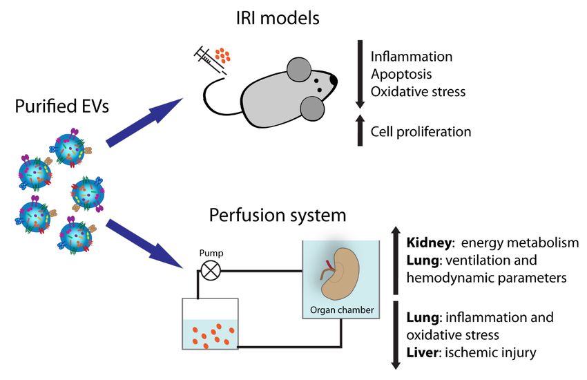
EVs, dissecting their possible application as an innovative therapeutic tool to precondition grafts before (Figure 1).
In particular,
transplant we as
as well describe their
to prevent use in pre-transplant
ischemic/reperfusion damage solid organ1).preservation
(Figure In particular, in
weassociation
describe theirwith
use
normothermic and hypothermic perfusion machines. In addition, their role in the
in pre-transplant solid organ preservation in association with normothermic and hypothermic perfusion limitation of IRI is
highlighted
machines. Infor kidney,
addition, liver,
their lung,
role in and heart.
the limitation Finally,
of IRI we present
is highlighted their immunomodulatory
for kidney, liver, lung, and heart.
properties
Finally, wein bone marrow
present transplantation. properties in bone marrow transplantation.
their immunomodulatory
Figure1.
Figure Extracellularvesicle
1. Extracellular vesicle(EV)
(EV)activities
activitiesin
insolid
solidorgan
organtransplantation.
transplantation.
2. Stem-Cell-Derived EVs and Regenerative Medicine
2. Stem-Cell-Derived EVs and Regenerative Medicine
EVs released by healthy cells are very heterogeneous in size and composition, and they can be
EVs released by healthy cells are very heterogeneous in size and composition, and they can be
classified based on their origin and dimension into two main categories: small EVs, ranging between
classified based on their origin and dimension into two main categories: small EVs, ranging between
30 and 100 nm, and large EVs, ranging between 50 and 1000 nm [14].
30 and 100 nm, and large EVs, ranging between 50 and 1000 nm [14].
Among small EVs, exosomes are the most characterized vesicles, considered to originate from
Among small EVs, exosomes are the most characterized vesicles, considered to originate from
multivesicular bodies after their fusion with the cell membrane [15]. However, other subtypes of small
multivesicular bodies after their fusion with the cell membrane [15]. However, other subtypes of
EVs different from the multivesicular-body-derived exosomes have been identified, for instance after
small EVs different from the multivesicular-body-derived exosomes have been identified, for
plasma membrane budding [14].
instance after plasma membrane budding [14].
Large EVs, also called microvesicles/ectosomes, comprise different populations of vesicles originating
Large EVs, also called microvesicles/ectosomes, comprise different populations of vesicles
from the budding of the plasma membrane [16]. The different EV populations express common and
originating from the budding of the plasma membrane [16]. The different EV populations express
specific surface markers. For instance, tetraspanins such as CD9, CD81, and CD63 are mainly expressed
common and specific surface markers. For instance, tetraspanins such as CD9, CD81, and CD63 are
by small EVs [14]. In addition, small EVs are characterized by the presence of molecules of the endosomal
mainly expressed by small EVs [14]. In addition, small EVs are characterized by the presence of
sorting complex required for transport (ESCRT), heat shock proteins (HSP70 and HSP90), and auxiliary
molecules of the endosomal sorting complex required for transport (ESCRT), heat shock proteins
proteins (ALIX, TSG101, and VPS4). In terms of variance, large EVs are specifically characterized by
(HSP70 and HSP90), and auxiliary proteins (ALIX, TSG101, and VPS4). In terms of variance, large
expression of the CD40 ligand [17,18]. The detailed composition of EV cargo has been deeply dissected
and several databases collecting these results are now available, such as EVpedia [19], Exocarta [20],Cells 2020, 9, 369 3 of 13
and Vesiclepedia [21]. EVs can be isolated from the majority of body fluids such as plasma and serum,
amniotic and seminal fluids, saliva, urine, or nasal and bronchial lavage fluids [9,22].
Is important to take into consideration that a limitation to consistent EV characterization is
the variability in EV isolation protocols. Depending on the size of EVs and on the fluids of origin,
different techniques can be utilized, including ultra-high-speed centrifugation, polymer precipitation,
immunoaffinity capture, or microfluidics-based techniques, among others [23]. Rigor criteria for EV
isolation and characterization were recently proposed by the International Society for Extracellular
Vesicles (ISEV) [14].
Stem-cell-derived EVs possess many characteristics in common with the originating cells;
for instance, they carry some transcription factors classically expressed by stem cells, such as Nanog
and Oct-4, as well as stem (CD133 and c-Kit) and mesenchymal markers (CD105, CD29, and CD73) [24].
It has been clearly demonstrated that stem-cell-derived EVs recapitulate the pro-regenerative capacity
of the cells of origin and, in particular, those derived from mesenchymal stromal cells (MSCs) appear
the ideal candidates to favor tissue regeneration. MSC-EVs may be isolated from MSCs derived from
different adult tissues such as bone marrow, peripheral and cord blood, adipose tissue, or neonatal
birth-associated tissues including placenta and umbilical cord [25]. Several studies have shown that
MSC-EVs possess strong pro-regenerative properties using preclinical models of renal, lung, liver, and
heart injuries, mimicking the beneficial effect of the cells themselves [14,26,27]. The activity of EVs
mainly results in the reduction of apoptosis, oxidative stress, and inflammation and in increase of cell
proliferation [24,28,29].
3. Normothermic and Hypothermic Perfusion Machines
In order to increase the number of successful transplants, the use of machine perfusion is currently
proposed to ameliorate the function of organs from marginal donors such as DCDs and ECDs. Dynamic
perfusion of organs appears a useful strategy to evaluate pretransplant graft function, limiting the
discard rate [30–32]. Moreover, this approach reduces the incidence of DGF in recipients receiving
organs from ECDs and DCDs.
At present, dynamic machine perfusion can be done in hypothermic (HMP) or in normothermic
(NMP) conditions with or without oxygen. Several studies have demonstrated that both HPM and NPM
are useful in the assessment of organ viability prior to transplantation [32–34]. Specifically, HMP is able
to reduce DGF and to increase the graft survival of organs harvested from ECDs, but conflicting results
have been reported on the beneficial effects of HMP on grafts from DCDs [35–40]. Another beneficial
effect of HMP is the removal of inflammatory mediators that may have detrimental effects on graft
function. The delivery of oxygen added to the hypothermic perfusate may help to restore adenosine
triphosphate (ATP) content [41–44]. Because of the unknown effects of this oxygenated perfusion on
transplanted patients, a large international randomized controlled trial has been planned to investigate
the beneficial effects of oxygenated short-term perfusion of kidneys from ECDs (Consortium for Organ
Preservation in Europe COPE Trials) [45].
As oxygenated machine perfusion, NMP may protect organs from IRI by restoring ATP levels [46,47].
In particular, ex vivo normothermic perfusion, consisting of circulation through the harvested organs of
warm oxygenated red-cell-based solution, is able to restore the metabolism and function of the graft
prior to transplantation [48–50]. NMP could offer a better evaluation of organ viability compared to
HMP, especially in kidney and liver grafts because of urine or bile production, together with a better
preservation of graft function [51].
Both HMP and NMP allow the delivery of targeted therapies to organs prior to transplantation.
In particular, these approaches offer the potential to explore the effects of several therapeutic strategies,
such as gene-silencing, nanoparticles, and cell therapies, in a fully functioning graft [52–57].Cells 2020, 9, 369 4 of 13
4. EVs for Kidney Transplant
An innovative EV-based application for organ preservation is the use of EVs in the perfusion
solution. A first report in the literature recently demonstrated that EVs released by MSCs, delivered in
the perfusate during organ cold perfusion (4 h), preserve and protect kidney function. Histological
and genetic analyses on EV-treated kidneys revealed upregulation of enzymes involved in energy
metabolism and reduction of global ischemic damage. In addition, the analysis of lactate, LDH,
and glucose in the effluent fluid confirmed a greater use of energy substrates by EV-treated kidneys,
supporting the report of improved functionality (Table 1) [58].
Moreover, an extensive number of publications have highlighted the beneficial effect of EVs
in preclinical models of IRI, further implying their possible application to limit organ damage [9].
In particular, EVs isolated from different MSC sources [59–62] have been shown to accelerate renal
recovery after damage, promoting cell proliferation and blocking inflammation and apoptosis when
intravenously injected after IR damage [63]. The mechanisms of action reported appear different
between the EV sources: MSC-EVs obtained from Wharton’s jelly stimulate tubular proliferation and
reduce inflammation and apoptosis via mitochondrial protection [61,62], while those from cord blood
promote tubular dedifferentiation and proliferation by the transfer of human HGF [60]. Moreover,
EVs isolated from bone marrow MSCs were protective mainly by suppressing inflammation when
injected under the renal capsule [64]. In addition, EVs obtained from MSCs isolated from glomeruli
have also been demonstrated to be capable of reducing ischemic damage [65].
Moreover, a recent publication demonstrated that EVs isolated from the venal perfusate of rats
subjected to remote ischemia preconditioning ameliorated renal function when injected into another
animal with IRI. To explore the underlying mechanism, authors tested in vivo, in the same IRI model,
the effect of EVs released by human proximal tubular cells cultured in hypoxia, supporting the thesis
that remote ischemia precondition activates a repairing program into tubular cells by the release of
pro-regenerative EVs [66].
Whereas all the studies mentioned above evaluated classical ischemic damage in models of
renal artery clamping, Wu and co-workers tested for the first time the effect of EVs in a rat model
of IRI after DCD renal transplantation [67]. The authors confirmed that Wharton’s jelly MSC-EVs,
intravenously injected after renal transplantation, mitigated renal damage, improving survival and
function. In particular, MSC-EVs were shown to reduce cell apoptosis and inflammation, to stimulate
HGF production, and subsequently to alleviate fibrosis [67].
Table 1. List of EV applications for organ preconditioning. Abbreviations: bone marrow (BM), human
liver stem cells (HLSCs).
Type of Time of
Organs EV Sources Results References
Perfusion Preconditioning
Kidney BM-MSCs Hypothermic 4h Preservation and protection Gregorini et al. [58]
Improvement of ventilation and
Lung BM-MSCs Normothermic 6h Gennai et al. [68]
hemodynamic parameters
Restoring permeability and
Lung BM-MSCs Normothermic 6h Park et al. [69]
reduction of inflammation
Attenuation of IR dysfunction and
Lung BM-MSCs Normothermic 1h Stone et al. [70]
immunomodulation
Reduction of inflammation and
Lung BM-MSCs Normothermic 3h Lonati et al. [71]
oxidative stress
Limitation of the progression of
Liver HLSCs Normothermic 4h Rigo et al. [72]
ischemic injury
5. EVs for Lung Transplantation
Adult lung transplantation is considered the most effective strategy for end-stage pulmonary
disease, although the reported 5-year survival rate is only 50% [73]. Infections, immunomodulation,
and IRI are in fact some of the aspects involved in lung transplant failure [74]. Through ex vivo lung
perfusion, donor lungs can be evaluated and reconditioned, while organs are perfused and ventilated [75].Cells 2020, 9, 369 5 of 13
The use of MSC-EVs has been proposed as a valid alternative for the rehabilitation of marginal human
lungs [68]. Upon administering MSC-EVs in the perfusion fluid, a dose-dependent increase of alveolar
fluid clearance, a decrease of lung weight gain, and an improvement of airway and hemodynamic
parameters were observed as compared to perfusion alone (Table 2). Moreover, the study showed
that CD44 was involved in the EV uptake mechanism, as the efficacy of MSC-EVs decreased with the
administration of anti-CD44 antibody.
A significant improvement of inflammatory conditions has also been ascribed to the EV effect on
lung bacterial infections. For example, MSC-EVs have been demonstrated to be effective in restoring
lung protein permeability and reducing inflammation in Escherichia-coli-endotoxin-induced acute
lung injury in mice. In particular, MSC-EV treatment restored protein permeability and reduced
inflammation, extravascular lung water, and total protein levels in the bronchoalveolar lavage fluid,
demonstrating a reduction in pulmonary edema [76]. On this path, in a recent work, the effects of
MSC-EVs were investigated in an ex vivo perfused human lung model, injured with severe E. coli
pneumonia [69]. The paper confirmed a significant increase of alveolar fluid clearance and decrease
in protein permeability, as well as the lowering of the bacterial load and the neutrophil count in the
injured alveolus (Table 2). MSC pretreatment with a toll-like-receptor 3 agonist before the isolation of
EVs increased their bactericidal activity.
Moreover, Stone and colleagues demonstrated the attenuation of IR dysfunction in lungs after
treatment with MSC-EVs both in vivo and in ex vivo perfusion systems [70]. In particular, they observed
a decrease of pro-inflammatory cytokines and upregulation of keratinocyte growth factor, PGE2, and
IL-10. Recently, in a mouse model of ex vivo lung perfusion, EV-treated organs showed decreased
vascular resistance and a rise of perfusate nitric oxide metabolites. Moreover, EV treatment prevented
the reduction in pulmonary ATP and increased the medium–high-molecular-weight hyaluronan in
the perfusate. The genes modulated in the pulmonary tissue by EV administration were involved in
anti-inflammatory and anti-oxidative stress pathways [71].
6. EVs for Liver Transplantation
The use of EVs released by stem cells as an innovative option to improve the viability of
pre-transplant livers was recently assessed in a model of ex vivo rat liver NMP. HLSC-EVs (EVs
isolated from human liver stem cells) were added to perfusate 15 min after the initiation of NMP and
administered for 4 h within the perfusate. The results showed that HLSC-EVs limited the progression
of ischemic injury, with a significant reduction of the levels of aspartate aminotransferase and alanine
aminotransferase and a decrease of histological damage compared with results of NMP alone (Table
2) [72]. Moreover, the authors demonstrated that HLSC-EVs were uptaken by hepatocytes, supporting
the thesis that EVs may recondition liver cells before transplantation [72].
Moreover, the potential therapeutic use of stem-cell-derived-EVs for liver regeneration, has been
also clearly demonstrated in pre-clinical models of liver IRI. In fact, hepatic ischemia and related
inflammation should be limited to avoid complication after liver transplantation [77]. The intravenous
injection of murine MSC-EVs prior to IRI reduced the area of necrosis and apoptosis with concomitant
increased liver function [77]. In addition, MSC-EVs have been shown to limit liver inflammation and
oxidative stress [77]. Similar results were obtained using EVs isolated from MSCs from inducible
pluripotent stem cells [78] or bone marrow [79]. Recently, Yao et al. demonstrated that human umbilical
cord MSC-EVs protect hepatic apoptosis post-IRI, modulating neutrophils and reducing oxidative
stress [80].
7. Stem-Cell-Derived EVs as Future Therapeutics in Heart Transplantation
EVs have been shown to be powerful allies against cardiovascular damage. Some important
interconnected effects related to EVs could improve the success of a heart transplantation, including
immunomodulatory properties, the improvement of heart function and vessel formation, and the
amelioration of myocardial function during IRI [81].Cells 2020, 9, 369 6 of 13
Much evidence confirms the hypothesis that cardiac progenitor cells release pro-regenerative
and anti-fibrotic EVs in response to hypoxic conditions [82,83], mainly due to their miRNA cargo [82].
Moreover, cardiac-progenitor-cell-derived EVs, released into their environment, can stimulate migration
of endothelial cells [84] and inhibit both cardiac fibroblast activation and collagen synthesis [85].
In parallel, MSC-EV treatment has also been proven as a therapeutic option to limit ischemic
damage in the heart. In particular, MSC-EV administration increased phosphorylated-Akt and
phosphorylated-GSK-3β, as well as ATP/NADH level, and could reduce phosphorylated-c-JNK and
inflammatory response in ischemic/reperfused hearts [86].
8. EVs for Islet Transplantation
Today, there are still many factors that limit the success of pancreatic islet transplantation, including
islet source limitation, sub-optimal engraftment, lack of oxygen and blood supply for transplanted
islets, and immune rejection [87]. In parallel with the other described organs, MSC-EVs may also be of
benefit for islet transplantation.
One of the primary reasons for apoptosis and reduced beta-cell function in transplants is hypoxic
damage. Recently, EVs from human-umbilical-cord-derived MSCs were shown to have a therapeutic
effect on the survival and function of neonatal porcine islets exposed to hypoxia [88]. The use of EVs,
in comparison with medium alone, enhanced the yield and survival of porcine islets, and showed
an improvement of the function through the amelioration of mitochondrial respiration efficiency [88].
In addition, Di Wen and colleagues showed that MSC-EV administration through delivery of
small RNAs promoted islet function and inhibited immune rejection [89]. In a mouse model, they used
MSC-EVs transfected with shFas and anti-miR-375 in order to silence Fas and miR-375 in human
islets, observing an improvement of islet viability and function. Moreover, the authors observed the
inhibition of peripheral blood mononuclear cell proliferation and the enhancement of T-cell regulatory
function. Based on these works, EVs from different sources appear of interest to increase the possibility
of successful islet transplantation.
9. Role of MSC-EVs in the Amelioration of Graft Versus Host Disease
EVs derived from bone marrow MSCs possess an immunosuppressive potential that can be
harnessed to treat graft versus host disease (GVHD), which today represents the greatest complication
after allogeneic transplantation. The majority of the literature on the subject has generically focused on
the effects of the whole MSC secretome, including EVs and soluble factors. Recently, the specific role of
EVs has been highlighted, showing an effect on innate and adaptive immunity (Table 2).
For example, in 2005, Aggarwal and Pittenger highlighted that the secretome, released by MSCs,
be responsible for modulation of immune reaction, involved in GVHD [90]. In fact, if co-cultured with
purified subpopulations of immune cells, human MSCs were able to switch an inflammatory response
into a tolerant phenotype. In particular, MSCs induced mature dendritic cells type 1 and type 2 to
decrease TNF-α and to increase IL-10 secretion, respectively; they also induced T helper 1 lymphocytes
and natural killer cells to decrease interferon (IFN) γ secretion. In addition, they enhanced a regulatory
response, causing the T helper 2 cells to increase secretion of IL-4, increasing the proportion of regulatory
T cells and producing prostaglandin (PG) E2 [90].
Moreover, soluble factors released by MSCs, such as vascular endothelial growth factor and IL-6,
were shown to inhibit T-cell proliferation and to be involved in a partial inhibition of dendritic cell
differentiation [91]. Selmani et al. not only confirmed the role of the MSC secretome in modulating
innate immunity, but they also sustained its strong modulation of adaptive immunity [92]. Moreover,
they reported that the nonclassic HLA class I molecule HLA-G is responsible for the immunomodulatory
properties of MSCs [92].
In a recent work, it was shown that bone marrow MSC-EVs recapitulate the therapeutic effects of
the cells against acute GVHD [93]. A systemic infusion of MSC-EVs in mice with acute GVHD was
associated with the suppression of CD4+ and CD8+ T cells and with the preservation of circulatingCells 2020, 9, 369 7 of 13
naive T cells, possibly due to the unique microRNA profiles of MSC-EVs. The analysis on microRNA
cargo in MSC-EVs identified that their target genes were involved in regulation of the cell cycle, T-cell
receptor signaling, and GVHD [93]. These findings suggest that MSC-EVs could be a new potential
therapeutic option to prevent GVHD, to be tested in future clinical trials.
Table 2. Immunomodulatory properties of MSC secretome/EVs.
Cell Types Actions Mechanisms Effector References
Decrease of TH1 secretion of
IFN-γ [91]
Increase of TH2 secretion of
Constitutive production
IL-4 [91]
of COX2 and PGE2
Increase of the proportion of S. Aggarwal et al. [91]
[91–93] Secretome [91–93]
T lymphocytes T-regs [91] Z Selmani et al. [93]
Secretion of TGF-β [91] EVs [94]
Suppression of T-naïve S. Fujii et al. [94]
Secretion of soluble
differentiation [94]
HLA-G5 [93]
Decrease in proliferation and
migration [94]
Decrease of CD4+ CD8+ [94]
Reversion of maturation of
DCs [92]
Decrease DC1 production of S. Aggarwal et al. [91]
DC Secretion of IL-6 [91] Secretome [91,92]
TNF-α [91] F. Djouad et al. [92]
Increase DC2 production of
IL-10 [91]
Secretion of indoleamine
Inhibition [91]
2,3-deoxygenase [91]
NK Alteration of secreted Secretome [91] S. Aggarwal et al. [91]
Secretion of PGE2 [91]
cytokines [91]
Secretion of TGF-β [91]
10. Conclusions
The organ demand is continuously increasing and there is a constant need to expand the pool of
donors. Increasing organ availability represents a major challenge in the field of transplantation.
Among the most recent innovative strategies, the use of EVs seems very promising. The application
of EVs in the perfusion solution, recently proposed for kidney, lung, and liver grafts, results in the
amelioration of donor organ viability and functionality. Moreover, consolidated results describe the
beneficial effects of EV administration in several preclinical models of IRI. In particular, stem-cell-derived
EVs have displayed strong pro-regenerative properties in different models of renal, lung, liver, and
heart injuries. IRI is an unavoidable consequence after transplants and the severity of this phenomenon
affects the graft outcome, leading to delayed graft function, graft rejection, chronic rejection, and chronic
graft dysfunction. The development of strategies to limit the progression of IRI is fundamental for the
success of transplants. Altogether, EVs appear the ideal candidate to target different aspects during
transplantation process.
Author Contributions: Writing—original draft preparation, A.R., C.G., L.B. and B.B.; writing—review and editing,
L.B., C.G. and B.B.; funding acquisition, B.B. All authors have read and agreed to the published version of
the manuscript.
Funding: This research was funded by, Regione Piemonte POR FESR 2014/2020—Grant “Bando Piattaforma
Tecnologica Salute e Benessere”, Project “Terapie Avanzate per Processi Fibrotici Cronici (EVER)”.
Conflicts of Interest: The authors declare no conflict of interest.
References
1. Wolfe, R.A.; Ashby, V.B.; Milford, E.L.; Ojo, A.O.; Ettenger, R.E.; Agodoa, L.Y.C.; Held, P.J.; Port, F.K.
Comparison of mortality in all patients on dialysis, patients on dialysis awaiting transplantation, and
recipients of a first cadaveric transplant. N. Engl. J. Med. 1999, 341, 1725–1730. [CrossRef] [PubMed]
2. 2017 Global Report—GODT. Available online: http://www.transplant-observatory.org/download/2017-
activity-data-report/ (accessed on 2 December 2019).Cells 2020, 9, 369 8 of 13
3. Branger, P.; Undine, S. Eurotransplant Annual Report 2018. 2018. Available online: https://www.eurotransplant.
org/organs/kidney/ (accessed on 5 February 2020).
4. NHS Organ Donation and Transplantation Activity Report 2018/19. 2019. Available
online: https://www.organdonation.nhs.uk/helping-you-to-decide/about-organ-donation/statistics-about-
organ-donation/transplant-activity-report/ (accessed on 5 February 2020).
5. Hart, A.; Smith, J.M.; Skeans, M.A.; Gustafson, S.K.; Wilk, A.R.; Castro, S.; Robinson, A.; Wainright, J.L.;
Snyder, J.J.; Kasiske, B.L.; et al. OPTN/SRTR 2017 Annual Data Report: Kidney. Am. J. Transplant. 2019, 19,
19–123. [CrossRef] [PubMed]
6. Abramowicz, D.; Oberbauer, R.; Heemann, U.; Viklicky, O.; Peruzzi, L.; Mariat, C.; Crespo, M.; Budde, K.;
Oniscu, G.C. Recent advances in kidney transplantation: A viewpoint from the Descartes advisory board.
Nephrol. Dial. Transplant. 2018, 33, 1699–1707. [CrossRef] [PubMed]
7. Sørensen, S.S. Rates of renal transplantations in the elderly-data from Europe and the US. Transplant. Rev.
2015, 29, 193–196. [CrossRef]
8. Nashan, B.; Abbud-Filho, M.; Citterio, F. Prediction, prevention, and management of delayed graft function:
Where are we now? Clin. Transplant. 2016, 30, 1198–1208. [CrossRef]
9. Grange, C.; Skovronova, R.; Marabese, F.; Bussolati, B. Stem Cell-Derived Extracellular Vesicles and Kidney
Regeneration. Cells 2019, 8, 1240. [CrossRef]
10. Quesenberry, P.J.; Aliotta, J.; Deregibus, M.C.; Camussi, G. Role of extracellular RNA-carrying vesicles in cell
differentiation and reprogramming. Stem Cell Res. Ther. 2015, 6, 1–10. [CrossRef]
11. Valadi, H.; Ekström, K.; Bossios, A.; Sjöstrand, M.; Lee, J.J.; Lötvall, J.O. Exosome-mediated transfer of
mRNAs and microRNAs is a novel mechanism of genetic exchange between cells. Nat. Cell Biol. 2007, 9,
654–659. [CrossRef]
12. Ratajczak, M.Z.; Ratajczak, J. Horizontal transfer of RNA and proteins between cells by extracellular
microvesicles: 14 years later. Clin. Transl. Med. 2016, 5. [CrossRef]
13. Deregibus, M.C.; Cantaluppi, V.; Calogero, R.; Lo Iacono, M.; Tetta, C.; Biancone, L.; Bruno, S.; Bussolati, B.;
Camussi, G. Endothelial progenitor cell—Derived microvesicles activate an angiogenic program in endothelial
cells by a horizontal transfer of mRNA. Blood 2007, 110, 2440–2448. [CrossRef]
14. Théry, C.; Witwer, K.W.; Aikawa, E.; Alcaraz, M.J.; Anderson, J.D.; Andriantsitohaina, R.; Antoniou, A.;
Arab, T.; Archer, F.; Atkin-Smith, G.K.; et al. Minimal information for studies of extracellular vesicles 2018
(MISEV2018): A position statement of the International Society for Extracellular Vesicles and update of the
MISEV2014 guidelines. J. Extracell. Vesicles 2018, 7, 1535750. [CrossRef] [PubMed]
15. Mathivanan, S.; Ji, H.; Simpson, R.J. Exosomes: Extracellular organelles important in intercellular
communication. J. Proteomics 2010, 73, 1907–1920. [CrossRef] [PubMed]
16. van der Pol, E.; Böing, A.N.; Harrison, P.; Sturk, A.; Nieuwland, R. Classification, functions, and clinical
relevance of extracellular vesicles. Pharmacol. Rev. 2012, 64, 676–705. [CrossRef] [PubMed]
17. Mobarrez, F.; Sjövik, C.; Soop, A.; Hållström, L.; Frostell, C.; Pisetsky, D.S.; Wallén, H. CD40L expression
in plasma of volunteers following LPS administration: A comparison between assay of CD40L on platelet
microvesicles and soluble CD40L. Platelets 2015, 26, 486–490. [CrossRef] [PubMed]
18. Jeppesen, D.K.; Fenix, A.M.; Franklin, J.L.; Higginbotham, J.N.; Zhang, Q.; Zimmerman, L.J.; Liebler, D.C.;
Ping, J.; Liu, Q.; Evans, R.; et al. Reassessment of Exosome Composition. Cell 2019, 177, 428–445.e18.
[CrossRef] [PubMed]
19. Kim, D.K.; Kang, B.; Kim, O.Y.; Choi, D.S.; Lee, J.; Kim, S.R.; Go, G.; Yoon, Y.J.; Kim, J.H.; Jang, S.C.; et al.
EVpedia: An integrated database of high-throughput data for systemic analyses of extracellular vesicles.
J. Extracell. Vesicles 2013, 2, 1–7. [CrossRef]
20. Mathivanan, S.; Fahner, C.J.; Reid, G.E.; Simpson, R.J. ExoCarta 2012: Database of exosomal proteins, RNA
and lipids. Nucleic Acids Res. 2012, 40, D1241-4. [CrossRef]
21. Kalra, H.; Simpson, R.J.; Ji, H.; Aikawa, E.; Altevogt, P.; Askenase, P.; Bond, V.C.; Borràs, F.E.; Breakefield, X.;
Budnik, V.; et al. Vesiclepedia: A Compendium for Extracellular Vesicles with Continuous Community
Annotation. PLoS Biol. 2012, 10, 8–12. [CrossRef] [PubMed]
22. Keller, S.; Ridinger, J.; Rupp, A.K.; Janssen, J.W.G.; Altevogt, P. Body fluid derived exosomes as a novel
template for clinical diagnostics. J. Transl. Med. 2011, 9, 86. [CrossRef]Cells 2020, 9, 369 9 of 13
23. Yang, X.-X.; Sun, C.; Wang, L.; Guo, X.-L. New insight into isolation, identification techniques and medical
applications of exosomes. J. Control. Release 2019, 308, 119–129. [CrossRef]
24. Bruno, S.; Chiabotto, G.; Favaro, E.; Deregibus, M.C.; Camussi, G. Role of extracellular vesicles in stem cell
biology. Am. J. Physiol. Cell Physiol. 2019, 317, C303–C313. [CrossRef] [PubMed]
25. Grange, C.; Tritta, S.; Tapparo, M.; Cedrino, M.; Tetta, C.; Camussi, G.; Brizzi, M.F. Stem cell-derived
extracellular vesicles inhibit and revert fibrosis progression in a mouse model of diabetic nephropathy.
Sci. Rep. 2019, 9, 4468. [CrossRef] [PubMed]
26. Heldring, N.; Mäger, I.; Wood, M.J.A.; Le Blanc, K.; Andaloussi, S.E.L. Therapeutic Potential of Multipotent
Mesenchymal Stromal Cells and Their Extracellular Vesicles. Hum. Gene Ther. 2015, 26, 506–517. [CrossRef]
27. Ferreira, J.R.; Teixeira, G.Q.; Santos, S.G.; Barbosa, M.A.; Almeida-Porada, G.; Gonçalves, R.M. Mesenchymal
Stromal Cell Secretome: Influencing Therapeutic Potential by Cellular Pre-conditioning. Front. Immunol.
2018, 9, 2837. [CrossRef]
28. Collino, F.; Bruno, S.; Incarnato, D.; Dettori, D.; Neri, F.; Provero, P.; Pomatto, M.; Oliviero, S.; Tetta, C.;
Quesenberry, P.J.; et al. AKI recovery induced by mesenchymal stromal cell-derived extracellular vesicles
carrying micrornas. J. Am. Soc. Nephrol. 2015, 26, 2349–2360. [CrossRef] [PubMed]
29. György, B.; Szabó, T.G.; Pásztói, M.; Pál, Z.; Misják, P.; Aradi, B.; László, V.; Pállinger, É.; Pap, E.; Kittel, Á.; et al.
Membrane vesicles, current state-of-the-art: Emerging role of extracellular vesicles. Cell. Mol. Life Sci. 2011,
68, 2667–2688. [CrossRef]
30. Matsuno, N.; Konno, O.; Mejit, A.; Jyojima, Y.; Akashi, I.; Nakamura, Y.; Iwamoto, H.; Hama, K.; Iwahori, T.;
Ashizawa, T.; et al. Application of machine perfusion preservation as a viability test for marginal kidney
graft. Transplantation 2006, 82, 1425–1428. [CrossRef]
31. Bissolati, M.; Gazzetta, P.G.; Caldara, R.; Guarneri, G.; Adamenko, O.; Giannone, F.; Mazza, M.; Maggi, G.;
Tomanin, D.; Rosati, R.; et al. Renal Resistance Trend during Hypothermic Machine Perfusion Is More
Predictive of Postoperative Outcome Than Biopsy Score: Preliminary Experience in 35 Consecutive Kidney
Transplantations. Artif. Organs 2018, 42, 714–722. [CrossRef]
32. Gelpi, R.; Paredes, D.; Rodríguez-Villar, C.; Roque, R.; Ruiz, A.; Adalia, R.; Peri-Cusí, L.; Sole, M.;
Oppenheimer, F.; Diekmann, F. The development of a predictive model of graft function in uncontrolled
donors after circulatory death: Validity of a pulsatile renal preservation machine cut-off value for kidney
acceptance. Nephrol. Dial. Transplant. 2019, 34, 531–538. [CrossRef]
33. Hameed, A.M.; Pleass, H.C.; Wong, G.; Hawthorne, W.J. Maximizing kidneys for transplantation using
machine perfusion: From the past to the future: A comprehensive systematic review and meta-analysis.
Medicine 2016, 95, e5083. [CrossRef]
34. Brat, A.; Pol, R.A.; Leuvenink, H.G.D. Novel preservation methods to increase the quality of older kidneys.
Curr. Opin. Organ Transplant. 2015, 20, 438–443. [CrossRef] [PubMed]
35. Gallinat, A.; Moers, C.; Smits, J.M.; Strelniece, A.; Pirenne, J.; Ploeg, R.J.; Paul, A.; Treckmann, J. Machine
perfusion versus static cold storage in expanded criteria donor kidney transplantation: 3-year follow-up
data. Transpl. Int. 2013, 26, 52–53. [CrossRef] [PubMed]
36. Ali, F.; Dua, A.; Cronin, D.C. Changing paradigms in organ preservation and resuscitation. Curr. Opin.
Organ Transplant. 2015, 20, 152–158. [CrossRef] [PubMed]
37. Matos, A.C.C.; Requiao Moura, L.R.; Borrelli, M.; Nogueira, M.; Clarizia, G.; Ongaro, P.; Durão, M.S.;
Pacheco-Silva, A. Impact of machine perfusion after long static cold storage on delayed graft function
incidence and duration and time to hospital discharge. Clin. Transplant. 2018, 32. [CrossRef] [PubMed]
38. Tedesco-Silva, H.; Mello Offerni, J.C.; Ayres Carneiro, V.; Ivani de Paula, M.; Neto, E.D.; Brambate Carvalhinho
Lemos, F.; Requião Moura, L.R.; Pacheco e Silva Filho, A.; de Morais Cunha, M.d.F.; Francisco da Silva, E.; et al.
Randomized Trial of Machine Perfusion Versus Cold Storage in Recipients of Deceased Donor Kidney
Transplants with High Incidence of Delayed Graft Function. Transplant. Direct 2017, 3, e155. [CrossRef]
[PubMed]
39. Delsuc, C.; Faure, A.; Berthiller, J.; Dorez, D.; Matillon, X.; Meas-Yedid, V.; Floccard, B.; Marcotte, G.;
Labeye, V.; Rabeyrin, M.; et al. Uncontrolled donation after circulatory death: Comparison of two kidney
preservation protocols on graft outcomes. BMC Nephrol. 2018, 19, 1–9. [CrossRef] [PubMed]Cells 2020, 9, 369 10 of 13
40. Czigany, Z.; Lurje, I.; Tolba, R.H.; Neumann, U.P.; Tacke, F.; Lurje, G. Machine perfusion for liver
transplantation in the era of marginal organs—New kids on the block. Liver Int. 2019, 39, 228–249.
[CrossRef]
41. Kron, P.; Schlegel, A.; De Rougemont, O.; Oberkofler, C.E.; Clavien, P.A.; Dutkowski, P. Short, cool, and well
oxygenated—HOPE for kidney transplantation in a rodent model. Ann. Surg. 2016, 264, 815–822. [CrossRef]
42. Minor, T.; Paul, A. Hypothermic reconditioning in organ transplantation. Curr. Opin. Organ Transplant. 2013,
18, 161–167. [CrossRef]
43. Kaths, J.M.; Paul, A.; Robinson, L.A.; Selzner, M. Ex vivo machine perfusion for renal graft preservation.
Transplant. Rev. 2018, 32, 1–9. [CrossRef]
44. Solhjou, Z.; Athar, H.; Xu, Q.; Abdi, R. Emerging therapies targeting intra-organ inflammation in
transplantation. Am. J. Transplant. 2015, 15, 305–311. [CrossRef] [PubMed]
45. COPE—Trials. Available online: http://cope-eu.com/workprogramme/trials.html (accessed on 2 December
2019).
46. Hessheimer, A.J.; Riquelme, F.; Fundora-Suárez, Y.; García Pérez, R.; Fondevila, C. Normothermic perfusion
and outcomes after liver transplantation. Transplant. Rev. 2019, 33, 200–208. [CrossRef] [PubMed]
47. Palomo-López, N.; Martín-Sastre, S.; Martín-Villén, L.; Ruiz de Azúa-López, Z.; Solis-Clavijo, D.;
Caballero-Gálvez, S.; Carballo-Caro, J.M.; Egea-Guerrero, J.J. Normothermic Regional Perfusion and Donation
after Circulatory Death (Controlled and Uncontrolled): Metabolic Differences and Kidney Transplantation
Evolution. Transplant. Proc. 2019, 51, 3044–3046. [CrossRef] [PubMed]
48. Hosgood, S.A.; Saeb-Parsy, K.; Wilson, C.; Callaghan, C.; Collett, D.; Nicholson, M.L. Protocol of a randomised
controlled, open-label trial of ex vivo normothermic perfusion versus static cold storage in donation after
circulatory death renal transplantation. BMJ Open 2017, 7, 1–7. [CrossRef] [PubMed]
49. Hosgood, S.A.; Thompson, E.; Moore, T.; Wilson, C.H.; Nicholson, M.L. Normothermic machine perfusion
for the assessment and transplantation of declined human kidneys from donation after circulatory death
donors. Br. J. Surg. 2018, 105, 388–394. [CrossRef]
50. Laing, R.W.; Mergental, H.; Yap, C.; Kirkham, A.; Whilku, M.; Barton, D.; Curbishley, S.; Boteon, Y.L.;
Neil, D.A.; Hübscher, S.G.; et al. Viability testing and transplantation of marginal livers (VITTAL) using
normothermic machine perfusion: Study protocol for an open-label, non-randomised, prospective, single-arm
trial. BMJ Open 2017, 7, 1–15.
51. Weissenbacher, A.; Hunter, J. Normothermic machine perfusion of the kidney. Curr. Opin. Organ Transplant.
2017, 22, 571–576. [CrossRef]
52. Brasile, L.; Stubenitsky, B.M.; Booster, M.H.; Arenada, D.; Haisch, C.; Kootstra, G. Transfection and transgene
expression in a human kidney during ex vivo warm perfusion. Transplant. Proc. 2002, 34, 2624. [CrossRef]
53. Chen, J.; Braet, F.; Brodsky, S.; Weinstein, T.; Romanov, V.; Noiri, E.; Goligorsky, M.S. VEGF-induced
mobilization of caveolae and increase in permeability of endothelial cells. Am. J. Physiol. Cell Physiol. 2002,
282, C1053–C1063. [CrossRef]
54. Chen, J.; Vemuri, C.; Palekar, R.U.; Gaut, J.P.; Goette, M.; Hu, L.; Cui, G.; Zhang, H.; Wickline, S.A. Antithrombin
nanoparticles improve kidney reperfusion and protect kidney function after ischemia-reperfusion injury. Am. J.
Physiol. Ren. Physiol. 2015, 308, F765–F773. [CrossRef]
55. DiRito, J.R.; Hosgood, S.A.; Tietjen, G.T.; Nicholson, M.L. The future of marginal kidney repair in the context
of normothermic machine perfusion. Am. J. Transplant. 2018, 18, 2400–2408. [CrossRef] [PubMed]
56. Hamaoui, K.; Gowers, S.; Boutelle, M.; Cook, T.H.; Hanna, G.; Darzi, A.; Smith, R.; Dorling, A.; Papalois, V.
Organ Pretreatment with Cytotopic Endothelial Localizing Peptides to Ameliorate Microvascular Thrombosis
and Perfusion Deficits in Ex Vivo Renal Hemoreperfusion Models. Transplantation 2016, 100, e128–e139.
[CrossRef] [PubMed]
57. Hamaoui, K.; Aftab, A.; Gowers, S.; Boutelle, M.; Cook, T.; Rudd, D.; Dobson, G.P.; Papalois, V. An ex vivo
comparison of adenosine and lidocaine solution and University of Wisconsin solution for hypothermic
machine perfusion of porcine kidneys: Potential for development. J. Surg. Res. 2017, 208, 219–229. [CrossRef]
[PubMed]
58. Gregorini, M.; Corradetti, V.; Francesca, E.; Rocca, C.; Milanesi, S.; Peloso, A.; Canevari, S.; Cecco, L.D.;
Dugo, M.; Antonietta, M.; et al. Perfusion of isolated rat kidney with Mesenchymal Stromal Cells/Extracellular
Vesicles prevents ischaemic injury MSC viability. J. Cell Mol. Med. 2017, 21, 3381–3393. [CrossRef]Cells 2020, 9, 369 11 of 13
59. Gatti, S.; Bruno, S.; Deregibus, M.C.; Sordi, A.; Cantaluppi, V.; Tetta, C.; Camussi, G. Microvesicles derived
from human adult mesenchymal stem cells protect against ischaemia-reperfusion-induced acute and chronic
kidney injury. Nephrol. Dial. Transplant. 2011, 26, 1474–1483. [CrossRef]
60. Gu, D.; Zou, X.; Ju, G.; Zhang, G.; Bao, E.; Zhu, Y. Mesenchymal Stromal Cells Derived Extracellular Vesicles
Ameliorate Acute Renal Ischemia Reperfusion Injury by Inhibition of Mitochondrial Fission through MIR-30.
Stem Cells Int. 2016, 2016, 2093940. [CrossRef]
61. Ju, G.Q.; Cheng, J.; Zhong, L.; Wu, S.; Zou, X.Y.; Zhang, G.Y.; Gu, D.; Miao, S.; Zhu, Y.J.; Sun, J.; et al.
Microvesicles derived from human umbilical cord mesenchymal stem cells facilitate tubular epithelial
cell dedifferentiation and growth via hepatocyte growth factor induction. PLoS ONE 2015, 10, e0121534.
[CrossRef]
62. Zou, X.; Zhang, G.; Cheng, Z.; Yin, D.; Du, T.; Ju, G.; Miao, S.; Liu, G.; Lu, M.; Zhu, Y. Microvesicles derived
from human Wharton’s Jelly mesenchymal stromal cells ameliorate renal ischemia-reperfusion injury in rats
by suppressing CX3CL1. Stem Cell Res. Ther. 2014, 5, 1–13. [CrossRef]
63. Grange, C.; Iampietro, C.; Bussolati, B. Stem cell extracellular vesicles and kidney injury. Stem Cell Investig.
2017, 4. [CrossRef]
64. Shen, B.; Liu, J.; Zhang, F.; Wang, Y.; Qin, Y.; Zhou, Z.; Qiu, J.; Fan, Y. CCR2 Positive Exosome Released by
Mesenchymal Stem Cells Suppresses Macrophage Functions and Alleviates Ischemia/Reperfusion-Induced
Renal Injury. Stem Cells Int. 2016, 2016, 1240301. [CrossRef]
65. Ranghino, A.; Bruno, S.; Bussolati, B.; Moggio, A.; Dimuccio, V.; Tapparo, M.; Biancone, L.; Gontero, P.;
Frea, B.; Camussi, G. The effects of glomerular and tubular renal progenitors and derived extracellular
vesicles on recovery from acute kidney injury. Stem Cell Res. Ther. 2017, 8, 1–15. [CrossRef] [PubMed]
66. Zhang, G.; Yang, Y.; Huang, Y.; Zhang, L.; Ling, Z.; Zhu, Y.; Wang, F.; Zou, X.; Chen, M. Hypoxia-induced
extracellular vesicles mediate protection of remote ischemic preconditioning for renal ischemia-reperfusion
injury. Biomed. Pharmacother. 2017, 90, 473–478. [CrossRef] [PubMed]
67. Wu, X.; Yan, T.; Wang, Z.; Wu, X.; Cao, G.; Zhang, C.; Tian, X.; Wang, J. Micro-vesicles derived from human
Wharton’s Jelly mesenchymal stromal cells mitigate renal ischemia-reperfusion injury in rats after cardiac
death renal transplantation. J. Cell. Biochem. 2018, 119, 1879–1888. [CrossRef] [PubMed]
68. Gennai, S.; Monsel, A.; Hao, Q.; Park, J.; Matthay, M.A.; Lee, J.W. Microvesicles Derived from Human
Mesenchymal Stem Cells Restore Alveolar Fluid Clearance in Human Lungs Rejected for Transplantation.
Am. J. Transplant. 2015, 15, 2404–2412. [CrossRef] [PubMed]
69. Park, J.; Kim, S.; Lim, H.; Liu, A.; Hu, S.; Lee, J.; Zhuo, H.; Hao, Q.; Matthay, M.A.; Lee, J.-W. Therapeutic
effects of human mesenchymal stem cell microvesicles in an ex vivo perfused human lung injured with
severe E. coli pneumonia. Thorax 2019, 74, 43–50. [CrossRef] [PubMed]
70. Stone, M.L.; Zhao, Y.; Smith, J.R.; Weiss, M.L.; Kron, I.L.; Laubach, V.E.; Sharma, A.K. Mesenchymal stromal
cell-derived extracellular vesicles attenuate lung ischemia-reperfusion injury and enhance reconditioning of
donor lungs after circulatory death. Respir Res. 2017, 18, 212. [CrossRef]
71. Lonati, C.; Bassani, G.A.; Brambilla, D.; Leonardi, P.; Carlin, A.; Maggioni, M.; Zanella, A.; Dondossola, D.;
Fonsato, V.; Grange, C.; et al. Mesenchymal stem cell-derived extracellular vesicles improve the molecular
phenotype of isolated rat lungs during ischemia/reperfusion injury. J. Heart Lung Transplant. 2019, 38,
1306–1316. [CrossRef]
72. Rigo, F.; De Stefano, N.; Navarro-Tableros, V.; David, E.; Rizza, G.; Catalano, G.; Gilbo, N.; Maione, F.;
Gonella, F.; Roggio, D.; et al. Extracellular Vesicles from Human Liver Stem Cells Reduce Injury in an Ex
Vivo Normothermic Hypoxic Rat Liver Perfusion Model. Transplantation 2018, 102, e205–e210. [CrossRef]
73. Dipchand, A.I.; Rossano, J.W.; Edwards, L.B.; Kucheryavaya, A.Y.; Benden, C.; Goldfarb, S.; Levvey, B.J.;
Lund, L.H.; Meiser, B.; Yusen, R.D.; et al. The Registry of the International Society for Heart and Lung
Transplantation: Eighteenth Official Pediatric Heart Transplantation Report—2015; Focus Theme: Early
Graft Failure. J. Hear. Lung Transplant. 2015, 34, 1233–1243. [CrossRef]
74. Krishnam, M.S.; Suh, R.D.; Tomasian, A.; Goldin, J.G.; Lai, C.; Brown, K.; Batra, P.; Aberle, D.R. Postoperative
complications of lung transplantation: Radiologic findings along a time continuum. Radiographics 2007, 27,
957–974. [CrossRef]
75. Meyer, K.C. Recent advances in lung transplantation. F1000Research 2018, 7, 1684. [CrossRef] [PubMed]Cells 2020, 9, 369 12 of 13
76. Zhu, Y.-G.; Feng, X.-M.; Abbott, J.; Fang, X.-H.; Hao, Q.; Monsel, A.; Qu, J.-M.; Matthay, M.A.; Lee, J.W.
Human mesenchymal stem cell microvesicles for treatment of Escherichia coli endotoxin-induced acute lung
injury in mice. Stem Cells 2014, 32, 116–125. [CrossRef] [PubMed]
77. Haga, H.; Yan, I.K.; Borelli, D.; Matsuda, A.; Parasramka, M.; Shukla, N.; Lee, D.D.; Patel, T.
Extracellular vesicles from bone marrow-derived mesenchymal stem cells protect against murine hepatic
ischemia/reperfusion injury. Liver Transpl. 2017, 23, 791–803. [CrossRef] [PubMed]
78. Du, Y.; Li, D.; Han, C.; Wu, H.; Xu, L.; Zhang, M.; Zhang, J.; Chen, X. Exosomes from Human-Induced
Pluripotent Stem Cell–Derived Mesenchymal Stromal Cells (hiPSC-MSCs) Protect Liver against Hepatic
Ischemia/Reperfusion Injury via Activating Sphingosine Kinase and Sphingosine-1-Phosphate Signaling
Pathway. Cell. Physiol. Biochem. 2017, 43, 611–625. [CrossRef] [PubMed]
79. Anger, F.; Camara, M.; Ellinger, E.; Germer, C.T.; Schlegel, N.; Otto, C.; Klein, I. Human Mesenchymal Stromal
Cell-Derived Extracellular Vesicles Improve Liver Regeneration after Ischemia Reperfusion Injury in Mice.
Stem Cells Dev. 2019, 28, 1451–1462. [CrossRef]
80. Yao, J.; Zheng, J.; Cai, J.; Zeng, K.; Zhou, C.; Zhang, J.; Li, S.; Li, H.; Chen, L.; He, L.; et al. Extracellular vesicles
derived from human umbilical cord mesenchymal stem cells alleviate rat hepatic ischemia-reperfusion
injury by suppressing oxidative stress and neutrophil inflammatory response. FASEB J. 2019, 33, 1695–1710.
[CrossRef]
81. Nawaz, M.; Fatima, F.; Vallabhaneni, K.C.; Penfornis, P.; Valadi, H.; Ekström, K.; Kholia, S.; Whitt, J.D.;
Fernandes, J.D.; Pochampally, R.; et al. Extracellular Vesicles: Evolving Factors in Stem Cell Biology.
Stem Cells Int. 2016, 2016, 1073140. [CrossRef]
82. Gray, W.D.; French, K.M.; Ghosh-Choudhary, S.; Maxwell, J.T.; Brown, M.E.; Platt, M.O.; Searles, C.D.;
Davis, M.E. Identification of therapeutic covariant microRNA clusters in hypoxia-treated cardiac progenitor
cell exosomes using systems biology. Circ. Res. 2015, 116, 255–263. [CrossRef]
83. Agarwal, U.; George, A.; Bhutani, S.; Ghosh-Choudhary, S.; Maxwell, J.T.; Brown, M.E.; Mehta, Y.; Platt, M.O.;
Liang, Y.; Sahoo, S.; et al. Experimental, systems, and computational approaches to understanding the
MicroRNA-mediated reparative potential of cardiac progenitor cell-derived exosomes from pediatric patients.
Circ. Res. 2017, 120, 701–712. [CrossRef]
84. Vrijsen, K.R.; Sluijter, J.P.G.; Schuchardt, M.W.L.; van Balkom, B.W.M.; Noort, W.A.; Chamuleau, S.A.J.;
Doevendans, P.A.F.M. Cardiomyocyte progenitor cell-derived exosomes stimulate migration of endothelial
cells. J. Cell. Mol. Med. 2010, 14, 1064–1070. [CrossRef]
85. Bracco Gartner, T.C.L.; Deddens, J.C.; Mol, E.A.; Magin Ferrer, M.; van Laake, L.W.; Bouten, C.V.C.;
Khademhosseini, A.; Doevendans, P.A.; Suyker, W.J.L.; Sluijter, J.P.G.; et al. Anti-fibrotic Effects of Cardiac
Progenitor Cells in a 3D-Model of Human Cardiac Fibrosis. Front. Cardiovasc. Med. 2019, 6, 52. [CrossRef]
86. Arslan, F.; Lai, R.C.; Smeets, M.B.; Akeroyd, L.; Choo, A.; Aguor, E.N.E.; Timmers, L.; van Rijen, H.V.;
Doevendans, P.A.; Pasterkamp, G.; et al. Mesenchymal stem cell-derived exosomes increase ATP levels,
decrease oxidative stress and activate PI3K/Akt pathway to enhance myocardial viability and prevent adverse
remodeling after myocardial ischemia/reperfusion injury. Stem Cell Res. 2013, 10, 301–312. [CrossRef]
[PubMed]
87. Khosravi-Maharlooei, M.; Hajizadeh-Saffar, E.; Tahamtani, Y.; Basiri, M.; Montazeri, L.; Khalooghi, K.;
Ashtiani, M.K.; Farrokhi, A.; Aghdami, N.; Nejad, A.S.H.; et al. THERAPY OF ENDOCRINE DISEASE: Islet
transplantation for type 1 diabetes: So close and yet so far away. Eur. J. Endocrinol. 2015, 173, R165–R183.
[CrossRef]
88. Nie, W.; Ma, X.; Yang, C.; Chen, Z.; Rong, P.; Wu, M.; Jiang, J.; Tan, M.; Yi, S.; Wang, W. Human
mesenchymal-stem-cells-derived exosomes are important in enhancing porcine islet resistance to hypoxia.
Xenotransplantation 2018, 25, e12405. [CrossRef] [PubMed]
89. Wen, D.; Peng, Y.; Liu, D.; Weizmann, Y.; Mahato, R.I.; Ph, D. Mesenchymal stem cell and derived exosome
as small RNA carrier and Immunomodulator to improve islet transplantation. J. Control. Release 2016, 238,
166–175. [CrossRef]
90. Biancone, L.; Bruno, S.; Deregibus, M.C.; Tetta, C.; Camussi, G. Therapeutic potential of mesenchymal stem
cell-derived microvesicles. Nephrol. Dial. Transplant 2012, 27, 3037–3042. [CrossRef] [PubMed]
91. Aggarwal, S.; Pittenger, M.F. Human mesenchymal stem cells modulate allogeneic immune cell responses.
Blood 2005, 105, 1815–1822. [CrossRef]Cells 2020, 9, 369 13 of 13
92. Djouad, F.; Charbonnier, L.-M.; Bouffi, C.; Louis-Plence, P.; Bony, C.; Apparailly, F.; Cantos, C.;
Jorgensen, C.; Noël, D. Mesenchymal stem cells inhibit the differentiation of dendritic cells through
an interleukin-6-dependent mechanism. Stem Cells 2007, 25, 2025–2032. [CrossRef]
93. Selmani, Z.; Naji, A.; Zidi, I.; Favier, B.; Gaiffe, E.; Obert, L.; Borg, C.; Saas, P.; Tiberghien, P.;
Rouas-Freiss, N.; et al. Human leukocyte antigen-G5 secretion by human mesenchymal stem cells is required
to suppress T lymphocyte and natural killer function and to induce CD4+CD25highFOXP3+ regulatory
T cells. Stem Cells 2008, 26, 212–222. [CrossRef]
94. Fujii, S.; Miura, Y.; Fujishiro, A.; Shindo, T.; Shimazu, Y.; Hirai, H.; Tahara, H.; Takaori-Kondo, A.; Ichinohe, T.;
Maekawa, T. Graft-Versus-Host Disease Amelioration by Human Bone Marrow Mesenchymal Stromal/Stem
Cell-Derived Extracellular Vesicles Is Associated with Peripheral Preservation of Naive T Cell Populations.
Stem Cells 2018, 36, 434–445. [CrossRef]
© 2020 by the authors. Licensee MDPI, Basel, Switzerland. This article is an open access
article distributed under the terms and conditions of the Creative Commons Attribution
(CC BY) license (http://creativecommons.org/licenses/by/4.0/).You can also read