Preclinical models of elbow injury and pathology - Annals of ...
←
→
Page content transcription
If your browser does not render page correctly, please read the page content below
Review Article
                                                                                                                                      Page 1 of 17

Preclinical models of elbow injury and pathology
Michael A. David1, Aaron M. Chamberlain2, Spencer P. Lake1,2,3
1
    Mechanical Engineering and Materials Science, 2Orthopaedic Surgery, 3Biomedical Engineering, Washington University in St. Louis, St. Louis,
MO, USA
Contributions: (I) Conception and design: All authors; (II) Administrative support: All authors; (III) Provision of study materials or patients: All
authors; (IV) Collection and assembly of data: MA David; (V) Data analysis and interpretation: MA David; (VI) Manuscript writing: All authors; (VII)
Final approval of manuscript: All authors.
Correspondence to: Spencer P. Lake, PhD. Mechanical Engineering and Materials Science, Washington University in St. Louis, St. Louis, MO, USA.
Email: lake.s@wustl.edu.

                   Abstract: The human elbow is a complex joint that is essential for activities of daily living requiring the
                   upper extremities; however, this complexity generates significant challenges when considering its response
                   to injury and management of treatment. The current understanding of elbow injury and pathologies lags
                   behind that of other joints and musculoskeletal tissues. Most research on the elbow joint is mainly focused
                   on the late-disease stages when irreversible damage has occurred. Consequentially, the specific contribution
                   and relative time course of different elbow tissues in disease progression, as well as optimized approaches
                   for treating such conditions, remains largely unknown. Given the challenge of studying elbow pathologies
                   in humans, preclinical models can serve as ideal alternatives. However, a limited number of preclinical
                   models exist to investigate elbow injury and pathology. This review highlights significant clinical elbow
                   diseases and the preclinical models currently available to recapitulate these diseases, while also providing
                   recommendations for the development of future preclinical models. Overall, this review will serve as a guide
                   for preclinical models studying injuries and pathologies of the elbow, with the long-term goal of developing
                   novel intervention strategies to improve the treatment of elbow diseases in human patients.

                   Keywords: Elbow; injury; pathology; preclinical models

                   Received: 18 January 2020; Accepted: 21 February 2020; Published: 15 January 2021.
                   doi: 10.21037/aoj.2020.02.09
                   View this article at: http://dx.doi.org/10.21037/aoj.2020.02.09

Introduction                                                                   immobilization, overuse, post-burn contracture, arthritis,
                                                                               and osteophytes/heterotopic ossification. Each elbow
Musculoskeletal tissues and joints are critical for daily
                                                                               condition can occur as a distinct entity or concomitantly
living. While decades of research have focused on the
                                                                               with other symptoms. Significant advancements have
physiology and pathophysiology of major musculoskeletal                        been achieved in how many of these elbow conditions are
joints (e.g., knee and shoulder), the elbow remains relatively                 treated; however, difficult challenges persist and will require
understudied. The elbow is a complex joint in terms of                         continued work to elucidate novel therapeutic strategies.
both anatomy and functionality. Due to its complex nature,                     A comprehensive presentation of the various therapies
the elbow is challenging to study and prone to injury                          currently in clinical use is beyond the scope of this review
and secondary pathologies. Thus, an urgent need exists                         but is discussed in many references used herein.
to understand the etiology and pathogenesis of elbow                              Although elbow pathologies have been clinical problems
pathologies and to develop novel therapeutic strategies to                     for decades, researchers have only recently started to
treat such conditions.                                                         identify and unravel the etiology and pathogenesis for some
   Clinically, the elbow is subjected to various pathologic                    of these conditions. Unfortunately, most elbow conditions
conditions: injury/dislocation, joint stiffness and contracture,               are studied at the late-disease stages when irreversible

© Annals of Joint. All rights reserved.                                              Ann Joint 2021;6:12 | http://dx.doi.org/10.21037/aoj.2020.02.09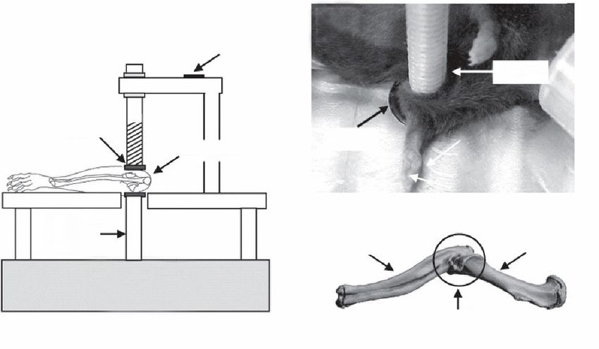
Page 2 of 17                                                                                                     Annals of Joint, 2021

damage has already occurred in the elbow (i.e., contracted           3−6× bodyweight (5,6,15,17,18). Overall, the elbow presents
elbow); warranting future work to understand early disease           itself as a complex and challenging joint to study in humans,
stages. It is challenging and often not feasible to study joint      as well as in other mammalian species.
conditions in humans, in part, because humans’ disease                  Given the complexity of the elbow joint, it is important
progresses relatively slowly and obtaining normal and                to highlight some of the similarities and differences of
diseased tissue is difficult. To overcome these limitations,         the elbow between humans and other commonly used
preclinical models (in vivo, in situ, ex vivo, in vitro) can serve   mammalian species for biomedical research (e.g., horses,
as valuable alternatives because of greater experimental             primates, dogs, rats, rabbits, and mice). Many mammals
control, faster disease progression, and increased access to         other than humans, whether bipedal or quadrupedal, have
tissue. Unfortunately, no preclinical model can recapitulate         elbows that are similar in anatomy to humans and can
every aspect of human elbow anatomy and pathology;                   perform the flexion-extension motion. However, elbows of
nonetheless, each model can provide valuable information             most mammals cannot pronate and supinate to the same
towards at least some understanding of elbow pathologies.            extent as human elbows do. A few exceptions include some
    Currently, only a handful of preclinical models have been        non-human primates and a specific breed of rats (i.e., Long-
used to investigate elbow injury and pathology, and as such          Evans) that can pronate and supinate over ranges that are
there are many opportunities for advances in this area. This         similar to humans (19,20). As a result, these species perhaps
review will discuss relevant considerations for using animal         are more clinically relevant to study the human elbow,
models to simulate human conditions, current preclinical             although other species may still be valuable for research
models of elbow diseases, then conclude by offering some             questions that are not as dependent on this type of motion.
ideas for potential future directions.                               Regardless, the selection of species and the joint is critical
                                                                     to consider when designing preclinical models to study the
                                                                     elbow’s normal physiology and pathophysiology.
Elbow anatomy in humans and other mammalian
species
                                                                     Current preclinical models of elbow injury and
The human elbow is one of the most complex and highly
                                                                     pathology
congruent joints, where three distinct bones (humerus, radius,
and ulna) join to form three articulating surfaces (1-8). These      In the following sections, common elbow injuries and
surfaces are surrounded by a joint capsule and numerous              pathologies and corresponding preclinical models that
ligaments (1-8), as well as several muscles (1,2,4,5,8,9).           have been used to recapitulate each disease are reviewed.
Intertwined among these tissues of the elbow is an extensive         Preclinical models considered and discussed herein were
network of nerves (4,8,10-13) and vasculature (14).                  included based upon the model’s relevance to pathologic
Collectively, these periarticular soft tissues allow for elbow       conditions of the elbow specifically; and, any therapeutic
stability and the motions of flexion-extension (normal               strategy using these preclinical models is not discussed.
range: ~0° to 150°; functional range: ~30° to 140°) and
pronation-supination (normal range: ~155°–175° total
                                                                     Trauma, immobilization, and post-traumatic contracture
motion between ~75°–85° pronation and ~80°–90°
                                                                     of the elbow
supination; functional range: ~100° total motion between
50° pronation and 50° supination) (1,2,5-8,15,16). The               Elbow trauma (e.g., dislocation and soft-tissue injury) and
elbow is vital to countless daily activities, such as opening a      subsequent complications (e.g., pain, joint immobilization,
door, typing, eating, personal hygiene, or using a telephone         contracture, stiffness, and arthritis) are some of the most
(5-7,15,16). The ability of the elbow to enable these                common elbow pathologies observed clinically (5,21-29).
activities is unlike other musculoskeletal joints (e.g., knee        Unfortunately, injuries are poorly tolerated in the elbow,
and hip) and is largely unique to humans, particularly in            leading to debilitating consequences for the patient (5,21-29).
how pronation and supination is utilized. During normal              The elbow can experience various types of injuries including
daily activities, the elbow experiences a wide range of              intra-articular fractures, soft tissue and ligament damage,
forces that largely originate from the periarticular tissues         as well as simple and complex dislocations (without or with
stabilizing the elbow (1,2,4-8,17). During heaving lifting           concurrent bony fracture, respectively) (5,21-29). Typically,
and dynamic loading, the elbow experiences stress as high as         the elbow is immobilized post-injury to stabilize the joint;

© Annals of Joint. All rights reserved.                                   Ann Joint 2021;6:12 | http://dx.doi.org/10.21037/aoj.2020.02.09
Annals of Joint, 2021 Page 3 of 17
Traumatic Joint Injury Immobilization
Intra-Articular
Patella Fracture
Femur Knee Immobilization
Cruciate in Flexion via K-wire
Ligaments
Fib
Menisci ula
Tibi Transection of Cruciate Ligaments
a
Plus Knee Hyperextension
Figure 1 Preclinical knee models of post-traumatic joint contracture. Schematic of knee models initially used in the mid-2000’s to study
post-traumatic joint contracture with respect to the elbow. In these rabbit and rat knee models, an invasive surgical procedure is undertaken
to cause an intra-articular fracture only, or in combination with transection of the cruciate ligaments and knee hyperextension, followed
by Kirschner wire (K-wire) placement for joint immobilization; this leads to permeant knee contracture and pathologic conditions seen in
contracted elbows clinically.
however, extended joint immobilization post-injury could traumatic joint contracture with respect to the elbow (41).
induced unwanted elbow contracture (23,26,28,30,31). In their preclinical model of joint contracture, a surgical
Generally, the most common elbow complaints following procedure was performed on the rabbit knee to induce
injury are increased elbow pain, stiffness, or limited range an intra-articular fracture combined with immobilization
of motion. via Kirschner wire (K-wires) (Figure 1). After 8 weeks of
A full picture of the post-injury response of elbow tissues immobilization, K-wires are removed, followed by a period
leading to elbow contracture/stiffness remains unclear. of remobilization (0,8,16, and 32 weeks), mimicking the
Some clinical evidence of pathological changes in the clinical treatment paradigm of elbow injuries of injury,
elbow synovial capsule suggests that the capsule becomes immobilization, and then remobilization. Using this
fibrotic and restricts the elbow range of motion (3,21,32,33). preclinical model, Hildebrand and colleagues identified a
Furthermore, in fibrosis of the elbow (21,33-35) and other reduced range of motion following immobilization (~30°)
joints (31,34,36), fibroblasts within the joint capsule have and remobilization (~25° at 8 weeks; ~10° at 16 and 32
been observed to take on a pro-fibrotic phenotype by weeks), as well as increased myofibroblasts, mast cells,
transitioning to myofibroblasts. Myofibroblasts are the neuropeptides, and pro-fibrotic factors in the capsule
effector cells that release pro-fibrotic factors and over- (41-45). Results from their work demonstrated that this
produce extracellular matrix, leading to capsule stiffness rabbit knee model could induce changes similarly seen in
and joint contracture. Indeed, studies have demonstrated contracted elbows of humans.
the presence of myofibroblasts and high levels of matrix Building upon the rabbit animal model developed by
metabolism in the contracted human elbow capsule Hildebrand and colleagues (41), Nesterenko and Abdel
(3,33-35,37-39). In contrast, at least one study did not et al. developed a similar yet more severe and permanent
observe increased number of myofibroblasts at chronic- joint contracture in the rabbit knee (46-48). In their
disease stages (40). This contradiction suggests that disease model, the cruciate ligaments are transected and the
stage and patient variability may complicate human findings; knee is hyperextended in addition to an intra-articular
preclinical models could allow for greater experimental fracture and K-wire immobilization (Figure 1). Indeed,
control to study myofibroblasts temporally. Generally, the their model produced more severe knee contracture and
primary goal of preclinical models recapitulating post- reduced range of motion at both the end of the 8 weeks
traumatic elbow contracture is to cause similar cellular (i.e., immobilization (~76°) and 16 weeks of remobilization
myofibroblasts) and tissue-level (i.e., capsular) changes and (~45°). Interestingly, Abdel et al. (47) observed a different
permanent loss of elbow joint motion post-trauma. pattern of myofibroblasts compared to Hildebrand et al.
Since the early 2000s, Hildebrand and colleagues (41), which may be related to differences in model severity.
have pioneered preclinical work in the context of post- Other groups have found similar findings using the more
© Annals of Joint. All rights reserved. Ann Joint 2021;6:12 | http://dx.doi.org/10.21037/aoj.2020.02.09Page 4 of 17 Annals of Joint, 2021
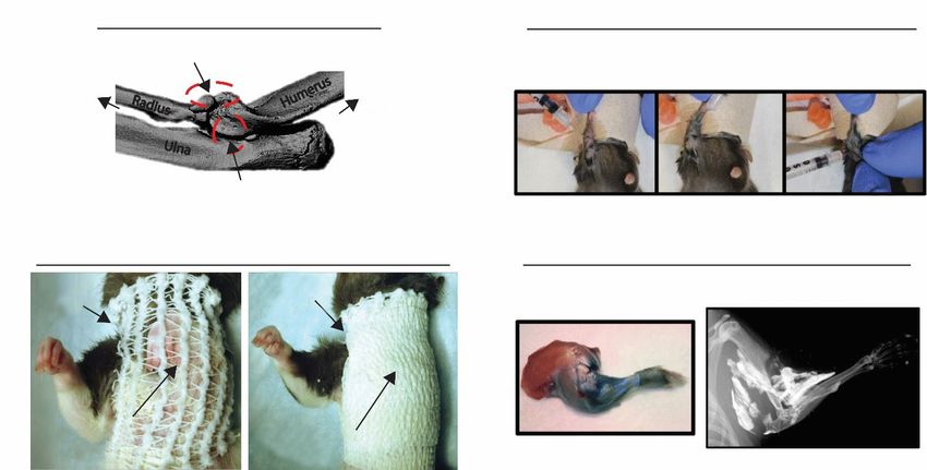
A Traumatic Joint Injury B Location of Toxin Injections Around the Elbow
Anterior Capsulotomy (i) (ii) (iii)
Extensors Flexors Proximal Forearm
er
Wr
ist ould
Sh
Lateral Collateral Ligament Transection
Immobilization Dispersion of Injected Solutions Around the EIbow
Netting Netting
Plus Blue Dye Barium Sulfate
Only Bandaging
(ii) (ii)
(i)
(i) (iii)
(iii)
Immobilized Immobilized
Forelimb Forelimb
Figure 2 Preclinical elbow models of post-traumatic joint contracture. (A) Micro-computed tomography reconstruction and animal images
highlight the soft-tissue injury and netting/bandages, respectively, to induce traumatic injury and immobilization in the rat elbow leading to
permanent elbow contracture. (B) Animal and radiographic images show the multiple injection locations of cardiotoxins around the mouse elbow.
These cardiotoxin injections, combined with researcher-induced plasminogen deficiency (not shown), leads to elbow contracture/stiffness. Image is
adapted from Moore-Lotridge et al., Journal of Experimental Orthopaedics, 2018, with permission (http://creativecommons.org/licenses/by/4.0/).
severe model by Nesterenko and Abdel et al. in the rat knee of IM and with a less severe loss after 6-weeks of FM in
(49-51). While knowledge obtained by these models has both flexion-extension (~43° and ~26°, respectively) and
been critical towards the understanding of post-traumatic pronation-supination (~40° and ~37°, respectively) motion
joint contracture, each of these studies utilized the knee (52-57). Ultimately, loss of motion caused permanent
joint. Knee models are useful towards understanding deficits in functional measures of grip strength and gait (58).
general concepts related to joint contracture; however, due We also confirmed the thickening of the capsule with
to anatomical and functional differences between the knee increased cellularity and presence of myofibroblasts, as well
and elbow, results may be limited in understanding aspects as altered matrix integrity and composition of both the
of elbow-specific injury and contracture. capsule and cartilage surfaces (52,53,55). Additionally, we
Nearly a decade after the initial work by Hildebrand determined that the major periarticular tissue contributors
et al. (41), our research group developed a preclinical model to contracture in our model were largely the anterior
of post-traumatic elbow contracture specific to the elbow capsule and lateral ligament complexes, not periarticular
(52,53). In our model, we utilized the Long-Evans rat, which muscles (56,57). Overall, our work has advanced the
has similar anatomy and functionality (flexion-extension field forward by creating the first animal model of joint
and pronation-supination movement) as the human elbow contracture using the elbow.
(19,20,52). To induce permeant post-traumatic elbow Soon after the development of our elbow-specific
contracture, we performed an anterior capsulotomy and model, Moore-Lotridge et al. created a mouse model
lateral collateral ligament transection, followed by 6 weeks of post-traumatic joint contracture (stiffness) of the
of immobilization (IM) using wrapping/bandage, and then elbow (59). In this model, injury to the periarticular
an additional 6 weeks of free mobilization (FM) (Figure 2A). soft-tissues (i.e., capsule ligaments, and surrounding
Our model caused a time-dependent reduction in the range musculature) of the elbow was induced by local
of motion of the elbow with a peak loss following 6-weeks injections of cardiotoxin combined with researcher-
© Annals of Joint. All rights reserved. Ann Joint 2021;6:12 | http://dx.doi.org/10.21037/aoj.2020.02.09Annals of Joint, 2021 Page 5 of 17
imposed plasminogen deficiency (Figure 2B). After the elbow, athletes such as pitchers, golfers, and tennis
28 days post-injury, this study showed a significant loss players are at exceptionally high risk (64-69). Elbow overuse
of elbow motion accompanied by an altered gait, muscle is often referred to as tennis or golfer elbow, but clinically
fibrosis and inflammation, thickening of the joint capsule, described as lateral and medial epicondylitis, respectively
and heterotopic ossification in the muscle (59). Given (64-69). While the terminology describing elbow overuse is
these findings, this mouse model of post-traumatic elbow still debated in the field, lateral epicondylitis is thought to
contracture replicates many of the same changes seen in occur in the absence of inflammation and resemble tendinosis
human contracted elbows. (64-69). Significant progress has been made clinically to
In addition to using an animal model of post-traumatic characterize the pathogenesis of elbow overuse, which has
joint contracture, Hildebrand and colleagues have explored identified important structural and cellular changes in these
other preclinical models of elbow contracture (60). Namely, tendons along with various neuropeptides thought to cause
they used an in vitro collagen gel contraction assay to pain (64-69).
mimic capsule contracture in the elbow (60). In this in vitro To date, only two preclinical models in the context of
model, Hildebrand et al. isolated primary capsule cells from elbow overuse exist. One study by Nakama and colleagues
contracted human elbow capsule and resuspended them in used a rabbit model to induce medial epicondylitis (71).
a collagen matrix with mast cells, another cell type thought In this model, the flexor digitorum profundus muscle,
to be involved in joint contracture (23,33,60,61). In these which transmits forces through a tendon that inserts into
studies, elbow capsule cells contracted the collagen gels, the medial epicondyle, was stimulated repeatedly for
which was enhanced with the addition of mast cells. In a set 80 hours a week for 14 weeks (71). Results from this study
of gel contraction studies by another group, Mattyasovszky demonstrated that the tendon at the insertion site of the
and colleagues found that inflammatory cytokines could medial epicondyle underwent significant increases in size,
modulate gel contraction of human capsule cells obtained as well as an increase in the number and size of tears within
from elbows undergoing arthroplasty (62,63). Results of these the tendon (71).
collagen gel contraction models have shown that capsule Another group developed a rat model to explore
cells from contracted human elbows are contractile and can repetitive, upper-extremity tasks required for reaching and
be modulated by mast cells and inflammatory cytokines to grabbing with the wrist, elbow, and shoulder (72). In this
influence the contraction of in vitro tissue analogs. model, rats repeatedly elevated their shoulder, fully extended
Overall, this section highlighted the progression of their elbow, and gripped with their wrist to obtain food
various preclinical models for post-traumatic elbow on cue, which caused repetitive muscular activation (72).
contracture. While each model uses different species, joints, While this group’s primary focus was on changes to the
injuries, and outcomes, they all each provided insights wrist, they did evaluate the elbow in this model and found
into the etiology and pathology of post-traumatic elbow that the elbow was unaffected (72).
contracture. Future work is necessary to fully elucidate the Each of the aforementioned preclinical models of
contributions of each elbow tissue, create new models to elbow overuse has provided preliminary insights into the
address other types of traumatic injuries, and determine etiology and pathogenesis of elbow overuse/epicondylitis/
the translatability of each preclinical model attempting to tendinopathy. Future investigations utilizing these
recapitulate the human condition. established, as well as novel, preclinical models are
warranted to elucidate the mechanisms involved fully with
elbow overuse. Perhaps other preclinical models previously
Elbow overuse
used to examine degenerative changes in other tendons,
Another common elbow pathology is elbow overuse, such as the Achilles or the rotator cuff in the shoulder, could
where repetitive use of the elbow can cause overstrain and provide insights to assist the study of elbow related tendon
microtrauma of ligaments around the elbow (64-66). Signs issues (73-75).
of elbow overuse are increased pain, point tenderness, and
difficulty in grasping items. Elbow overuse is common in
Elbow arthritis
individuals whose jobs and household tasks are repetitive and
require the elbow and other motions involving extensive wrist Arthritis occurs in the elbow and can take the form of either
use (65-70). While many individuals are at risk for overusing idiopathic osteoarthritis (OA), post-traumatic osteoarthritis
© Annals of Joint. All rights reserved. Ann Joint 2021;6:12 | http://dx.doi.org/10.21037/aoj.2020.02.09Page 6 of 17 Annals of Joint, 2021
A B
Strain
gauge
Stator
Stator
Elbow
Elbow
Loading
C
rod Ulna Humerus
Piezoelectric
actuator
Loading
Site
Figure 3 Preclinical elbow model to study cartilage metabolism and osteoarthritis. Schematic of the experimental setup (A), animal image (B),
and micro-computed tomography reconstruction (C) highlight the loading location in a mouse elbow used to study cartilage metabolism and
osteoarthritis. Image is reprinted from Sun et al., Connective Tissue Research, 2012, with permission from the publisher (Taylor & Francis Ltd,
http://www.tandfonline.com).
(PTOA), and rheumatoid arthritis (RA). All three types A few preclinical models exist addressing elbow arthritis.
of arthritis ultimately lead to cartilage degeneration, pain, In the context of idiopathic OA, preclinical studies have
loss of joint function, and a need for arthroplasty. While evaluated cartilage damage, joint space narrowing, gait, and
OA in the elbow is rare (~2% of all joints with OA and of lameness in pig (93), cats (94-97), and dogs (98-106) with
patients with elbow arthritis), the etiology and pathogenesis signs of elbow OA. Although informative, these studies
remain unclear (17,18,76-83). There are some risk factors for recruited animals with naturally occurring OA and were
OA such as aging and overuse. A more common risk factor unable to control the onset of degenerative changes. An
for developing elbow OA is a traumatic joint injury, which alternative model with more experimental control is the
accelerates the development of OA; referred to as PTOA, mouse elbow/ulna loading models, which have historically
this condition is also relatively uncommon in the elbow been used to study the bone response of the elbow/ulna
(17,18,76,77,79,84-86). Despite the rarity, OA and PTOA under controlled loading rate and magnitude (107,108).
in the elbow are debilitating for patients once the disease Recently, an elbow loading model was used to evaluate
initiates and progresses. Although reporting on prognosis is cartilage metabolism in the proximal ulna and distal
mixed, treated intra-articular elbow fractures can lead to at humerus following mechanical stimuli (Figure 3) (109).
least a 50% chance of developing PTOA as early as 10 years Additional studies, however, are needed to validate the OA-
post-injury (17,84), which are similar odds in other joints like phenotype of the elbow in this model. In the context
such as the knee and ankle (86,87). This high occurrence is of PTOA, Dunham et al. minimally evaluated changes in
concerning since elbow trauma frequently occurs in younger the cellularity and matrix integrity of the cartilage (55).
(28,29,79,85,88) and active individuals including athletes No preclinical models currently exist to study RA in the
(29,79) and military service members (89,90). Besides OA elbow. Although limited effort has been made to study
and PTOA, the next most common form of arthritis affecting elbow arthritis using preclinical models, the etiology and
the elbow is RA, inflammatory-driven arthritis (17,76,91). RA pathogenesis of elbow arthritis remain unclear.
isolated to the elbow alone occurs in ~5% of patients with RA Some knowledge about elbow arthritis and guidance
(92); however, ~20–50% of patients with RA in other joints on preclinical models could be inferred from arthritis in
or organs also have signs RA in the elbow (76,92). other joints. For instance, countless preclinical models have
© Annals of Joint. All rights reserved. Ann Joint 2021;6:12 | http://dx.doi.org/10.21037/aoj.2020.02.09Annals of Joint, 2021 Page 7 of 17
been created to study arthritis in the knee, including OA to form along the joint margins of the ulna, radius, and
(110-112), PTOA (110,112-114), and RA (115-118). Most humerus (79,85,127-129). The formation of osteophytes
commonly, in vivo models of OA and PTOA include the use ultimately causes significant pain and reduces the joint
of chemicals, genetic manipulation, aging, or surgically and space, limiting the elbow range of motion (79,85,127-129).
non-surgically induced traumatic injuries, while models of In addition to osteophyte formations, HO in the elbow has
RA include injections of compounds inducing an immune recently gained attention in the literature (27,130-133). HO
response. To study interactions between various tissues of is an aberrant bone formation that can contribute to elbow
the joint that are otherwise challenging in vivo, researchers pain and reduction in elbow range of motion. Although
have turned to ex vivo and in vitro preclinical models of osteophytes and HO are prevalent in diseased elbows, how
arthritis (112,119). While these established models of osteophytes and HO form and what their full role in various
arthritis in other joints can provide insights for models elbow pathologies remains unknown.
of elbow arthritis, researchers should be aware that the Unfortunately, the preclinical evaluation of HO and
cartilage structure and cellularity is different between joints, osteophytes in the elbow has been limited. To date, only one
potentially causing differences in cartilage response to preclinical study has addressed the presence of HO in elbow
arthritic stressors (120). contracture/stiffness (Figure 4A) (59), and only a few have
evaluated osteophytes in elbow osteoarthritis (Figure 4B)
(96,97,100-103,105). To study osteophytes and HO in
Post-burn elbow contracture
the elbow, researchers can study the current preclinical
Post-burn contracture is defined as a contracture of the models of elbow pathologies mentioned above or other
skin or joint following a significant burn. Contracture can preclinical models that may be appropriate. For example,
develop following a burn in many parts of the body, including researchers have performed intra-articular injection of pro-
the knee (121) and non-musculoskeletal tissues (122); fibrotic factors into mouse knees, causing the formation of
however, the elbow is one of the most commonly affected osteophytes (134). Although these pro-fibrotic factors can
joints (123-125). While the skin and other soft tissues may induce osteophytes in the knee, it is not known whether
play a role in post-burn contracture, the pathogenesis and the same effects would be seen in the elbow. Overall, a
scope of tissues involved in this condition in the elbow need exists for preclinical models to investigate HO and
remain unclear. One of the most common symptoms that osteophytes in all elbow pathologies.
occur following burns is the formation of heterotopic
ossifications (see next section), which is thought to reduce
Osteochondritis dissecans
the range of motion in the elbow (123-125).
Current preclinical models of post-burn injury include Another common clinical elbow pathology is
the use of various species such as mice and rats; however, osteochondritis dissecans (OCD) (17,135-137). OCD is the
no model has been used to address post-burn elbow process by which the articular cartilage separates from the
contracture (126). Caution must be taken when choosing underlying subchondral bone, leading to fragmentations
a species to study post-burn elbow contracture, partly of cartilage in the joint space, pain, and a reduced range
because the structure of the skin and the post-burn severity of motion in the elbow (17,29,135-137). In the elbow,
and response in other species may not translate to humans OCD is typically observed in young, over-head throwing
(122,126). In the future, the development of preclinical athletes at the humeral capitellum (17,29,135-137).
models will be necessary to shed insights into the etiology The etiology of OCD formation and its role in elbow
and pathogenesis of post-burn elbow contracture. pathologies remains to be elucidated (17,29,135,136).
It has been suggested that genetics, repetitive injury,
subchondral bone abnormalities (e.g., loss of vasculature and
Osteophyte formation and heterotopic ossification in elbow
becoming necrotic), and excessive force applied to the cartilage
pathologies
potentially contribute to OCD symptoms (17,29,135,136).
Among many of the elbow pathologies clinically, there is Regardless of the etiology, OCD may make the surrounding
the concurrent formation of osteophytes and heterotopic cartilage prone to further degeneration, leading to arthritis (17).
ossification (HO). Osteophytes are defined as bony Unfortunately, no preclinical study has investigated the
outgrowths or spurs that form; in the elbow, these tend etiology or pathogenesis of OCD in the elbow. OCD has
© Annals of Joint. All rights reserved. Ann Joint 2021;6:12 | http://dx.doi.org/10.21037/aoj.2020.02.09Page 8 of 17 Annals of Joint, 2021
A Healthy Elbow Contracted/Stiff Elbow B Healthy Elbow Arthritic Elbow
Mediolateral flexed view
Shoulder
Elbow
Craniocaudal view
Wrist/Hand
Figure 4 Preclinical models evaluating heterotopic ossification and osteophytes in elbow. (A) Micro-computed tomography reconstruction
demonstrates severe heterotopic ossification (white arrows) following a soft-tissue injury to induce elbow contracture/stiffness in mice.
Image is adapted from Moore-Lotridge et al., Journal of Experimental Orthopaedics, 2018, with permission (http://creativecommons.org/
licenses/by/4.0/). (B) Radiographic images highlight an arthritic elbow with osteophytes (white arrows) accompanying naturally occurring
osteoarthritis in dogs. Image is adapted from Hurlbeck et al., Research in Veterinary Science, 2014, with permission from Elsevier.
been researched in preclinical models of other joints, such (4,8,10-13). While the nerve supply for each elbow tissue
as the knee (138-140). However, it should be cautioned that is different, neuroinflammatory pathways, which involve
the etiology and pathogenesis of OCD in other joints could neuropeptides (substance-P) and mast cells (23,33,42,61),
be different than in the elbow, partly because of differences may play a role in elbow pain. It remains to be determined
in biomechanical forces experienced (e.g., weight vs. non- if other inflammatory and nerve cells are also involved
weight bearing), structure, and cellularity of different joint’s with neuroinflammatory pathways. In addition to sensing
cartilage (120,141). To investigate OCD in the elbow, pain, nerves of the elbow are prone to complications such
preclinical models could be non-invasive, repeated motion as nerve entrapment and neuropathy, particularly after an
overuse models; however, these models don’t exist for the injury, capsulectomy, or arthroplasty (22,142-145).
elbow. Given that the etiology and pathogenesis of OCD in To date, no preclinical models of elbow pathologies
the elbow is unknown, current and novel preclinical models exist that have directly evaluated pain and innervation. The
of the elbow, especially trauma and overuse models, may most relevant preclinical studies addressing any degree of
note that OCD lesions develop. pain and innervation are those preclinical studies that have
indirectly assessed the role of innervation through studying
neuropeptides (substance-P) and mast cells in post-
Elbow pain and innervation
traumatic elbow contracture (23,33,42,61). However, a few
Most elbow pathologies are associated with some degree of studies have begun to directly address elbow innervation
pain; however, the pathomechanisms causing pain are still under non-pathological conditions in monkey (146) and
up for debate in the elbow and have received little attention. rat elbows (147,148). Thus, future work is warranted to
One potential source of pain could arise from the extensive understand the etiology and pathogenesis of pain and
network of nerves and nerve endings throughout the elbow innervation in every elbow pathology.
© Annals of Joint. All rights reserved. Ann Joint 2021;6:12 | http://dx.doi.org/10.21037/aoj.2020.02.09Annals of Joint, 2021 Page 9 of 17
Future considerations for preclinical models of to follow the recommended guidelines of animal research:
elbow diseases reduction, refinement, and replacement (149).
Ex vivo and in vitro models can serve as alternatives
This review has demonstrated and summarized in Table 1
to in vivo and in situ models of elbow pathologies and be
that only a limited number of preclinical models exist to
used to achieve many samples while minimizing the need
study elbow diseases. Future studies are warranted to refine
for many animals. Furthermore, ex vivo and in vitro allow
and create new preclinical models to elucidate the etiology
the following: (I) more control of the experimental design
and pathogenesis of common elbow pathologies. Research
and variables involved; (II) the ability to isolate and study
groups interested in pursuing future work in elbow
the interaction of certain experimental variables between
pathologies might consider looking to other joints with
tissues/cells; and (III) the introduction of endogenous
more mature research results to gain valuable guidance for
factors (e.g., growth factors and inflammatory cytokines) in
developing new preclinical models of elbow pathologies. To
a controlled manner. However, a downside to both ex vivo
help facilitate the implementation of novel preclinical elbow
and in vitro models is that the tissues/cells are removed from
models, the following section will highlight some critical the body, and thus, results may not necessarily translate to
points to consider and will provide some suggestions for the in vivo condition. Nonetheless, all models have positive
appropriate next steps. and negative aspects and can each help provide insight into
the etiology and pathogenesis of elbow conditions.
Considerations for refining and developing preclinical
models of elbow pathologies Considerations for experimental outcomes and model
An important consideration when refining or developing a variables
preclinical model for the elbow is to specify which elbow An essential aspect of any elbow preclinical model is the
pathology will be studied. As highlighted previously, experimental outcomes used to assess a model's ability
each elbow pathology involves different tissues and cells, to mimic aspects of elbow pathologies. Importantly, it
as well as having a different time scales of etiology and is critical for these outcome measures to be well defined
pathogenesis; thus, a “gold-standard” for preclinical models and in-depth enough to understand the disease process,
would be to closely mimic the clinical observations of the but also universal enough and not too specialized, such
specific elbow condition under investigation. that outcome measures can be compared across research
After deciding an elbow pathology to study, the next laboratories. Outcome measures can range from the
likely consideration is which type of preclinical model to molecular and cellular level to the tissue and functional
use: in vivo, in situ, ex vivo, or in vitro model systems. Each level; such a multi-level approach will allow for easier
type of model has important trade-offs worth considering. translation to the clinic. Preclinical studies should
For example, in vivo and in situ models allow for the study continue to assess these outcome measures but implement
and interaction of all tissues in the body, including the full additional approaches that have not been applied to
biological response, enabling for the closest translation of the elbow. For instance, non-invasive imaging, such as
findings to the human disease. A critical consideration when micro-computed tomography, magnetic resonance, and
choosing an in vivo and in situ model is how closely the ultrasound techniques, could be used to assess structural
anatomy and functionality of the animal of choice mimic and compositional changes in the elbow. Additionally,
the human elbow. Additionally, another consideration is local and systemic biochemical outcome measures could
whether to use an invasive or non-invasive approach to be defined, such as the establishment of synovial fluid and
induce a traumatic elbow injury. An invasive model requires serum biomarkers of elbow pathology.
a surgical incision that may induce an unwanted and A limitation of this overview relates to the obvious
confounding biological response that would otherwise not challenge to discuss every type of elbow pathology and
be present in the human; thus, non-invasive animal models potential confounding variables in detail. The effects of
would be more clinically relevant. While in vivo models are species sex, age, and other conditions, such as diabetes, joint
more representative of the human situation, they require dysplasia, changes to the microbiome, and sedentary versus
the use of animals, which are accompanied by ethical active lifestyle, could impact the progression of elbow
concerns and other requirements (149). Thus, it is critical pathologies and are worth considering. Furthermore, it’s
© Annals of Joint. All rights reserved. Ann Joint 2021;6:12 | http://dx.doi.org/10.21037/aoj.2020.02.09Page 10 of 17 Annals of Joint, 2021
Table 1 Summary of major clinical pathologies in the elbow joint and current preclinical models
Clinical elbow Preclinical Species and joint Outcome
Model description Key references
pathology model type studied measurements
Trauma, In vivo Surgically induced fracture plus New Zealand Range of motion, Hildebrand et al. 2004
immobilization, immobilization via K-Wires for White female histology, (41), 2004 (45), 2006 (43),
and post- 8 weeks; removal of K-Wires rabbit knee immunohistochemistry, 2008 (42,44)
traumatic followed by 0, 8, 16 or 32 weeks of gene expression, protein
contracture remobilization expression
In vivo Surgically induced fracture and knee New Zealand Range of motion, Nesterenko et al.
hyperextension plus immobilization White female histology, 2009 (46); Abdel et al.
via K-Wires for 8 weeks; removal of rabbit knee immunohistochemistry, 2012 (47,48)
K-Wires followed by 0 or 16 weeks gene expression
of remobilization
In vivo Surgically induced fracture Lewis female rat Range of motion, Li et al. 2013 (49);
and knee hyperextension plus knee histology, Sun et al. 2016 (50)
immobilization via sutures for immunohistochemistry,
8 weeks protein expression
In vivo Surgically induced fracture and knee Sprague Dawley Range of motion Baranowski et al.
hyperextension plus immobilization male rat knee 2018 (51)
via K-Wires for 4 weeks; removal
of K-Wires followed by 8 weeks of
remobilization
In vivo Surgically induced anterior Long-Evans male Range of motion, grip Lake et al. 2016 (52);
capsulotomy and lateral collateral rat elbow strength, gait, histology, Dunham et al.
ligament transection plus immunohistochemistry, 2017 (53,54), 2018
immobilization via wrapping/ gene expression (55,56), 2019 (57); Reiter
bandage for 6 weeks; removal of et al. 2019 (58)
wrapping/bandage followed by
addition 6 weeks of remobilization
In vivo Local injections of cardiotoxin C57BL/6 male Range of motion, grip Moore-Lotridge et al.
around the elbow combined with mouse elbow strength, gait, histology, 2018 (59)
researcher-imposed plasminogen immunohistochemistry,
deficiency micro-computed
tomography
In vitro Encapsulation of isolated capsule Contracted Gel contraction Hildebrand et al.
cells with and without mast cells human elbow 2014 (60)
into collagen matrix to allow for capsule cells
collagen gel contraction with and without
human mast cell
line
In vitro Encapsulation of isolated capsule Human elbow Immunohistochemistry, Mattyasovszky et al.
cells with and without inflammatory capsule cells cell viability, collagen 2017 (62,63)
cytokines into collagen matrix to from elbow gel contraction, gene
allow for collagen gel contraction arthroplasty expression
Overuse In vivo Forearm muscle stimulation to New Zealand Histology Nakama et al. 2005 (71)
induce tendinopathy/epicondylitis White female
at elbow medial epicondyle rabbit forearm
In vivo Repetitive movement of shoulder, Sprague Dawley Cytokine array Driban et al. 2011 (72)
elbow, and wrists to reach food female rat forearm
Table 1 (continued)
© Annals of Joint. All rights reserved. Ann Joint 2021;6:12 | http://dx.doi.org/10.21037/aoj.2020.02.09Annals of Joint, 2021 Page 11 of 17
Table 1 (continued)
Clinical elbow Preclinical Species and joint Outcome
Model description Key references
pathology model type studied measurements
Idiopathic In vivo Naturally occurring osteoarthritis Pig, cat, and dog Gait, histology, Pig: Kirk et al. 2008 (93)
osteoarthritis elbow immunohistochemistry,
Cat: Freire et al. 2014 (94)
gene expression,
and 2011 (96), Ryan et al.
radiography, pain,
2013 (95), Lascelles et al.
cytokine array
2010 (97)
Dog: Kapatkin
et al. 2006 (98), Spahni
et al. 2009 (99), Clements
et al. 2009 (100), Alves
et al. 2017 (101),
Hurlbeck et al. 2014
(102), Goldhammer et al.
2010 (103), Kunst
et al. 2014 (105),
Bockstahler et al.
2009 (106)
In vivo Elbow loading via indentation on C57BL/6 female Gene expression, Sun et al. 2012 (109)
proximal ulna and distal humerus mouse elbow enzyme activity
Post-traumatic In vivo Surgically induced anterior Long-Evans male Histology Dunham et al. 2018 (55)
osteoarthritis capsulotomy and lateral collateral rat elbow
ligament transection plus
immobilization via wrapping/
bandage for 6 weeks; removal of
wrapping/bandage followed by
addition 6 weeks of remobilization
Osteophytes In vivo Naturally occurring osteoarthritis Cat and dog Micro-computed Cat: Freire et al. 2011 (96),
elbow tomography Lascelles et al. 2010 (97)
Dog: Clements et al.
2009 (100), Alves et al.
2017 (101), Hurlbeck et al.
2014 (102), Goldhammer
et al. 2010 (103), Kunst
et al. 2014 (105)
Heterotopic In vivo Local injections of cardiotoxin around C57BL/6 male Micro-computed Moore-Lotridge et al.
ossification the elbow combined with researcher- mouse elbow tomography and 2018 (59)
imposed plasminogen deficiency histology
Rheumatoid N/A N/A N/A N/A N/A
arthritis
Post-burn N/A N/A N/A N/A N/A
contracture
Osteochondritis N/A N/A N/A N/A N/A
dissecans
Pain and In vivo Surgically induced fracture plus New Zealand Immunohistochemistry Hildebrand et al.
innervation immobilization via K-Wires for White female 2008 (42)
8 weeks; removal of K-Wires rabbit knee
followed by 0, 8, 16 or 32 weeks of
remobilization
© Annals of Joint. All rights reserved. Ann Joint 2021;6:12 | http://dx.doi.org/10.21037/aoj.2020.02.09Page 12 of 17 Annals of Joint, 2021
worthwhile to consider clinical complications with post- of Health, during the conduct of the study; AMC reports
surgical treatments, implants, devices, and infection. grants from Zimmer, grants from National Institute of
Health, personal fees from DePuy, personal fees from
Arthrex, personal fees from Wright Medical, outside the
Conclusion
submitted work; SP served as the unpaid Guest Editor of
The human elbow is one of the most complicated the series and reports grants from National Institutes of
musculoskeletal joints and critical for many daily activities Health, during the conduct of the study. The authors have
that require the upper extremity; yet, to date, the elbow no other conflicts of interest to declare.
is a relatively understudied musculoskeletal joint. Due to
its complex nature, the elbow has high susceptibility to Ethical Statement: The authors are accountable for all
injury and pathologies, such as joint contracture, stiffness, aspects of the work in ensuring that questions related
and arthritis. Unfortunately, limited therapies exist to treat to the accuracy or integrity of any part of the work are
elbow pathologic conditions, which only offer limited appropriately investigated and resolved.
clinical success. To develop alternative therapeutic strategies
for elbow conditions, it is essential to understand the Open Access Statement: This is an Open Access article
etiology and pathogenesis of common elbow pathologies distributed in accordance with the Creative Commons
better; preclinical models serve as ideal alternatives to Attribution-NonCommercial-NoDerivs 4.0 International
pursue such topics. This review has highlighted major License (CC BY-NC-ND 4.0), which permits the non-
clinical elbow diseases and the preclinical models currently commercial replication and distribution of the article with
available to recapitulate these diseases, while also providing the strict proviso that no changes or edits are made and the
recommendations for future preclinical models. Overall, this original work is properly cited (including links to both the
review could serve as the foundation for preclinical models formal publication through the relevant DOI and the license).
to study the etiology and pathogenesis of elbow pathologies See: https://creativecommons.org/licenses/by-nc-nd/4.0/.
with the goal of better understanding elbow function and
joint health, and for developing therapeutic intervention
References
strategies to improve treatment of elbow conditions.
1. King GJW, Morrey BF, An KN. Stabilizers of the elbow. J
Shoulder Elbow Surg 1993;2:165-74.
Acknowledgments
2 Safran MR, Baillargeon D. Soft-tissue stabilizers of the
Funding: This review was supported by the National elbow. J Shoulder Elbow Surg 2005;14:179S-85S.
Institute of Arthritis and Musculoskeletal and Skin Diseases 3 Cohen MS, Schimmel DR, Masuda K, et al. Structural and
of the National Institutes of Health under award number biochemical evaluation of the elbow capsule after trauma. J
R01AR071444. Shoulder Elbow Surg 2007;16:484-90.
4 Martin S, Sanchez E. Anatomy and biomechanics of the
elbow joint. Semin Musculoskelet Radiol 2013;17:429-36.
Footnote
5 Chalmers PN, Chamberlain AM. Biomechanics of the
Provenance and Peer Review: This article was commissioned Elbow. Unstable Elb., Cham: Springer International
by the Guest Editor (Spencer Lake) for the series “Emerging Publishing, 2017:13-26.
Trends in Elbow Injury, Pathology and Treatment” 6 LaStayo PC, Lee MJ. The Forearm Complex: Anatomy,
published in Annals of Joint. The article has undergone Biomechanics and Clinical Considerations. J Hand Ther
external peer review. 2006;19:137-44.
7 Kapandji A. Biomechanics of pronation and supination of
Conflicts of Interest: All authors have completed the ICMJE the forearm. Hand Clin 2001;17:111-22, vii.
uniform disclosure form (available at http://dx.doi. 8 Alcid JG, Ahmad CS, Lee TQ. Elbow anatomy and
org/10.21037/aoj.2020.02.09). The series “Emerging structural biomechanics. Clin Sports Med 2004;23:503-17.
Trends in Elbow Injury, Pathology and Treatment” was 9 An KN, Hui FC, Morrey BF, et al. Muscles across the elbow
commissioned by the editorial office without any funding or joint: A biomechanical analysis. J Biomech 1981;14:659-
sponsorship. MAD reports grants from National Institutes 61, 663-9.
© Annals of Joint. All rights reserved. Ann Joint 2021;6:12 | http://dx.doi.org/10.21037/aoj.2020.02.09Annals of Joint, 2021 Page 13 of 17 10 Gardner E. The innervation of the elbow joint. Anat Rec traumatic injury. J Shoulder Elbow Surg 2011;20:39-44. 1948;102:161-74. 25 Englert C, Zellner J, Koller M, et al. Elbow Dislocations: 11 Bekler H, Riansuwan K, Vroeman JC, et al. Innervation of A Review Ranging from Soft Tissue Injuries to the Elbow Joint and Surgical Perspectives of Denervation: Complex Elbow Fracture Dislocations. Adv Orthop A Cadaveric Anatomic Study. J Hand Surg Am 2013;2013:951397. 2008;33:740-5. 26 Lindenhovius ALC, Jupiter JB. The Posttraumatic Stiff 12 Cavalheiro CS, Filho MR, Rozas J, et al. Anatomical study Elbow: A Review of the Literature. J Hand Surg Am on the innervation of the elbow capsule. Rev Bras Ortop 2007;32:1605-23. 2015;50:673-9. 27 Bruno RJ, Lee ML, Strauch RJ, et al. Posttraumatic elbow 13 Nourbakhsh A, Hirschfeld AG, Schlatterer DR, et al. stiffness: evaluation and management. J Am Acad Orthop Innervation of the elbow joint: A cadaveric study. J Hand Surg 2002;10:106-16. Surg Am 2016;41:85-90. 28 Lu X, Yan G, Lu M, et al. Epidemiologic features 14 Yamaguchi K, Sweet FA, Bindra R, et al. The Extraosseous and management of elbow dislocation with associated and Intraosseous Arterial Anatomy of the Adult Elbow. J fracture in pediatric population. Medicine (Baltimore) Bone Joint Surg Am 1997;79:1653-62. 2017;96:e8595. 15 Fornalski S, Gupta R, Lee TQ. Anatomy and 29 Andelman S, DiPrinzio E, Hausman M. Elbow injuries in Biomechanics of the Elbow Joint. Tech Hand Up Extrem the pediatric athlete. Ann Joint 2018;3:21. Surg 2003;7:168-78. 30 Schippinger G, Seibert FJ, Steinböck J, et al. Management 16 Morrey BF, Askew LJ, Chao EY. A biomechanical study of simple elbow dislocations: Does the period of of normal functional elbow motion. J Bone Joint Surg Am immobilization affect the eventual results? Langenbecks 1981;63:872-7. Arch Surg 1999;384:294-7. 17 Heijink A, Vanhees M, van den Ende K, et al. 31 Wong K, Trudel G, Laneuville O. Noninflammatory Joint Biomechanical considerations in the pathogenesis of Contractures Arising from Immobility: Animal Models to osteoarthritis of the elbow. Knee Surg Sports Traumatol Future Treatments. Biomed Res Int 2015;2015:848290. Arthrosc 2016;24:2313-8. 32 Gallay SH, Richards RR, O’Driscoll SW. Intraarticular 18 Kroonen LT, Piper SL, Ghatan AC. Arthroscopic capacity and compliance of stiff and normal elbows. Management of Elbow Osteoarthritis. J Hand Surg Am Arthroscopy 1993;9:9-13. 2017;42:640-50. 33 Hildebrand KA. Posttraumatic Elbow Joint Contractures: 19 Whishaw IQ, Gorny B, Foroud A, et al. Long-Evans and Defining Pathologic Capsular Mechanisms and Sprague-Dawley rats have similar skilled reaching success Potential Future Treatment Paradigms. J Hand Surg Am and limb representations in motor cortex but different 2013;38:2227-33. movements: some cautionary insights into the selection of 34 Mattyasovszky SG, Hofmann A, Brochhausen C, et al. The rat strains for neurobiological motor research. Behav Brain effect of the pro-inflammatory cytokine tumor necrosis Res 2003;145:221-32. factor-alpha on human joint capsule myofibroblasts. 20 Sacrey L-AR, Alaverdashvili M, Whishaw IQ. Similar Arthritis Res Ther 2010;12:R4. hand shaping in reaching-for-food (skilled reaching) in 35 Germscheid NM, Hildebrand KA. Regional Variation Is rats and humans provides evidence of homology in release, Present in Elbow Capsules after Injury. Clin Orthop Relat collection, and manipulation movements. Behav Brain Res Res 2006;450:219-24. 2009;204:153-61. 36 Usher KM, Zhu S, Mavropalias G, et al. Pathological 21 Charalambous CP, Morrey BF. Posttraumatic Elbow mechanisms and therapeutic outlooks for arthrofibrosis. Stiffness. J Bone Joint Surg Am 2012;94:1428-37. Bone Res 2019;7:9. 22 Mellema JJ, Lindenhovius ALC, Jupiter JB. The 37 Hildebrand KA, Zhang M, van Snellenberg W, et al. posttraumatic stiff elbow: an update. Curr Rev Myofibroblast Numbers are Elevated in Human Elbow. Musculoskelet Med 2016;9:190-8. Clin Orthop Relat Res 2004;(419):189-97. 23 Monument MJ, Hart DA, Salo PT, et al. Posttraumatic 38 Hildebrand KA, Zhang M, Hart DA. High Rate of Joint elbow contractures: targeting neuroinflammatory Capsule Matrix Turnover in Chronic Human Elbow fibrogenic mechanisms. J Orthop Sci 2013;18:869-77. Contractures. Clin Orthop Relat Res 2005;439:228-34. 24 Myden C, Hildebrand K. Elbow joint contracture after 39 Hildebrand KA, Zhang M, Hart DA. Myofibroblast © Annals of Joint. All rights reserved. Ann Joint 2021;6:12 | http://dx.doi.org/10.21037/aoj.2020.02.09
Page 14 of 17                                                                                                    Annals of Joint, 2021

     Upregulators are Elevated in Joint Capsules in                  52 Lake SP, Castile RM, Borinsky S, et al. Development
     Posttraumatic Contractures. Clin Orthop Relat Res                  and use of an animal model to study post-traumatic
     2007;456:85-91.                                                    stiffness and contracture of the elbow. J Orthop Res
40   Doornberg JN, Bosse T, Cohen MS, et al. Temporary                  2016;34:354-64.
     Presence of Myofibroblasts in Human Elbow Capsule               53 Dunham CL, Castile RM, Havlioglu N, et al. Persistent
     After Trauma. J Bone Joint Surg Am 2014;96:e36.                    motion loss after free joint mobilization in a rat model of
41   Hildebrand KA, Sutherland C, Zhang M. Rabbit knee                  post-traumatic elbow contracture. J Shoulder Elbow Surg
     model of post-traumatic joint contractures: The long-              2017;26:611-8.
     term natural history of motion loss and myofibroblasts. J       54 Dunham CL, Castile RM, Chamberlain AM, et al.
     Orthop Res 2004;22:313-20.                                         Pronation-Supination Motion Is Altered in a Rat Model
42   Hildebrand KA, Zhang M, Hart DA. Joint capsule                     of Post-Traumatic Elbow Contracture. J Biomech Eng
     mast cells and neuropeptides are increased within four             2017;139:1-7.
     weeks of injury and remain elevated in chronic stages of        55 Dunham CL, Castile RM, Havlioglu N, et al. Temporal
     posttraumatic contractures. J Orthop Res 2008;26:1313-9.           Patterns of Motion in Flexion-extension and Pronation-
43   Hildebrand KA, Zhang M, Hart DA. Joint capsule matrix              supination in a Rat Model of Posttraumatic Elbow
     turnover in a rabbit model of chronic joint contractures:          Contracture. Clin Orthop Relat Res 2018;476:1878-89.
     Correlation with human contractures. J Orthop Res               56 Dunham CL, Chamberlain AM, Meyer GA, et al. Muscle
     2006;24:1036-43.                                                   does not drive persistent posttraumatic elbow contracture
44   Hildebrand KA, Zhang M, Germscheid NM, et al.                      in a rat model. Muscle and Nerve 2018;58:843-51.
     Cellular, matrix, and growth factor components of the joint     57 Dunham CL, Castile RM, Chamberlain AM, et al. The
     capsule are modified early in the process of posttraumatic         Role of Periarticular Soft Tissues in Persistent Motion
     contracture formation in a rabbit model. Acta Orthop               Loss in a Rat Model of Posttraumatic Elbow Contracture.
     2008;79:116-25.                                                    J Bone Joint Surg Am 2019;101:e17.
45   Hildebrand KA, Holmberg M, Shrive N. A New Method               58 Reiter A, Kivitz GJ, Castile RM, et al. Functional Measures
     to Measure Post-Traumatic Joint Contractures in the                of Grip Strength and Gait Remain Altered Long-Term
     Rabbit Knee. J Biomech Eng 2003;125:887.                           in a Rat Model of Post-Traumatic Elbow Contracture. J
46   Nesterenko S, Morrey ME, Abdel MP, et al. New rabbit               Biomech Eng 2019;141:071001.
     knee model of posttraumatic joint contracture: Indirect         59 Moore-Lotridge SN, Oelsner WK, Ihejirika Y, et al.
     capsular damage induces a severe contracture. J Orthop             Novel preclinical murine model of trauma-induced elbow
     Res 2009;27:1028-32.                                               stiffness. J Exp Orthop 2018;5:36.
47   Abdel MP, Morrey ME, Barlow JD, et al. Myofibroblast            60 Hildebrand KA, Zhang M, Befus AD, et al. A
     cells are preferentially expressed early in a rabbit model of      myofibroblast-mast cell-neuropeptide axis of fibrosis in
     joint contracture. J Orthop Res 2012;30:713-9.                     post-traumatic joint contractures: An in vitro analysis of
48   Abdel MP, Morrey ME, Grill DE, et al. Effects of joint             mechanistic components. J Orthop Res 2014;32:1290-6.
     contracture on the contralateral unoperated limb in a           61 Monument MJ, Hart DA, Salo PT, et al.
     rabbit knee contracture model: A biomechanical and                 Neuroinflammatory Mechanisms of Connective
     genetic study. J Orthop Res 2012;30:1581-5.                        Tissue Fibrosis: Targeting Neurogenic and Mast Cell
49   Li F, Liu S, Fan C. Lentivirus-mediated ERK2                       Contributions. Adv Wound Care (New Rochelle)
     siRNA reduces joint capsule fibrosis in a rat model                2015;4:137-51.
     of post-traumatic joint contracture. Int J Mol Sci              62 Mattyasovszky SG, Mausbach S, Ritz U, et al. Influence
     2013;14:20833-44.                                                  of the anti-inflammatory cytokine interleukin-4 on
50   Sun Y, Li F, Fan C. Effect of pERK2 on extracellular               human joint capsule myofibroblasts. J Orthop Res
     matrix turnover of the fibrotic joint capsule in a post-           2017;35:1290-8.
     traumatic joint contracture model. Exp Ther Med                 63 Mattyasovszky SG, Mausbach S, Ritz U, et al. Cytokine
     2016;11:547-52.                                                    Interferon-γ suppresses the function of capsule
51   Baranowski A, Schlemmer L, Förster K, et al. A novel rat           myofibroblasts and induces cell apoptosis. J Orthop Res
     model of stable posttraumatic joint stiffness of the knee. J       2017;35:2524-33.
     Orthop Surg Res 2018;13:185.                                    64 Inagaki K. Current concepts of elbow-joint disorders and

© Annals of Joint. All rights reserved.                                   Ann Joint 2021;6:12 | http://dx.doi.org/10.21037/aoj.2020.02.09
You can also read