Preclinical study to improve microbubble mediated drug delivery in cancer using an ultrasonic probe with an interchangeable acoustic lens
←
→
Page content transcription
If your browser does not render page correctly, please read the page content below
www.nature.com/scientificreports
OPEN Preclinical study to improve
microbubble‑mediated
drug delivery in cancer
using an ultrasonic probe
with an interchangeable acoustic
lens
Seunghyun Lee1,2,3,6, Hoyoon Jeon4,5,6, Shinyong Shim4, Maesoon Im4, Jinsik Kim5,
Jung Hoon Kim1,2,3* & Byung Chul Lee4*
Focused ultrasound with microbubbles (FUS-MBs) has shown that it can lead to an efficient drug
delivery system (DDS) involving the oscillation and destruction of the MB but is limited in drug
delivery due to its narrow pressure field. However, unfocused ultrasound with MBs (UUS-MBs) and an
interchangeable acoustic lens can tune and enhance the pressure field for MB destruction to overcome
the disadvantages of FUS-MB DDSs. We designed a lens suitable for an ultrasound-phased array
probe and studied the optimal treatment conditions for MB destruction in vitro through an optical
imaging setup. The DDS effects were evaluated in a rat hepatoma model using doxorubicin (DOX)
treatment. A concave lens with a radius of curvature of 2.6 mm and a thickness of 4 mm was selected
and fabricated. UUS-MBs with the acoustic lens at 60 Vpp for 32 cycles and a PRF of 1 kHz could induce
MB destruction, promoting the DDS even under fluidic conditions. In the animal experiment, the UUS-
MBs in the acoustic lens treatment group had a higher concentration of DOX in the tumor than the
control group. Our system suggests uses an acoustic lens to increase DDS effectiveness by providing
sufficient ultrasound irradiation to the MBs.
Drug delivery systems (DDSs), which can enhance therapeutic effects, have gained considerable interest as
potential noninvasive adjuvant treatments1–4. Chemotherapy, a type of cancer treatment using a drug, has been
adopted to overcome the difficulty of removing metastasized cancer through surgery. However, conventional
cytotoxic agents such as doxorubicin (DOX), which is a widely used chemotherapeutic agent, have low response
rates and high complication rates with respect to their cytotoxicity to normal tissues5,6. Therefore, it is crucial
to improve drug delivery to a specific cancer site without increasing the dose to normal cells and tissues. As
drug delivery strategies7–9, mechanical force-triggered DDSs have been approached as a means of releasing or
activating the drug at the target site. Ultrasound has significant potential for treating cancer with the capability
to focus on specific target sites inside the body with fewer side effects.
Focused ultrasound with microbubbles (FUS-MBs), which are commonly used in clinical practice due to
their proven safety, have shown highly efficient therapeutic effects with complex phenomena that involve the
oscillation and destruction of M Bs10,11. Recent research has shown improvements in cancer drug delivery to
malignant tumors with FUS-MBs12,13. Despite these promising results, fixed-focused ultrasound with a nar-
row pressure field might limit accurate drug delivery by aligning blood vessels to the lesion due to the vascular
1
Department of Radiology, Seoul National University Hospital, 101 Daehak‑ro, Jongno‑gu, Seoul 03080, Republic
of Korea. 2Department of Radiology, Seoul National University College of Medicine, Seoul 03080, Republic of
Korea. 3Institute of Radiation Medicine, Seoul National University Medical Research Center, 103 Daehak‑ro,
Jongno‑gu, Seoul 03080, Republic of Korea. 4Brain Science Institute, Korea Institute of Science and Technology
(KIST), Seoul 02792, Republic of Korea. 5Department of Medical Biotechnology, Dongguk University, Seoul 04620,
Republic of Korea. 6These authors contributed equally: Seunghyun Lee and Hoyoon Jeon. *email: jhkim2008@
gmail.com; bclee@kist.re.kr
Scientific Reports | (2021) 11:12654 | https://doi.org/10.1038/s41598-021-92097-z 1
Vol.:(0123456789)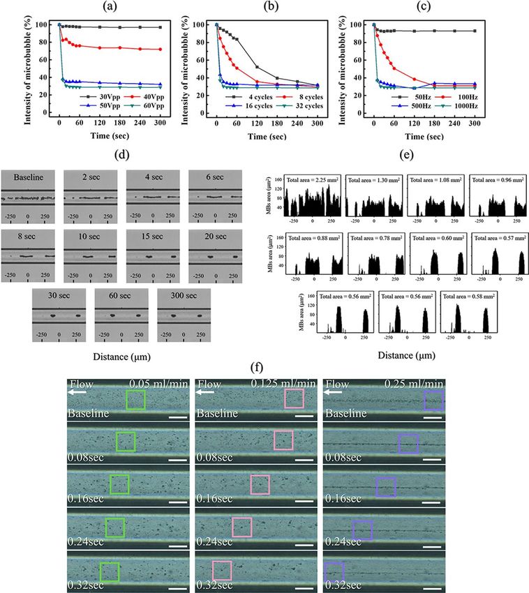
www.nature.com/scientificreports/
Figure 1. Schematic of the focused ultrasound-mediated drug delivery system (FUS-mediated DDS) and
unfocused ultrasound-mediated drug delivery system (UUS-mediated DDS) with the designed acoustic lens.
structure of cancer. Cancer reconstructs the appearance of blood and lymphatic vessels in extraordinary ways
that are not confined to the initial disease area. These unpredictable vascular structures have limitations with
respect to irradiating blood vessels using focused ultrasound with a narrow focal point. In this case, a monitoring
system for finding the focal target spot is n ecessary14,15. Thus, a complex FUS-MB DDS requires an ultrasonic
array transducer, front-end electronics to control each array element, and other medical imaging systems for
monitoring the spots being stimulated. In addition, thermal damage due to the high energy of focused ultrasound
is an inevitable problem16,17.
Previous studies have been employed to address these limitations via unfocused ultrasound with MBs (UUS-
MBs), and the effectiveness of UUS-MBs has also been demonstrated. For example, Beccaria et al.18 and Yao
et al.19 published papers reporting that UUS-MBs could effectively deliver drugs to an extensively targeted area.
However, Kovacs et al. asserted that tumor migration, not observed with FUS-MBs, was derived from drug deliv-
ery in an extensive area20. Thus, for drug delivery with UUS-MBs, the prevention of unexpected damage caused by
widespread excessive ultrasonic irradiation is essential. Unlike conventional unfocused transducers, ultrasound
with an acoustic lens can tune the pressure field depending on the type of interchangeable acoustic lens according
to the purpose of the D DS21,22. Simple attachment of the inexpensive lens on the purchased diagnostic ultrasound
probe can also reduce the tremendous cost for new therapeutic ultrasound probes. Thus, we hypothesized that
ultrasound with an acoustic lens might produce an efficient strategy for safe extensive drug delivery to the target
lesion without damaging other normal tissues, additionally giving a cost-effective and straightforward solution.
This work aimed to enhance DDS effectiveness by eliminating alignment issues using broadly spreading
acoustic pressure fields customized to the tumor size, as shown in Fig. 1. In addition, we tuned the acoustic
pressure intensity with an acoustic lens to minimize normal tissue damage. Our approach utilizes an interchange-
able and attachable compound acoustic lens in front of a conventional diagnostic ultrasonic probe and a MB
injection mixed with a cytotoxic drug. The acoustic lens dimensions were designed to be suitable for ultrasound
phased array probes through a commercial finite element method (FEM) simulation tool in preparation for the
unfocused pressure field. To avoid unexpected damage by a widespread pressure field, we optimized the pressure
field according to the tumor size and fabricated a lens to deliver the anticancer drug via uniform low-intensity
ultrasound. When the pressure was sufficient to induce ultrasound-mediated MB destruction (UMMD), we
studied the optimal treatment conditions for MB destruction in vitro through an optical imaging setup. Based
on these results, the effects of the DDS against cancer by UUS-MBs were evaluated in a xenograft rat hepatoma
model using DOX treatment.
Results
Acoustic characterization of the attachable divergent acoustic lens. Based on the results of the
FEM simulation to apply the uniformly distributed pressure fields to the lesion, a concave lens with a radius of
curvature (ROC) of 2.6 mm and a thickness of 4 mm was selected and fabricated. The acoustic pressure fields
with and without the acoustic lens are depicted in Fig. 2a–f. When the designed concave acoustic lens was
applied to the probe, the pressure field was uniformly distributed with the beam profile following the introduc-
tion of the acoustic lens compared to the pressure level without the lens. The 1 cm × 3 cm area of interest (AOI)
is marked with a dashed box in Fig. 2b,c,e,f. We calculated the median values and standard deviations of each
pressure distribution along the Z-axis line at each location X in the AOI. The graphs in Fig. 2g–j show each result.
From the simulation results shown in Fig. 2g,i, the acoustic pressure dropped to almost half of the maximum
value with the lens while uniform pressure distribution was acquired. The standard deviation of the calculated
median value at each X position was reduced from 0.21 to 0.01. From the measurement data, the uniform pres-
sure distribution showed similar results from the simulation and the standard deviation decreased from 0.18
to 0.04 (Fig. 2h,j). Thus, we confirmed that the designed concave lens could uniformly distribute the acoustic
pressure in the AOI.
Scientific Reports | (2021) 11:12654 | https://doi.org/10.1038/s41598-021-92097-z 2
Vol:.(1234567890)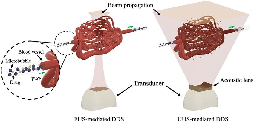
www.nature.com/scientificreports/
Figure 2. The acoustic pressure fields under either with or without the acoustic lens. Acoustic field
propagations without (top) and with (bottom) the concave-shaped acoustic lens. (a) Schematic of the measured
domain without the lens. (b) Simulated and (c) measured results of the pressure field without the lens. (d)
Schematic of the measured domain with the lens. (e) Simulated and (f) measured results of the pressure field
with the lens. The mean value distributions of (g) simulated and (h) measured acoustic pressure along one
Z-axis at each X position without the lens. The mean value distributions of (i) simulated and (j) measured
acoustic pressure along one Z-axis at each X position with the lens. The dotted box represents the area of interest
(AOI).
In vitro microbubble destruction experiment using an unfocused uniform ultrasound beam
with the lens. Since acoustic attenuation through the lens material caused the pressure to decrease to 55%
that of the maximum peak pressure, MB destruction must be confirmed under reduced pressure. Figure 3 shows
the in vitro test results for destroying MBs by variation in the acoustic parameters such as cycle, pulse repetition
frequency (PRF), and pressure amplitude. To study the threshold of optimized acoustic parameters for UMMD
in the AOI, MBs were injected into the cylindrical vessel phantom with a diameter 235 μm. Various acoustic
parameters were irradiated within the AOI at a distance of 40 mm, where the pressure was relatively low. The
inertial cavitation of MBs, in which MBs violently collapse in a liquid, was measured through an intensity com-
parison baseline from the captured images. The ratios of image intensity for comparing UMMD under various
cycles, PRF, and pressure amplitude are plotted in Fig. 3a–c. First, the MBs in the mimic vessel phantom were
irradiated in the absence of flow conditions and measured with one variable, keeping the other conditions were
fixed. Figure 3a shows that a minimum acoustic pressure of 50–60 V pp is required to burst MBs. The threshold
voltage (40 Vpp) applied to the conventional ultrasonic probe with the lens to start MB collapse corresponded to
330 kPa. Figure 3b shows that the destroyed MBs depend on the number of cycles. Although all of the different
numbers of cycles resulted in a 70% decrease in MB intensity within 5 min, 16–32 cycles, which burst 70% of the
MBs within 20 s, were appropriate to induce a large amount of UMMD at once. Figure 3c shows that a PRF of
500–1000 Hz is needed. PRFs of 50 Hz and 100 Hz were not sufficient to induce MB rupture in bulk at one time.
Figure 3d,e present MB destruction in a straight-line microchannel with the acoustic parameters maximized
(60 Vpp or 500 kPa, 32 pulse cycles, and a PRF of 1 kHz) for 5 min. Figure 3d shows that UMMD was saturated
within 15 s, and the aggregated MBs seemed to remain stable even after 5 min. The variation in the MBs was
also quantified using these captured images. We counted the number of pixels that corresponded to the contrast
value of the MBs. As shown in Figure 3e, it was confirmed that 75% of the total pixel area decreased after 20 s of
ultrasonic irradiation. However, after sufficient time passed after the ultrasonic irradiation, the acoustic radia-
tion force continuously moved the aggregated MB cluster, although the total pixel area or the unaffected MBs
remained unchanged. Additionally, to create a circulatory system similar to that in the human body, we observed
whether the MBs responded to ultrasound in fluidic flow with the same UMMD conditions. Figure 3f presents
the optical MB captured images with a time interval of 0.08 s under three different flow velocities (1.92 cm/s or
0.05 ml/min, 4.80 cm/s or 0.125 ml/min, and 9.60 cm/s or 0.25 ml/min). We applied the same acoustic conditions
to the MBs in the microchannel with the flow rate setup. As a result, the MBs burst under the same conditions
even at a flow rate of 0.25 ml/min (9.60 cm/s) and affected large areas, as shown in Figure 3f.
The enhanced drug delivery system via UUS‑MBs with the attachable lens. To measure the
enhanced chemotherapy effects using UUS-MBs with the attachable lens, we compared the DOX concentra-
tion within the tumor in a rat xenograft hepatoma model (Fig. 4). The experiment was conducted by dividing
the samples into three groups according to irradiation time (5, 10, 30 min) with input voltage conditions of 60
Vpp, 32 cycles, and a PRF of 1 kHz to find a suitable treatment time. As shown in Fig. 5, the DOX concentra-
tion increased by approximately 25% after lens treatment for 5 min (236.9 ± 74.0 ng/ml vs. 295.7 ± 93.8 ng/ml,
P = 0.022). This DOX concentration increment drops to approximately 13% when the treatment lasts 10 min.
After 30 min of UUS irradiation, the DOX concentration in the tumor was not significantly different from that
in the control group. The UUS-MB group had a relatively high DOX concentration in the tumor due to the UUS-
Scientific Reports | (2021) 11:12654 | https://doi.org/10.1038/s41598-021-92097-z 3
Vol.:(0123456789)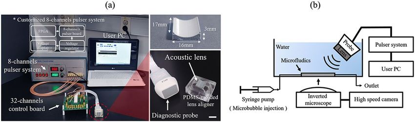
www.nature.com/scientificreports/
Figure 3. Experimental results using different variables to observe the changes in microbubbles with optical
microscopy in vitro. Image intensity of the microbubbles versus time under several various pulse conditions,
such as (a) voltage input (with fixed conditions of 32 pulse cycles and PRF of 1 kHz), (b) the number of
pulse cycles (with fixed conditions of voltage input of 60 Vpp and PRF of 1 kHz), and (c) PRF (with fixed
conditions of voltage input of 60 Vpp and 32 pulse cycles) under static conditions (n = 2). (d) Optical images
of the microbubbles in the straight-line microchannel sonicated with the lens-attached probe applying the
input voltage conditions of 60 V pp, 32 cycles, and PRF of 1 kHz for 5 min. No fluidic flow was applied. (e)
Analysis of the optical images determined by calculating the pixel area of the microbubbles based on the
intensity. (f) Optical images of the microbubble clusters with three different flow velocity setups of 0.05 ml/min,
0.125 ml/min, and 0.25 ml/min for 0.32 s of sonication in a straight line 235 μm diameter microchannel (scale
bar = 100 μm).
MB-induced DDS compared to the control group, but there was no significant difference in the tumor mass
among any of the groups (all P > 0.05).
Discussion
Many kinds of research have shown that FUS-MBs can enhance the effects of DDSs and this approach is conveni-
ent for mechanical force-triggered D DSs23–25. However, due to the abnormal vascular structure in the tumor and
in extensive tumors, FUS-MBs have a temporal limitation in exposing all the tumor areas. This study suggested
that UUS-MBs with the designed acoustic lens could be a solution for treating large tumors. We designed acoustic
lens fitting to a conventional ultrasonic transducer to enhance DDS effectiveness by adjusting the pressure field
for tumor size and evaluating the chemotherapeutic feasibility against cancer.
An acoustic lens, which is a mechanical aid used to adjust the acoustic propagation path, was used to control
the local pressure field. Polymer-based acoustic lenses are easy to design and manufacture because acoustic
propagation is controlled according to the geometric shape of the acoustic lens based on Snell’s law. The lens was
designed based on the geometric parameters that can uniformly irradiate the acoustic field among the results
obtained by the FEM method. In this work, a polydimethylsiloxane (PDMS)-based acoustic lens was attached
to a commercial diagnostic ultrasound transducer with biocompatible features. As shown in Fig. 2, ultrasound
treatment with the acoustic lens, unlike the control group, could result in uniform acoustic pressure in the AOI
regardless of its position. To destroy MBs in all areas of the AOI, we searched for the optimum acoustic param-
eters that caused the MBs to explode even in relatively low sound pressure areas.
MBs, as contrast agents for enhancing ultrasound imaging, reach the target within a few seconds, which
is fast enough to be used for clinical diagnostics. Moreover, we can use MBs to induce high mechanical stress
Scientific Reports | (2021) 11:12654 | https://doi.org/10.1038/s41598-021-92097-z 4
Vol:.(1234567890)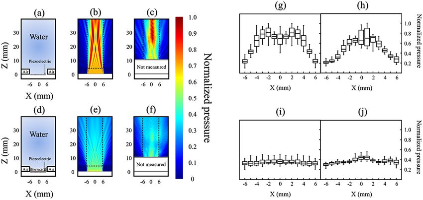
www.nature.com/scientificreports/
Figure 4. Animal experiment. (a) Photograph of the subcutaneous hepatoma rat model. (b) An illustration of
the in vivo experimental setup. Doxorubicin (DOX) was firstly injected via the tail vein, and then the ultrasound
treatment system was applied during each treatment time at one side. Microbubbles (MBs) were immediately
injected after DOX injection. Ultrasound irradiation occurred immediately after injecting the MBs and DOX.
The rats were sacrificed 30 min after the start of sonication, and the tumors were extracted to measure the DOX
concentration. (c) Photograph of the contrast-enhanced ultrasound images after injection of the MBs. The
dotted circle represents the concentration of MBs in the tumor over time. Contrast-enhanced ultrasound images
were only obtained to determine the ultrasound-treated time. (d) Photograph of the extracted tumors from the
subcutaneous hepatoma of rat models (scale bar = 1 cm).
upon their collapse. This shock wave can generate locally high pressure and temperature, which is called inertial
cavitation. Many kinds of research on UMMD have demonstrated that ultrasound-mediated MBs can increase
the effectiveness of DDSs and induce blood–brain barrier opening via this inertial cavitation26–28. Thus, we simi-
larly determined MBs as sonosensitizers to enhance chemotherapy and chose a commercial product, SonoVue
(Bracco, Millan, Italy), which is widely used in the clinic. Since the effects and characteristics of MBs are not fully
understood, their behavior under several ultrasound conditions must be investigated for use as the safest sono-
sensitizer in DDSs. Some previous studies have utilized a real-time optical microscope to monitor only a single
MB or a few M Bs29–31. However, it is essential to observe that many MBs interact with mechanical waves because
a high dose of MBs is injected in the clinic. Very few experiments have been conducted to explore the interaction
between a relatively large amount of MBs and ultrasound, as the limited number of MBs tested experimentally
could limit their further applications. Therefore, we adopted microfluidic technology to construct an artificial
blood vessel structure, and this confined channel could provide a similar in vivo environment to test the interac-
tion between the large number of MBs and several different ultrasonic excitation conditions. This experiment
was also conducted under physiologically realistic fluidic conditions compared to the in vivo circulation system.
Real-time optical imaging measurements confirmed that UUS-MBs with an acoustic lens at 60 Vpp for 32
cycles and a PRF of 1 kHz for 5 min could induce UMMD to promote the DDS even in fluidic conditions. It
was also verified that the interaction time between the MBs and ultrasound is crucial for the DDS. First, in vitro
experiments under static conditions were conducted for various pulse cycle numbers, PRFs and voltage inputs
to find the optimal acoustic parameters for inertial cavitation, as shown in Fig. 3. Abrupt inertial cavitation (at
a voltage of 50–60 Vpp, 16–32 pulse cycles, or a PRF of 500–1000 Hz) exhibited promising effects, as determined
by a reduction in the intensity of microbubbles compared to stable cavitation (a voltage input of 30 V pp or a PRF
of 50 Hz) and slow inertial cavitation (a voltage of 50–60 V pp, 4–8 pulse cycles or a PRF of 100 Hz). The optimal
conditions placed a relatively strong acoustic radiation force on the microbubbles within 20 s, thereby disrupt-
ing the microbubble shells. Although the maximum pressure conditions were applied to the aggregated MBs to
clean them thoroughly, it was hardly possible to remove the remaining MBs. We think that secondary Bjerknes
force under the fixed acoustic pressure field prevented the static aggregated MBs from rupturing because the
Scientific Reports | (2021) 11:12654 | https://doi.org/10.1038/s41598-021-92097-z 5
Vol.:(0123456789)www.nature.com/scientificreports/
Figure 5. Doxorubicin concentration within the tumor in a rat xenograft hepatoma model. (a) Doxorubicin
concentration in the subcutaneous hepatoma rat models with unfocused ultrasound-mediated microbubbles.
(b) Graph of the change in DOX concentration according to ultrasound treatment.
aggregated MBs generated a so-called cushioning effect so that they can mitigate each other’s vigorous oscilla-
tion. On the other hand, MB aggregation and saturation phenomena decreased under fluidic conditions, but the
effects of the ultrasonic waves appeared to be different from those under static conditions. This phenomenon
resulted from an insufficient amount of time to see the interaction of ultrasound and flowing MBs within the
observation area. This suggests that the long-term interaction between the MBs and ultrasound will be crucial
in stimulating nearby tissue structures by DDS with ultrasound-mediated MBs. As one strategy to address this
issue, our ultrasound system using the acoustic lens might serve as an exclusive approach that provides sufficient
time to destroy moving MBs in the tumor.
Our ultrasound treatment system is capable of diverging pressure fields, and the acoustic lens can increase
drug delivery effectiveness by providing an ultrasound irradiation time to MBs. The fabricated interchangeable
and attachable acoustic lens demonstrated a low and uniformly acoustic pressure in AOI and optimized MB
destruction in vitro experiment, which was at 60 Vpp for 32 cycles and a PRF of 1 kHz for 5 min. Based on
these results, the effects of the DDS were evaluated in a xenograft rat hepatoma model using DOX treatment
when the ultrasound treatment was applied for 5 min, 10 min, and 30 min. Most MBs in rats did not show avid
enhancement in the tumor after 5 min but needed the further extended treatment up to 30 min to exclude little
circulating MBs effect in the rat. Therefore, we divided the treatment groups into 5 min, 10 min, and 30 min
(Fig. 4). UUS-MBs with the acoustic lens can promoted the DDS in vivo compared to the control group. Since
sufficient irradiation time was required in the fluidic conditions, as shown in Fig. 3f, ultrasonic irradiation was
conducted in consideration of the MB lifetime (< 30 min). The UUS-MBs in the acoustic lens treatment group
had a higher concentration of DOX in the tumor than the control group. Although all groups showed an increase
of DOX concentration in ultrasound-treated tumors, only the 5-min group showed a statistically significant
increase of DOX in the tumor. In terms of extracted tumor weight, there was a slightly lower weight in the tumor
on the ultrasound-treated side, but the DOX concentration (ng/ml) was analyzed using the volume of solvent
adjusted by tumor extract weight (Fig. 5). Therefore, the DOX concentration might be a reliable value adjusted
and normalized by tumor weight.
However, there were some limitations for the interpretation in animal experiment. First, there was no data
about the initial and final tumor volume measurement by using 3D imaging modality. It might be an essential
factor when dividing rats into groups by random distribution. Over 1 week after tumor implantation, the tumors
were only measured using calipers to check the tumor formation. Among the bilaterally implanted rats, rats who
reached above 10 mm of tumor length at both sides after 7 days were only included and then randomly distributed
three groups. When the size of both tumors was measured, if either side was less than 10 mm, it was not included
in the study. Therefore, the experiment was started under the assumption that there was no difference in the size
of bilateral tumors. In fact, the length of the protruding subcutaneous tumor did not mean the accurate tumor
volume. If the tumor characteristics such as tumor necrosis or viable tumor portion as well as tumor volume be
evaluated through the contrast-enhanced imaging study such as MB-induced ultrasound imaging modality, the
evidence of DDS effectiveness would be more strengthened in vivo study, because it might be a critical factor
for the DOX distribution in the tumor. Second, it is difficult to mention that DOX concentrations increase as
Scientific Reports | (2021) 11:12654 | https://doi.org/10.1038/s41598-021-92097-z 6
Vol:.(1234567890)www.nature.com/scientificreports/
Figure 6. Complete ultrasonic system for the experimental study. (a) Photographs of the UUS-mediated DDS
with the attachable acoustic lens (scalar bar = 1 cm) and the custom-built ultrasonic pulser system. (b) An
illustration of the in vitro experimental setup. Ultrasonic waves were emitted with an incident angle of 45° to
prevent a standing wave effect in the microchannel.
the ultrasound-treated time increases. To minimize the possible differences between subjects with tumors that
had undergone the ultrasound treatment and subjects with tumors not treated with the ultrasound treatment,
the tumors were implanted at one side and the other side thighs of the same subject. Therefore, the intrinsic
factors such as blood circulation were minimized to compare only the ultrasound effect with DOX injection in
the same rats. There was a similar DOX concentration at the non-treated side (236.9 ± 74.0 and 236.5 ± 100.1 ng/
ml) and higher DOX concentration at the ultrasound-treated side (295.7 ± 93.8 and 267.5 ± 98.7 ng/ml) between
5- and 10-min groups. However, there was a higher DOX concentration at the non-treated side (247.7 ± 93.7 ng/
ml) and lower DOX concentration (252.8 ± 81.7 ng/ml) at the ultrasound-treated side in the 30-min group. The
accurate and logical basis for explaining this phenomenon has not been found in this study. Third, the ultra-
sound treatment device with the attachable lens could not show tumor imaging simultaneously because of the
customized ultrasound system, which did not capture real-time ultrasound images. However, adequate genera-
tion of sonication could be monitored using the uniform wave generating by the customized ultrasound device.
Further development of the ultrasound treatment device should be added to the real-time imaging module for
the assessment of tumor volume and physiologic characteristics. This is easily achievable because we used the
conventional diagnostic ultrasonic probe with the simple detachment of the acoustic lens. Finally, the focus of the
in vivo study was the difference in final DOX concentration in tumors treated with and without the ultrasound
treatment system when DOX was injected through the tail vein to treat tumors on both sides (assumed to be
approximately the same size) in the same rat. However, it might also be necessary to measure the DOX concen-
tration in normal tissues to find evidence of improving drug delivery to certain cancer cells without increasing
the dose to normal cells to obtain the clinical usefulness of this DDS system.
Conclusion
In summary, our ultrasound system is capable of diverging pressure fields and is suitable for this tumor size, and
using this acoustic lens can increase drug delivery effectiveness by providing sufficient ultrasound irradiation
time to the MBs. Future research is required to optimize acoustic lens designs and acoustic parameters, includ-
ing the duration to maximize UMMD in blood vessels. Furthermore, ongoing animal studies using our system
for cancer treatment are currently being conducted to evaluate the clinical safety of our microbubble-mediated
treatment approach.
Methods
Custom‑built ultrasonic pulser system. A photograph of the total ultrasonic system, including the
attachable acoustic lens and a custom-built ultrasound pulser system, is shown in Fig. 6a. Thirty-two elements
out of the 64 elements from a diagnostic phased array transducer (SP1-5; Alpinion Medical Systems, Seoul,
Korea) were used to radiate ultrasound to excite the MBs. The custom-built 8-channel pulser system applied
several voltage levels with a center frequency of 2 MHz to this conventional ultrasonic diagnostic probe. The pul-
ser system was controlled by a field-programmable gate array (FPGA) on a personal computer (PC) to encode
the acoustic parameters using a universal serial bus (USB) connector. We programmed the FPGA to control the
ultrasonic pulse parameters, including the number of cycles, the PRF, and the probe center frequency.
Fabrication of the concave acoustic lens. The acoustic field distribution was simulated using the finite
element method (FEM) with COMSOL simulation software (COMSOL Multiphysics 5.2, COMSOL, MA, USA).
A two-dimensional FEM model under continuous wave mode was used to reduce the model complexity and
computational costs. The acoustic beam profiles were assessed using a hydrophone (HGL-200, Onda, CA, USA)
in the presence and absence of the acoustic lens. After the hydrophone measurement, we modified the FEM
model based on the experimental measurements. The polydimethylsiloxane (PDMS, Sylgard 160, Dow Corn-
ing, MI, USA) acoustic lens was replicated from a 3-D printed plastic mold designed from the final simulation.
Optical observation of ultrasound‑mediated microbubbles. The in vitro experimental setup is
shown in Fig. 6b. We positioned the ultrasonic transducer with the lens slanted at 45° to the microfluidic chips’
surface because we tried to eliminate the standing waves or the wave interference from the reflected surface of
Scientific Reports | (2021) 11:12654 | https://doi.org/10.1038/s41598-021-92097-z 7
Vol.:(0123456789)www.nature.com/scientificreports/
the water tank. Also, this helped to set the light source to directly observe the microbubble dynamics from the
inverted microscope, while we kept the relative position between the channel and the incidence angle remained
perpendicular. Ultrasound-mediated destruction of the MBs in the straight-line microchannel chip was moni-
tored using an inverted microscope (IX73, Olympus, Tokyo, Japan). Optical images were captured using both an
EM-CCD camera (ImagEMX2; Hamamatsu Photonics, Hamamatsu, Japan) at 70 frames per s and a high-speed
camera (Phantom V2512; AMTEK VISION RESEARCH, NJ, USA) at 5000 frames per s. The PDMS micro-
fluidic chips were fabricated using stainless steel (SUS) microneedles as a template. The microchannel (diam-
eter = 235 μm) was connected to two tube adapters (diameter = 800 μm). Under static experimental conditions,
5.04 mg/ml ultrasound contrast agent (SonoVue, Bracco, Millan, Italy) was injected into the microchannel. Both
the outlet and inlet of the microchannel were sealed with epoxy to halt the flow. Under the fluidic experimental
conditions, fluid flow and MB injection were conducted using a syringe pump (Ultra 4400, Harvard Apparatus,
MA, USA) and a 1-ml syringe. The dynamics of the MB destruction within the microchannel were evaluated
with four different flow velocity setups. Before investigating the MB with fluid flow, the time of observation was
set to 0.32 s due to the limitations of the accessible time for tracking the MBs with the fastest flow velocity setup.
The MB variation within the microchannel was quantified using custom-written image analysis code in
MATLAB software (Natick, MA, USA). The captured images were loaded into MATLAB, and the RGB images
were converted to grayscale intensity images. In the 235 μm-diameter microchannel (900 × 235 μm2), we counted
the number of pixels with a cutoff value less than that of the pixel contrast value of the outer wall of the MBs.
Based on this image processing setup, the relative intensity of the MBs was extracted from the captured images
every 10 s or 1 min.
Sonication by the diagnostic probe with the attachable acoustic lens with the maximal acous‑
tic parameters. Maximal unfocused ultrasound sonication was delivered at 2 MHz with 60 Vpp in bursts
of 32 cycles at a 1 kHz repetition time. From the AOI, the results through the hydrophone indicated that the
mechanical index (MI) was 0.17 that of the average with a standard deviation of 0.03, and the Ispta (spatial-peak-
temporal-average intensity) was 0.412 W/cm2 that of the average with a standard deviation of 0.124.
Rat hepatoma model preparation and treatment. This study was approved by the Institutional Ani-
mal Care and Use Committee of the Seoul National University Hospital (IACUC; No. 18-0124-S1A1(3)) and
was performed in accordance with the ARRIVE guideline. All methods were carried out in accordance with
relevant guidelines and regulations. The N1-S1 (CRL-1604; ATCC, Manassas, VA) hepatoma tumor cell line was
cultured in RPMI-1640 (Welsen, Daegu, Korea). The medium was supplemented with 10% fetal bovine serum
and a 1% penicillin/streptomycin mixture (Gibco, Grand Island, NY, USA). Cell viability was tested with Trypan
blue staining to confirm cell viability of > 90% before tumor implantation. To establish a xenografted rat tumor
model, 1 × 10⁷ N1-S1 cells were collected and subcutaneously injected into the bilateral backs of Sprague-Dawley
rats weighing approximately 300 g. Cyclosporine A (20 mg/kg/day; Chong Kun Dang Pharmaceutical Corp.,
Seoul, Korea) was subcutaneously injected 1 day before tumor implantation and for 4 days postoperatively in
order to prevent spontaneous regression of the N1-S1 cells.
Over 1 week, the animals were weighed, and the tumors were measured using calipers. When the tumor length
reached 10 mm after 7 days, the rats were divided into three groups: Group A (DOX + LENS 5 min, n = 10), Group
B (DOX + LENS 10 min, n = 10), and Group C (DOX + LENS 30 min, n = 10) (Fig. 4a,b). Contrast-enhanced
ultrasound was performed by a radiologist (J.H.K.) with 18 years of clinical experience in order to determine the
treatment time for DOX delivery using a GE LOGIQ E9 Ultrasound System (GE Healthcare, Wauwatosa, WI,
USA) with the following parameters: a transducer frequency of 9 MHz; a frame rate of 13 Hz; a dynamic range
of 60; an MI of 0.14; a gain of 24; and a depth of 2.0 cm (Fig. 4c). DOX (3 mg/kg) was injected via the tail vein
in all groups. SonoVue (0.3 mL in 0.1 ml of saline for each rat) through the tail vein and the LENS device was
applied for the treatment at each treatment time. For uniform exposure of hepatoma tumor cells, the rats were
positioned in the center of our customized ultrasound system. Thirty minutes after injection of the microbubbles,
all rats were sacrificed, and the tumors were removed (Fig. 4d).
Measurement of doxorubicin concentrations in the tumors. The tumors were carefully removed
from the subcutaneous layer of the rat thighs. They were then used for the analysis of DOX concentration accord-
ing to previously published procedures. Briefly, the tissues were homogenized in acidic alcohol (3% hydrochlo-
ride, 48.5% ethanol, 48.5% double-distilled water) with a microtissue homogenizer, and DOX was extracted for
24 h in the dark at 4 °C. The homogenates were then centrifuged at 5000 rpm for 10 min at 4 °C, and the super-
natants were collected. To quantify DOX, the level of fluorescence for each of the samples was measured using
liquid chromatography/tandem mass spectrometry (LC–MS/MS) [an Agilent 1260 Infinity Binary LC (Agilent
Technologies, Santa Clara, CA, USA) and an API 4000 QTRAP system (AB Sciex, Framingham, MA, USA)], and
the DOX concentration of each sample was calculated according to the weight of the corresponding tumor. All
statistical analyses were performed using SPSS version 21.0 (SPSS, Chicago, IL, USA). The DOX concentration
of each group was analyzed using the Wilcoxon-signed rank test at each treatment group.
Data availability
The datasets generated and/or analyzed during the current study are available from the corresponding author
upon reasonable request.
Received: 3 March 2021; Accepted: 4 June 2021
Scientific Reports | (2021) 11:12654 | https://doi.org/10.1038/s41598-021-92097-z 8
Vol:.(1234567890)www.nature.com/scientificreports/
References
1. Banchereau, J. & Palucka, K. Immunotherapy: Cancer vaccines on the move. Nat. Rev. Clin. Oncol. 15, 9–10 (2018).
2. Baskar, R., Lee, K. A., Yeo, R. & Yeoh, K. W. Cancer and radiation therapy: Current advances and future directions. Int. J. Med. Sci.
9, 193–199 (2012).
3. Shi, X., Zhang, C. Y., Gao, J. & Wang, Z. Recent advances in photodynamic therapy for cancer and infectious diseases. Wiley
Interdiscip. Rev. Nanomed. Nanobiotechnol. 11, e1560 (2019).
4. Wan, G. Y. et al. Recent advances of sonodynamic therapy in cancer treatment. Cancer Biol. Med. 13, 325–338 (2016).
5. El-Rayes, B. F. et al. Phase I study of liposomal doxorubicin (Doxil) and cyclophosphamide in solid tumors. Invest. New Drugs.
23, 57–62 (2005).
6. Thomas, M. B. et al. Systemic therapy for hepatocellular carcinoma: Cytotoxic chemotherapy, targeted therapy and immunotherapy.
Ann. Surg. Oncol. 15, 1008–1014 (2008).
7. Hu, Q., Chen, Q. & Gu, Z. Advances in transformable drug delivery systems. Biomaterials 178, 546–558 (2018).
8. Zhao, C. Y., Cheng, R., Yang, Z. & Tian, Z. M. Nanotechnology for cancer therapy based on chemotherapy. Molecules 23, 826
(2018).
9. Zhao, Z. et al. Delivery strategies of cancer immunotherapy: Recent advances and future perspectives. J. Hematol. Oncol. 12, 126
(2019).
10. Phillips, L. C., Dhanaliwala, A. H., Klibanov, A. L., Hossack, J. A. & Wamhoff, B. R. Focused ultrasound-mediated drug delivery
from microbubbles reduces drug dose necessary for therapeutic effect on neointima formation—Brief report. Arterioscler. Thromb.
Vasc. Biol. 31, 2853–2855 (2011).
11. Treat, L. H. et al. Targeted delivery of doxorubicin to the rat brain at therapeutic levels using MRI-guided focused ultrasound. Int.
J. Cancer. 121, 901–907 (2007).
12. Gong, Y. et al. Low-intensity focused ultrasound mediated localized drug delivery for liver tumors in rabbits. Drug Deliv. 23,
2280–2289 (2016).
13. Wu, S. K., Tsai, C. L., Huang, Y. & Hynynen, K. Focused ultrasound and microbubbles-mediated drug delivery to brain tumor.
Pharmaceutics. 13, 15 (2020).
14. Carmeliet, P. & Jain, R. K. Angiogenesis in cancer and other diseases. Nature 407, 249–257 (2000).
15. Stacker, S. A. et al. Lymphangiogenesis and lymphatic vessel remodelling in cancer. Nat. Rev. Cancer. 14, 159–172 (2014).
16. Lentacker, I., De Cock, I., Deckers, R., De Smedt, S. C. & Moonen, C. T. Understanding ultrasound induced sonoporation: Defini-
tions and underlying mechanisms. Adv. Drug Deliv. Rev. 72, 49–64 (2014).
17. Tran, B.C., Seo, J., Fowlkes, J.B., Cain, C. Microbubble enhanced threshold reductions for tissue damage using high intensity
ultrasound. in 2001 IEEE Ultrasonics Symposium, Vol. 2, 1389–1392 (2001).
18. Beccaria, K. et al. Opening of the blood–brain barrier with an unfocused ultrasound device in rabbits. J. Neurosurg. 119, 887–898
(2013).
19. Yao, L. et al. Facilitated brain delivery of poly (ethylene glycol)-poly (lactic acid) nanoparticles by microbubble-enhanced unfocused
ultrasound. Biomaterials 35, 3384–3395 (2014).
20. Kovacs, Z. et al. Prolonged survival upon ultrasound-enhanced doxorubicin delivery in two syngenic glioblastoma mouse models.
J. Control Release. 187, 74–82 (2014).
21. Chang, C. L. et al. Acoustic lens for capacitive micromachined ultrasonic transducers. J. Micromech. Microeng. 24, 085007 (2014).
22. Engholm, M., Beers, C., Bouzari, H., Jensen, J. A. & Thomsen, E. V. Increasing the field-of-view of row-column-addressed ultra-
sound transducers: Implementation of a diverging compound lens. Ultrasonics 88, 97–105 (2018).
23. Horise, Y. et al. Sonodynamic therapy with anticancer micelles and high-intensity focused ultrasound in treatment of canine cancer.
Front. Pharmacol. 10, 545 (2019).
24. Treat, L. H., McDannold, N., Zhang, Y., Vykhodtseva, N. & Hynynen, K. Improved anti-tumor effect of liposomal doxorubicin
after targeted blood–brain barrier disruption by MRI-guided focused ultrasound in rat glioma. Ultras. Med. Biol. 38, 1716–1725
(2012).
25. Liu, H. L., Fan, C. H., Ting, C. Y. & Yeh, C. K. Combining microbubbles and ultrasound for drug delivery to brain tumors: Current
progress and overview. Theranostics. 4, 432–444 (2014).
26. Burgess, A. & Hynynen, K. Drug delivery across the blood-brain barrier using focused ultrasound. Expert Opin. Drug Deliv. 11,
711–721 (2014).
27. Tsai, H. C. et al. Safety evaluation of frequent application of microbubble-enhanced focused ultrasound blood–brain-barrier
opening. Sci. Rep. 8, 17720 (2018).
28. Burgess, A., Shah, K., Hough, O. & Hynynen, K. Focused ultrasound-mediated drug delivery through the blood-brain barrier.
Expert Rev. Neurother. 15, 477–491 (2015).
29. Chen, H., Kreider, W., Brayman, A. A., Bailey, M. R. & Matula, T. J. Blood vessel deformations on microsecond time scales by
ultrasonic cavitation. Phys. Rev. Lett. 106, 034301 (2011).
30. Faez, T., Skachkov, I., Versluis, M., Kooiman, K. & de Jong, N. In vivo characterization of ultrasound contrast agents: Microbubble
spectroscopy in a chicken embryo. Ultras. Med. Biol. 38, 1608–1617 (2012).
31. Cosgrove, D. Ultrasound contrast agents: An overview. Eur. J. Radiol. 60, 324–330 (2006).
Acknowledgements
The authors would like to specially thank Deog Moon Rho (KIST) for the lens fabrication and Min Tack Oh
(KIST) for the custom-built ultrasound pulser system assembly. This work was supported by the National
Research Foundation of Korea (NRF) Grant funded by the Korea government (MSIT) (No. 2020R1C1C1008716).
This work was also supported in part by the Brain Research Program through the NRF funded by the MSIT (No.
2016M3C7A1913845) and the KIST Institutional Program (2E30964).
Author contributions
S.L., design, collection and assembly of data, data analysis and interpretation, manuscript writing, financial sup-
port; H.J., conception and design, data analysis and interpretation, manuscript writing; S.S., design, data analysis
and interpretation; M.I., design, data analysis and interpretation; J.K., design, data analysis and interpretation;
J.H.K., design, collection and assembly of data, data analysis and interpretation; B.C.L., design, collection and
assembly of data, data analysis and interpretation, manuscript writing, financial support.
Funding
This work was supported by the National Research Foundation of Korea (NRF) Grant funded by the Korea gov-
ernment (MSIT) (No. 2020R1C1C1008716). This work was also supported in part by the Brain Research Program
through the NRF funded by the MSIT (No. 2016M3C7A1913845) and the KIST Institutional Program (2E30964).
Scientific Reports | (2021) 11:12654 | https://doi.org/10.1038/s41598-021-92097-z 9
Vol.:(0123456789)www.nature.com/scientificreports/
Competing interests
The authors declare no competing interests.
Additional information
Correspondence and requests for materials should be addressed to J.H.K. or B.C.L.
Reprints and permissions information is available at www.nature.com/reprints.
Publisher’s note Springer Nature remains neutral with regard to jurisdictional claims in published maps and
institutional affiliations.
Open Access This article is licensed under a Creative Commons Attribution 4.0 International
License, which permits use, sharing, adaptation, distribution and reproduction in any medium or
format, as long as you give appropriate credit to the original author(s) and the source, provide a link to the
Creative Commons licence, and indicate if changes were made. The images or other third party material in this
article are included in the article’s Creative Commons licence, unless indicated otherwise in a credit line to the
material. If material is not included in the article’s Creative Commons licence and your intended use is not
permitted by statutory regulation or exceeds the permitted use, you will need to obtain permission directly from
the copyright holder. To view a copy of this licence, visit http://creativecommons.org/licenses/by/4.0/.
© The Author(s) 2021
Scientific Reports | (2021) 11:12654 | https://doi.org/10.1038/s41598-021-92097-z 10
Vol:.(1234567890)You can also read