Prevalence of right ventricular dysfunction and impact on all cause death in hospitalized patients with COVID 19: a systematic review and ...
←
→
Page content transcription
If your browser does not render page correctly, please read the page content below
www.nature.com/scientificreports
OPEN Prevalence of right ventricular
dysfunction and impact
on all‑cause death in hospitalized
patients with COVID‑19:
a systematic review
and meta‑analysis
Bernadette Corica1,7, Alberto Maria Marra2,3,7, Stefania Basili1, Roberto Cangemi1,
Antonio Cittadini2, Marco Proietti 4,5,6,8* & Giulio Francesco Romiti1,8
The Coronavirus Disease (COVID-19) pandemic imposed a high burden of morbidity and mortality. In
COVID-19, direct lung parenchymal involvement and pulmonary microcirculation dysfunction may
entail pulmonary hypertension (PH). PH and direct cardiac injury beget right ventricular dysfunction
(RVD) occurrence, which has been frequently reported in COVID-19 patients; however, the prevalence
of RVD and its impact on outcomes during COVID-19 are still unclear. This study aims to evaluate
the prevalence of RVD and associated outcomes in patients with COVID-19, through a Systematic
Review and Meta-Analysis. MEDLINE and EMBASE were systematically searched from inception to
15th July 2021. All studies reporting either the prevalence of RVD in COVID-19 patients or all-cause
death according to RVD status were included. The pooled prevalence of RVD and Odds Ratio (OR) for
all-cause death according to RVD status were computed and reported. Subgroup analysis and meta-
regression were also performed. Among 29 studies (3813 patients) included, pooled prevalence of RVD
was 20.4% (95% CI 17.1–24.3%; 95% PI 7.8–43.9%), with a high grade of heterogeneity. No significant
differences were found across geographical locations, or according to the risk of bias. Severity of
COVID-19 was associated with increased prevalence of RVD at meta-regression. The presence of RVD
was found associated with an increased likelihood of all-cause death (OR 3.32, 95% CI 1.94–5.70).
RVD was found in 1 out of 5 COVID-19 patients, and was associated with all-cause mortality. RVD may
represent one crucial marker for prognostic stratification in COVID-19; further prospective and larger
are needed to investigate specific management and therapeutic approach for these patients.
Severe acute respiratory syndrome coronavirus 2 (SARS-CoV-2) and its associated disease (COVID-19), plagued
the world during 2020, with the World Health Organization declaring a pandemic state earlier in the year1. By
13th July 2021, an estimated 4 million deaths were attributed to COVID-19 w orldwide2, with an extremely high
healthcare, social, and economic burden. Most of the disease’s mortality and severity have been attributed to
respiratory complications of the disease; indeed, patients may develop severe pneumonia up to Acute Respiratory
Distress Syndrome (ARDS). Beyond direct alveolar involvement, also lung microcirculation seems to be affected
in these patients. Autopsies studies revealed a pattern of pulmonary endothelial dysfunction with increased
1
Department of Translational and Precision Medicine, Sapienza – University of Rome, Rome, Italy. 2Department
of Translational Medical Sciences, “Federico II” University of Naples, Naples, Italy. 3Center for Pulmonary
Hypertension, Thoraxklinik at Heidelberg University Hospital, Heidelberg, Germany. 4Department of Clinical
Sciences and Community Health, University of Milan, Milan, Italy. 5Geriatric Unit, IRCCS Istituti Clinici Scientifici
Maugeri, Via Camaldoli 64, 20138 Milan, Italy. 6Liverpool Centre for Cardiovascular Science, University of
Liverpool and Liverpool Heart & Chest Hospital, Liverpool, UK. 7These authors contributed equally: Bernadette
Corica and Alberto Maria Marra. 8These authors jointly supervised this work: Marco Proietti and Giulio Francesco
Romiti. *email: marco.proietti@unimi.it
Scientific Reports | (2021) 11:17774 | https://doi.org/10.1038/s41598-021-96955-8 1
Vol.:(0123456789)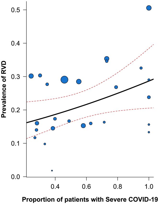
www.nature.com/scientificreports/
inflammatory infiltrate and microvascular thrombosis3. All the conditions mentioned above may lead to the
development of increased pulmonary pressures and right ventricle overload.
On the other hand, cardiovascular complications have been early addressed as one concern in these patients4.
isease5, cardiovascular disease, includ-
While several factors may influence the severity and clinical course of the d
ing arrhythmia, myocardial disfunction and myocardial injury, have been repeatedly identified as potential key
detrimental factors in patients with COVID-196–8. Lung parenchymal involvement, pulmonary microvascular
pathologic changes, right ventricular pressure overload, and direct myocardial injury exert a synergic detrimental
effect on the right ventricular function.
Indeed, right ventricular dysfunction (RVD)9 has been described as a potential predictor of poor outcomes
in small preliminary studies, but its prevalence and associated outcomes in patients with COVID-19 are far from
being elucidated. Clarification of the prevalence of RVD, and its associated outcomes in patients with COVID-
19, may promote the implementation of tailored strategies for the screening, prevention, and treatment of right
ventricular impairment.
Amid this pandemic, systematic review and meta-analysis have been depicted as essential tools to provide
a timely and comprehensive synthesis of evidence during the COVID-19 p andemic10. Moving from this, this
systematic review and meta-analysis aimed to estimate the prevalence of RVD among patients with COVID-19
and to explore its impact on all-cause mortality.
Materials and methods
This systematic review has been performed according to the Preferred Reporting Items for Systematic Reviews
and Meta-Analyses (PRISMA) guidelines and recommendations. The protocol was registered into the interna-
tional register of systematic reviews PROSPERO, N. CRD42021227946.
Search strategy. Systematic and comprehensive literature research was performed on MEDLINE and
EMBASE databases, from inception to 15th July 2021. The search strategy included a combination of terms
related to the research question, including ‘right ventricular dysfunction’, ‘COVID-19’, and ‘SARS-CoV-2’. The
full search strategy is available in Supplementary Material, Table S1.
Study selection. All records retrieved from the database search were systematically assessed by two inde-
pendent authors (BC and GFR) according to titles and abstracts; articles included after this phase were subse-
quently screened for full-text eligibility. Study selection was performed with the use of a standardized web-based
platform (Covidence). Any disagreement during each phase was discussed collegially. Inclusion criteria were:
(i) any study reporting the prevalence of RVD; or (ii) any study reporting outcomes in patients with COVID-19
according to the RVD status (i.e., number of patients with and without RVD who died). Exclusion criteria were:
(i) case reports, conference abstracts, editorial, comments, systematic reviews, meta-analyses and guidelines, (ii)
studies that enrolled less than 30 COVID-19 patients, and (iii) articles in languages other than English, Italian
or Spanish.
References of the included studies were additionally searched for other relevant articles that were not retrieved
from the literature search. In the case of two or more studies based on the same cohort of subjects and exploring
the same outcome(s), only the most recently published were selected and included.
Outcomes definition. Primary outcomes were defined as (i) prevalence of RVD, as defined in the original
studies, and (ii) all-cause death according to RVD status. When RVD was not clearly defined in the original stud-
ies or multiple definitions were reported, we considered RVD according to the study-defined TAPSE cut-off, if
available, to increase the homogeneity of RVD definition among studies included. Among the “all-cause death”
definition, we also included the in-hospital mortality and 30-day mortality, as defined in the original studies.
Data extraction and quality assessment. Data from the studies included were independently extracted
by two co-authors (BC and GFR) using a standardized electronic form. Data on sample size, number of individu-
als with and without RVD, mortality, and follow-up time were extracted. We collected data about study design
and cohort baseline characteristics (i.e., age, sex, associated comorbidities including hypertension, diabetes mel-
litus, congestive heart failure (CHF)), and data on the severity of the disease or intensity of care received (i.e.
intensive care unit (ICU), mechanically ventilated patients), when available. Proportion of patients with severe
COVID-19 disease enrolled was also extracted; we defined severity according to the original studies definitions,
when available, or according to a diagnosis of ARDS, or need for mechanical ventilation.
All studies included were independently evaluated by two co-authors (BC and GFR) to evaluate their quality
and assess the risk of bias. We assessed the risk of bias separately for the two primary outcomes of the study. For
studies reporting the prevalence of RVD, we used a customized version of the Newcastle–Ottawa Scale (NOS)
for cross-sectional studies, composed of 5 items across 3 domains (selection, comparability, outcomes), with
a maximum of 5 points. Each study with a score ≤ 3 was considered at high risk of bias. We used a customized
version of the NOS for cohort studies for studies reporting outcomes, composed of 8 items across 3 domains
(selection, comparability, outcomes), with a maximum of 9 points. Each study with a score ≤ 6 was considered
at high risk of bias.
Publication bias was assessed for studies reporting outcomes according to RVD status. Funnel plots were
visually inspected for asymmetricity; furthermore, Egger’s test was also performed and reported.
Scientific Reports | (2021) 11:17774 | https://doi.org/10.1038/s41598-021-96955-8 2
Vol:.(1234567890)www.nature.com/scientificreports/
Statistical analysis. Pooled prevalence of RVD, 95% confidence intervals (CI) and 95% prediction inter-
vals (PI) were estimated using a generalized linear mixed m odel11. 95% PI represents a predicted range of the
true effect in an individual or new study and provide useful information on the variability of the effect in differ-
ent clinical settings12,13.
Outcomes from the original studies and according to RVD status were pooled and compared using random-
effect models; restricted maximum likelihood was used to estimate tau for this outcome.
Pooled estimates were reported as Odds Ratios (OR) and 95% confidence intervals (CI). The inconsistency
index (I2) was calculated to measure heterogeneity. According to pre-specified cut-offs, low heterogeneity was
defined as an I 2 of < 25%, moderate heterogeneity when I 2 falls between 25 and 75%, and high heterogeneity
when I2 was > 75%.
For each primary outcome, a “leave-one-out” sensitivity analysis was performed, by iteratively removing 1
study at a time to analyse their influence on pooled estimate and heterogeneity.
We also performed two subgroup analyses for the prevalence of RVD, according to the geographical loca-
tion of the included studies, and the risk of bias. No subgroup analysis was performed for the all-cause death,
according to the low number of studies included in this analysis. To evaluate the potential impact of COVID-19
severity on the prevalence of RVD, we also performed an univariable meta-regression analysis.
All the statistical analyses were performed using R version 4.1.0 (The R Foundation, 2021), with the use of
‘meta’, ‘metafor’ and ‘dmetar’ packages.
Results
A total of 350 studies were retrieved from the literature search (146 from MEDLINE and 204 from EMBASE).
After the selection process, a total of 29 articles were included in the a nalysis14–42 (Fig. S1 in Supplementary
Materials).
Systematic review of the included studies. Table 1 shows the baseline characteristics of the stud-
ies included in the meta-analysis. 29 studies reported data about the prevalence of RVD14–42, while 7 reported
about all-cause mortality according to RVD status17,19,20,30,33,35,36. 11 studies were held in Europe16,18–21,33–36,38,40,
8 in North America23,25,26,28,29,37,41,42, 3 in Asia30–32, and 7 in other geographical locations14,15,17,22,24,27,39, including
2 multinational studies24,27. Cohorts included were quite homogeneous in terms of mean age of the included
patients (ranging from 52 years old to 68 years old); among sex, males were generally more represented than
females (up to 84%). Among studies reporting outcome, the definition of all-cause mortality comprised ICU-
death17, in-hospital mortality35, 30-day m ortality36, 90-day m ortality19 or unspecified all-cause death20,30,33.
The definition of RVD was heterogeneous across studies, both in terms of parameters and cut-offs used to
defined RVD. 13 studies used a combination of several parameters15,18,19,21,24,28,30,33–35,38,40,42, 8 studies defined
RVD according to TAPSE cut-off14,16,22,23,31,32,36,39, while other or unclear definitions of RVD was used in 8
studies17,20,25–27,29,37,41. In one study, a surrogate of Right Ventricular-Arterial uncoupling, which in turn relates
the degree of RVD to the increase of pulmonary pressure, was a ddressed20. For 4 studies, we assumed RVD
according to the reported TAPSE cut-offs, to mitigate heterogeneity in the RVD definition among s tudies14,16,22,39.
The risk of bias for each study was reported in Tables S2 and S3 in Supplementary Materials, respectively for
studies assessing the prevalence of RVD, and for studies reporting outcomes. Among studies reporting preva-
lence, 13 were defined at high risk of bias, while 5 studies were defined at high risk of bias among those reporting
outcomes. Selection bias and definition of RVD were the most common concerns among the included studies.
Meta‑analysis of the included studies. Prevalence of RVD. Among 3813 patients included in the anal-
ysis, pooled prevalence of RVD was found as high as 20.4%, with a high degree of heterogeneity between studies.
PI was between 7.8% and 43.9% (Fig. 1). No significant differences were observed in the prespecified subgroup
analysis according to geographical location or bias risk (Table S4).
The prespecified leave-one-out sensitivity analysis showed overall consistency of the main results, with little
to no influence of individual studies on pooled estimates or heterogeneity (Fig. S2).
To evaluate the impact of COVID-19 severity (defined as the proportion of patients defined as “severe” or
“critical” in the original study, or when a stratification was not available, as those patients with ARDS or mechani-
cally ventilated) on the prevalence of RVD, we performed an univariable meta-regression analysis. The results
are graphically reported in Fig. 2; the proportion of patients with severe COVID-19 disease was significantly
associated with the prevalence of RVD in the cohorts included (p = 0.040, R 2 = 22.4%).
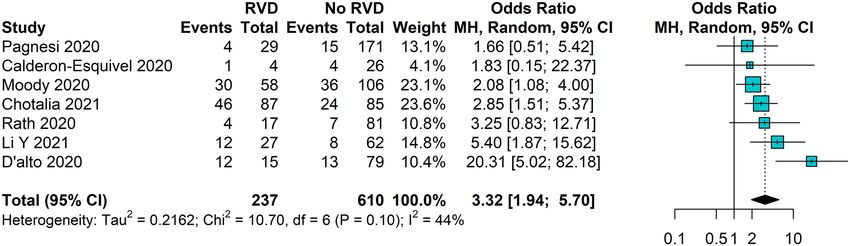
All‑cause death according to RVD. Seven studies reported all-cause death occurrence according to RVD status,
with a total of 847 patients included in the analysis. RVD was associated with a significantly increased likelihood
of all-cause death (OR 3.32, 95% CI 1.94–5.70), with a moderate grade of heterogeneity between studies (Fig. 3).
The leave-one-out analysis showed that excluding the study from D’Alto et al.20 reduces the pooled estimate, with
no heterogeneity among the 6 remaining studies ( I2 = 0%, Fig. S3). No publication bias was detected (Egger’s test
p = 0.446, Fig. S4).
Discussion
COVID-19 disease was defined as the third leading cause of death in the United States by October 202043. At
this stage of the pandemic, early identification of patients at higher risk of clinical deterioration is critical for
proper prognostic stratification and delivering the best care. Cardiovascular complications, including myocardial
dysfunction, has been described as a potential predictor of adverse outcomes44; although most studies focused
Scientific Reports | (2021) 11:17774 | https://doi.org/10.1038/s41598-021-96955-8 3
Vol.:(0123456789)www.nature.com/scientificreports/
Follow
Geographical Incl. Cohort Age up time
Study location criteria (N) RVD (N) (mean) Males (%) ICU (%) HTN (%) T2DM (%) Definition of RVD Outcomes (days)
Barman COVID-19
Other 90 15 56,4 51 32 35 15 TAPSE ≤ 16 mm Prevalence –
(2020)14 patients
COVID-19
TAPSE < 16 mm,
Bitar patients
Other 77 9 53* 83 100 26 32 RV S’ < 9.5 cm/s, Prevalence –
(2021)15 admitted to
RV FAC < 35%
ICU
COVID-19
patients
Bleakley
Europe underwent 84 20 52 74 100 36 22 TAPSE < 17 mm Prevalence –
(2020)16
mechanical
ventilation
COVID-19
Calderon- Prevalence,
patients
Esquivel Other 30 4 59 7 100 23 13 Unclear ICU mor- NR
admitted to
(2020)17 tality
ICU
COVID-19
patients
TAPSE < 17 mm,
Ceriani admitted
Europe 55 1 58,5* 64 0 36 13 RV S’ < 9.5 cm/s, Prevalence –
(2021)18 to medium
RV FAC < 35%
intensity
unit
COVID-19 Prevalence,
Chotalia TAPSE < 17 m, RV
Europe patients 172 87 59* 77 100 37 31 all-cause 90
(2021)19 FAC < 35%
with ARDS death
Right Ventricular-
arterial uncou- Prevalence,
D’alto COVID-19
Europe 94 15 63,6 75 39 67 17 pling (TAPSE/ all-cause NR
(2020)20 patients
PASP < 0.635 mm/ death
mmHg)
COVID-19
TAPSE < 16 mm,
Doyen patients
Europe 43 14 60 84 100 33 28 or RV S’ < 9,5 cm/s Prevalence –
(2020)21 admitted to
or RV FAC < 35%
ICU
COVID-19
Garcia-
patients
Cruz Other 82 22 56* 62 100 48 44 TAPSE < 17 mm Prevalence –
admitted to
(2020)22
ICU
COVID-19
Gibson patients
North America 32 5 56 66 100 50 41 TAPSE < 18 mm Prevalence –
(2021)23 admitted to
ICU
Giustino COVID-19 TAPSE < 17 mm or
Other 298 62 63 67 NR 59 37 Prevalence –
(2020)24 patients RV S′ < 9,5 cm/s
Iyengar-
COVID-19
Kapuganti North America 59 9 66,8 NR NR NR NR Unclear Prevalence –
patients
(2020)25
COVID-19
Jain patients
North America 52 18 59,9 60 100 69 37 Visual Prevalence –
(2021)26 admitted to
ICU
Karagodin COVID-19
Other 509 148 60* NR NR NR NR RVFWS > -20% Prevalence –
(2021)27 patients
TAPSE < 16 mm
Kim COVID-19
North America 268 41 64 66 68 63 41 and RV Prevalence –
(2020)28 patients
Sʹ < 10 mm/s
Krishna COVID-19
North America 179 54 59.8 62 62 60 37 Visual Prevalence –
(2021)29 patients
COVID-19 TAPSE, RV S’, RV Prevalence,
Li Y
Asia with previ- 89 27 66 57 22 79 19 FAC below pre- All-cause NR
(2021)30
ous CVD specified cut-offs mortality
COVID-19
Li YL
Asia patients 49 8 64,7 51 100 35 27 TAPSE < 17 mm Prevalence –
(2021)31
with ARDS
Liaqat COVID-19 TAPSE (unclear
Asia 181 29 44,6 59 NR 18 18 Prevalence –
(2021)32 patients cut-off)
Prevalence,
Moody COVID-19 TAPSE < 17 mm, 31
Europe 164 58 61 78 73 41 32 all-cause
(2020)33 patients or RV FAC < 35% (14- 42)
death
COVID-19
Norden patients Combined (RV
Europe 31 9 58 77 100 39 16 Prevalence –
(2021)34 admitted to Score)
ICU
Continued
Scientific Reports | (2021) 11:17774 | https://doi.org/10.1038/s41598-021-96955-8 4
Vol:.(1234567890)www.nature.com/scientificreports/
Follow
Geographical Incl. Cohort Age up time
Study location criteria (N) RVD (N) (mean) Males (%) ICU (%) HTN (%) T2DM (%) Definition of RVD Outcomes (days)
Non-ICU Prevalence,
Pagnesi TAPSE < 17 or RV
Europe COVID-19 200 29 66* 65 0 42 18 in-hospital 9 (4–14)
(2020)35 S’ < 9,5 cm/s
patients mortality
Prevalence,
Rath COVID-19
Europe 98 17 68 63 NR 70 24 TAPSE < 20 mm 30-day 30
(2020)36 patients
mortality
Schott COVID-19
North America 65 18 60 58 58 58 35 Unclear Prevalence –
(2020)37 patients
Soulat- TAPSE < 17 mm,
COVID-19
Dufour Europe 445 65 68 66 NR 60 29 RV S’ < 9.5 cm/s, Prevalence –
patients
(2021)38 RV FAC < 35%
Szekely COVID-19
Other 100 14 66,1 63 NR 57 29 TAPSE < 17 mm Prevalence –
(2020)39 patients
Van den TAPSE < 17 mm
COVID-19
Heuvel Europe 51 5 63* 8 31 41 18 or RV S’ veloc- Prevalence –
patients
(2020)40 ity < 10 cm/s
Vasudev COVID-19
North America 45 5 61,4 51 NR 64 55 Unclear Prevalence –
(2020)41 patients
Wats COVID-19 Combined (Visual,
North America 214 61 66,5 63 NR 68 36 Prevalence –
(2021)42 patients RV S’)
Table 1. Baseline characteristics of the included studies. ARDS acute respiratory distress syndrome, CVD
cardiovascular disease, HTN hypertension, ICU intensive care unit, NR not reported, PASP pulmonary arterial
systolic pressure, RVD right ventricular dysfunction, RVEF right ventricle ejection fraction, RV FAC right
ventricle fractional area change, RV FWS right ventricle free wall strain, RV S’ right ventricle systolic velocity,
TAPSE tricuspid annular plane systolic excursion, T2DM type 2 diabetes mellitus. *Median.
on left ventricular impairment, some reports clearly underlines a potential detrimental role of RVD in patients
with COVID-19.
Our study reports a comprehensive and updated systematic review and meta-analysis on the prevalence of
RVD and associated outcomes in patients with COVID-19. Overall, we found that the prevalence of RVD may
be as high as almost 1 out of 5 patients. Among the studies included, we observed a largely ranging prevalence of
RVD, possibly reflecting the heterogeneity in the sensitivity of the methods used to define RVD. Bleakley et al.16
observed that specific phenotypes of RVD may be present in patients with COVID-19, and that definition accord-
ing to TAPSE may have low sensitivity to detect RVD in this clinical scenario. The severity of the disease may also
represent one key factor influencing the prevalence of RVD among COVID-19 patients, although evidence is
limited. In our meta-analysis, we reported a large variation of PI, up to 44%; this information may be particularly
useful to interpret the findings of our study: our results indicates that, depending on the method used to define
RVD and the clinical setting, the prevalence of RVD in patients with COVID-19 may be higher than expected.
This hypothesis is confirmed by our meta-regression, which found that the proportion of severe COVID-19
patients enrolled was a significant predictor for higher prevalence of RVD in the studies included. The relatively
low R2 found for this association suggests that other factors are important in determining the prevalence of RVD,
but we were unfortunately unable to analyze them, and to perform multivariable meta-regressions, due to data
availability. Beyond that, and although this analysis has some clear limitation (the heterogeneous definition of
severe disease, and the study-level nature of this association), the results of our meta-regression may support a
mechanistic link between severe disease and RVD. However, further studies are needed to confirm this associa-
tion, and to evaluate the impact of other risk factors on the risk of RVD in COVID-19 patients.
Several physiopathological hypotheses sustain association between COVID-19 and RVD. COVID-19 related
ARDS is likely to be often complicated by RVD, given the direct alveolar injury and the associated ventilatory
strategies such as hyper-inflated lungs and permissive hypercapnia45. Moreover, a direct detrimental viral effect
on pulmonary microcirculation up to a pattern of endothelium with endothelial d ysfunction46 and increased vas-
cular inflammatory infiltrate was reported in autopsies from COVID-19 p atients3. As a matter of fact, an interplay
between COVID-19, angiotensin-converting enzyme 2, and pulmonary hypertension have been postulated47,48.
Furthermore, COVID-19 has been linked to an increased risk of venous thromboembolism (VTE) and pul-
monary embolism49, with the highest risk for patients with severe disease; moving from these evidence, VTE
may represent a critical cause of deterioration of RV function and performance50. Taken all pathophysiological
mechanisms together (parenchymal involvement, endothelial damage, and pulmonary embolism), right ven-
tricular overload with increased pulmonary pressures is likely to occur frequently. Furthermore, COVID-19 is
associated with direct myocardial injury through many different mechanisms, including inflammation, micro-
vascular dysfunction, hypoxia, and i schemia44, with also a COVID-19 related myocarditis d escribed51. Although
these manifestations may be more frequently causing left ventricular dysfunction, it is possible that they also
have a role, although often overlooked, in the onset of RVD.
Beyond these hypotheses, our findings demonstrated that patients with COVID-19 and RVD are exposed
to an excess of mortality than patients without RVD. Our results are in line to what has been observed in other
clinical settings characterized by respiratory infectious diseases; indeed, reduced right ventricular function was
reported as a risk factor for adverse events in community-acquired pneumonia52, as well as in patients with
Scientific Reports | (2021) 11:17774 | https://doi.org/10.1038/s41598-021-96955-8 5
Vol.:(0123456789)www.nature.com/scientificreports/
Figure 1. Pooled Prevalence of RVD among patients with COVID-19. CI confidence intervals, GLMM
generalized linear mixed model.
ARDS45. This information may be crucial for clinicians dealing with patients with COVID-19. In fact, bedside
ultrasound examination has become increasingly important in this pandemic, for the assessment of disease
progression, lung-heart interactions, and hemodynamic instability53, in a context where access to second-line
diagnostics tools is often reduced by logistic constraints or severity of the disease, as in the critically ill patients.
According to our data, careful assessment of RV function, which is often undervalued and overlooked in a fast
approach to cardiac ultrasound, may provide useful information and may drive specific therapeutic approaches.
Since most of the included cohorts reported about patients in ICU settings, these findings may not be immedi-
ately translated to all patients with COVID-19; however, further studies are required to confirm these results,
and to explore whether a standardized screening program for RVD dysfunction, as well as tailored therapeutic
approaches, may significantly improve the prognosis of these patients.
Limitations. This study has several limitations. First, most of the studies included in this meta-analysis are
retrospective or based on small cohorts, some of which were found at high risk of bias. This limitation, which may
have affected our results and estimates, is mainly due to the nature of the research question, and the pandemic
scenario in which these studies were conducted. Moreover, the heterogeneity in the definition of RVD may have
distorted our estimates on the pooled prevalence of the disease. To overcome these limitation (at least partially),
we reported PI, which gave a broader sense of the possible distribution of the actual prevalence in patients with
COVID-19. Furthermore, our leave-one-out analysis showed consistency of our results after excluding one study
at a time. Our study was not designed to assess factors that may influenced the association between COVID-19
and RVD; also, some baseline characteristics were missing or not reported in the original studies, and most stud-
ies did not provide details on the severity or grade of RVD. This limited our ability to evaluate the influence of
specific variables on the prevalence of RVD, or stratify our results according to RVD grading. We attempted to
estimate the impact of COVID-19 severity on RVD prevalence through a meta-regression analysis, which may
Scientific Reports | (2021) 11:17774 | https://doi.org/10.1038/s41598-021-96955-8 6
Vol:.(1234567890)www.nature.com/scientificreports/
Figure 2. Meta-regression analysis for the prevalence of RVD according to severity of COVID-19. RVD right
ventricular dysfunction.
Figure 3. All-cause death according to RVD status in patients with COVID-19. CI confidence intervals; RVD
right ventricular dysfunction.
help in understanding the association between severity of COVID-19 and burden of RVD. However, we were
not able to perform multivariable meta-regression with other risk factors, due to data availability; therefore, the
results of the meta-regression analysis should be interpreted with caution. Most of the patients were recruited in
ICUs and/or underwent mechanical ventilation, and these factors may have influenced the assessment of RVD
in the original studies and, in turn, our results. However, these patients represent a relevant part of individuals
with COVID-19, so that these findings are highly relevant to everyday practice. We were only able to analyse
the association between RVD and all-cause mortality in COVID-19 patients, since the original studies did not
report sufficient data on the causes of death observed. Further studies are required to analyse the impact of RVD
on different cause of mortality, including cardiovascular and COVID-19 related mortality.
Conclusion
Among patients with COVID-19, RVD can be found in almost 1 out of 5 patients; the prevalence may be influ-
enced by the severity of COVID-19 disease, but these results need confirmation in further studies. Patients with
RVD showed a threefold higher likelihood of all-cause death, compared to patients with normal RV function.
RVD may represent one important and often overlooked marker for prognostic stratification in COVID-19;
further studies are needed to clarify this association and investigate the specific management and therapeutic
approach for these patients.
Scientific Reports | (2021) 11:17774 | https://doi.org/10.1038/s41598-021-96955-8 7
Vol.:(0123456789)www.nature.com/scientificreports/
Received: 28 April 2021; Accepted: 12 August 2021
References
1. World Health Organization. WHO Director-General’s Opening Remarks at the Media Briefing on COVID-19-11 March 2020 (WHO,
2021).
2. World Health Organization. Weekly Epidemiological Update on COVID-19—13 July 2021 (WHO, 2021).
3. Varga, Z. et al. Endothelial cell infection and endotheliitis in COVID-19. The Lancet 395, 1417–1418 (2020).
4. Long, B., Brady, W. J., Koyfman, A. & Gottlieb, M. Cardiovascular complications in COVID-19. Am. J. Emerg. Med. 38, 1504–1507
(2020).
5. Jordan, R. E., Adab, P. & Cheng, K. K. Covid-19: Risk factors for severe disease and death. BMJ 368, 1–2 (2020).
6. Shi, S. et al. Association of cardiac injury with mortality in hospitalized patients with COVID-19 in Wuhan, China. JAMA Cardiol.
5, 802–810 (2020).
7. Gupta, A. K. et al. Current perspectives on coronavirus disease 2019 and cardiovascular disease: A white paper by the JAHA edi-
tors. J. Am. Heart Assoc. 9, e017013 (2020).
8. Romiti, G. F., Corica, B., Lip, G. Y. H. & Proietti, M. Prevalence and impact of atrial fibrillation in hospitalized patients with
COVID-19: A systematic review and meta-analysis. J. Clin. Med. 10, 2490 (2021).
9. D’Andrea, A. et al. Right ventricular function and pulmonary pressures as independent predictors of survival in patients with
COVID-19 pneumonia. Cardiovasc. Imaging 13, 2467–2468 (2020).
10. Romiti, G. F., Corica, B., Cangemi, R., Basili, S. & Raparelli, V. Need for innovative and timely synthesis of evidence during Covid-
19 outbreak. Eur. J. Intern. Med. 77, 165–166 (2020).
11. Stijnen, T., Hamza, T. H. & Özdemir, P. Random effects meta-analysis of event outcome in the framework of the generalized linear
mixed model with applications in sparse data. Stat. Med. 29, 3046–3067 (2010).
12. IntHout, J., Ioannidis, J. P. A., Rovers, M. M. & Goeman, J. J. Plea for routinely presenting prediction intervals in meta-analysis.
BMJ Open 6, e010247 (2016).
13. Riley, R. D., Higgins, J. P. T. & Deeks, J. J. Interpretation of random effects meta-analyses. BMJ 342, 964–967 (2011).
14. Barman, H. A. et al. Echocardiographic features of patients with COVID-19 infection: A cross-sectional study. Int. J. Cardiovasc.
Imaging. https://doi.org/10.1007/s10554-020-02051-9 (2020).
15. Bitar, Z. I., Shamsah, M., Bamasood, O. M., Maadarani, O. S. & Alfoudri, H. Point-of-care ultrasound for COVID-19 pneumonia
patients in the ICU. J. Cardiovasc. Imaging 29, 60–68 (2021).
16. Bleakley, C. et al. Right ventricular dysfunction in critically ill COVID-19 ARDS. Int. J. Cardiol. https://doi.org/10.1016/j.ijcard.
2020.11.043 (2020).
17. Calderón-Esquivel, N. et al. Correlación de variables ecocardiográficas y biomarcadores en pacientes graves con COVID-19. Cir.
Cir. 89, 1–6 (2020).
18. Ceriani, E. et al. Early echocardiographic findings in patients hospitalized for COVID-19 pneumonia: A prospective, single center
study. Intern. Emerg. Med. https://doi.org/10.1007/s11739-021-02733-9 (2021).
19. Chotalia, M. et al. Right ventricular dysfunction and its association with mortality in coronavirus disease 2019 acute respiratory
distress syndrome. Crit. Care Med. https://doi.org/10.1097/ccm.0000000000005167 (2021).
20. D’Alto, M. et al. Right ventricular-arterial uncoupling independently predicts survival in COVID-19 ARDS. Crit. Care 24, 1–10
(2020).
21. Doyen, D. et al. Characteristics of cardiac injury in critically ill patients with coronavirus disease 2019. Chest. https://doi.org/10.
1016/j.chest.2020.10.056 (2020).
22. García-Cruz, E. et al. Critical care ultrasonography during COVID-19 pandemic: The ORACLE protocol. Echocardiography 37,
1353–1361 (2020).
23. Gibson, L. E. et al. Right ventricular strain is common in intubated COVID-19 patients and does not reflect severity of respiratory
illness. J. Intens. Care Med. https://doi.org/10.1177/08850666211006335 (2021).
24. Giustino, G. et al. Characterization of myocardial injury in patients with COVID-19. J. Am. Coll. Cardiol. 76, 2043–2055 (2020).
25. Iyengar-Kapuganti, R. L. et al. Point-of-care ultrasound findings and clinical outcomes in patients with COVID-19. J. Am. Soc.
Echocardiogr. 33, 1416–1417 (2020).
26. Jain, R. et al. Comprehensive echocardiographic findings in critically ill COVID-19 patients with or without prior cardiac disease.
J. Patient Cent. Res. Rev. 8, 68–76 (2021).
27. Karagodin, I. et al. Echocardiographic correlates of in-hospital death in patients with acute COVID-19 infection: The world alli-
ance societies of echocardiography (WASE-COVID) STUDY. J. Am. Soc. Echocardiogr. https://doi.org/10.1016/j.echo.2021.05.010
(2021).
28. Kim, J. et al. Prognostic utility of right ventricular remodeling over conventional risk stratification in patients with COVID-19. J.
Am. Coll. Cardiol. 76, 1965–1977 (2020).
29. Krishna, H. et al. Cardiac abnormalities in COVID-19 and relationship to outcome. Mayo Clin. Proc. 96, 932–942 (2021).
30. Li, Y. et al. Echocardiographic characteristics and outcome in patients with COVID-19 infection and underlying cardiovascular
disease. Front. Cardiovasc. Med. https://doi.org/10.3389/fcvm.2021.642973 (2021).
31. Li, Y. L. et al. Acute right ventricular dysfunction in severe COVID-19 pneumonia. Rev. Cardiovasc. Med. 21, 635–641 (2020).
32. Liaqat, A., Ali-Khan, R. S., Asad, M. & Rafique, Z. Evaluation of myocardial injury patterns and ST changes among critical and
non-critical patients with coronavirus-19 disease. Sci. Rep. 11, 4828 (2021).
33. Moody, W. E. et al. Impact of right ventricular dysfunction on mortality in patients hospitalized with COVID-19, according to
race. CJC Open. https://doi.org/10.1016/j.cjco.2020.09.016 (2020).
34. Norden, N. et al. Cardiac involvement in critically ill and mechanically ventilated patients with COVID-19-a prospective, obser-
vational echocardiographic study. Am. J. Cardiovasc. Dis. 11, 253–261 (2021).
35. Pagnesi, M. et al. Pulmonary hypertension and right ventricular involvement in hospitalised patients with COVID-19. Heart 106,
1324–1331 (2020).
36. Rath, D. et al. Impaired cardiac function is associated with mortality in patients with acute COVID-19 infection. Clin. Res. Cardiol.
109, 1491–1499 (2020).
37. Schott, J. P. et al. Transthoracic echocardiographic findings in patients admitted with SARS-CoV-2 infection. Echocardiography
37, 1551–1556 (2020).
38. Soulat-Dufour, L. et al. Prognostic value of right ventricular dilatation in patients with COVID-19: A multicentre study. Eur. Heart
J. Cardiovasc. Imaging. https://doi.org/10.1093/ehjci/jeab067 (2021).
39. Szekely, Y. et al. Spectrum of cardiac manifestations in COVID-19: A systematic echocardiographic study. Circulation 142, 342–353
(2020).
40. van den Heuvel, F. M. A. et al. Cardiac function in relation to myocardial injury in hospitalised patients with COVID-19. Neth.
Heart J. 28, 410–417 (2020).
41. Vasudev, R. et al. The utility of bedside echocardiography in critically ill COVID-19 patients: Early observational findings from
three Northern New Jersey hospitals. Echocardiography 37, 1362–1365 (2020).
Scientific Reports | (2021) 11:17774 | https://doi.org/10.1038/s41598-021-96955-8 8
Vol:.(1234567890)www.nature.com/scientificreports/
42. Wats, K. et al. Association of right ventricular dysfunction and pulmonary hypertension with adverse 30-day outcomes in COVID-
19 patients. Pulm. Circ. 11, 204589402110070 (2021).
43. Woolf, S. H., Chapman, D. A. & Lee, J. H. COVID-19 as the leading cause of death in the United States. JAMA. https://doi.org/10.
1001/jama.2020.24865 (2020).
44. Lang, J. P. et al. A current review of COVID-19 for the cardiovascular specialist. Am. Heart J. 226, 29–44 (2020).
45. Vieillard-Baron, A., Price, L. C. & Matthay, M. A. Acute cor pulmonale in ARDS. Intens. Care Med. 39, 1836–1838 (2013).
46. Libby, P. & Lüscher, T. COVID-19 is, in the end, an endothelial disease. Eur. Heart J. 41, 3038–3044 (2020).
47. Park, J. F., Banerjee, S. & Umar, S. In the eye of the storm: The right ventricle in COVID-19. Pulm. Circ. 10, 204589402093666
(2020).
48. Cao, Y., Zhang, M., Guo, Y. & Zhang, Y. The overlooked chamber in coronavirus disease 2019. ESC Heart Fail. 7, 3483–3486 (2020).
49. Bompard, F. et al. Pulmonary embolism in patients with COVID-19 pneumonia. Eur. Respir. J. 56, 2001365 (2020).
50. Middeldorp, S. et al. Incidence of venous thromboembolism in hospitalized patients with COVID-19. J. Thromb. Haemost. 18,
1995–2002 (2020).
51. Pirzada, A., Mokhtar, A. T. & Moeller, A. D. COVID-19 and myocarditis: What do we know so far? CJC Open 2, 278–285 (2020).
52. Biteker, F. S. et al. Prognostic value of transthoracic echocardiography and biomarkers of cardiac dysfunction in community-
acquired pneumonia. Clin. Microbiol. Infect. 22, e1–e6 (2016).
53. Lazzeri, C., Bonizzoli, M., Batacchi, S. & Peris, A. Echocardiographic assessment of the right ventricle in COVID-related acute
respiratory syndrome. Intern. Emerg. Med. https://doi.org/10.1007/s11739-020-02494-x (2020).
Author contributions
B.C., A.M.M., M.P. and G.F.R. conceived and designed the study, and drafted the first version of the manuscript;
B.C. and G.F.R. acquired and analyzed the data; B.C., A.M.M., S.B., R.C., A.C., M.P. and G.F.R. revised the
manuscript critically and gave final approval of the version to be submitted.
Competing interests
AC: unrestricted grants from Merck-Serono; AMM: advisory board and lecture fees from MSD. SB received
research grants from MSD, outside the scope of this study. The other authors have nothing to disclose.
Additional information
Supplementary Information The online version contains supplementary material available at https://doi.org/
10.1038/s41598-021-96955-8.
Correspondence and requests for materials should be addressed to M.P.
Reprints and permissions information is available at www.nature.com/reprints.
Publisher’s note Springer Nature remains neutral with regard to jurisdictional claims in published maps and
institutional affiliations.
Open Access This article is licensed under a Creative Commons Attribution 4.0 International
License, which permits use, sharing, adaptation, distribution and reproduction in any medium or
format, as long as you give appropriate credit to the original author(s) and the source, provide a link to the
Creative Commons licence, and indicate if changes were made. The images or other third party material in this
article are included in the article’s Creative Commons licence, unless indicated otherwise in a credit line to the
material. If material is not included in the article’s Creative Commons licence and your intended use is not
permitted by statutory regulation or exceeds the permitted use, you will need to obtain permission directly from
the copyright holder. To view a copy of this licence, visit http://creativecommons.org/licenses/by/4.0/.
© The Author(s) 2021
Scientific Reports | (2021) 11:17774 | https://doi.org/10.1038/s41598-021-96955-8 9
Vol.:(0123456789)You can also read