Probiotic properties of a phytase producing Pediococcus acidilactici strain SMVDUDB2 isolated from traditional fermented cheese product, Kalarei ...
←
→
Page content transcription
If your browser does not render page correctly, please read the page content below
www.nature.com/scientificreports

            OPEN           Probiotic properties of a phytase
                           producing Pediococcus acidilactici
                           strain SMVDUDB2 isolated from
                           traditional fermented cheese
                           product, Kalarei
                           Deepali Bhagat1, Neelu Raina1, Amit Kumar2, Meenu Katoch3, Yugal Khajuria4,
                           Parvez Singh Slathia1 & Preeti Sharma1*
                           The nutritional challenge faced by the monogastric animals due to the chelation effects of phytic
                           acid, fuel the research on bioprospecting of probiotics for phytase production. Pediococcus acidilactici
                           SMVDUDB2 isolated from Kalarei, exhibited extracellular phytase activity of 5.583 U/mL after
                           statistical optimization of fermentation conditions viz. peptone (1.27%); temperature (37 °C); pH (6.26)
                           and maltose (1.43%). The phytase enzyme possessed optimum pH and temperature of 5.5 and 37 °C,
                           respectively and was thermostable at 60 °C. The enzyme was purified 6.42 fold with a specific activity
                           of 245.12 U/mg with hydrophobic interaction chromatography. The purified enzyme had Km and Vmax
                           values of 0.385 mM and 4.965 μmol/min respectively, with sodium phytate as substrate. The strain
                           depicted more than 80% survival rate at low pH (pH 2.0, 3.0), high bile salt concentration (0.3 and
                           0.5%), after gastrointestinal transit, highest hydrophobicity affinity with ethyl acetate (33.33 ± 0%),
                           autoaggregation (77.68 ± 0.68%) as well as coaggregation (73.57 ± 0.47%) with Staphylococcus
                           aureus (MTCC 3160). The strain exhibited antimicrobial activity against Bacillus subtilis (MTCC
                           121), Mycobacterium smegmatis (MTCC 994), Staphylococcus aureus (MTCC 3160), Proteus vulgaris
                           (MTCC 426), Escherichia coli (MTCC 1652) and Lactobacillus rhamnosus (MTCC 1408). The amount
                           of exopolysaccharide produced by the strain was 2 g/L. This strain having the capability of phytate
                           degradation and possessing probiotic traits could find application in food and feed sectors.

                           With increasing health and nutritional issues, the demand of functional foods is expected to reach 96 billion USD
                           by 20201. Probiotics are playing an important role in functional foods as they tend to increase the nutritional
                           value of food besides increasing the organoleptic properties. LAB (Lactic acid bacteria) producing phytase in
                           the gut can have therapeutic and nutritional aspect associated with them. Phytase in human food will increase
                           the availability of essential divalent mineral ions and release of myo-inositols which can serve as therapeutic
                           measure for various diseases like Crohn’s disease, Alzehmier’s disease, and irritable bowel syndrome. Phytase
                           (myo-inositol hexakisphosphatephosphohydrolases) hydrolyses the phosphoric monoester bonds in phytic acid,
                           a major component of phosphorous in plant diets has a strong chelation with divalent minerals such as Ca2+,
                           Mg2+, Zn2+ and Fe2+. The beneficial properties like enzymatic activity, susceptibility or resistance to selected
                           antibiotics associated with probiotic strains depend upon the source of isolation of the bacterial strain2. So it is
                           very important to have an appropriate beneficial bacterial strain in food products as the variation in beneficial
                           properties of bacterial strains are related to strain specificity3. Reports have suggested that P. acidilactici inhabit
                           the entire digestive tract of humans due to their ability to survive in extreme range of pH, temperatures, and
                           osmotic pressures. The importance of P. acidilactici has increased with some of the strains of this species have

                           1
                             School of Biotechnology, Shri Mata Vaishno Devi University, Katra, Jammu & Kashmir, 182320, India.
                           2
                             Instrumentation Division, CSIR - Indian Institute of Integrative Medicine, Canal Road, Jammu, 180001, India.
                           3
                            Microbial Biotechnology Division, CSIR - Indian Institute of Integrative Medicine, Canal Road, Jammu, 180001, India.
                           4
                            School of Physics, Shri Mata Vaishno Devi University, Katra, Jammu & Kashmir, 182320, India. *email: preeti.res@
                           gmail.com

Scientific Reports |   (2020) 10:1926 | https://doi.org/10.1038/s41598-020-58676-2                                                              1
www.nature.com/scientificreports/ www.nature.com/scientificreports
been cited as probiotic bacteria4. P. acidilactici strain FT28 has been reported for in vitro probiotic properties, a
probiotic strain with higher nutrient digestibility, heamato- biochemical and antioxidant properties when com-
pared to Lactobacillus acidophilus NCDC155. There are reports6,7 wherein strains have produced bacteriocins
against Bacillus subtilis and fungi. P. acidilactici (KTU05-7) and Pediococcus pentosaceus KTU05-8, KTU05-9.
KTU05-8 and KTU05-9 have shown highest extracellular phytase activity7 although report on production of exo-
polysaccharides (EPS) from these strains was absent.Thus, searching new strains of LAB with wider applications
is an increasing need of the hour with traditional fermented foods of Southeast Asian countries which are less
extensively explored, and some of the species from these sources could be of commercial potential8. P. acidilactici
Kp10, an isolate from a traditional dried curd has an ability to be used into food products because of following
properties viz. cell surface hydrophobicity, resistance to proteolytic activities, enzymatic and adhesion associ-
ated with it3. P. acidilactici M76 an isolate from Korean traditional rice wine, Makgeolli, was reported for pro-
duction of exopolysaccharide and as antitumor, antioxidant, antiulcer, antibacterial, immune-stimulating, blood
glucose-regulating, and UV radiation protective agent9. Most of the EPS-producing LAB strains produce heter-
opolysaccharides and homopolysaccharides composed of glucose, galactose and rhamnose and others containing
only either D-fructose or D-glucose10. Health benefits associated with some of the β-D-glucans polysaccharides
are antithrombotic, antitumor, or immunomodulatory effect. The effect of EPS, a 2-substituted-(1-3)-β-D-glucan
production was observed in case of Pediococcus damnosus (strain 2.6) with the change in concentration of glu-
cose, temperature, and bacto casamino acids11. Strains of Pediococcus reported for production of bacteriocins
include P. acidilactici Kp103 and P. acidilactici12. P. acidilactici with the capacity for the production of antimicro-
bial peptide and interesting properties such as resistance to heat, cold, pH and proteolytic treatments have given
tremendous importance to the strain13. P. acidilactici B14 strain has shown resistance to vancomycin and the
gene is considered to be inherent in Pediococcus genus13 but in some strains P. acidilactici Kp103 the strain was
found to be susceptible to vancomycin. Looking into diversified properties of Pediococcus genus, more and more
properties of P. acidilactici can be explored to obtain a strain having high industrial potentials. P. acidilactici strain
capable of utilizing pentose sugars such as xylose and arabinose, have shown high resistant towards inhibitors
from pretreated lignocellulosic biomass, such as furan derivatives, phenolic compounds and weak acids4 during
bioethanol production from lignocellulosic biomass. In our study P. acidilactici SMVDUDB2 is capable of pro-
ducing phytase, an exogenous enzyme suggesting potentially important mechanism of probiotic functionality and
the ability to survive in gastrointestinal tract conditions; its adhesive properties and potential of producing EPS
proves its wider industrial potential.
Results
Isolation, selection and identification of phytase producing LAB. Twenty potential LAB isolates
obtained from Kalarei samples were capable of surviving at 6.5% NaCl concentrations at pH 3.0 in MRS medium.
These 20 presumptive probiotic strains were gram positive and catalase negative and almost all had a phytate
degrading ability. The one exhibiting highest extracellular phytate degrading ability (5.18 ± 0.09 U/mL) was iden-
tified as P. acidilactici based on genotypic identification viz.16 S rRNA gene sequencing and designated as P.
acidilactici SMVDUDB2. The gene sequence has been submitted in NCBI GenBank and the accession number
given is MK280750.
Phytase studies. Phytase production has been earmarked as an essential criterion of probiotics. Qualitative
analysis of phytase in case of P. acidilactici SMVDUDB2 showed a zone formation of 8 mm which was higher than
L. rhamnosus MTCC 1408 (5 mm) on phytase screening medium (PSM) after 48 h at 37 °C while with spectropho-
tometric analysis the enzyme activity was 5.18 ± 0.09 U/mL and for L. rhamnosus (MTCC 1408) phytase activity
was 3.69 ± 0.12 U/mL.
Statistical optimization of phytase production. In order to enhance the phytase production from P.
acidilactici SMVDUDB2 statistical optimization of various physico-chemical factors was done. Plackett Burman
Design (PBD) facilitated the screening of important factors contributing positively to the phytase production14.
Studies have shown an effect of maltose, peptone, inoculum age, inoculum size, initial pH, incubation tempera-
ture and fermentation period on phytase production. Variation in response (phytase production) was observed
ranging from 4.93-5.37 U/mL (Supplementary Table 1). The analysis of regression coefficient values illustrated
that an incubation temperature, an initial pH, maltose and peptone have significant positive effect on phytase
production. The significance of the model (Supplementary Table 2) was stated from results of analysis of the
variance (ANOVA) based on PBD.
Determination of the optimal levels and the interaction of the selected variables for an enhanced phytase
production in case of P. acidilactici SMVDUDB2 was studied using Response Surface Methodology (RSM) and
Central Composite Design (CCD). The predicted and observed values of 30 experimental runs is shown in
Supplementary Table 3 and the result of analysis of variance (ANOVA) based on CCD experiments is depicted in
Supplementary Table 4. The phytase production was predicted by the following regression equation:
Y = + 5.37 − 0.34 (A) − 0.10(B) + 0.072(C) − 0.34(D) − 0.48(A2)
− 0.22(B2 ) − 0.54(C2 ) − 0.43(D2 ) + 5.625E − 003(AB) − 0.047(AC)
+ 0.16(AD) − 0.047(BC) − 0.047(BD) − 0.31(CD)
where Y is the predicted response of phytase production and A, B, C and D represent peptone, temperature, pH
and maltose respectively.
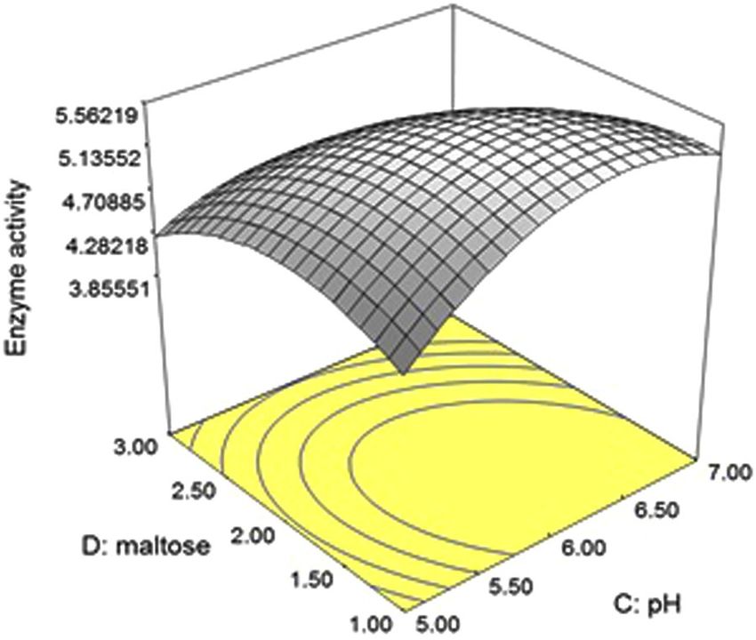
Among two positive interacting variables (maltose and pH) an increase in activity was observed with an
increase in maltose and pH as shown in the response surface (3D) plot (Fig. 1). The significance of the model was
Scientific Reports | (2020) 10:1926 | https://doi.org/10.1038/s41598-020-58676-2 2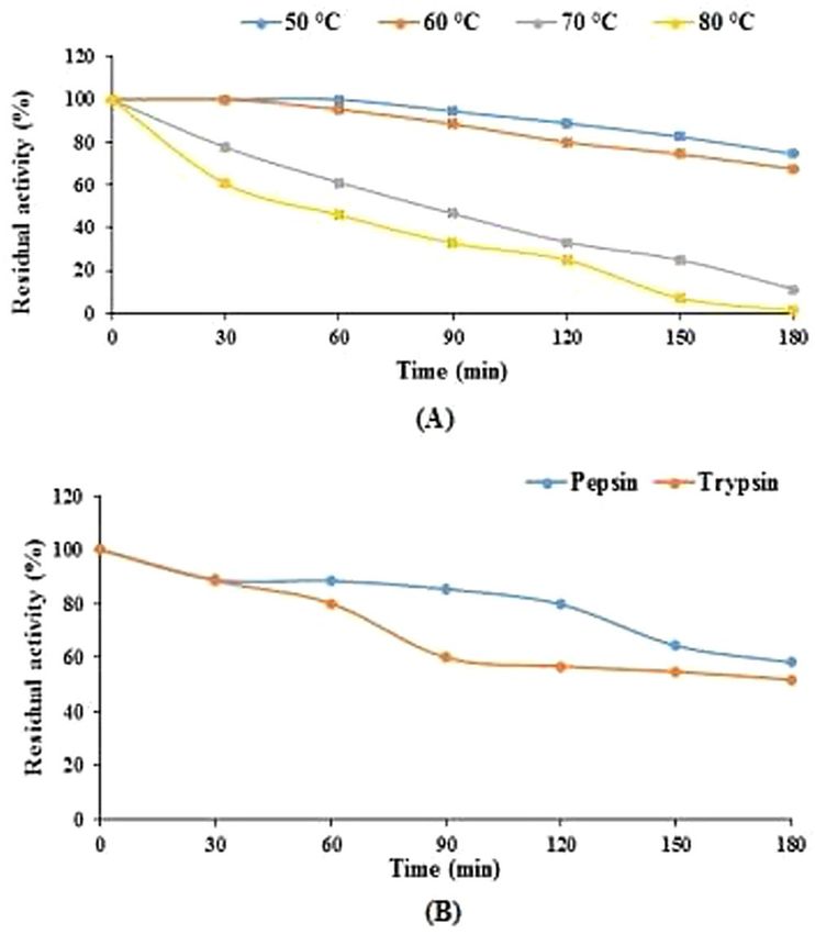
www.nature.com/scientificreports/                                                               www.nature.com/scientificreports

                           Figure 1. RSM plot showing interactive effect of maltose and pH on phytase production for P. acidilactici
                           SMVDUDB2.

                           stated by F value 5.27, the probability of the F value is due to noise, the chances for which are only 0.14%. The
                           values of “prob > F” less than 0.05 indicated that in this case A, D, A2, C2, D2, CD model terms are significant.
                           The “Lack of Fit F-value” of 4.61 indicated that a “Lack of Fit F-value” is not significant relative to the pure error.
                           The reliability of the model was stated by coefficient of variation (CV) value 13.87. A high correlation between
                           the predicted and observed response was attributed to the fitness of the model examined by the determination
                           coefficient i.e. R square (0.8310) which is closer to the value of 1.0. In present case the model designated a preci-
                           sion ratio of 6.724, greater than 4 which is the most desirable value indicating an adequate signal response. Point
                           prediction tool of RSM was used for validation of the statistical model. The model was validated by performing
                           the experiment under optimum value of 4 significant variables, peptone (1.27%); temperature (37 °C); pH (6.26)
                           and maltose (1.43%) and the precise closeness of predicted response (5.563 U/mL) and actual response (5.583 U/
                           mL) for phytase production, indicated the suitability of the model. Statistical model has been used to enhance the
                           production of phytase from several microorganisms15–17 but, here in we are reporting the optimization of extra-
                           cellular phytase from P. acidilactici using RSM.

                           Phytase purification and characterization.                Extracellular phytase from P. acidilactici SMVDUDB2 was
                           purified using ammonium sulphate precipitation, hydrophobic column chromatography and determined by
                           HPLC analysis as per literature studies18. The purification process resulted in 6.42 fold increase in specific activity
                           (245.12 U/mg) with 9.02% yield (Supplementary Table 5). SDS-PAGE and zymogram analysis (Supplementary
                           Fig. 1) confirmed the molecular weight of the protein to be 62 kDa, homogeneity of the purified enzyme has been
                           shown in Supplementary Fig. 2
                               The activity of enzyme was maintained over a broad pH range (2.5–12.5) (Supplementary Fig. 3A) and tem-
                           perature (20–60 °C) (Supplementary Fig. 3B). Enzyme optimum activity was observed at pH 5.5 and temperature
                           37 °C. The enzyme exhibited good thermostability (Fig. 2A) and the reduction in relative enzyme activity was
                           25.57% and 32.6% after 3 h of an incubation period at 50 °C and 60 °C respectively, while 38.89% and 54.16%
                           reduction was observed after 1 hour incubation period at 70 °C and 80 °C respectively (Fig. 2A).
                               The enzyme had strong proteolytic resistance towards pepsin and trypsin at the ratio of 1:1 (pepsin or
                           trypsin/phytase, v/v). Above 80% of phytase activity was retained after 1 hour incubation period in pepsin and
                           trypsin solution, which reduced to 58.21% and 51.81% after 3 h incubation period in pepsin and trypsin solution
                           (Fig. 2B).
                               The substrate specificity studies for extracellular phytase from P. acidilactici SMVDUDB2 revealed that the rel-
                           ative enzyme activity observed was 100% for sodium phytate followed by p- nitrophenyl phosphate (80.12 ± 0%),
                           glucose-6-phosphate (79.33 ± 0.79%) and sodium β-glycerophosphate (19.99 ± 0%). The maximum enzyme
                           specificity was towards sodium phytate, Km and Vmax values were calculated as 0.385 mM and 4.965 μmol/min,
                           respectively (Supplementary Fig. 4) for the same.

                           Probiotic Properties
                           Ability to withstand simulated gastrointestinal tract (GIT) conditions. P. acidilactici SMVDUDB2
                           showing maximum phytase production was explored for probiotic potentials and its survival rate at high acidic
                           pH (2.0, 3.0), in presence of high bile salts (0.3% and 0.5%), under simulated gastric (pepsin 3 mg/mL, pH 3) and
                           intestinal conditions (trypsin 1 mg/mL, pH 8) was higher as compared standard strain L. rhamnosus MTCC 1408
                           (Fig. 3A). The survival rate above 90% was observed at pH 3.0, 0.3% bile salt concentration and under simulated
                           gastrointestinal conditions in presence of proteolytic enzymes viz. pepsin and trypsin (Fig. 3A). While at pH 2.0
                           in presence of 0.5% bile salt concentration there was a slight decrease in the survival rate (above 80%).

Scientific Reports |   (2020) 10:1926 | https://doi.org/10.1038/s41598-020-58676-2                                                              3
www.nature.com/scientificreports/ www.nature.com/scientificreports
Figure 2. Thermostability and proteolysis resistance of the P. acidilactici SMVDUDB2 phytase. (A)
Thermostability of the purified enzyme. For thermostability, incubation of the enzyme was carried out at 50 and
60 °C for 0–180 min, separately. (B) Resistance of purified enzyme towards pepsin and trypsin. Residual phytase
activity of the purified phytase was determined after treating with pepsin or trypsin/phytase in the ratio of 1:1
(v/v) for an incubation period of 0–180 min. Data are mean ± SE (n = 3).
Figure 3. Probiotic properties of P. acidilactici SMVDUDB2 (A) Survival of P. acidilactici SMVDUDB2
and standard probiotic strain L. rhamnosus (MTCC 1408) under simulated gastrointestinal conditions. (B)
Hydrophobicity percent of P. acidilactici SMVDUDB2 and standard probiotic strain L. rhamnosus (MTCC
1408) with different organic solvents. Data are mean ± SE (n = 3).
Scientific Reports | (2020) 10:1926 | https://doi.org/10.1038/s41598-020-58676-2 4www.nature.com/scientificreports/ www.nature.com/scientificreports
Coaggregation %
Autoaggregation % B. subtilis M. smegmatis S. aureus P. vulgaris E. coli
4h 24 h 4h 24 h 4h 24 h 4h 24 h 4h 24 h 4h 24 h
P.
22.76 ± 0.47 77.68 ± 0.68 21.3 ± 0.29 71.18 ± 0.0 18.37 ± 0.19 67.92 ± 0.2 23.32 ± 0.16 73.57 ± 0.47 6.35 ± 0.32 61.67 ± 0.03 16.53 ± 0.25 69.55 ± 0.33
acidilactici
L.
26.31 ± 0.34 74.67 ± 0.36 24.29 ± 0.94 58.74 ± 0.2 31.11 ± 0.31 61.64 ± 0.8 38.84 ± 0.23 70.74 ± 0.6 28.68 ± 0.56 79.06 ± 0.4 39.55 ± 0.37 85.93 ± 0.21
rhamnosus
Table 1. Study of autoaggregation and coaggregation ability (%) of P. acidilactici SMVDUDB2 and standard
probiotic bacteria strain L. rhamnosus (MTCC 1408) with various bacterial pathogens. Data are mean ± SE
(n = 3). Significant difference between 4 h and 24 h (p < 0.05) by the Student’s t test.
Hydrophobicity, autoaggregation and coaggregation studies. In the present study maximum affin-
ity (33.33 ± 0%) was observed towards ethyl acetate (Fig. 3B), an alkalic and monopolar solvent revealing oxidiz-
ing (acidic) nature of LAB3. The autoaggregation ability was maximum (77.68 ± 0.68%) and a gradual increase is
reported after 24 h incubation at 37 °C (Table 1). As per earlier reports P. acidilactici has shown autoaggregation
of 47% after 2 h incubation at 37 °C19, but a varying level of coaggregation potential was observed with standard
MTCC cultures belonging to gram positive and gram negative pathogenic group, maximum (73.57 ± 0.47%)
coaggregation ability was observed with S. aureus.
L. rhamnosus MTCC 1408 exhibited relatively higher (above 50%) hydrophobicity than P. acidilactici
SMVDUDB2 and maximum affinity was observed with chloroform (73.45 ± 0.47%) while the least affinity was
observed with ethyl acetate (52.29 ± 0.67%) indicating the nonacidic and poor electron acceptor property of the
strain19. L. rhamnosus (MTCC 1408) exhibited good autoaggregation ability (74.67 ± 0.36%) after 24 h of incuba-
tion at 37 °C and the strain was highly coaggregated with E. coli (85.93 ± 0.21%).
Antibiotic susceptibility. Behaviour of P. acidilactici SMVDUDB2 was studied in relation to antibiotics,
as elimination of the risk of transmission of antibiotic-resistance genes is an important selection criterion for
probiotic bacteria20. P. acidilactici SMVDUDB2 was found to be sensitive towards cell wall synthesis, (ampicillin,
amoxicillin, cefotaxime, cefoperazone, cephalothin, cefepime, imipenem) protein synthesis, (streptomycin, tet-
racycline, erythromycin, nitrofurantoin, gentamicin, amikacin) RNA synthesis (rifampicin) and DNA synthesis
(moxifloxacin) inhibitors. The resistance against norfloxacin, penicillin-G, co-trimoxazole, nalidixic acid, cef-
podoxime, ceftazidime and vancomycin was seen. The standard strain L. rhamnosus MTCC 1408 was found to
be susceptible towards norfloxacin, cefpodoxime, cell wall synthesis, protein synthesis, RNA synthesis and DNA
synthesis inhibitors mentioned above, however resistance against cefepime, penicillin-G, co-trimoxazole, nali-
dixic acid, ceftazidime and vancomycin was observed for this strain also.
Antagonistic activity. Minimum inhibitory concentration (MIC) was determined by using cell free extract
(CFE) to study an inhibitory effect against all the pathogenic genera in case of fraction-A (non-neutralized
cell-free supernatant) and Fraction B (pH neutralized supernatant) In our case the inhibitory effect was due
to organic acid production and no pediocin-like substance was produced by the reported strain. P. acidilactici
SMVDUDB2 has shown lactic acid yield (18.32 ± 0.30 mg/mL) comparable with L. rhamnosus MTCC 1408 strain
(16.99 ± 0.17 mg/mL). MIC value of the potential probiotic LAB strain was 6% to inhibit the growth of Bacillus
subtilis, Mycobacterium smegmatis, Staphylococcus aureus, and Proteus vulgaris while 8% of MIC was observed
against Escherichia coli. The importance of organic acids as strong inhibitors against Gram negative bacteria has
been reported, whereby the lactic acid acts as permeator of the outer membrane of these bacteria13,21.
A higher MIC value was observed i.e. 10% to inhibit B. subtilis and 12% to inhibit M. smegmatis, S. aureus, P.
vulgaris and E. coli was observed in case of MTCC 1408.
Exopolysaccharide (EPS) production. The present strain reported 2 g/L of EPS. The FTIR spectra of EPS
from P. acidilactici SMVDUDB2 is presented in Fig. 4. A continuous absorption at around 3245 cm−1 is assigned
to the hydroxyl group (O-H), which is a characteristic of carbohydrate ring. Strong bands around 2924 cm−1 cor-
responded to stretching vibration of the methylene group (C-H), usually present in hexoses22. The band at 1647
cm−1 was associated to the stretching vibration of carboxyl group (C = O)23. Absence of peak around 1529.8 cm–1
which corresponds to an amino group was absent in our case. A peak at 1214 cm−1 corresponded to the presence
of C-O stretching in ether or alcohol groups24. The peak around 1025 cm−1 recommended the presence of C-O-H
link bond position22. The spectra exhibited the typical bands of polysaccharides which is within the fingerprint
region (1200–950 cm−1)23. In addition the peak at around 809.3 cm−1 was characteristic for mannose22,25. Our
observation reported the presence of mannose monomer in EPS from P. acidilactici SMVDUDB2 and absence of
glucuronic acid or diacetyl ester monomeric unit.
Discussion
The ability of phytase to hydrolyze phytic acid and release phosphate from phytate26 has highlighted the lactic
acid fermentations of corn, lentils and peas. Lactic acid bacterial strains designated as probiotics isolated from
dairy product sources have fulfilled the Food and Drug Administration (FDA) category and recognized as safe.
“GRAS” status has been assigned to them and they have been found to be fit for use in fermented dairy and
non-dairy products27. To apply the effect of probiotics, these microorganisms should be in an adequate amount
i.e. 106 colony-forming units (CFU) per gram or milliliter of product when consumed. Their survival in GIT
Scientific Reports | (2020) 10:1926 | https://doi.org/10.1038/s41598-020-58676-2 5www.nature.com/scientificreports/ www.nature.com/scientificreports
Figure 4. FTIR spectrum of purified EPS from P. acidilactici SMVDUDB2 in the range of 400–4000 cm−1.
conditions should be such that it provides therapeutic benefits along with phytate degrading ability which is an
important criteria of potential probiotic strains28. Intrinsic resistance towards vancomycin is a trait reported in
probiotic groups, belonging to Lactobacilli, Pediococci and Leuconostoc genera29,30. Resistance against vancomycin
and cefotaxime in case of P. acidilactici B14 strain has been reported and vancomycin resistance gene is inherited
in Pediococcus genus13.
Pediococcus is commonly known LAB belonging to homofermentative group capable of catabolizing one mole
of glucose in presence of limited oxygen in Embden–Meyerhof–Parnas (EMP) pathway to yield lactic acid only31.
Reports have shown that production of lactic acid results in lowering of the pH in case of LAB, resulting in low-
ering or stopping the growth of bacterial pathogens and sometimes even killing them32. No pediocin activity was
exhibited by P. acidilactici SMVDUDB2, but statistical optimization of medium components and fermentation
parameters may result in bacteriocin production33. According to a recent report intracellular stimulation by inter-
nalized inulin nanoparticles lead to enhancement in pediocin production by P. acidilactici12.
In present study, P. acidilactici SMVDUDB2 has fulfilled an essential prerequisite ability of probiotic strain
to survive in harsh GIT environment with survival rate higher than the literature reports (P. acidilactici B14)
survival rate was 45.9% at pH 2.5; 72.4% in 0.3% bile salt concentration and 95.8% after passing through gastro-
intestinal tract viz. pH 4.013.
Our strain has shown diversified properties such as phytase production, EPS producing ability and a survival
rate more than 80% at low pH, high bile salt concentrations (0.3% and 0.5%). Its adhesive property is expressed in
terms of hydrophobicity. Highest affinity was seen with ethyl acetate whereas lowest with xylene. The lowest affin-
ity (11.85%) to xylene in our case is comparable to P. acidilactici (10.4%)19. Autoaggregation and coaggregation
properties of these probiotic good bacteria make them predominant over the pathogenic bacteria as they outpass
the competition with pathogenic coubterparts for the points of attachment on the GIT and because of the com-
petition for nutrients34. Adhesion and colonization of intestinal wall by probiotic bacteria is based on the surface
properties of the strain20. Variations in the properties do exist at strain, species and genus levels and the health
benefits are associated with a strain of a particular genus or species which may not be extrapolated to other mem-
bers of same genus/species. Therefore, each bacterial probiotic strain needs to be investigated. Reports have sug-
gested that coaggregation ability is strain specific against indicator strains and the present study is very much in
accordance with earlier reports35. Food associated LAB are known as potential producers of EPS31 and their EPS
can be used as prebiotics36. Prebiotics are non-digestible polysaccharides, which act as substrates for fermentation
by gut symbionts and stimulate the colonization of beneficial bacteria. However, owing to the lack of expansive
knowledge on EPSs from the food-associated LABs, EPSs have remained largely underexploited9. Structural anal-
ysis of P. acidilactici M76 by using HPLC, thin-layer chromatography (TLC) and FTIR has confirmed presence
of a glucan consisting of glucose units9. On comparison of FTIR graph similar peaks were observed in our study,
however the absence of glucuronic acid and diacetyl ester in EPS was ascertained by the missing characteristic
absorption peak around the region 1700–1770 cm−1 24. P. pentosaceus P 773, isolated from a spoiled beer pro-
duced 3.9 g/L of EPS in presence of 100 g/L of sucrose for 48 h 37 after 48 h fermentation period37.The constituents
of EPS were glucose and fructose residues present in ratio of 3:1. P. acidilactici M76 produced more than 2.0 g/L
of EPS9 comparable to yields obtained from P. acidilactici SMVDUDB2 indicating the potential of the strain for
industrial applications.
The enhanced extracellular phytase (5.583 U/ml) production was the result of two positive interactive varia-
bles viz maltose and pH, exhibiting proteolytic resistance against pepsin and trypsin enzymes in the gastrointes-
tinal conditions. The enzyme reported has shown maximum substrate specificity with sodium phytate as in case
of Lactobacillus coryniformis wherein the values of Km and Vmax were 0.32 mM and 4.83 μmol/min respectively38.
A good thermostability of an enzyme, an important criteria of interest growing in industrial as well as scientific
communities towards thermo-acido-philic enzymes17 has been fulfilled in case of P. acidilactici SMVDUDB2.
In conclusion, P. acidilactici SMVDUDB2 isolated from Kalarei exhibited excellent probiotic properties as
compared to standard probiotic strain L. rhamnosus (MTCC 1408). Phytase producing ability of this probiotic
strain is considered as one of the potentially important mechanism of probiotic functionality, its high surviv-
ing ability in gastrointestinal tract conditions; its adhesive properties and potential of producing EPS under
Scientific Reports | (2020) 10:1926 | https://doi.org/10.1038/s41598-020-58676-2 6www.nature.com/scientificreports/ www.nature.com/scientificreports
Low level High level
Symbol Code Variables Units (−1) (+1)
A Fermentation period h 43 53
B Inoculum age h 43 53
C Inoculum size % (v/v) 5 15
D Incubation temperature °C 32 42
E Initial pH — 5.5 6.5
F Maltose % (w/v) 0.5 1.5
G Peptone % (w/v) 0.5 1.5
H, J, K, L Virtual factors — −1 +1
Table 2. Experimental levels of variables for phytase production in Plackett–Burman design.
statistically optimized enzyme conditions proves its wide industrial potential. The proteolytic resistance property
of enzyme along with tolerance over wide pH range and temperature contributes to repository of phytase with
different origin, properties and has a bright scope for broader application in industries.
Materials and Methods
Media, chemicals and bacterial cultures. Microbiological media, chemicals and biochemicals used
in present study were purchased from Sigma- Aldrich Chemical Ltd. (St. Louis, MO, USA), HiMedia Pvt. Ltd.
(Mumbai, India) and SD Fine Chemicals (Mumbai, India). Antibiotics used for susceptibility test were procured
from HiMedia Pvt. Ltd. (Mumbai, India). The microbial strains: Bacillus subtilis (MTCC 121), Mycobacterium
smegmatis (MTCC 994), Staphylococcus aureus (MTCC 3160), Proteus vulgaris (MTCC 426), Escherichia
coli (MTCC 1652) and Lactobacillus rhamnosus (MTCC 1408) were purchased from Microbial Type Culture
Collection (MTCC), Institute of Microbial Technology (IMTECH), Chandigarh, India.
Isolation of lactic acid bacteria (LAB). Twelve samples of Kalarei, an indigenous fermented cheese prod-
uct, were collected from urban and rural areas of Jammu region for isolation of probiotic bacteria. For isolation of
LAB serially diluted Kalarei samples were spread on de Man, Rogosa and Sharpe (MRS) agar medium and incu-
bated at 37 °C for 48 h under anaerobic conditions. Presumptive identification was done based on gram staining,
microscopic examination and catalase production. The strains were sub-cultured and maintained on MRS agar
slants. For long term preservation glycerol stock (50%, v/v) were maintained at −80 °C in MRS.
Screening of cultures for phytase enzyme. Phytase studies were carried out by growing the bacterial
cultures in modified MRS broth (MRS-MOPS) in which inorganic phosphate (KH2PO4) was replaced by 0.65 g/L
of sodium phytate 0.1 M 3-(N-Morpholino) propanesulfonic acid (MOPS). In order to reduce the final phosphate
content and to promote phytase production, in the above stated media, the contents of glucose, beef extract and
yeast extract were reduced to 10, 4 and 2 g/L, respectively39. Modified MRS-MOPS medium was inoculated with
10% (v/v) of 48 h grown cultures (108 CFU/mL) and incubated under anaerobic conditions at 37 °C.
For studying qualitative phytate degrading ability plate assay method was followed17,40. The phytase screen-
ing media (PSM) was composed of 1.5% glucose, 0.1% sodium phytate, 0.2% NH 4NO3, 0.05% KCl, 0.05%
MgSO4.7H2O, 0.03% MnSO4, 0.03% FeSO4.7H2O and 2.0% agar (pH 7.5). In order to ensure that the zone forma-
tion is due to phytate hydrolysis rather than an acid production by the LAB strains, the agar plates were washed
using distilled water and petri plates were flooded with 2% (w/v) aqueous cobalt chloride solution. After 5 min
of incubation at room temperature the cobalt chloride solution was replaced with a freshly prepared solution
containing equal volumes of 6.25% (w/v) aqueous ammonium molybdate) solution and 0.42% (w/v) ammonium
meta vanadate solution. After 5 min incubation, the ammonium molybdate/ammonium vanadate solution was
removed and the plates were examined for zone of hydrolysis17.
For quantitative phytase assay, the amount of liberated inorganic phosphate ions from sodium phytate was
measured. The presence of crude extracellular phytase was determined using a reaction mixture consisting of
800 µL sodium phytate (1% w/v) prepared in 0.2 M sodium acetate buffer (pH 5.5) and 200 µL of crude enzyme
extract. After an incubation period of 1 h at 37 °C, the reaction was stopped by heating the reaction mixture for
10 min at 100 °C. The liberated phosphate ions were quantified by spectrophotometric method40, for this 100 μL
of assay mixture was mixed with 900 μL of colour reagent comprised of 1.0 M H2SO4, 10% ascorbic acid, 2.5%
ammonium molybdate (3:1:0.1) (v/v) and after 20 mins of an incubation at 50 °C, absorbance was measured at
820 nm. A standard curve was plotted by using KH2PO4. One unit of phytase activity was defined as the amount
of enzyme that released 1 μmol of phosphate per minute under the assay conditions. Specific phytase activity was
defined as U per mg of protein. Protein concentration in the enzyme preparation and during purification steps
was determined by Bradford method41 using bovine serum albumin as standard. The culture producing highest
amount of phytase was selected for further studies.
Statistical optimization of fermentation conditions for phytase production. Plackett–Burman
design (PBD) was employed to determine significant variables for which medium components (carbon and nitro-
gen sources) and cultivation parameters (initial pH, inoculum age, inoculum size, incubation temperatures and
incubation periods) were taken into consideration. Each parameter was examined at two levels (high level and a
low level) as illustrated in (Table 2). A total of 12 experimental runs were generated and analysed by using Design
Expert 6 software (State-Ease Inc., Minneapolis, U.S.A.).
Scientific Reports | (2020) 10:1926 | https://doi.org/10.1038/s41598-020-58676-2 7www.nature.com/scientificreports/ www.nature.com/scientificreports
Symbol code Variables Unit Low level High level
A Peptone % (w/v) 1 2
B Incubation temperature °C 35 40
C Initial pH — 5 7
D Maltose % (w/v) 1 3
Table 3. Level of the different independent variables used in CCD for phytase production from P. acidilactici
SMVDUDB2.
Central composite design (CCD) of RSM was used for 4 variables (incubation temperature, initial pH, malt-
ose and peptone) identified with significant impact on phytase production. Lower and higher levels of selected
variables were depicted in Table 3 and 30 experimental runs were executed. Statistical model was validated based
on point prediction method.
Purification and characterization of phytase enzyme. Partial purification of extracellular phytase
was carried out using ammonium sulphate precipitation followed by hydrophobic chromatographic studies using
Phenyl-Sepharose CL-4B hydrophobic column matrix42.
The extracellular enzyme was separated and quantified by a Shimadzu HPLC system (Kyoto, Japan) consisting
of an LC-10 ATvp pump, SIL-10 ADvp automatic sampling unit (auto sampler), CTO-10 and SCL-10 Avp as the
system controller. Class VP software (version 6.10) was used for data analysis and data processing. The samples
were analyzed at 30 °C on RP-18.5 μm, 250 × 4 mm i.d. Merck (Darmstadt, Germany) column. UV detection was
performed at 210 nm. The analysis was carried out using a mobile phase comprised of 0.1% trifluroacetic acid in
water (solvent A) and acetonitrile (solvent B) at a flow rate of 1.0 mL/min.
The apparent molecular mass of the purified enzyme was estimated by SDS-PAGE according to Laemmli43.
Electrophoresis was carried out in 12% acrylamide gel using vertical Mini-PROTEAN gel system (Bio-Rad,
China). The proteins were stained with Coomassie brilliant blue. The zymogram was prepared by soaking the
gel first in 1% Triton X-100 for 1 h at room temperature and then 0.2 M sodium acetate buffer (pH 5.5) at 4 °C for
a period of 1 h. Phytase activity was detected by incubating the gel for 16 h in a 0.2 M sodium acetate buffer (pH
5.5) containing 0.4% (w/v) sodium phytate. Activity bands were visualized by immerging the gel in a 2% (w/v)
aqueous cobalt chloride solution. After 5 min of incubation at room temperature, the cobalt chloride solution was
replaced with a freshly prepared solution containing equal volumes of a 6.25% (w/v) aqueous ammonium molyb-
date solution and 0.42% (w/v) ammonium vanadate solution. Phytase activity was evident as zones of clearing in
an opaque background44.
The optimized pH of purified enzyme was determined over the range of 2.5–12.5 using glycine-HCL buffer
(pH 2.5), acetate buffer (pH 3.5–5.5), citrate buffer (pH 6.5), glycine–NaOH buffer (pH 8.5–10.5) and KCl-NaOH
buffer (pH 12.5).
For optimum temperature studies enzyme substrate complex mixture was incubated at various tempera-
tures viz. 20, 30, 37, 50 and 60 °C. Thermostability test for enzyme was determined by preincubating the purified
enzyme at 50 °C, 60 °C, 70 °C and 80 °C for 3 h. The residual activity of thermotolerant enzyme was calculated after
a regular interval of 30 min.
Phytase resistance to proteolytic enzymes (pepsin and trypsin) was evaluated according to the method
reported in literature45 with little modifications. The purified enzyme (10 µg/mL) was incubated in 0.2 M
glycine-HCl buffer (pH 2.5) containing pepsin (10 µg/mL) in the ratio of 1:1, and 0.2 M Tris-HCl buffer (pH 8.0)
containing trypsin (10 µg/mL) in the ratio of 1:1 at 37 °C for a period of 3 h. As a control, the purified enzyme was
incubated in the same condition as stated above but lacking pepsin or trypsin and the percentage residual activity
was calculated after a regular interval of 30 min.
Substrate specificity of purified extracellular phytase enzyme was determined using various phosphorylated
substrates like sodium phytate, glucose-6-phosphate, sodium β-glycerophosphate and p- nitrophenyl phosphate
at 3 mM concentration. Km and Vmax were measured from Lineweaver-Burk plot using different concentrations of
sodium phytate (0.1–1.8 mM) prepared in sodium acetate buffer (0.2 M, pH 5.5).
Molecular identification of selected LAB isolate. Bacterial genomic DNA of potential phytase produc-
ing LAB isolate was extracted using DNA purification kit. Identification was based on 16 S rRNA gene sequenc-
ing. The universal primers used were (lac1–27 F 5′-AGAGTTTGATCCTGGCTCAG-3′ and lac 1-1492 R 5′
TACGGYTACCTTGTTACGACT-3′). The conditions in PCR programme were as follows: initial denaturation at
94 °C for 2 min, denaturation at 94 °C for 30 s, annealing at 55 °C for 1 min, extension at 72 °C for 1 min and final
extension at 72 °C for 10 min. The PCR product obtained was sequenced by AgriGenome Labs Pvt. Ltd., Kerala.
Basic Local Alignment Search Tool (BLAST) was used to analyse the homology of sequence and the sequence
was submitted in NCBI GenBank. Phylogenetic tree made from sequenced 16 S rRNA region of the isolate and
evolutionary analyses were conducted using Clustal Omega.
Probiotic properties. Tolerance to simulated gastrointestinal tract (GIT) conditions. Survival rate was
determined at low pH (2.0 and 3.0), high bile salt concentration (0.3% and 0.5%) under simulated GIT conditions
as per earlier reports46,47 and a comparative study was carried out with standard probiotic bacterial strain MTCC
1408. Evaluation of pathogenicity factor was carried out by studying the lipolytic effect of the isolate13.
Scientific Reports | (2020) 10:1926 | https://doi.org/10.1038/s41598-020-58676-2 8www.nature.com/scientificreports/ www.nature.com/scientificreports
Hydrophobicity, autoaggregation and coaggregation studies. Organic solvents viz. ethyl acetate, xylene, chloro-
form were used for hydrophobicity studies, autoaggregation and coaggregation28 was carried out using MTCC
cultures (B. subtilis, M. smegmatis, S. aureus, P. vulgaris and E. coli) for evaluating adhesive properties. Similar
studies were carried out with standard probiotic strain.
Antibiotic susceptibility test. For antibiotic susceptibility test, disc diffusion method was employed as per earlier
reports48. Petriplates were incubated under anaerobic conditions at 37 °C for 24 h. The plates were examined for
zone of inhibition around the antibiotic disks. Twenty two antibiotic discs viz. rifampicin (5 mcg), moxifloxa-
cin (5 mcg), ampicillin (10 mcg), amoxicillin (10 mcg), imipenem (10 mcg), gentamicin (10 mcg), norfloxacin
(10 mcg), penicillin-G (10 mcg), erythromycin (15 mcg), streptomycin (25 mcg), co-trimoxazole (25 mcg), cefo-
taxime (30 mcg), cephalothin (30 mcg), cefepime (30 mcg), tetracycline (30 mcg), amikacin (30 mcg), nalidixic
acid (30 mcg), cefpodoxime (30 mcg), ceftazidime (30 mcg) and vancomycin (30 mcg)), cefoperazone (75 mcg),
nitrofurantoin (300 mcg), were screened against standard probiotic strain MTCC 1408 as well as LAB isolated
from Kalarei.
Antagonistic activity and organic acid production. Minimum inhibitory concentration (MIC) of cell free extract
(CFE) for standard strain MTCC 1408 and LAB isolate was determined49 against standard MTCC cultures,
B. subtilis (MTCC 121), M. smegmatis (MTCC 994), S. aureus (MTCC 3160), P. vulgaris (MTCC 426), E. coli
(MTCC 1652) grown on nutrient agar medium. For studying antagonistic effect and to determine the presence of
bacteriocin like inhibitory substance (BLIS), 48 h grown culture was centrifuged (10,000 × g for 10 min at 4 °C)
and the filter sterilized cell free extracts (CFEs) were obtained. Standard MTCC cultures (1% of the bacterial solu-
tion having 108 cfu/mL) were inoculated in Luria-Bertani broth (E. coli, S. aureus) and nutrient broth (B. subtilis,
M. smegmatis and P. vulgaris) containing CFE of LAB (0, 2, 4, 6, 8, 10 and 12%) and incubated for a period of 24 h
at 37 °C under aerobic conditions49. The filter sterilized CFEs were assayed as follows: Fraction A, cell-free super-
natant and Fraction B, consisted of pH neutralized supernatant (pH 7.0) for detection of inhibition by organic
acids32. MIP was monitored by measuring optical density (OD600 nm).
Organic acid (lactic acid) production was determined by titration method50 for selected LAB isolate as well
as MTCC 1408. Amount of organic acid produced from 48 h grown culture in MRS medium at 37 °C was deter-
mined by using titration method described by AOAC50. Ten millilitre of supernatant from LAB and 3 drops of
phenolphthalein as indicator was added for titration against 0.1 M NaOH. The NaOH from the burette was added
slowly to the samples until colour changes to pink. Total titratable acidity of lactoc acid (LA) was measured by
the following formula:
Total titratable acidity of LA (mg/mL) = mL NaOH × N NaOH × M.E ÷ volume of sample used, where total
volume of NaOH used was represented in mL, N denotes normality of NaOH used and mass equivalent (M.E)
represents equivalent factor which is equal to 90.08 mg of lactic acid51.
Exopolysaccharide (EPS) production. The LAB isolate from Kalarei was inoculated in MRS broth supplemented
with 1% maltose as carbon source and incubated at 37 °C for 48 h. EPS were isolated from CFE collected after
centrifugation at 10000 × g for 30 min at 4 °C followed by ethanol precipitation, dialysis and lyophilization52. The
concentration of EPS was determined by phenol-sulphuric acid methodology53. The EPS sample was analyzed by
Fourier- transform infrared (FTIR) spectroscopy. Sample was prepared by mixing lyophilized EPS sample with
KBr and the spectra were recorded with a light source in the range of 4000–400 cm−1 36.
Received: 20 August 2019; Accepted: 30 December 2019;
Published: xx xx xxxx
References
1. Research, T. M. Global Probiotic Market to reach worth of US$96 billion by 2020 End, Expanding Availability in Retail Stores and New
Product Launches impart Momentum to Growth, https://www.globenewswire.com/news-release/2018/06/06/1517768/0/en/Global-
Probiotic-Market-to-reach-worth-of-US-96-billion-by-2020-End-Expanding-Availability-in-Retail-Stores-and-New-Product-
Launches-impart-Momentum-to-Growth.html (2018).
2. Zielinska, D. & Kolozyn-Krajewska, D. Food-Origin Lactic Acid Bacteria May Exhibit Probiotic Properties: Review. BioMed research
international 2018, 5063185, https://doi.org/10.1155/2018/5063185 (2018).
3. Abbasiliasi, S. et al. In vitro assessment of Pediococcus acidilactici Kp10 for its potential use in the food industry. BMC microbiology
17, 121, https://doi.org/10.1186/s12866-017-1000-z (2017).
4. Callejon, S., Sendra, R., Ferrer, S. & Pardo, I. Recombinant laccase from Pediococcus acidilactici CECT 5930 with ability to degrade
tyramine. PloS one 12, e0186019, https://doi.org/10.1371/journal.pone.0186019 (2017).
5. Dowarah, R., Verma, A. K., Agarwal, N., Singh, P. & Singh, B. R. Selection and characterization of probiotic lactic acid bacteria and
its impact on growth, nutrient digestibility, health and antioxidant status in weaned piglets. PloS one 13, e0192978, https://doi.
org/10.1371/journal.pone.0192978 (2018).
6. Cizeikiene, D., Juodeikiene, G., Paskevicius, A. & Bartkiene, E. Antimicrobial activity of lactic acid bacteria against pathogenic and
spoilage microorganism isolated from food and their control in wheat bread. Food Control 31, 539–545 (2013).
7. Cizeikiene, D., Juodeikiene, G., Bartkiene, E., Damasius, J. & Paskevicius, A. Phytase activity of lactic acid bacteria and their impact
on the solubility of minerals from wholemeal wheat bread. International journal of food sciences and nutrition 66, 736–742, https://
doi.org/10.3109/09637486.2015.1088939 (2015).
8. Abbasiliasi, S. et al. Isolation of Pediococcus acidilactici Kp10 with ability to secrete bacteriocin-like inhibitory substance from milk
products for applications in food industry. BMC microbiology 12, 260–260, https://doi.org/10.1186/1471-2180-12-260 (2012).
9. Song, Y. R., Cha, Y. S. & Baik, S. H. Exopolysaccharide produced by Pediococcus acidilactici M76 isolated from the Korean
traditional rice wine, Makgeolli. Journal of microbiology and biotechnology 23, 681–688 (2013).
10. Duenas, M., Munduate, A., Perea, A. & Irastorza, A. Exopolysaccharide production by Pediococcus damnosus 2.6 in a semidefined
medium under different growth conditions. International journal of food microbiology 87, 113–120 (2003).
11. Duenas-Chasco, M. T. et al. Structural analysis of the exopolysaccharide produced by Pediococcus damnosus 2.6. Carbohydrate
research 303, 453–458 (1997).
Scientific Reports | (2020) 10:1926 | https://doi.org/10.1038/s41598-020-58676-2 9www.nature.com/scientificreports/ www.nature.com/scientificreports
12. Kim, W. S. et al. A new way of producing pediocin in Pediococcus acidilactici through intracellular stimulation by internalized
inulin nanoparticles. Scientific reports 8, 5878, https://doi.org/10.1038/s41598-018-24227-z (2018).
13. Ribeiro, M. C. D. O. et al. Evaluation of probiotic properties of Pediococcus acidilactici B14 in association with Lactobacillus
acidophilus ATCC 4356 for application in a soy based aerated symbiotic dessert. Brazilian Archives of Biology and Technology 57,
755–765 (2014).
14. Neira-Vielma, A. A. et al. Optimized production of phytase by solid-state fermentation using triticale as substrate and source of
inducer. African Journal of Biotechnology 17, 81–90 (2018).
15. Joshi, S. & Satyanarayana, T. Optimization of heterologous expression of the phytase (PPHY) of Pichia anomala in P. pastoris and its
applicability in fractionating allergenic glycinin from soy protein. Journal of industrial microbiology & biotechnology 41, 977–987,
https://doi.org/10.1007/s10295-014-1407-6 (2014).
16. Bhavsar, K., Kumar, V. R. & Khire, J. M. High level phytase production by Aspergillus niger NCIM 563 in solid state culture: response
surface optimization, up-scaling, and its partial characterization. Journal of industrial microbiology & biotechnology 38, 1407–1417,
https://doi.org/10.1007/s10295-010-0926-z (2011).
17. Chanderman, A., Puri, A. K., Permaul, K. & Singh, S. Production, characteristics and applications of phytase from a rhizosphere
isolated Enterobacter sp. ACSS. Bioprocess and biosystems engineering 39, 1577–1587, https://doi.org/10.1007/s00449-016-1632-7
(2016).
18. Gunashree, B. S. & Venkateswaran, G. Extracellular phytase from Aspergillus niger CFR 335: purification and characterization. J
Food Sci Technol 52, 4558–4564, https://doi.org/10.1007/s13197-014-1304-z (2015).
19. Xu, H., Jeong, H. S., Lee, H. Y. & Ahn, J. Assessment of cell surface properties and adhesion potential of selected probiotic strains.
Letters in applied microbiology 49, 434–442, https://doi.org/10.1111/j.1472-765X.2009.02684.x (2009).
20. Giri, S. S., Sen, S. S., Saha, S., Sukumaran, V. & Park, S. C. Use of a potential probiotic, Lactobacillus plantarum L7, for the
preparation of a rice-based fermented beverage. Frontiers in microbiology 9, 473 (2018).
21. Servin, A. L. Antagonistic activities of lactobacilli and bifidobacteria against microbial pathogens. FEMS microbiology reviews 28,
405–440, https://doi.org/10.1016/j.femsre.2004.01.003 (2004).
22. Shen, J.-W., Shi, C.-W. & Xu, C.-P. Exopolysaccharides from Pleurotus pulmonarius: Fermentation optimization, characterization
and antioxidant activity. Food Technology and Biotechnology 51, 520 (2013).
23. Abid, Y. et al. Production and structural characterization of exopolysaccharides from newly isolated probiotic lactic acid bacteria.
International journal of biological macromolecules 108, 719–728, https://doi.org/10.1016/j.ijbiomac.2017.10.155 (2018).
24. Kanamarlapudi, S. & Muddada, S. Characterization of Exopolysaccharide Produced by Streptococcus thermophilus CC30. BioMed
research international 2017, 4201809, https://doi.org/10.1155/2017/4201809 (2017).
25. Mathlouthi, M. & Koenig, J. L. Vibrational spectra of carbohydrates. Advances in carbohydrate chemistry and biochemistry 44, 7–89
(1986).
26. Zhao, Y., Zhu, L., Lin, C., Shen, Z. & Xu, C. Transgenic soybean expressing a thermostable phytase as substitution for feed additive
phytase. Scientific reports 9, 14390, https://doi.org/10.1038/s41598-019-51033-y (2019).
27. Ghosh, T., Beniwal, A., Semwal, A. & Navani, N. K. Mechanistic Insights Into Probiotic Properties of Lactic Acid Bacteria Associated
With Ethnic Fermented Dairy Products. Frontiers in microbiology 10, 502, https://doi.org/10.3389/fmicb.2019.00502 (2019).
28. Andrabi, S. T., Bhat, B., Gupta, M., Bajaj, B. K. J. P. & Proteins, a. Phytase-producing potential and other functional attributes of lactic
acid bacteria isolates for prospective probiotic applications. 8, 121–129 (2016).
29. Jena, P. K. et al. Isolation and characterization of probiotic properties of Lactobacilli isolated from rat fecal microbiota. Microbiology
and immunology 57, 407–416, https://doi.org/10.1111/1348-0421.12054 (2013).
30. Manini, F. et al. Characterization of lactic acid bacteria isolated from wheat bran sourdough. LWT-food Science and Technology 66,
275–283 (2016).
31. Patel, S., Majumder, A. & Goyal, A. Potentials of exopolysaccharides from lactic Acid bacteria. Indian journal of microbiology 52,
3–12, https://doi.org/10.1007/s12088-011-0148-8 (2012).
32. Angmo, K., Kumari, A. & Bhalla, T. C. Antagonistic activities of lactic acid bacteria from fermented foods and beverage of Ladakh
against Yersinia enterocolitica in refrigerated meat. Food bioscience 13, 26–31 (2016).
33. Suganthi, V. & Mohanasrinivasan, V. Optimization studies for enhanced bacteriocin production by Pediococcus pentosaceus
KC692718 using response surface methodology. Journal of food science and technology 52, 3773–3783, https://doi.org/10.1007/
s13197-014-1440-5 (2015).
34. Plessas, S., Bosnea, L., Alexopoulos, A. & Bezirtzoglou, E. Potential effects of probiotics in cheese and yogurt production: A review.
Engineering in Life Sciences 12, 433–440 (2012).
35. Balakrishna, A. In vitro evaluation of adhesion and aggregation abilities of four potential probiotic strains isolated from guppy
(Poecilia reticulata). Brazilian Archives of Biology and Technology 56, 793–800 (2013).
36. Palaniyandi, S., Damodharan, K., Suh, J.-W. & Yang, S. H. Functional Characterization of an Exopolysaccharide Produced by
Bacillus sonorensis MJM60135 Isolated from Ganjang. Journal of microbiology and biotechnology 28, 663–670 (2018).
37. Semjonovs, P. & Zikmanis, P. Evaluation of novel lactose-positive and exopolysaccharide-producing strain of Pediococcus
pentosaceus for fermented foods. European Food Research and Technology 227, 851–856 (2008).
38. Demir, Y., Dikbas, N. & Beydemir, S. Purification and Biochemical Characterization of Phytase Enzyme from Lactobacillus
coryniformis (MH121153). Mol Biotechnol 60, 783–790, https://doi.org/10.1007/s12033-018-0116-1 (2018).
39. Raghavendra, P. & Halami, P. M. Screening, selection and characterization of phytic acid degrading lactic acid bacteria from chicken
intestine. Int J Food Microbiol. 133, 129–134, https://doi.org/10.1016/j.ijfoodmicro.2009.05.006 (2009).
40. Mittal, A. et al. Isolation and biochemical characterization of acido-thermophilic extracellular phytase producing bacterial strain for
potential application in poultry feed. Jundishapur Journal of Microbiology 4, 273–282 (2011).
41. Bradford, M. M. A rapid and sensitive method for the quantitation of microgram quantities of protein utilizing the principle of
protein-dye binding. Anal Biochem 72, 248–254 (1976).
42. Soni, S. K., Magdum, A. & Khire, J. M. Purification and characterization of two distinct acidic phytases with broad pH stability from
Aspergillus niger NCIM 563. World journal of microbiology & biotechnology 26, 2009–2018, https://doi.org/10.1007/s11274-010-
0385-8 (2010).
43. Laemmli, U. K. Cleavage of structural proteins during the assembly of the head of bacteriophage T4. Nature 227, 680–685 (1970).
44. Bae, H. D., Yanke, L. J., Cheng, K. J. & Selinger, L. B. A novel staining method for detecting phytase activity. Journal of microbiological
methods 39, 17–22 (1999).
45. Zhang, G. Q. et al. Purification, characterization, and cloning of a novel phytase with low pH optimum and strong proteolysis
resistance from Aspergillus ficuum NTG-23. Bioresource technology 101, 4125–4131, https://doi.org/10.1016/j.biortech.2010.01.001
(2010).
46. Guo, Z. et al. In vitro comparison of probiotic properties of Lactobacillus casei Zhang, a potential new probiotic, with selected
probiotic strains. 42, 1640–1646 (2009).
47. Tulumoglu, S. et al. Probiotic properties of lactobacilli species isolated from children’s feces. Anaerobe 24, 36–42, https://doi.
org/10.1016/j.anaerobe.2013.09.006 (2013).
48. Masalam, B. et al. Isolation, Molecular Characterization and Probiotic Potential of Lactic Acid Bacteria in Saudi Raw and Fermented
Milk. 2018 (2018).
Scientific Reports | (2020) 10:1926 | https://doi.org/10.1038/s41598-020-58676-2 10www.nature.com/scientificreports/ www.nature.com/scientificreports
49. AL-Saadi, Z. N. Estimation of Minimum Inhibitory Concentration (MIC) and Minimum Bactericidal Concentration (MBC) of
Cell-Free Extracts of Bifidobacterium Species Against Methicillin-Resistant Staphylococcus aureus in vitro. American Journal of
Biomedical and Life Sciences 4, 75–80 (2016).
50. Chemists, A. A. & Horwitz, W. Official methods of analysis. Vol. I. 15th ed. AOAC, Arlington, VA 489 (1990).
51. Wakil, S. & Ajayi, O. O. Production of lactic acid from Starchy-based food substrates. Journal of Applied Biosciences 71, 5673–5681
(2013).
52. Khalil, E. S. et al. Probiotic characteristics of exopolysaccharides-producing Lactobacillus isolated from some traditional Malaysian
fermented foods. CyTA Journal of Food 16, 287–298 (2018).
53. Dubois, M., Gilles, K. A., Hamilton, J. K., Rebers, P. T. & Smith, F. Colorimetric method for determination of sugars and related
substances. Analytical chemistry 28, 350–356 (1956).
Acknowledgements
The authors would like to thank Department of Biotechnology, Government of India (Award no. BT/Bio-
CARe/03/636/2011–12) and University Grant Commission for Rajiv Gandhi National Fellowship [Award no.
17.1/2011–12/RGNF-SC-JAM-5449/ (SA-III/Website)]. The authors are thankful to Dr. Yugal Khajuria and
Anshul Uppal, School of Physics, Shri Mata Vaishno Devi University for doing the FTIR analysis.
Author contributions
The corresponding author acknowledges the support and keen interest shown by all the contributing authors of
the manuscript. Designing of experiments and manuscript preparation was carried out by Preeti Sharma and
Parvez Singh Slathia. Experimental bench work was done by Deepali Bhagat and Neelu Raina. Meenu Katoch and
Amit carried out HPLC analysis of the enzyme. FTIR analysis was carried out by Yugal Khajuria.
Competing interests
The authors declare no competing interests.
Additional information
Supplementary information is available for this paper at https://doi.org/10.1038/s41598-020-58676-2.
Correspondence and requests for materials should be addressed to P.S.
Reprints and permissions information is available at www.nature.com/reprints.
Publisher’s note Springer Nature remains neutral with regard to jurisdictional claims in published maps and
institutional affiliations.
Open Access This article is licensed under a Creative Commons Attribution 4.0 International
License, which permits use, sharing, adaptation, distribution and reproduction in any medium or
format, as long as you give appropriate credit to the original author(s) and the source, provide a link to the Cre-
ative Commons license, and indicate if changes were made. The images or other third party material in this
article are included in the article’s Creative Commons license, unless indicated otherwise in a credit line to the
material. If material is not included in the article’s Creative Commons license and your intended use is not per-
mitted by statutory regulation or exceeds the permitted use, you will need to obtain permission directly from the
copyright holder. To view a copy of this license, visit http://creativecommons.org/licenses/by/4.0/.
© The Author(s) 2020
Scientific Reports | (2020) 10:1926 | https://doi.org/10.1038/s41598-020-58676-2 11You can also read