Promotion of biological nitrogen fixation activity of an anaerobic consortium using humin as an extracellular electron mediator - Nature
←
→
Page content transcription
If your browser does not render page correctly, please read the page content below
www.nature.com/scientificreports
OPEN Promotion of biological nitrogen
fixation activity of an anaerobic
consortium using humin
as an extracellular electron
mediator
Sujan Dey1,6, Takanori Awata2,6, Jumpei Mitsushita1, Dongdong Zhang3,5, Takuya Kasai1,3,
Norihisa Matsuura4 & Arata Katayama1,3*
Nitrogen fertiliser is manufactured using the industrial Haber–Bosch process, although it is extremely
energy-consuming. One sustainable alternative technology is the electrochemical promotion of
biological nitrogen fixation (BNF). This study reports the promotion of BNF activity of anaerobic
microbial consortia by humin, a solid-phase humic substance, at any pH, functioning as an
extracellular electron mediator, to levels of 5.7–11.8 times under nitrogen-deficient conditions. This
was evidenced by increased acetylene reduction activity and total nitrogen content of the consortia.
Various humins from different origins promoted anaerobic BNF activity, although the degree of
promotion differed. The promotion effected by humin differed from the effects of chemical reducing
agents and the effects of supplemental micronutrients and vitamins. The promotion of anaerobic BNF
activity by only reduced humin without any other electron donor suggested that humin did not serve
as organic carbon source but as extracellular electron mediator, for electron donation to the nitrogen-
fixing microorganisms. The next generation sequencing (NGS) of partial 16S rRNA genes showed the
predominance of Clostridiales (Firmicutes) in the consortia. These findings suggest the effectiveness
of humin as a solid-phase extracellular electron mediator for the promotion of anaerobic BNF activity,
potentially to serve for the basis for a sustainable technology.
Fixation of atmospheric nitrogen to ammonia is crucial to maintain the global nitrogen cycle and provide N
fertiliser for the growth of plants, including crops. At present, the majority of nitrogen fertilisers are produced
by the Haber–Bosch process1. However, ammonia production by the Haber–Bosch process is extremely energy-
consuming, utilising 1–2% of total world energy-use and releasing approximately 2.5% of global CO2 emissions2,3.
Biological nitrogen fixation (BNF) is expected to become an alternative to the Haber–Bosch process as a means of
ammonia production because of its lower energy consumption. For the application of BNF to produce ammonia,
high activity of nitrogen-fixing microorganisms is essential because of the slow kinetics of biological production
of ammonia4.
The process of BNF is carried out by diazotrophs, using nitrogenase, an enzyme catalysing the biological
N-reducing reaction (Eq. 1).
N2 + 8H+ + 8e− + 16ATP → 2NH3 + H2 + 16ADP + 16Pi (1)
Nitrogenase is a complex oxidoreductase that is classified into three different types based on the heterometal
cofactor present in the active site of the enzyme complex5,6. There is a wide range of diazotrophs, including free
living (consisting of aerobic, microaerophilic, and anaerobic) and symbiotic bacteria, cyanobacteria, and archaea.
1
Graduate School of Engineering, Nagoya University, Chikusa‑ku, Nagoya 464‑8603, Japan. 2National Institute
for Land and Infrastructure Management, Asahi 1, Tsukuba, Ibaraki 305‑0804, Japan. 3Institute of Materials and
Systems for Sustainability, Nagoya University, Chikusa‑ku, Nagoya 464‑8603, Japan. 4School of Geosciences and
Civil Engineering, Kanazawa University, Kakuma‑machi, Kanazawa, Ishikawa 920‑1192, Japan. 5Present address:
Ocean College, Zhejiang University, Zhoushan 316021, China. 6These authors contributed equally: Sujan Dey and
Takanori Awata. *email: katayama.arata@nagoya-u.jp
Scientific Reports | (2021) 11:6567 | https://doi.org/10.1038/s41598-021-85955-3 1
Vol.:(0123456789)www.nature.com/scientificreports/
Some species of Geobacter, for example G. sulfurreducens and G. metallireducens, have also been reported as
N-fixers7,8. These broadly distributed diazotrophs fix atmospheric nitrogen under complex and specific condi-
tions, including the type of host plants and a range of environmental factors.
In recent years, efforts have been made to increase the activity of nitrogenase by direct input of electrons:
electrocatalysis9, bioelectrocatalysis using immobilised n itrogenase10, photocatalysis11–15, and nitrogen reduction
by transition metal catalysis mimicking n itrogenase16–18. These have been devoted to finding efficient alternatives
for the Haber–Bosch process to synthesise ammonia under ambient conditions. However, in these approaches,
limitations arise from low conversion efficiencies, poor selectivity and expensive c atalysts19. Moreover, irrevers-
ible damage to nitrogenase occurs when it is exposed to O 2 through immobilisation, making bioelectrocatalysis
untenable for N reduction. Nitrogenase loosed the activity also by changes in the conditions such as pH. In
addition, the ammonia synthesised in these approaches must be converted to fertiliser to make it available for
plants, another energy-consuming chemical reaction. These makes the use of purified enzymes more expensive
and difficult in handlings, compared to self-regulated intact c ell20. Therefore, promotion of BNF in living, intact,
microorganisms, rather than nitrogen fixation of purified enzymes, can be a more sustainable approach to the
Haber–Bosch process. Because the reaction rates of BNF are dependent on energy for dinitrogen reduction to
ammonia, the supply of electrons to nitrogen-fixing microorganisms through extracellular electron mediators
may increase the efficiency of the BNF reaction (Eq. 1), thus accelerating BNF.
Recently, various extracellular electron mediators including humic substances and artificial electron mediators
have been reported to accelerate different microbial reactions in relation with carbon and nitrogen c ycles21–27.
Some artificial electron mediators, methyl viologen and other viologens, have been used in dinitrogen fixation for
the bio-electrochemical synthesis of a mmonia28. However, methyl viologen and other viologens are water soluble,
toxic to the environment, and human h ealth29. Water-soluble and toxic extracellular electron mediators can be
washed away from the reactor and into the environment, decreasing the applicability of the mediator. Conse-
quently, insoluble, chemically stable, non-toxic materials should be candidates as extracellular electron mediators
for promoting microbial processes. Humin is an environmentally harmless organo-mineral humic substance
extracted from soil, insoluble in any pH conditions, formed by the decomposition of organic materials. Cur-
rently, humin has been reported as an extracellular electron mediator for different microbial reducing reactions,
including reductive d ehalogenation30,31, nitrate reduction to ammonia, iron r eduction32, and denitrification33.
Although humin has advantageous characteristics as an extracellular electron mediator, including its stability in
nature and no loss in its application, the effect of humin on BNF has not been reported to-date.
Thus, the objective of this study is to examine if the BNF activity/efficiency of anaerobic consortia can be
promoted by supplying extracellular electrons through an extracellular electron mediator-humin.
Results
Promotion of BNF activity in anaerobic consortia by humin. Two nitrogen-fixing anaerobic micro-
bial consortia were enriched from Kamajima paddy soil by transfer using an anaerobic, modified nitrogen-
deficient, Ashby medium (anaerobic MNDA medium) with and without supplementation of humin every two
weeks (herein referred to as humin consortium and no-humin consortium, respectively). Anaerobic BNF activ-
ity of the consortia was determined using both acetylene reduction activity (ARA) and the increase in nitrogen
content in the consortia. The enriched consortia exhibited stable ARA after the 11th generation of enrichment
(Fig. S1). Thus, consortia after the 11th generation were subjected to the experiments in this study. Figure 1a
shows the effect of humin on ARA in the consortium. The humin consortium exhibited higher ARA than the no-
humin consortium, regardless of the ARA test conditions with and without humin. In addition, both consortia
showed higher ARA under test conditions with humin than those without humin. The promotion of anaerobic
BNF activity was also confirmed by the increase in total N in the consortia after incubation at 30 °C for 14 days
(Fig. 1b). Extensive promotion of anaerobic BNF activity was observed in the humin consortium (342% to the
control) compared with the no-humin consortium (127% to the control). No increase was observed in humin
only (without inoculum), indicating no chemical nitrogen fixation. These results clearly suggest that the promo-
tion of anaerobic BNF activity in the humin consortium required both the consortium and humin. It should be
noted that anaerobic BNF activity was promoted by humin beyond the level observed under nitrogen-deficient
conditions. Figure 1a shows that humin addition promoted ARA by 90% for no-humin consortium and by 40%
for humin consortium in the test. In comparison between the no-humin consortium tested without humin and
the humin consortium with humin, ARA was higher by 6.7 times in the latter. Based on the relative amount of
total nitrogen (Fig. 1b), counting the nitrogen fixation in the no-humin consortium (27%) and the contribution
of nitrogen content of the humin itself (23%), the humin consortium promoted the BNF reaction by 292%, 11.8
times of the no-humin consortium, under nitrogen-deficient conditions.
Ubiquitous promotion effect of Humin on anaerobic BNF activity. Effects of various humins
extracted from different source soils and sediment were examined using the ARA test applied to the no-humin
consortium (Fig. 2). All humins significantly promoted the ARA of the no-humin consortium compared with
the no-humin consortium without humin. Among the five humins, the largest promotion was observed in the
consortium with Kamajima humin, extracted from Kamajima paddy soil. Therefore, Kamajima humin was used
for further studies.
Effect of redox state of humin on anaerobic BNF activity. The BNF activity of washed and starved
no-humin consortium was examined by the ARA test using Helium (He)-bubbled, anaerobic, mannitol-free,
MNDA medium supplemented with Kamajima humin in different redox states, in comparison with the no-
humin control medium. Figure 3 shows that the consortium tested with the reduced humin only exhibited
Scientific Reports | (2021) 11:6567 | https://doi.org/10.1038/s41598-021-85955-3 2
Vol:.(1234567890)www.nature.com/scientificreports/
Figure 1. (a) ARA of the humin and no-humin consortia using testing conditions with (+) and without humin
(−); (b) changes in the relative total nitrogen content in the cultures under four conditions during 14 days of
incubation: humin consortium, no-humin consortium, humin only and control (medium only, as 100%). The
humin used here was Kamajima humin.
Figure 2. Effects of various humins on ARA of the no-humin consortium. All humins examined promoted
ARA of the no-humin consortium. Kamajima humin yielded the highest ARA in the no-humin consortium.
ARA, which increased two fold, in proportion with the increase in the amount of reduced humin added to
the medium. However, the consortium tested with oxidised humin, intact humin, and without humin did not
show any activity. The promotion of BNF activity of the consortium was also made apparent in the significant
(P < 0.05) increase in total nitrogen content in the consortium, but only for the consortium with reduced humin,
after 14 days of incubation in the anaerobic mannitol-free MNDA medium at 30 °C (Fig. 4). Total nitrogen con-
tent in consortia incubated under other conditions did not increase after the incubation, and remained almost
the same as that on day zero.
Scientific Reports | (2021) 11:6567 | https://doi.org/10.1038/s41598-021-85955-3 3
Vol.:(0123456789)www.nature.com/scientificreports/
Figure 3. ARA of the no-humin consortium tested with reduced, oxidised and intact humin and without
humin. The humin used here was Kamajima humin. Humin concentration was set at two levels as shown by the
symbols “1x” and “2x”, indicating 15 g/L and 30 g/L, respectively. ND not detected.
Figure 4. Effect of humin preparation on the nitrogen fixation activity of the no-humin consortium, tested
using reduced, oxidised and intact humin, as shown by the change in relative nitrogen content during 14 days of
incubation. Gray bars denote the nitrogen contents at 0 day (100%), and black bars denote those after 14 days.
The humin used here was Kamajima humin.
Effects of reducing agents on anaerobic BNF activity. The effect of three commonly used reducing
agents (Na2S, cysteine, and titanium (III) trinitriloacetate (Ti-NTA)), on the BNF activity of the no-humin con-
sortium was examined by the ARA test in anaerobic MNDA medium, with and without humin. Figure 5 shows
that the reducing agents did not promote, but rather inhibited, the BNF activity of the no-humin consortium.
The introduction of cysteine and Ti-NTA did not significantly change ARA of the no-humin consortium, while
the no-humin consortium tested with humin (positive control) exhibited much higher ARA. The BNF activity of
the consortium was higher in the medium with humin in addition to cysteine or Ti-NTA, but still less than that
of the positive control. The consortium tested with Na2S showed no ARA, regardless of the addition of humin,
indicating an inhibitory effect. These findings suggest that the promoting effect of humin on BNF activity dif-
fered from that of the reducing agents.
Effect of micronutrients and vitamins on anaerobic BNF activity. The effect of micronutrients and
vitamins on BNF activity was examined by the ARA test in anaerobic MNDA medium supplemented with trace
element solution SL-10 and vitamin solution under conditions with and without humin. Figure 6 shows that the
ARA of the humin consortium and no-humin consortium tested with and without humin under the conditions
supplemented with micronutrients and vitamins. Compared with Fig. 1a, the supplementation increased ARA
by 52 ± 10% as average of four test conditions. However, the supplementation of micronutrients and vitamins did
not change the promoting effect of humin on ARA in the consortia. The addition of humin increased ARA by
77% for the no-humin consortium, and by 40% for the humin consortium, respectively. In comparison between
the no-humin consortium tested without humin and the humin consortium with humin, ARA was higher by
5.7 times in the latter. Higher ARA was observed in the humin consortium and under the test conditions with
humin (Fig. 6), the same trend as Fig. 1a. These results suggest that humin does not work as a source of micronu-
trients and vitamins to promote BNF activity. Iron, cobalt, manganese, chromium, zinc and nickel are commonly
Scientific Reports | (2021) 11:6567 | https://doi.org/10.1038/s41598-021-85955-3 4
Vol:.(1234567890)www.nature.com/scientificreports/
Figure 5. Effect of reducing agents on ARA of the no-humin consortium. The no-humin consortium tested
with addition of cysteine or Ti-NTA as reducing agent showed less ARA than the no-humin consortium tested
with humin (Kamajima humin, positive control). ARA of the no-humin consortium tested with cysteine or
Ti-NTA was increased by the addition of humin, although the value was still lower than the positive control. The
reducing agent Na2S inhibited ARA of the consortium regardless of the addition of humin. ND not detected.
Figure 6. ARA of the no-humin and humin consortia tested in the medium supplemented with micronutrients
and vitamins with (+) and without humin (−). The humin used here was Kamajima humin.
Figure 7. Copy numbers of the 16S rRNA gene (gray bar) and nifH gene (black bar) in the no-humin
consortium and in the humin consortium in the qPCR. The amplicons of the genes were used to estimate
the total and N-fixing population. The primer sets used are described in the materials and methods. ND not
detected.
rigins34. All these trace elements were provided in the medium
detected trace elements in humin of different o
supplemented with SL-10 solution.
Microbial community structure. Figure 7 shows copy numbers of the 16S rRNA gene, as an indicator of
total bacterial population, and of the nifH gene, as an indicator of nitrogen-fixing microorganisms, estimated by
quantitative PCR (qPCR). Copy numbers of 16S rRNA gene were estimated to exceed 1 09 copies/ml in both of
consortia, although the humin consortium had 100 times larger numbers than the no-humin consortium, indi-
Scientific Reports | (2021) 11:6567 | https://doi.org/10.1038/s41598-021-85955-3 5
Vol.:(0123456789)www.nature.com/scientificreports/
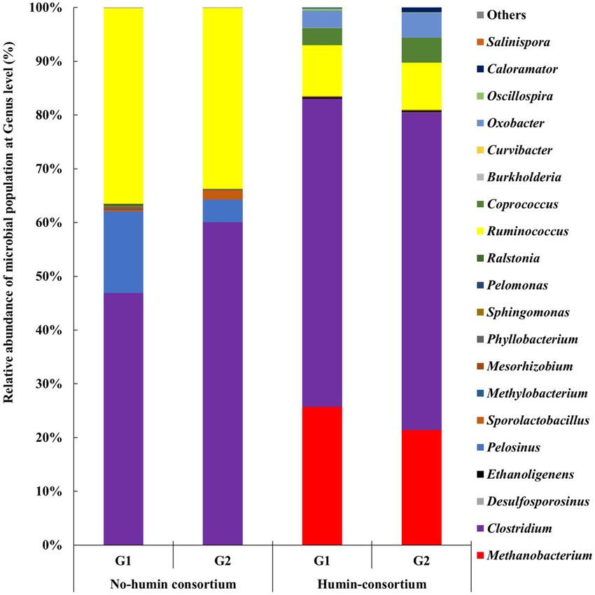
Figure 8. Community structures of the no-humin and humin consortia (35th generation) based on 16S rRNA
gene sequencing. The data shows the community structures of two replicates (G1 and G2) of the consortia,
individually. “Others” denotes the taxonomic groups with less than 0.02% abundance. In the figure, taxonomic
groups are arranged as N-fixers, nifH-positive microorganisms and non N-fixers from bottom to top.
cating the larger microbial population in the humin consortium. The nifH gene was only detected in the humin
consortium at 105 copies/ml, and not detected in the no-humin consortium. These suggested that nitrogen-
fixing microorganisms were not present at a detectable level by the qPCR analysis adopted in the no-humin con-
sortium but were present in the humin consortium. However, the observed small population of nitrogen-fixing
microorganisms did not agree with the stable BNF activity detected both in the humin and no-humin consortia
following enrichment.
The microbial community structures in the humin and no-humin consortia were determined by next genera-
tion amplicon sequence of partial 16S rRNA genes using Miseq (Ilmina) (Fig. 8). Bacteria, mostly Firmicutes,
accounting for 73–77% of the relative abundance, dominated the humin consortium. Clostridium, Ruminococcus
and Oxobacter, belonging to Clostridiales, were the major genera of detected Firmicutes. Other Clostridiales
such as Desulfocporosinus, Ethanoligenens, Coprococcus, Oscillospira and Caloramator were detected as the minor
populations. Other than Clostridiales, the detected genera of Bacteria were Sporolactobacillus (Bacillales), Phyl-
lobacterium (Rhizobiales), Sphingomonas (Sphingomonadales) and Ralstonia (Burkholderiales) accounting for
several percentage. Archaea, mostly Methanobacterium, occupied 22–26% of relative abundance in the humin
Scientific Reports | (2021) 11:6567 | https://doi.org/10.1038/s41598-021-85955-3 6
Vol:.(1234567890)www.nature.com/scientificreports/
consortium. The no-humin consortium consisted of mostly Bacteria, in which Firmicutes, mainly Clostridium,
Pelosinus and Ruminococcus, which belong to Clostridiales, occupied over 96% of the relative abundance. The
minor populations detected were Sporolactobacillus (Bacillales), Methylobacterium (Rhizobiales), Mesorhizobium
(Rhizobiales),, Phyllobacterium (Rhizobiales), Sphingomonas (Sphingomonadales), Pelomonas (Burkholderi-
ales), Ralstonia (Burkholderiales), Burkholderia (Burkholderiales), Curvibacter (Burkholderiales), and Salinispora
(Burkholderiales). Archaea was present only as negligible abundance, accounted for 0.02% in the no-humin
consortium. The same dominant microbial genera, Clostridium and Ruminococcus, were observed in both the
humin and no-humin consortia between the 35th (Fig. 8) and the 11th generation (Fig. S2), consistent with
the stable ARA of consortia after the 11th generation (Fig. S1). Among the observed genera of both consortia,
Methanobacterium, Clostridium, Desulfosporosinus, Ethanoligenens, Pelosinus, Sporolactobacillus, Methylobacte-
rium, Mesorhizobium, Phyllobacterium, Sphingomonas, Pelomonas and Ralstonia belong to cluster I, cluster II, and
cluster III of the N-fixing phylogeny and are known to synthesise nitrogenase, including alternative nitrogenases,
for fixing atmospheric N 235–48. Ruminococcus, Coprococcus, Burkholderia and Curvibacter have not been reported
as N-fixing microorganisms but are known to carry nifH-like sequences belonging to Cluster IV of the N-fixing
bacterial group35. These results suggest that more than 93% of the microbial species, in both consortia, were
considered nifH positive, contrary to the results of qPCR using nifH-specific primers. Oxobacter, Oscillospira and
Caloramator observed in humin consortium, and Oxobacter and Salinispora observed in no-humin consortium,
have not been demonstrated to possess the nif gene or BNF activity.
The Shannon diversity index49 was 0.50 for the humin consortium, with an evenness of 0.17, and was 1.1 for
the no-humin consortium, with an evenness of 0.38. The Chao 1 diversity i ndex50, calculated using operational
taxonomic units (OTUs), was 19.1 for the humin consortium and 19.3 for the no-humin consortium. These
results indicated that although both consortia were enriched with specific microbial species, fewer microbial
diversity were present in the humin consortium, than in the no-humin consortium.
Discussion
Nitrogen fixation in the consortia was promoted with humin. The increase in nitrogen content during 14 days
of incubation was significantly higher (by 292%, 11.8 times) in the humin consortium than in the no-humin
consortium (Fig. 1b). The nitrogen content in the medium containing humin without inoculation did not show
any increase, suggesting no chemical nitrogen fixation by humin. These results suggested that humin promoted
the BNF activity of microorganisms. The higher ARA with humin (≥ 40%), regardless of the consortia (Fig. 1a),
suggested that the promoting effect of humin was not attributable to the larger population in the humin con-
sortium (Fig. 7) but to increased BNF activity of the microbial cells. This conclusion was also supported by the
increased ARA (Fig. 3) and the increase in nitrogen content (Fig. 4) by only reduced humin using the same no-
humin consortium. The promotion effect of humin on the BNF activity is also suggested to be distributed widely
in the environment, although the extent of the promotion effect would vary, as shown in Fig. 2.
The mechanism of the promotion effect of humin was examined. The promotion of BNF activity was not
achieved by the addition of reducing agents, N a2S, cysteine, and Ti-NTA, or by the addition of mixtures of the
reducing agents and humin (Fig. 5). This indicated that the levels of reduction of the medium did not explain
the effect of humin. The promotion of BNF activity by humin was also observed even in anaerobic MNDA
medium supplemented with micronutrients and vitamins (Fig. 6), suggesting that the promoting effect of humin
was not due to the supply of micronutrients. Comparative studies using electrochemically reduced humin, oxi-
dised humin, and intact humin under the conditions without any other electron donor, demonstrated that only
reduced humin promoted BNF activity of the washed non-humin consortium, as shown by ARA (Fig. 3) and the
increase in nitrogen content (Fig. 4). No ARA and no increase in nitrogen content were observed with intact and
oxidised humin. These results suggested that humin did not serve as organic carbon source donating electrons
by the metabolism in cell for the BNF promotion. Rather, humin functioned as extracellular electron mediator
for donating electrons to the N-fixing microorganisms. Quinone and sulphur containing moieties have been
suggested as redox-active centers of humin34. The number of electron equivalents donated by reduced humin
was calculated, which promoted the BNF activity of the consortium. The stoichiometry of BNF is given as four
electron equivalents required for producing one mole of ammonia from atmospheric N 2 (Eq. 1). Therefore, the
number of electron equivalents donated from humin was estimated to be 236 ± 25 µEq/g-humin for nitrogen fixa-
tion, based on the increase of total nitrogen 248 ± 26 µg-N/bottle with 0.3 g of humin (Fig. 4). It can be concluded
that intact humin did not serve as organic carbon source but as an extracellular electron mediator in the micro-
bial consortium, thereby promoting BNF, whereby intact humin was reduced by humin-reducing bacteria. This
reduced humin subsequently donated electrons to N-fixers of the consortium, resulting in increased BNF activity.
Although the extracellular electron mediating function of humin has been reported for various microbial
reactions30–33, this is the first study to demonstrate a significant promotion of BNF by humin acting as an
extracellular electron mediator. Although the promotion of nitrogen reduction to ammonia using nitrogenase
with bioelectrochemical a ctivation10 and catalysts or nitrogenase hybrids with photochemical a ctivation11–15 has
been demonstrated previously, this is the first demonstration of promotion of BNF of whole microbial cells to
levels exceeding the biochemical capacity of the cells. The redox potential of half reaction of N 2/NH4+ has been
reported as − 278 mV ( E ′) . The viologens, artificial mediators useful for donating electrons to nitrogenase,
0 52
have been reported to require a redox potential lower than − 360 mV (versus a normal hydrogen electrode) to
be effective53. Such low redox potential of humin have been suggested in C O2-reducing acetogenesis assisted by
humin (− 290 mV, E 0′ of C
O2/CH3COOH)54. The isoelectric potential of humin has been also reported ranging
from − 400 to + 272 mV (versus standard hydrogen electrode) in the cyclic voltammetry analysis of h umin34.
These suggested that humin has redox active moieties with lower (more negative) redox potential, which is
thermodynamically feasible for nitrogen fixing reaction.
Scientific Reports | (2021) 11:6567 | https://doi.org/10.1038/s41598-021-85955-3 7
Vol.:(0123456789)www.nature.com/scientificreports/
In addition to the BNF promotion, the previous reports also demonstrated that humin promoted
denitrification33 and nitrate reduction to a mmonia32 as extracellular electron mediator. Humic substances (nat-
ural organic matter) have been also shown to serve as terminal electron acceptors to fuel anammox reaction
(anaerobic ammonium oxidation to N 2)51. Given the wide distribution of humic substances including humin, it is
suggested that humin and/or humic substances would play a vital role for global nitrogen cycle under anaerobic
environments, not only promoting BNF but also nitrogen loss to atmosphere.
The analysis of community structures demonstrated that both consortia were dominated by Clostridium
(Fig. 8). Clostridium is an anaerobic N-fixer. The promotion of BNF by humin in both consortia could be
attributed to extracellular electron donation from humin to Clostridium. Methanobacterium (Archaea) was
only abundant in the humin consortium. Methanobacterium, an anaerobic N-fixer as well as hydrogenotrophic
methanogen55, may obtain the extracellular electrons from humin, resulting in the propagation in the humin
consortium. Ethanoligenens and Desulfosporosinus were detected only in the humin consortium although they
are minor populations (0.35%), suggesting that they are humin-dependent microorganisms. Ethanoligenens and
Desulfosporosinus were reported to fix nitrogen using an alternative n itrogenase44. The roles of Ruminococcus,
Coprococcus and Curvibacter remain unknown because they carry nifH-like genes belonging to Cluster IV, which
are not involved in nitrogen fixation35.
More than 93% genera in both consortia harbour the nifH gene35,36,41–44,56. However, qPCR analysis (Fig. 7)
demonstrated that the N-fixing microbial population was significantly smaller (10–5) than that of the total micro-
bial population in the humin consortium and even not detected in the no-humin consortium. Although qPCR
assays using different primer sets (F2/ R6, PolF/PolR, and IGK /NifH3)57 were undertaken to quantify the nifH
genes, zero, or poor, amplification was observed in all attempts (Fig. S3). This could be due to the low sensitivity
of these primer sets for nifH genes of the consortia. In preliminary trials to isolate anaerobic microorganisms
from the consortia, 18 cultures were all positive in ARA (data not shown), suggesting a dominance of N-fixing
microorganisms in the consortia. Further studies should be undertaken to identify the isolates and nif genes.
Oxobacter, Caloramator, and Oscillospira without N-fixing ability in the humin consortium indicated the avail-
ability of N sources other than nitrogen gas for growth. An increase in ammoniacal nitrogen concentration
during the incubation was observed in both consortia, which would be released as excess NH3 by the N-fixers
in the community (Fig. S4).
In conclusion, this study demonstrated that humin promoted the BNF activity of N-fixing anaerobic consor-
tia by 5.7–11.8 times as shown by the increases in ARA and the nitrogen content. Humin did not function as a
reducing agent or as a source of micronutrients or vitamins, or as organic carbon source, but as an extracellular
electron mediator to donate electrons to N-fixing consortia. The consortium enriched with humin was dominated
by Clostridium, Ruminococcus, and Methanobacterium, while the consortium enriched without humin was domi-
nated by Clostridium, Ruminococcus, and Pelosinus as possible N-fixers. Further studies are needed to identify
humin-promoted specific N-fixers. Moreover, the mechanism of the electron mediating function of humin for
N-fixers is still under study. However, considering the promotion of BNF activity under nitrogen-deficient condi-
tions and the ubiquitous distribution of humin, the implementation of humin-promoted atmospheric N-fixation
would contribute to the development of a sustainable technology. In addition, considering the promoting effects
of humin on denitrification and nitrate reduction to ammonia, humin would be playing an important role for
global N-cycle, especially under anaerobic environment.
Materials and methods
Humin preparation. Humin was extracted from the following surface soils or sediments: Kamajima paddy
soil, Nagoya University farm upland soil, Ibakaki upland soil, Yatomi paddy soil, and Arako river sediment,
as previously reported58, with some modifications. Briefly, the soils and sediment were air-dried and sieved
through 1 mm mesh. The sieved soils or sediment (100 g) were repeatedly washed by shaking with a solution
(24 h; compositions defined below), centrifugation (8000 g, 15 min, 24 °C), followed by decantation. The wash-
ing chemical solutions were as follows: ten times with 150 mL of 2% HF, ten times with 150 mL of 0.1 M NaOH,
and twenty times with 150 mL of ultrapure water. After washing, pH was adjusted to 7.5 using 0.1 M NaOH. The
pH-adjusted humin was freeze-dried, ground using a ceramic mortar and pestle, and then used experimentally.
The C, H, and N contents of humin were determined using a CHN analyser (Yanaco MT-5 CHN-corder, Yanaco
New Science Inc., Kyoto, Japan) and the results are shown in Table S1.
Enrichment of humin and no‑humin consortia with nitrogen‑fixing activity. Kamajima paddy
soil was collected on 6th January 2009 and used as an inoculum. 250 mL of a modified nitrogen-deficient Ashby
(MNDA) medium59 and 150 mL of water-saturated soil was mixed in a 600 ml glass bottle and subsequently
sealed with a butyl rubber stopper and aluminium cap. The headspace was flushed with nitrogen gas to achieve
anaerobic conditions. The MNDA medium was composed of mannitol (20 g/L), K2HPO4 (0.2 g/L), MgSO4·7H2O
(0.2 g/L), NaCl (0.2 g/L), K2SO4 (0.1 g/L), and CaCO3 (5 g/L). After seven weeks of incubation at 30 °C under
static conditions, 2 mL of the culture was aseptically transferred to a 50 mL-volume glass vial containing 20 mL
of anaerobic MNDA medium. A supplement of 15 g/L of humin was added prior to the preparation of anaero-
bic MNDA medium in the vial. Thus, two anaerobic consortia were enriched: one using the anaerobic MNDA
medium supplemented with humin (humin consortium) and another using the same medium but without
humin (no-humin consortium). Transfer occurred once every four weeks until the 6th generation, and then
reduced to every two weeks to increase the frequency of culture transfer. The culture transfer was maintained to
the 11th generation to obtain enriched N-fixing microbial consortia with stable ARA. The BNF activity of the
consortia was examined using either ARA or the increase in nitrogen in the culture.
Scientific Reports | (2021) 11:6567 | https://doi.org/10.1038/s41598-021-85955-3 8
Vol:.(1234567890)www.nature.com/scientificreports/
Acetylene reduction activity test. ARA is the widely used indicator of the biological N-fixation activ-
ity of diazotrophs60,61. Therefore, the ARA test was undertaken to examine nitrogen fixation activity indirectly
because acetylene is reduced to ethylene by nitrogenase, which is also responsible for BNF62. The ARA test was
undertaken as follows. Two sets of 10 ml-volume glass vials (one set supplemented with 0.03 g of humin in each
vial, another set having no humin) were flushed with helium gas, then, added aseptically with 2 mL of the He-
bubbled anaerobic MNDA medium. After that, 2 mL of inoculum from the humin-consortium and no-humin
consortium was separately inoculated into each of the two sets of the vials. Finally, all the vials were spiked with
200 µL of acetylene gas and incubated under static conditions at 30 °C for one week. After incubation, 100 µL
of the gaseous sample was drawn from the headspace using a Pressure-LokR, VICI Precision Analytical Syringe
(Baton Rouge, LA USA). The concentrations of acetylene and ethylene in the sample were determined using
a GC-14B gas chromatograph (Shimadzu, Kyoto, Japan) equipped with a Molecular Sieve-5A column (60/80
mesh and 3 m in length) and a flame ionisation detector. Nitrogen gas was used as the carrier gas at a flow rate of
60 mL/min. H2 gas and compressed air were provided for the detector at flow rates of 50 mL/min and 450 mL/
min, respectively. The temperatures of the column, injector, and detector were 80 °C, 100 °C, and 100 °C, respec-
tively.
Determination of nitrogen concentration. Biological nitrogen fixation activity was determined
directly by measuring the increase in concentration of nitrogen in the culture. For this determination, the indi-
vidual consortia were sealed in a 50 mL vial containing 30 mL of the anaerobic MNDA medium with or without
the supplementation of humin (15 g/L), bubbled with nitrogen gas for 90 min, and the headspace was flushed
for 5 min. The medium was inoculated aseptically with 3 mL of one of the consortia. Sampling was undertaken
by taking 10 mL of the consortium using a syringe prior to incubation. After two weeks of incubation at 30 °C,
an additional 10 mL of the consortium was collected in the same manner. These samples were transferred to a
ceramic mortar and dried at 105 °C for two days. Dried samples were ground and approximately 2000 µg of sam-
ple used for the determination of nitrogen content. The elemental composition of C, H, and N of dried samples
was determined using a CHN analyser. All measurements were replicated a minimum of three times.
Effects of various humins on ARA. The effects of various humins on BNF activity of the no-humin con-
sortium were examined using the ARA test. Humin sources were as follows: Kamajima humin, Nagoya Univer-
sity farm (NU farm) humin, Ibakaki humin, Yatomi humin, and Arako humin. The ARA test of the no-humin
consortium without the addition of humin was provided as the control. The effects were evaluated using tripli-
cate experiments.
Effects of reducing agents on ARA. Effects of reducing agents on N-fixation activity of the no-humin
consortium were examined by the addition of different reducing agents, including 15 mM Na2S, 1.7 mM
L-cysteine hydrochloride, 0.025 mM titanium (III) trinitriloacetate-(Ti-NTA), which were added to the anaero-
bic MNDA medium, with and without the supplementation of Kamajima humin (15 g/L). ARA tests were con-
ducted at least three times.
Effect of micronutrients and vitamins on ARA. The effects of micronutrients and vitamins on ARA
were examined because the anaerobic MNDA medium did not contain micronutrients and vitamins. Micronu-
trients and vitamins were added as 1 ml of trace element SL-10 solution63 and 10 mL of vitamin solution64 to 1
L of the anaerobic MNDA medium to make the improved Ashby medium. The ARA test was then performed
using the no-humin consortium in the improved Ashby medium with and without supplementation of Kama-
jima humin (15 g/L).
Effect of humin prepared under oxidised and reduced conditions on BNF. The effects of different
redox state of humin on the BNF activity of the no-humin consortium were examined experimentally. Reduced
and oxidised Kamajima humin were prepared in 200 mL of 0.5 M NaCl solution using an electrochemical system
under anaerobic conditions, in a vinyl anaerobic chamber equipped with a vacuum airlock and two glove ports
(Coy-7450000, COY, Grass Lake, MI, USA). The electrochemical system consisted of a potentiostat (Automatic
Polarisation System HSV-110, Hokuto Denko, Osaka, Japan), two twisted platinum electrodes (0.8 mm in diam-
eter and 1 m in length) as working and counter electrodes, and a Ag/AgCl reference electrode (+ 0.199 V versus
the standard hydrogen electrode (SHE)). Redox potential was maintained at − 0.4 V or + 0.4 V (versus SHE) for
24 h to reduce or oxidise 2 g of autoclaved humin, respectively. After equilibrium was attained, the oxidised or
reduced humin was collected by filtration and dried using a vacuum pump. In this test, the washed and starved
non-humin consortium was used. The non-humin consortium was incubated at 30 °C for 2 weeks in anaero-
bic MNDA medium in which the buffer CaCO3 was replaced with 30 mM HEPES (anaerobic MNDA HEPES
medium). After incubation, the consortium was washed three times by suspending it in mannitol-free anaerobic
MNDA HEPES medium and by decanting the supernatant after centrifuging the consortium at 2500×g for 5 min
at 4 °C. The washed consortium was then kept for starvation in the mannitol-free anaerobic MNDA medium
for two days. After washing and starvation pre-treatments, the no-humin consortium was subjected to a test
evaluating the effect of different redox states of humin on BNF activity. Biological nitrogen fixation activity
was evaluated using both the ARA test with He-bubbled, organic carbon source-free (mannitol-free), anaerobic
MNDA medium and the increase in nitrogen content using N 2-bubbled, organic carbon source-free (mannitol-
free), anaerobic MNDA medium, by incubating at 30 °C under static condition for two weeks. All tests were
conducted a minimum of three times.
Scientific Reports | (2021) 11:6567 | https://doi.org/10.1038/s41598-021-85955-3 9
Vol.:(0123456789)www.nature.com/scientificreports/
qPCR for total and nitrogen‑fixing bacteria in the consortia. Microbial DNA was extracted using
a FastDNA SPIN kit for soil (MP Biomedicals, Santa Ana, CA, USA) according to the manufacturer’s instruc-
tions. Populations of total and N-fixing bacteria in the consortia were estimated by qPCR using a universal
primer set targeting the 16S rRNA gene (27F: 5′-AGAGTTTGATCCTGGCTCAG-3′, and 1492R: 5′-GGTTAC
CTTGTTACGACTT-3′)65 and specific primer set targeting the nifH gene, PolF and PolR. PolF was the forward
primer ( 5′-TGCGAYCCSAARGCBGACTC-3′: Y = C and T; S = C and G; R = A and G; B = G, T, and C) and PolR
the reverse primer (5′-ATSGCCATCATYTCRCCGGA-3′: S = C and G; Y = C and T; R = A and G)57. qPCR was
performed using the LightCycler system (Roche Diagnostics, Basel, Switzerland). The reaction mixtures (final
volume 20 µL) contained 12.4 µL PCR grade H 2O, 1.6 µL of 25 mM MgCl2, 1 µL of 10 μM forward primer, 1 µL
of 10 μM reverse primer, 2 µL LightCycler FastStart DNA Master SYBR Green I (Roche Molecular Biochemi-
cals, Mannheim, Germany) and 2 µL template microbial DNA. To generate a standard curve, DNA fragments
of 16S rRNA and nifH genes were amplified by PCR using Ex Taq polymerase (TaKaRa Bio Inc., Kusatsu, Shiga,
Japan), and the PCR products were purified using a QIAEX II Gel Extraction Kit (Qiagen, Düsseldorf, Germany)
according to the manufacturer’s instructions. A dilution series of the purified DNA fragment of each gene was
amplified to generate a standard curve. The PCR cycling conditions consisted of a pre-incubation step at 95 °C
for 10 min followed by a quantification step consisting of 45 cycles of denaturation at 95 °C for 10 s, annealing
at 55 °C for 10 s, and elongation at 72 °C for 10 s. After amplification, the melting curve was obtained by heating
the PCR products at 95 °C, followed by cooling at 65 °C for 15 s and then gradually increasing the temperature
to 95 °C at the rate of 0.1 °C/ s. Cooling was undertaken at 40 °C for 30 s.
Analysis of community structure in consortia. The partial 16S rRNA amplicon sequence was per-
formed for the DNA fragments obtained by PCR amplification with the primer set targeting the V3-V4 region:
Pro341F (5′- CCT ACG GGN BGC ASC AG-3′) and Pro805R (5′- GAC TAC NVG GGT ATC TAA TCC-3′)66.
The PCR mixtures contained 5 µL of template DNA (5 ng/µL), 2.5 µL of Pro341F and Pro805R primers (2 µM
each), and 12.5 µL of KAPA HiFi HotStart Ready mix (KAPA Biosystems, Wilmington, MA, USA). The PCR
reaction was performed as follows: initial activation at 94 °C for 30 s, followed by 10 cycles at 94 °C for 10 s, 60 °C
for 30 s, 72 °C for 30 s, followed by 10 cycles at 94 °C for 10 s, 59 °C for 30 s, 72 °C for 30 s, followed by 10 cycles
at 94 °C for 10 s, 58 °C for 30 s, 72 °C for 30 s, and a final extension at 72 °C for 4 min. Purification of the PCR
products was carried out using the AMPure XP kit (Beckman Coulter Genomics Inc., Brea, CA, USA) according
to the manufacturer’s instructions, and the PCR products were confirmed using 1% agarose gel electrophoresis.
The concentration of purified DNA was determined using a QuantiFluor dsDNA System (Promega Corpora-
tion, Fitchburg, WI, USA), and sequencing of the purified DNA was performed using a Miseq platform with a
Miseq reagent kit v3 (600 cycle, Illumina Inc., San Diego, CA, USA). A chimaera check for the base sequences
of each read obtained from the analysis was carried out using USEARCH v6.167. Sequence reads with more than
97% similarity were classified into the same operational taxonomic unit (OTU), and OTU picking, and a cluster
analysis was performed in QIIME 2 68. Finally, OTUs were identified using the Green-gene database (ver. 13_8)
as a reference69.
Received: 14 September 2020; Accepted: 5 March 2021
References
1. Galloway, J. N. et al. Transformation of the nitrogen cycle : Recent trends, questions, and potential solutions. Science 320, 889–892
(2008).
2. Zhou, F. et al. Electro-synthesis of ammonia from nitrogen at ambient temperature and pressure in ionic liquids. Energy Environ.
Sci. 10, 2516–2520 (2017).
3. Kandemir, T., Schuster, M. E., Senyshyn, A., Behrens, M. & Schlögl, R. The Haber-Bosch process revisited: On the real structure
and stability of ‘ammonia iron’ under working conditions. Angew. Chemie - Int. Ed. 52, 12723–12726 (2013).
4. Vitousek, P. M., Menge, D. N. L., Reed, S. C., Cleveland, C. C. & Vitousek, P. M. Biological nitrogen fixation: Rates, patterns and
ecological controls in terrestrial ecosystems. Phil. Trans. R. Soc. B 368, 20130119 (2013).
5. Hu, Y., Lee, C. C. & Ribbe, M. W. Vanadium nitrogenase: A two-hit wonder?. Dalt. Trans. 41, 1118–1127 (2012).
6. Eady, R. R. Structure-function relationships of alternative nitrogenases. Chem. Rev. 96, 3013–3030 (1996).
7. Coppi, M. V., Leang, C., Sandler, S. J. & Lovley, D. R. Development of a genetic system for Geobacter sulfurreducens. Appl. Environ.
Microbiol. 67, 3180–3187 (2001).
8. Bazylinski, D. A., Dean, A. J., Schüler, D., Phillips, E. J. P. & Lovley, D. R. N
2-dependent growth and nitrogenase activity in the
metal-metabolizing bacteria, Geobacter and Magnetospirillum species. Environ. Microbiol. 2, 266–273 (2000).
9. Lan, R., Irvine, J. T. S. & Tao, S. Synthesis of ammonia directly from air and water at ambient temperature and pressure. Sci. Rep.
3, 1145 (2013).
10. Milton, R. D. et al. Nitrogenase bioelectrocatalysis: Heterogeneous ammonia and hydrogen production by MoFe protein. Energy
Environ. Sci. 9, 2550–2554 (2016).
11. Brown, K. A. et al. Light-driven dinitrogen reduction catalyzed by a CdS:nitrogenase MoFe protein biohybrid. Science 352, 448–450
(2016).
12. Zhu, D., Zhang, L., Ruther, R. E. & Hamers, R. J. Photo-illuminated diamond as a solid-state source of solvated electrons in water
for nitrogen reduction. Nat. Mater. 12, 836–841 (2013).
13. Oshikiri, T., Ueno, K. & Misawa, H. Selective dinitrogen conversion to ammonia using water and visible light through plasmon-
induced charge separation. Angew. Chemie - Int. Ed. 128, 4010–4014 (2016).
14. Liu, J. et al. Nitrogenase-mimic iron-containing chalcogels for photochemical reduction of dinitrogen to ammonia. Proc. Natl.
Acad. Sci. U. S. A. 113, 5530–5535 (2016).
15. Ali, M. et al. Nanostructured photoelectrochemical solar cell for nitrogen reduction using plasmon-enhanced black silicon. Nat.
Commun. 7, 11335 (2016).
Scientific Reports | (2021) 11:6567 | https://doi.org/10.1038/s41598-021-85955-3 10
Vol:.(1234567890)www.nature.com/scientificreports/
16. Anderson, J. S., Rittle, J. & Peters, J. C. Catalytic conversion of nitrogen to ammonia by an iron model complex. Nature 501, 84–87
(2013).
17. Arashiba, K., Miyake, Y. & Nishibayashi, Y. A molybdenum complex bearing PNP-type pincer ligands leads to the catalytic reduc-
tion of dinitrogen into ammonia. Nat. Chem. 3, 120–125 (2011).
18. Schrock, R. R. Catalytic reduction of dinitrogen to ammonia at a single molybdenum center. Acc. Chem. Res. 38, 955–962 (2005).
19. Cherkasov, N., Ibhadon, A. O. & Fitzpatrick, P. A review of the existing and alternative methods for greener nitrogen fixation.
Chem. Eng. Process. Process Intensif. 90, 24–33 (2015).
20. Ortiz-medina, J. F., Grunden, A. M., Hyman, M. R. & Call, D. F. Nitrogen gas fixation and conversion to ammonium using microbial
electrolysis cells. ACS Sustain. Chem. Eng. 7, 3511–3519 (2019).
21. Lovley, D. R., Coates, J. D., Blunt-Harris, E. L., Phillips, E. J. P. & Woodward, J. C. Humic substances as electron acceptors for
microbial respiration. Nature 382, 445–448 (1996).
22. Martinez, C. M., Alvarez, L. H., Celis, L. B. & Cervantes, F. J. Humus-reducing microorganisms and their valuable contribution
in environmental processes. Appl. Microbiol. Biotechnol. 97, 10293–10308 (2013).
23. Valenzuela, E. I. & Cervantes, F. J. The role of humic substances in mitigating greenhouse gases emissions: Current knowledge and
research gaps. Sci. Total Environ. 750, 141677 (2021).
24. Valenzuela, E. I. et al. Humic substances mediate anaerobic methane oxidation linked to nitrous oxide reduction in wetland sedi-
ments. Front. Microbiol. 11, 587 (2020).
25. Valenzuela, E. I. et al. Anaerobic methane oxidation driven by microbial reduction of natural organic matter in a tropical wetland.
Appl. Environ. Microbiol. 83, e00645-e717 (2017).
26. Lovley, D. R., Fraga, J. L., Coates, J. D. & Blunt-Harris, E. L. Humics as an electron donor for anaerobic respiration. Environ.
Microbiol. 1, 89–98 (1999).
27. Lovley, D. R. Bug juice: Harvesting electricity with microorganisms. Nat. Rev. Microbiol. 4, 497–508 (2006).
28. Milton, R. D. et al. Bioelectrochemical Haber-Bosch process: An ammonia-producing H2/N2 fuel cell. Angew. Chemie - Int. Ed.
56, 2680–2683 (2017).
29. Kamel, F. Paths from pesticides to Parkinson’s. Science 341, 722–723 (2013).
30. Zhang, C. & Katayama, A. Humin as an electron mediator for microbial reductive dehalogenation. Environ. Sci. Technol. 46,
6575–6583 (2012).
31. Laskar, M., Awata, T., Kasai, T. & Katayama, A. Anaerobic dechlorination by a humin-dependent pentachlorophenol-dechlorinating
consortium under autotrophic conditions induced by homoacetogenesis. Int. J. Environ. Res. Public Health 16, 2873 (2019).
32. Zhang, D., Zhang, C., Xiao, Z., Suzuki, D. & Katayama, A. Humin as an electron donor for enhancement of multiple microbial
reduction reactions with different redox potentials in a consortium. J. Biosci. Bioeng. 119, 188–194 (2015).
33. Xiao, Z. et al. Enhanced denitrification of Pseudomonas stutzeri by a bioelectrochemical system assisted with solid-phase humin.
J. Biosci. Bioeng. 122, 85–91 (2016).
34. Pham, M. D., Kasai, T., Yamaura, M. & Katayama, A. Humin : No longer inactive natural organic matter. Chemosphere 269, 128697
(2021).
35. Raymond, J., Siefert, J. L., Staples, C. R. & Blankenship, R. E. The natural history of nitrogen fixation. Mol. Biol. Evol. 21, 541–554
(2004).
36. Lambert, B. et al. Identification and plant interaction of a Phyllobacterium sp., a predominant rhizobacterium of young sugar beet
plants. Appl. Environ. Microbiol. 56, 1093–1102 (1990).
37. Sy, A. et al. Methylotrophic Methylobacterium bacteria nodulate and fix nitrogen in symbiosis with legumes. J. Bacteriol. 183,
214–220 (2001).
38. Marques, J. M. et al. Nitrogen fixing and phosphate mineralizing bacterial communities in sweet potato rhizosphere show a
genotype-dependent distribution. Diversity 11, 231 (2019).
39. Xie, C. H. & Yokota, A. Sphingomonas azotifigens sp. nov., a nitrogen-fixing bacterium isolated from the roots of Oryza sativa. Int.
J. Syst. Evol. Microbiol. 56, 889–893 (2006).
40. Yuan, B., Yoshikane, Y., Yokochi, N., Ohnishi, K. & Yagi, T. The nitrogen-fixing symbiotic bacterium Mesorhizobium loti has and
expresses the gene encoding pyridoxine 4-oxidase involved in the degradation of vitamin B6. FEMS Microbiol. Lett. 234, 225–230
(2006).
41. Chen, W. M., James, E. K., Prescott, A. R., Kierans, M. & Sprent, J. I. Nodulation of Mimosa spp. by the β-proteobacterium Ralstonia
taiwanensis. Mol. Plant-Microbe Interact. 16, 1051–1061 (2003).
42. Verma, S. C., Chowdhury, S. P. & Tripathi, A. K. Phylogeny based on 16S rDNA and nifH sequences of Ralstonia taiwanensis strains
isolated from nitrogen-fixing nodules of Mimosa pudica, India. Can. J. Microbiol. 50, 313–322 (2004).
43. Rojas, A., Holguin, G., Glick, B. R. & Bashan, Y. Synergism between Phyllobacterium sp. (N2-fixer) and Bacillus licheniformis
(P-solubilizer), both from a semiarid mangrove rhizosphere. FEMS Microbiol. Ecol. 35, 181–187 (2001).
44. Addo, M. A. & Dos Santos, P. C. Distribution of nitrogen-fixation genes in prokaryotes containing alternative nitrogenases. Chem-
BioChem 21, 1749–1759 (2020).
45. Sachdev, D., Nema, P., Dhakephalkar, P., Zinjarde, S. & Chopade, B. Assessment of 16S rRNA gene-based phylogenetic diversity
and promising plant growth-promoting traits of Acinetobacter community from the rhizosphere of wheat. Microbiol. Res. 165,
627–638 (2010).
46. Thajudeen, J. et al. Nitrogen fixing bacterial diversity in a tropical estuarine sediments. World J. Microbiol. Biotechnol. 33, 41 (2017).
47. Bouffaud, M. L., Renoud, S., Moenne-Loccoz, Y. & Muller, D. Is plant evolutionary history impacting recruitment of diazotrophs
and nifH expression in the rhizosphere?. Sci. Rep. 6, 21690 (2016).
48. Bae, H. S., Morrison, E., Chanton, J. P. & Ogram, A. Methanogens are major contributors to nitrogen fixation in soils of the Florida
Everglades. Appl. Environ. Microbiol. 84, e02222-e2317 (2018).
49. Shannon, C. E. A mathematical theory of communication. Bell Syst. Techn. J. 27, 379–423 (1948).
50. Chao, A. Nonparametric estimation of the number of classes in a population. Scand. J. Stat. 11, 265–270 (1984).
51. Rios-Del Toro, E. E., Valenzuela, E. I., Ramirez, J. E., Lopez-Lozano, N. E. & Cervantes, F. J. Anaerobic ammonium oxidation linked
to microbial reduction of natural organic matter in marine sediments. Environ. Sci. Technol. Lett. 5, 571–577 (2018).
52. Fukagawa, M., Murakami, S. & Nakanishi, H. Oxidation reduction potential of microorganisms reaction in activated sludge process.
Proc. Environ. Sanit. Eng. Res. 28, 105–112 (1992).
53. Badalyan, A., Yang, Z. & Seefeldt, L. C. A voltammetric study of nitrogenase catalysis using electron transfer mediators. ACS Catal.
9, 1366–1372 (2019).
54. Laskar, M., Kasai, T., Awata, T. & Katayama, A. Humin assists reductive acetogenesis in absence of other external electron donor.
Int. J. Environ. Res. Public Health 17, 4211 (2020).
55. Boone, D. R. Genus I. Methanobacterium. In Bergey’s Manucal of Systematic Bacteriology 2nd edn (eds Boone, D. R. et al.) 215–218
(Springer, Berlin, 2001).
56. Igai, K. et al. Nitrogen fixation and nifH diversity in human gut microbiota. Sci. Rep. 6, 31942 (2016).
57. Gaby, J. C. & Buckley, D. H. A comprehensive evaluation of PCR primers to amplify the nifH gene of nitrogenase. PLoS ONE 7,
e42149 (2012).
58. Zhang, C. et al. Characterization of humins from different natural sources and the effect on microbial reductive dechlorination of
pentachlorophenol. Chemosphere 131, 110–116 (2015).
Scientific Reports | (2021) 11:6567 | https://doi.org/10.1038/s41598-021-85955-3 11
Vol.:(0123456789)www.nature.com/scientificreports/
59. Ashby, S. F. Some observatons on the assimilation of atmospheric nitrogen by a free living soil organism—Azotobacter chroococcum
of Beijerinck. J. Agric. Sci. 2, 35–51 (1907).
60. Hardy, R. W. F., Burns, R. C. & Holsten, R. D. Applications of the acetylene-ethylene assay for measurement of nitrogen fixation.
Soil Biol. Biochem. 5, 47–81 (1973).
61. Hardy, R. W. F., Holsten, R. D., Jackson, E. K. & Burns, R. C. The acetylene–ethylene assay for N2 fixation: Laboratory and field
evaluation. Plant Physiol 43, 1185–1207 (1968).
62. Burris, R. H. Minireview: Nitrogenases. J. Biol. Chem. 266, 9339–9342 (1991).
63. Widdel, F., Kohring, G.-W. & Mayer, F. Studies on dissimilatory sulfate-reducing bacteria that decompose fatty acids. Arch. Micro-
biol. 134, 286–294 (1983).
64. Holliger, C. et al. Dehalobacter restrictus gen. nov. and sp. nov., a strictly anaerobic bacterium that reductively dechlorinates tetra-
and trichloroethene in an anaerobic respiration. Arch. Microbiol. 169, 313–321 (1998).
65. Dojka, M. A., Hugenholtz, P., Haack, S. K. & Pace, N. R. Microbial diversity in a hydrocarbon- and chlorinated-solvent- contami-
nated aquifer undergoing intrinsic bioremediation. Appl. Environ. Microbiol. 64, 3869–3877 (1998).
66. Takahashi, S., Tomita, J., Nishioka, K., Hisada, T. & Nishijima, M. Development of a prokaryotic universal primer for simultaneous
analysis of Bacteria and Archaea using next-generation sequencing. PLoS ONE 9, e105592 (2014).
67. Edgar, R. C. Search and clustering orders of magnitude faster than BLAST. Bioinformatics 26, 2460–2461 (2010).
68. Bolyen, E. et al. Reproducible, interactive, scalable and extensible microbiome data science using QIIME 2. Nat. Biotechnol. 37,
852–857 (2019).
69. McDonald, D. et al. An improved Greengenes taxonomy with explicit ranks for ecological and evolutionary analyses of bacteria
and archaea. ISME J. 6, 610–618 (2012).
Acknowledgements
This research was financially supported in part by a Grant-in-Aid for Scientific Research (17H01899, 18K11707,
20H04363, 20K15431, JRP with NSFC FY2019) from the Japan Society for the Promotion of Science (JSPS),
by the co-operative study program of IMaSS, Nagoya University (2018, 2019, and 2020 projects), the JFE 21st
Century Foundation (2019 project), the Iwatani Naoji Foundation (2020 research project), and by the Institute
for Fermentation, Osaka (L-2019-3-003). The authors thank Toyoko Demachi and Shozo Ohta in Institute of
Materials and Systems for Sustainability, Nagoya University, and Tomoko Atsumi, in Graduate School of Bio-
agricultural Sciences, Nagoya University, for their technical assistance.
Author contributions
S.D. and T.A. contributed equally as the first authors to this study. T.A., D.Z., and A.K. made conception. S.D.,
T.A., D.Z., T.K. and A.K. designed the experiments. S.D., T.A., J.M., D.Z., and N.M. performed data acquisition,
analysis and interpretation. T.A., T.K. and A.K. supervised the research. S.D. and T.A. drafted the manuscript.
T.K. and A.K. revised the manuscript. All the authors have read and approved the final version of the manuscript.
Competing interests
The authors declare no competing interests.
Additional information
Supplementary Information The online version contains supplementary material available at https://doi.org/
10.1038/s41598-021-85955-3.
Correspondence and requests for materials should be addressed to A.K.
Reprints and permissions information is available at www.nature.com/reprints.
Publisher’s note Springer Nature remains neutral with regard to jurisdictional claims in published maps and
institutional affiliations.
Open Access This article is licensed under a Creative Commons Attribution 4.0 International
License, which permits use, sharing, adaptation, distribution and reproduction in any medium or
format, as long as you give appropriate credit to the original author(s) and the source, provide a link to the
Creative Commons licence, and indicate if changes were made. The images or other third party material in this
article are included in the article’s Creative Commons licence, unless indicated otherwise in a credit line to the
material. If material is not included in the article’s Creative Commons licence and your intended use is not
permitted by statutory regulation or exceeds the permitted use, you will need to obtain permission directly from
the copyright holder. To view a copy of this licence, visit http://creativecommons.org/licenses/by/4.0/.
© The Author(s) 2021
Scientific Reports | (2021) 11:6567 | https://doi.org/10.1038/s41598-021-85955-3 12
Vol:.(1234567890)You can also read