Promotion of Hair Growth by Conditioned Medium from Extracellular Matrix/Stromal Vascular Fraction Gel in C57BL/6 Mice
←
→
Page content transcription
If your browser does not render page correctly, please read the page content below
Hindawi
Stem Cells International
Volume 2020, Article ID 9054514, 11 pages
https://doi.org/10.1155/2020/9054514

Research Article
Promotion of Hair Growth by Conditioned Medium from
Extracellular Matrix/Stromal Vascular Fraction Gel in
C57BL/6 Mice

          Shune Xiao, Yurong Deng, Xiaojin Mo, Zhiyuan Liu, Dali Wang, Chengliang Deng ,
          and Zairong Wei
          Department of Plastic Surgery, Affiliated Hospital of Zunyi Medical University, Zunyi, Guizhou, China

          Correspondence should be addressed to Chengliang Deng; cheliadeng@sina.com and Zairong Wei; zairongwei@163.com

          Received 15 March 2020; Revised 11 May 2020; Accepted 25 May 2020; Published 13 June 2020

          Academic Editor: Katia Mareschi

          Copyright © 2020 Shune Xiao et al. This is an open access article distributed under the Creative Commons Attribution License,
          which permits unrestricted use, distribution, and reproduction in any medium, provided the original work is properly cited.

          Adipose-derived stem cell- (ADSC-) based regenerative medicine has expanded to include the treatment of hair loss. However, stem
          cell therapy remains a relatively recent technique, and reports of its use for treating alopecia are rare. ADSCs exert biological
          functions via the paracrine actions of various growth factors and cytokines. Conditioned medium from ADSCs (ADSCs-CM) is
          a cell-free suspension rich in growth factors and cytokines that has demonstrated a significant role in stimulating hair growth,
          with encouraging outcomes in terms of hair regeneration and hair growth. Extracellular matrix/stromal vascular fraction gel
          (ECM/SVF-gel) is an ADSC- and adipose native extracellular matrix-enriched product for cytotherapy. In this study, we
          compared the effects of CM from ECM/SVF-gel (ECM/SVF-CM) and from stem cells (SVF-CM) on hair growth in mice.
          ECM/SVF-CM stimulated hair growth more than SVF-CM, through promoting the proliferation of dermal papilla cells and cells
          in the bulge, neovascularization, and anagen induction. ECM/SVF-CM might, thus, provide an effective and improved strategy
          for promoting hair growth. These data provide a theoretical foundation for the clinical administration of ECM/SVF-CM for the
          treatment of hair loss.

1. Introduction                                                        of subcutaneous adipose tissue, which have been extensively
                                                                       researched and have been applied in tissue engineering and
Hair loss is one of the most common complaints presenting              regenerative medicine [4, 5]. Furthermore, the field of
to dermatologists and plastic surgeons and is usually associ-          ADSC-based regenerative medicine has now expanded to
ated with a major psychological impact on the affected                  include the treatment of hair loss. Cellular therapy with
patient. Androgenic alopecia is a major cause of hair loss,            human autologous SVF ADSCs was recently reported for alo-
with an estimated 70% of men and 40% of women affected                  pecia areata, with encouraging outcomes including increased
by androgenic alopecia and hair loss at some point in their            hair density and thickness and decreased pull-test results [6].
lives [1]. Conservative treatments for alopecia include the            However, stem cell therapy is relatively novel and subject to
use of drugs, such as finasteride and minoxidil, and surgical           regulatory surveillance, and reports of its use for the treat-
hair transplantation [2]. However, these treatments are asso-          ment of alopecia are rare. ADSCs exert their biological func-
ciated with their own adverse reactions and are ineffective in          tions via the paracrine actions of various growth factors and
some patients.                                                         cytokines [7]. Innovative approaches to hair regeneration
    Alternative safe and effective therapeutic approaches are           involve the production of conditioned medium from ADSCs
therefore required. Using the patient’s own stem cells to              (ADSCs-CM) and use of this cell-free suspension enriched
regenerate hair growth is a promising alternative therapeutic          with nutritious growth factors and cytokines secreted by
strategy [3]. Adipose-derived stem cells (ADSCs) are mesen-            ADSCs [8]. ADSCs-CM has been reported to be rich in
chymal stem cells within the stromal vascular fraction (SVF)           growth factors and cytokines, including vascular endothelial
2                                                                                                       Stem Cells International

growth factor (VEGF), basic fibroblast growth factor (bFGF),       conducted according to the guidelines of the National Health
platelet-derived growth factor (PDGF), hepatocyte growth          and Medical Research Council (China).
factor (HGF), keratinocyte growth factor (KGF), and
insulin-like growth factor I (IGF-I) [8]. These growth factors    2.2. Preparation of CMs from ECM/SVF-Gel and SVF.
and cytokines potentially modulate the host environment           ECM/SVF-gel and SVF suspension were prepared as
and thereby play significant roles in hair growth [9]. Previous    described previously [19]. Briefly, the Coleman adipose tissue
studies found that human hair regrowth and density                was mechanically emulsified by shifting between two 10 mL
increased significantly after the application of ADSCs-CM          syringes connected by a Luer-Lok connector with an internal
[10–13].                                                          diameter of 2.4 mm for 1 min (10 mL/s). After centrifugation
    The generation of specific CM rich in growth factors/cy-       at 2000 × g/min for 3 min, the emulsion was divided into
tokines to support hair regeneration and growth is the            three layers: a top oil layer, middle ECM/SVF-gel layer, and
essence of therapeutic strategies, and several approaches         bottom layer with a small amount of fluid. For SVF prepara-
can be used to increase the levels of ADSC growth factors/cy-     tion, the Coleman adipose tissue was minced and incubated
tokines. A low dose of UVB radiation was shown to upregu-         with 0.075% collagenase I (Sigma-Aldrich, St. Louis, MO,
late the secretion of ADSC-derived growth factors, and            USA) at 37°C for 40 min. After terminating the digestion,
treatment with UVB-irradiated ADSCs-CM improved hair              the cell suspension was filtered through a 100 μm mesh cell
regeneration and modulated the hair cycle in mice [14]. Hyp-      strainer, centrifuged at 500 × g for 3 min, and the cell pellet
oxia also enhanced the paracrine effects of ADSCs [15].            was resuspended in the Dulbecco’s modified Eagle’s medium
Other strategies to increase the secretion of growth factors/-    (DMEM; Gibco, Carlsbad, CA, USA). ECM/SVF-gel and
cytokines include growing the ADSCs in 3D or spheroid cul-        SVF suspension containing the same numbers of cells
tures or culturing the cells in laminin- or Matrigel-coated       (5 × 10⁵ ) were cultured in 5 mL DMEM supplemented with
culture vessels [16–18]. Lu et al. described an injectable adi-   10% fetal bovine serum (FBS; Gibco) and 1% penicillin-
pose tissue-derived stem cell and adipose native ECM enrich-      streptomycin (Gibco) at 37°C with 5% CO2 for 24 h. The
ment product (ECM/SVF-gel) [19] generated from the                medium was replaced with serum-free medium to obtain
Coleman adipose tissue using a pure mechanical process,           CM. After a further 24 h, the supernatant was collected from
which eliminated adipocytes while preserving ADSCs/SVF            the ECM/SVF and SVF cultures and filtered through a
[19]. They recently demonstrated that ECM/SVF-gel exhib-          syringe filter unit (40 mm) to remove cell and tissue debris.
ited greater effects on wound healing compared with SVF            The aliquots were stored at −20°C until use (Figure 1).
suspension, mainly as a result of the high concentration of
                                                                  2.3. Growth Factor Assays. VEGF, bFGF, PDGF, and KGF in
growth factors secreted by the ECM/SVF-gel [20]. Further-
                                                                  the ECM/SVF and SVF CMs were assayed using a Quanti-
more, CM from ECM/SVF-gel was rich in growth factors
                                                                  kine enzyme-linked immunosorbent assay kit (Sigma-
and improved wound healing compared with conventional
                                                                  Aldrich) according to the manufacturer’s instructions.
SVF-CM [21].
    We therefore hypothesized that CM from ECM/SVF-gel            2.4. Hair Growth Activity In Vivo. Seven-week-old C57BL/6
culture might be an attractive source of multiple growth fac-     mice were anesthetized by intraperitoneal injection of
tors for hair growth. To test this hypothesis, we prepared CM     10 g/L pentobarbital sodium (0.4 mL/100 g), and their back
from ECM/SVF-gel (ECM/SVF-CM) and SVF cultures                    hair was shaved with clippers and then completely removed
(SVF-CM) and compared the effects of these CMs on hair             using hair remover cream (Reckitt Benckiser France,
growth. We studied the effects of the CMs on the prolifera-        CEDEX, France). Phosphate-buffered saline (PBS; control),
tion of human dermal papilla cells (DPCs) in vitro by the Cell    ECM/SVF-CM, and SVF-CM, respectively, were then
Counting Kit-8 (CCK8) assay and investigated their effects         injected subcutaneously in the dorsal skin of each mouse
on hair growth in C57BL/6 mice in vivo. Furthermore, we           once per week for 3 weeks to examine the hair-growth activ-
identified growth factors present in the CMs potentially           ities of the CMs. Skin darkening was monitored, and the hair-
responsible for this stimulatory effect.                           growth score was evaluated according to the method
                                                                  described by Vegesna et al. [22]. Briefly, a score of 0 indicated
                                                                  no change in hair growth compared with the hair-loss induc-
2. Materials and Methods                                          tion day, and a score of 10 indicated full hair growth over the
                                                                  entire site where the hair had been removed. Each group con-
2.1. Animals and Ethical Approval. Wild-type, 7-week-old          sisted of 10 mice.
C57BL/6 mice were provided by the Animal Experimental
Center of the Army Military Medical University (Chongqing,        2.5. Isolation, Culture, and Immunofluorescence Identification
China). Human adipose tissue was obtained during fat-             of Human DPCs. Normal human scalp hair follicles were
grafting procedures, and normal human scalp hair follicles        obtained during hair transplantation procedures, and all fol-
were obtained during hair transplantation procedures, after       licles were in the anagen phase. DPCs were isolated and
obtaining written informed consent. All protocols were            expanded as described previously [23]. Briefly, follicle bulbs
approved by the Ethics Committee of the Affiliated Hospital         were transected and dermal papillae (DP) were microdis-
of Zunyi Medical University. All animal experiments were          sected from the bulbs and transferred to cell culture in
approved by the Institutional Animal Care and Use Commit-         DMEM supplemented with 10% (v/v) FBS and 1% (v/v)
tee of the Affiliated Hospital of Zunyi Medical University and      penicillin-streptomycin. After 1 week of culture at 37°C with
Stem Cells International                                                                                                                               3

                                                                t   ion
                                                             ara
                                                         rep
                                                       lp                             ECM/SVF-gel
                                                  ge
                                               F-
                                           /SV
                                       M
                                  EC

                                       Coleman adipose tissue

                                                                                                              ECM/SVF-gel culture

                                                                                 CM of ECM/SVF

                                                                                       (a)

                                                                          SVF cells

                                             Coleman adipose tissue

                                                                                                                 SVF cells culture

                                                                                      CM of SVF

                                                                                       (b)

Figure 1: Schematic diagram of the preparation of ECM/SVF-CM (a) and SVF-CM (b). ECM: extracellular matrix; SVF: stromal vascular
fraction; CM: conditioned medium.

5% CO2, the DPCs were treated with 0.25% (wt/vol) trypsin-                                   were fixed with 4% paraformaldehyde (Gibco) for 30 min at
EDTA (Gibco) and split into two equal volumes for subcul-                                    4°C, rinsed with PBS (Gibco), and permeabilized with 5%
ture. DPCs at passage 3 were used in subsequent experiments.                                 Triton X-100 (Sigma) for 10 min, followed by blocking with
    Immunofluorescence staining was used to identify the                                      5% bovine serum albumin (BSA; Gibco) for 30 min. The
characteristics of the DPCs, and alkaline phosphatase                                        DPCs were then stained with the following primary antibod-
(ALP) and β-catenin were examined as molecular markers                                       ies at 4°C overnight: rabbit anti-β-catenin (1 : 200, Abcam,
of DPCs. For immunofluorescence staining, cultured DPCs                                       Cambridge, UK) and rabbit anti-ALP (1 : 100, Abcam). After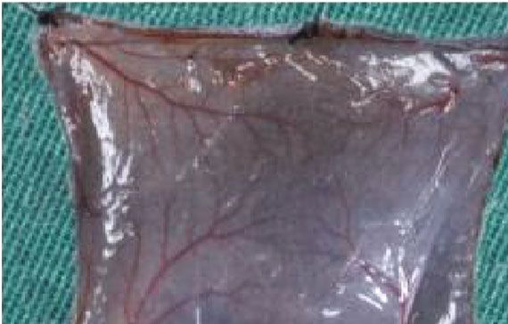
4                                                                                                               Stem Cells International

                                                    ⁎⁎⁎                                               ⁎⁎⁎
                                                                                   150
                                2500

                                2000

                                                                    VEGF (pg/mL)
                                                                                   100
                 bFGF (pg/mL)

                                1500

                                1000
                                                                                    50
                                500

                                  0                                                 0
                                       ECM/SVF-CM         SVF-CM                         ECM/SVF-CM         SVF-CM
                                                                                                      ⁎⁎⁎
                                 250                                               80
                                                    ⁎⁎⁎
                                200
                                                                                    60
                 PDGF (pg/mL)

                                                                    KGF (pg/mL)
                                150
                                                                                    40
                                100

                                                                                    20
                                 50

                                  0                                                  0
                                       ECM/SVF-CM         SVF-CM                         ECM/SVF-CM         SVF-CM

Figure 2: Quantification of growth factor expression. Quantification of VEGF, bFGF, PDGF, and KGF expression in SVF-CM and
ECM/SVF-CM. ∗∗∗ P < 0:01.

thorough washing with PBS, the cells were stained with Alexa            diaminobenzidine (Invitrogen) and hematoxylin, dehy-
Fluor488-conjugated goat anti-rabbit immunoglobulin G                   drated, transparentized, and mounted. Ki67-positive cells of
secondary antibody (1 : 200, Abcam) and DAPI (1 : 500,                  serial sections of specimens were counted with a microscope
Sigma) at room temperature for 1 h. Immunofluorescent                    at high magnification. Statistical analysis was performed
images were recorded using a fluorescence microscopy sys-                based on five high-powered fields per sample.
tem (IX71 FL, Olympus, Japan).
                                                                        2.8. Angiogenesis Activity In Vivo. We determined the effects
2.6. DPC Proliferation Assay. DPCs at passage 3 were seeded             of ECM/SVF-CM and SVF-CM on angiogenesis in vivo by
into 96-well culture plates at a density of 4000 cells per well         examining the vascularization of the hair-regeneration sites
and cultured in DMEM supplemented with 10% FBS incu-                    2 weeks after the injection of CM. Angiogenesis levels were
bated at 37°C with 5% CO2 for 6 h to permit cell adhesion,              evaluated on the basis of gross observation of the inner skin
to study the effects of the CMs on cell proliferation. The cells         of the hair-regrowth site and immunohistochemistry stain-
were then washed repeatedly with PBS and cultured in each               ing for CD31. Immunohistochemistry staining for CD31
of the two CMs or in serum-free medium (control group)                  was carried out as above, using anti-CD31 (1 : 200, Abcam).
for 1, 3, or 5 days, respectively. DPC proliferation was deter-         Neovascularization of serial sections of specimens was
mined by CCK8 assay (Sigma). The absorbance of the culture              counted with a microscope at high magnification.
medium was measured at 450 nm using a multilabel counter
(n = 3).                                                                2.9. Western Blot Protein Assay. Protein samples were pre-
                                                                        pared in RIPA lysis buffer (Gibco), and 30 μg aliquots were
2.7. Proliferative Activity of Cells in the Bulge Region. Half of       separated by sodium dodecyl sulfate-polyacrylamide gel
the mice in each group were sacrificed 2 weeks after injection.          electrophoresis and blotted onto polyvinylidene fluoride
The dorsal skin was harvested and fixed in 4% paraformalde-              membranes. After blocking, the membranes were incu-
hyde (Sigma) for at least 24 h, embedded in paraffin, and                 bated with rabbit anti-Wnt5a (1 : 1000, Abcam), rabbit
4 μm sections were cut. The proliferative activity of the cells         anti-Wnt10b (1 : 1000, Abcam), and anti-glyceraldehyde
in the bulge region of the hair follicle was examined in depar-         3-phosphate dehydrogenase (1 : 1000, Abcam) monoclonal
affinized sections stained with anti-Ki-67 (1 : 100, Abcam).              control antibody at 4°C for 24 h. Membrane-bound pri-
Specimens were pretreated by heating followed by blocking               mary antibodies were detected by incubation with second-
with 1% BSA (Sigma) and incubation with primary antibody                ary antibodies (1 : 5000, Abcam) at room temperature for
at 4°C overnight. The sections were then incubated with                 1 h. The results were analyzed using an enhanced chemilu-
secondary antibody (1 : 250, Abcam) for 30 min, stained with            minescence kit (Invitrogen, Grand Island, NY, USA) and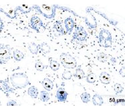
Stem Cells International                                                                                                                          5

             Control                    SVF-CM                 ECM/SVF-CM

 0d

 7d

14 d

                                                                                                           15
                                                                                                                                              #

                                                                                       Hair growth score
                                                                                                                                             ⁎⁎
                                                                                                           10                        #
                                                                                                                                    ⁎⁎

21 d                                                                                                                   #                      ⁎
                                                                                                           5          ⁎⁎
                                                                                                                                    ⁎
                                                                                                                       ⁎
                                                                                                            0
                                                                                                                0      1          2           3   4
                                                                                                                           Weeks of injection

                                                                                                                    Control
                                                                                                                    CM-SVF
                                                                                                                    CM-SVF/ECM
                                         (a)                                                                                  (b)

Figure 3: Hair-growth effect of ECM/SVF-CM in hair-loss-induced C57BL/6 mice. The hair was removed from the backs of C57BL/6 mice
and the hair-growth rate was monitored for 3 weeks. Mice were injected with ECM/SVF-CM, SVF-CM, and PBS (control), respectively, once
per week for 3 weeks. Gross view was observed by photographs (a), and hair-growth scoring was measured (b). Mice in the ECM/SVF-CM
injection group (n = 10) showed significantly increased hair regeneration compared with the SVF-CM injection group (∗∗ P < 0:05, n = 10)
and negative control group (#P < 0:01, n = 10). The SVF-CM injection group also showed significantly increased hair regeneration
compared with the control group (∗ P < 0:05). The data shown represent one of each group, and experiments were performed three times
independently.

analyst/PC densitometry software (Bio-Rad Laboratories,              C57BL/6 mice. CM-injected mice showed enhanced hair-
Hercules, CA, USA).                                                  growth rates at 1 and 2 weeks after injection, compared with
                                                                     the negative control group injected with PBS. Moreover, mice
2.10. Statistical Analysis. Differences among treatments              injected with ECM/SVF-CM had more rapid hair growth
were assessed by one-way analysis of variance (ANOVA)                than mice injected with SVF-CM (Figures 3(a) and 3(b)).
followed by Tukey–Kramer’s test for independent groups.              The ECM/SVF-CM group developed dark pigment on the
A P value < 0.05 was regarded as significant.                         initially pink hairless skin 1 week after injection, while the
                                                                     skin was less dark in the SVF-CM group and remained pink
3. Results                                                           in the negative control group (Figure 3(a)). Hair recovery
                                                                     extent on the dorsal skin and hair length were both sig-
3.1. Growth Factor Levels Increased in CM from ECM/SVF.              nificantly affected in the ECM/SVF-CM injection group at
We investigated the regulatory factors in ECM/SVF and                2 weeks after injection (Figure 3(a)), and 95%–100% hair
SVF CMs responsible for stimulating mouse hair growth.               regeneration to full length was observed in the ECM/SVF-
Levels of VEGF, bFGF, PDGF, and KGF were all significantly            CM group, but not in the SVF-CM group (70%–75%) at
higher in ECM/SVF-CM than in SVF-CM (Figure 2).                      3 weeks after injection. These results suggest that both
                                                                     CMs enhanced hair growth and that CM from ECM/SVF
3.2. Hair-Growth-Promoting Effects of ECM/SVF-CM in a                 had a stronger ability to simulate hair growth.
C57BL/6 Mouse Model. We compared the differences in hair
growth after weekly injection of ECM/SVF-CM and SVF-CM               3.3. ECM/SVF-CM Increased Proliferation of Human DPCs.
to determine their effects on hair growth efficiency in                 Hair follicle regeneration is governed by the interaction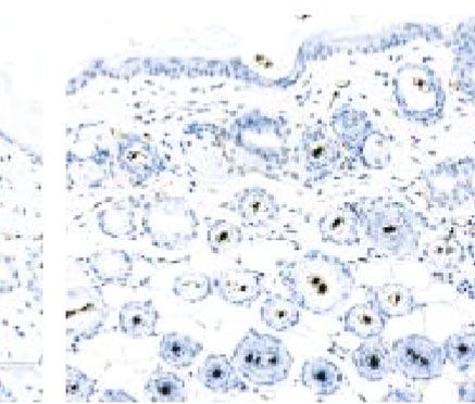
6                                                                                                                             Stem Cells International

                                         (a)                                                                  (b)

                                         (c)                                                                  (d)
                                                                                                         ⁎⁎
                                                                                                          ⁎
                                                                       2.0
                                                                                                     ⁎
                                               Absorbance (OD 450nm)

                                                                                           ⁎⁎
                                                                       1.5                      ⁎
                                                                                       ⁎
                                                                       1.0

                                                                       0.5

                                                                       0.0
                                                                             Day 1     Day 3         Day 5

                                                                             Control
                                                                             CM-SVF
                                                                             CM-SVF/ECM
                                                                                     (e)

Figure 4: Morphology of DPs and DPCs. Immunofluorescence identification of DPCs and proliferation-promoting effect of the two CMs on
DPCs. Phase-contrast micrograph showing oval appearance 2 days after inoculation, with most DPs attached with a few DPCs around them
(a). DPCs showing spindle-shaped morphology (b). ALP (c) and β-catenin (d) were highly expressed in DPCs as shown by
immunofluorescence staining. The viabilities of DPCs in the CM-treated groups were significantly higher than in the control group at
days 3 and 5, and the viability in the ECM/SVF-CM-treated group was significantly higher than in the SVF-CM-treated group (e). Scale
bar = 100 μm. (∗ P < 0:05, ∗∗ P < 0:01).

between epithelial precursor cells residing in the bulge region                            ated in an adherent fashion with spindle-like morphology
and mesenchymal cells located at the base of the hair follicle,                            (Figure 4(b)). To identify the intrinsic characteristics of
known as the DP [24]. We investigated the effects of CMs on                                 DPCs, signature markers associated with the hair-inducing
DPC proliferation. DPCs were isolated, cultured, and sub-                                  ability of DPCs, including ALP and β-catenin, were identified
jected to immunofluorescence analysis. The DPs were oval                                    by immunofluorescence staining. ALP and β-catenin were
and most dermal DPs were attached with a few DPCs around                                   highly expressed in DPCs (Figures 4(c) and 4(d)). The effects
them at 2 days after incubation (Figure 4(a)). DPCs prolifer-                              of the CMs on DPC proliferation are shown in Figure 4(e).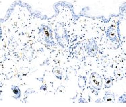
Stem Cells International                                                                                                                                         7

                    Control                                                                   SVF-CM                                 ECM/SVF-CM

                                                                                                       ⁎⁎⁎
                                            The number of Ki67-positive cell
                                                                               40
                                                                                                               ⁎⁎

                                                                               30
                                                                                               ⁎⁎

                                                                               20

                                                                               10

                                                                               0
                                                                                    Control         SVF-CM          ECM/SVF-CM

Figure 5: Immunohistochemical staining of Ki-67 in bulge cells. The number of Ki-67-positivity cell was significantly more in both the
ECM/SVF-CM- and SVF-CM-treated groups compared with the control group, with the highest cell viability in the ECM/SVF-CM-
treated group (∗∗ P < 0:05, ∗∗∗ P < 0:01). Scale bar = 20 μm.

The relative viabilities of DPCs in the CM-treated groups                                           3.6. Analysis of Wnt5a and Wnt10b Expression Involvement
were significantly higher than in the control group at days                                          in Hair-Growth Promotion by ECM/SVF-CM. To investigate
3 and 5, while the viability of the ECM/SVF-CM-treated                                              the signaling mechanism underlying the induction of anagen
cells was significantly higher than that of the SVF-CM-                                              phase in the CM groups, we detected signature proteins asso-
treated cells.                                                                                      ciated with anagen induction by western blot. Mice treated
                                                                                                    with CMs exhibited anagen induction, as evidenced by high
3.4. ECM/SVF-CM Increased Proliferation of Cells in the                                             expression of Wnt5a and Wnt10b (Figure 7). In addition,
Bulge Region of Hair Follicles. Previous studies revealed the                                       the ECM/SVF-CM-treated group had significantly higher
existence of a pool of hair follicle stem cells in the bulge                                        Wnt5a and Wnt10b expression levels compared with the
region [25]. These cells provide abundant progeny that are                                          group treated with SVF-CM. These results suggest that
responsible for sustaining the intact structure of the hair fol-                                    CMs activate certain functions in the anagen phase, and that
licle during continuous hair cycles [25]. We evaluated Ki-67                                        CMs stimulate hair follicle growth through the Wnt pathway.
expression in the bulge cells by immunohistochemical stain-
ing to determine the effects of the CMs on cell proliferation in                                     4. Discussion
this region of the hair follicle. Cell proliferation rates in both
the ECM/SVF-CM- and SVF-CM-treated groups were                                                      To optimize the CM therapy strategy for stimulating hair
higher than those in the control group, with the highest cell                                       growth, we introduced a novel adipose tissue-derived
viability in the ECM/SVF-CM-treated group (Figure 5).                                               stem cell and adipose native ECM enrichment product
                                                                                                    (ECM/SVF-gel) and compared the effects of ECM/SVF-
3.5. ECM/SVF-CM Increased Neoangiogenesis in C57BL/6                                                CM and SVF-CM on hair growth. The results suggested that
Mice. We investigated vascularization of the hair-regeneration                                      ECM/SVF-CM exerted hair growth-promoting effects via
sites 2 weeks after injection, to determine the effects of the                                       the proliferation of DPCs and cells in the bulge, and by neo-
CMs on angiogenesis in vivo. Blood vessels in the inner dor-                                        vascularization and anagen induction.
sal skin in the CM-treated groups were increased compared                                               Hair follicles undergo cyclic transformation from rapid
with the control group (Figure 6(a)). Mice in the ECM/SVF-                                          growth (anagen) to regression (catagen) and then progress
CM-treated group showed clear blood vessel branching and                                            to relative quiescence (telogen). The hair-growth cycle is
a significantly enhanced level of mature vessel formation                                            fine-regulated by epithelial–dermal interactions [26]. In ana-
(Figure 6(a)). We confirmed this by immunohistochemistry                                             gen, hair growth is governed by the proliferation of follicular
analysis of CD31. As expected, CD31 expression levels were                                          cells, mainly composed of epithelial cells residing in the
significantly higher in the ECM/SVF-CM-treated compared                                              bulge and DPCs located at the base of the hair follicle [26].
with the SVF-CM-treated and control groups (Figure 6(b)).                                           In addition, hair growth can be induced by modulating the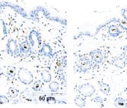
8                                                                                                            Stem Cells International

                 ECM/SVF-CM                                     SVF-CM                                      Control

                                                                  (a)
                ECM/SVF-CM                                      SVF-CM                                       Control

                                                                  ⁎⁎⁎

                                            40                             ⁎⁎
                                                           ⁎⁎
                                            30

                                            20

                                            10

                                             0
                                                 ECM/SVF-CM      SVF-CM         Control
                                                                  (b)

Figure 6: Stimulatory effect of ECM/SVF-CM on angiogenesis. Vascularization of the hair-regeneration sites was analyzed 2 weeks after
injection. The inner skin of the hair-regrowth site showed mature blood vessel branching (a). Among the three groups, the ECM/SVF-
CM-injection group showed markedly enhanced formation of mature vessels (a). Immunohistochemical staining of CD31 showed more
new blood vessels in the ECM/SVF-CM-injection compared with the SVF-CM-injection and control groups. Arrow indicates blood vessels
(b). (∗∗ P < 0:05, ∗∗∗ P < 0:01). Scale bar = 50 μm.

hair cycle, for example by delayed catagen induction and                our previous studies, we found that the percentage of
prolonged anagen, or by transition from telogen to anagen               MSC-specific cell markers (CD90+, CD73+, or CD105+) in
[27, 28]. Localized supplementation of growth factors and               cultured SVF cells were more than 95%, while the percent-
cytokines favoring hair growth was reported to promote hair             age of hematopoietic-specific cell markers (CD34-, CD11b-,
regeneration [8]. Previous reports show that ADSC-CM                    CD19-, or CD45-) was negative [30–32]. In this study, CMs
promoted hair growth by regulating the cell cycle and induc-            were collected from the medium of ECM/SVF and SVF
ing the anagen phase of the hair cycle, thereby promoting               after cultured for 48 h. Therefore, we consider that the bio-
the proliferation of DPCs and possibly epithelial cells [9].            logical efficacy of CMs mainly attributed to ADSCs. The
Our current results are consistent with those findings.                  expression of growth factors (bFGF, VEGF, PDGF, and
    The paracrine effect is one of the most important thera-             KGF) was found in ECM/SVF-CM and SVF-CM which
peutic advantages of stem cell therapy [9]. Secretory factors           may play important roles in regulating cell functions during
derived from ADSCs related to hair-growth stimulation,                  hair growth. In particular, bFGF promotes hair growth via
including VEGF, bFGF, PDGF, HGF, KGF, and IGF, posi-                    DPC proliferation and increased hair follicle size [33] and
tively regulate hair follicle activity and promote hair growth          also has a positive effect on the hair growth cycle by induc-
[8]. The population of SVF includes endothelial cells, endo-            ing anagen phase and elongating the hair shaft [34]. PDGF
thelial progenitor cells, pericytes, preadipocytes, macro-              induces and maintains anagen phase and provides a stem cell
phage, and ADSCs [29]. However, cells were almost                       niche to regulate the hair cycle [35]. KGF prevents hair folli-
excluded except for ADSCs after in vitro cell culture. In               cle cell death by promoting protein synthesis [36], and HGF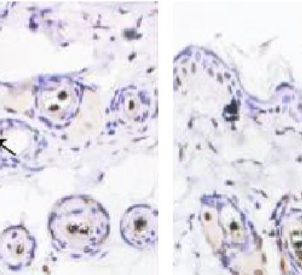
Stem Cells International                                                                                                                  9

                            ECM/SVF-CM SVF-CM          Control

                       Wnt5a
                                                                                                                      ⁎⁎⁎
                                                                                                2.0                     ⁎⁎⁎
                                                                                                        ⁎⁎⁎

                                                                  Relative protein expression
                                                                                                          ⁎⁎⁎      ⁎⁎⁎
                                                                                                1.5   ⁎⁎⁎
                      Wnt10b

                                                                                                1.0

                                                                                                0.5
                     GAPDH

                                                                                                0.0
                                                                                                       Wnt5a       Wnt-10b
                                                                                                      PBS
                                                                                                      SVF-CM
                                                                                                      ECM/SVF-CM

Figure 7: Involvement of Wnt pathway in the stimulatory effect of hair growth by ECM/SVF-CM. Both ECM/SVF-CM and SVF-CM
markedly increased Wnt5a and Wnt10b expression levels compared with the control group, with the highest expression in the ECM/SVF-
CM-treated group (∗∗∗ P < 0:05).

contributes to anagen maintenance by retarding hair follicle                 higher cell potency in ECM/SVF-gel. However, further
regression and suppressing follicle keratinocyte apoptosis                   research is still necessary to verify the precise mechanisms
[37]. Enhancement of angiogenesis is usually accompanied                     underlying the higher expression of growth factors in
by positive hair growth promotion [38]. VEGF is a powerful                   ECM/SVF-CM compared with SVF-CM.
mediator of angiogenesis that accelerates hair growth and                        Although we found the beneficial effect of ECM/SVF-CM
increases follicular size by initiating angiogenesis, resulting              on hair growth, some related limitations still exist. First,
in the formation of a new network of capillaries carrying                    we did not compare ECM/SVF with an in vitro analogue.
blood, oxygen, and nutrients to the hair follicle [39]. Angio-               Second, the components of CMs were complex, and mass
genesis around hair follicles was previously shown to be                     spectrometry analysis of the components stimulated by
related to the hair cycle [40]. Significant angiogenesis occurs               ECM/SVF-CM would be necessary. Furthermore, we did
during the hair-growth anagen phase, while vessels around                    not explain the interaction between cells and ECM.
the hair follicles are reduced and degenerate during the hair
growth-retardation catagen and telogen phases [41]. In the                   5. Conclusions
current study, the hair-growth effect of CM from ECM/SVF
was associated with a stimulatory effect on angiogenesis, as                  In conclusion, this study provides the first evidence for the
evidenced by a marked increase in vessels following treat-                   promoting effects of CM from ECM/SVF on hair growth in
ment. Inducing angiogenesis may also induce the anagen                       a mouse model, acting via the proliferation of DPCs and cells
phase of the hair cycle as a hair growth-stimulating process.                in the bulge, as well as by enhancing angiogenesis and anagen
Therefore, we speculated that the promotion of hair growth                   induction. ECM/SVF-CM can be stored with long-term sta-
in the two CM-treatment groups may be due to the growth                      bility and can be produced on a large scale, making it a poten-
factors in the extract.                                                      tially low-cost therapy. Further studies are needed to confirm
    Optimization of stem cell-CM to upregulate key growth                    the use of CM from ECM/SVF as an effective and improved
factors is an important strategy to achieve encouraging out-                 strategy for the treatment of hair loss.
comes in terms of hair regeneration and hair growth. The
current study showed that VEGF, bFGF, PDGF, and KGF                          Data Availability
secretions were all significantly increased in ECM/SVF-CM
compared with SVF-CM, resulting in more effective stimula-                    The data used to support the findings of this study are
tion of hair growth. This result may be attributed to the ben-               included within the article.
eficial effects of native ECM on tissue-resident stem cells.
Natural ECM provides a physical scaffold for cells and retains                Conflicts of Interest
the natural state of the cells, thereby providing a favorable
environment for attached cells to maintain optimal cell sur-                 The authors declare no competing financial interests.
vival and efficacy [21]. In addition to providing structural
support for cells, ECM also acts as a dynamic microenviron-                  Authors’ Contributions
ment, regulating important biological functions of cells [21].
In this study, we found that ECM/SVF-CM had a higher con-                    All authors agree with the manuscript content. Zairong Wei
centration of growth factors than the SVF-CM, indicating a                   and Shune Xiao designed the experiments, Yurong Deng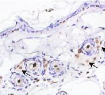
10                                                                                                                  Stem Cells International

performed the cellular biology experiments and statistical                     International Journal of Dermatology, vol. 54, no. 6, pp. 730–
analysis. Zhiyuan Liu and Xiaojin Mo conducted the animal                      735, 2015.
assay. Dali Wang helped perform the analysis with construc-             [12]   K. Narita, H. Fukuoka, T. Sekiyama, H. Suga, and K. Harii,
tive discussions. Chengliang Deng provided many sugges-                        “Sequential scalp assessment in hair regeneration therapy
tions during manuscript preparation, and Shune Xiao wrote                      using an adipose-derived stem cell-conditioned medium,”
the manuscript. Zairong Wei and Chengliang Deng are                            Dermatologic Surgery, vol. 46, no. 6, pp. 819–825, 2020.
cocorresponding authors. Shune Xiao and Yurong Deng con-                [13]   H. Fukuoka and H. Suga, “Hair regeneration treatment using
tributed equally and are co-first authors.                                      adipose-derived stem cell conditioned medium: follow-up
                                                                               with trichograms,” Eplasty, vol. 15, article e10, 2015.
Acknowledgments                                                         [14]   Y. M. Jeong, Y. K. Sung, W. K. Kim et al., “Ultraviolet B pre-
                                                                               conditioning enhances the hair growth-promoting effects of
This work was supported by the National Natural Science                        adipose-derived stem cells via generation of reactive oxygen
Foundation of China (no. 81801921), Science and Technol-                       species,” Stem Cells and Development, vol. 22, no. 1, pp. 158–
                                                                               168, 2013.
ogy Program of Zunyi (HZ,2019-51 and 2018-9), and the
PhD Fund of Scientific Research Foundation of Zunyi Medi-                [15]   B. S. Park, W. S. Kim, J. S. Choi et al., “Hair growth stimulated
                                                                               by conditioned medium of adipose-derived stem cells is
cal University (2018-14).
                                                                               enhanced by hypoxia: evidence of increased growth factor
                                                                               secretion,” Biomedical Research, vol. 31, no. 1, pp. 27–34,
References                                                                     2010.
                                                                        [16]   S. H. Bhang, S. Lee, J. Y. Shin, T. J. Lee, H. K. Jang, and B. S.
 [1] E. Yim, K. L. B. Nole, and A. Tosti, “5α-Reductase inhibitors in
                                                                               Kim, “Efficacious and clinically relevant conditioned medium
     androgenetic alopecia,” Current Opinion in Endocrinology,
                                                                               of human adipose- derived stem cells for therapeutic angio-
     Diabetes, and Obesity, vol. 21, no. 6, pp. 493–498, 2014.
                                                                               genesis,” Molecular Therapy, vol. 22, no. 4, pp. 862–872, 2014.
 [2] S. Kassira, D. Z. Korta, L. W. Chapman, and F. Dann, “Review
                                                                        [17]   Y. J. Cho, H. S. Song, S. Bhang et al., “Therapeutic effects of
     of treatment for alopecia totalis and alopecia universalis,”
                                                                               human adipose stem cell-conditioned medium on stroke,”
     International Journal of Dermatology, vol. 56, no. 8, pp. 801–
                                                                               Journal of Neuroscience Research, vol. 90, no. 9, pp. 1794–
     810, 2017.
                                                                               1802, 2012.
 [3] P. Gentile and S. Garcovich, “Advances in regenerative stem
                                                                        [18]   S. J. Kim, J. Park, H. Byun et al., “Hydrogels with an embossed
     cell therapy in androgenic alopecia and hair loss: Wnt path-
                                                                               surface: an all-in-one platform for mass production and cul-
     way, growth-factor, and mesenchymal stem cell signaling
                                                                               ture of human adipose-derived stem cell spheroids,” Biomate-
     impact analysis on cell growth and hair follicle development,”
                                                                               rials, vol. 188, pp. 198–212, 2019.
     Cell, vol. 8, no. 5, p. 466, 2019.
 [4] G. Storti, M. G. Scioli, B. S. Kim, A. Orlandi, and V. Cervelli,   [19]   Y. Yao, Z. Dong, Y. Liao et al., “Adipose extracellular matrix/-
     “Adipose-derived stem cells in bone tissue engineering: useful            stromal vascular fraction gel: a novel adipose tissue-derived
     tools with new applications,” Stem Cells International,                   injectable for stem cell therapy,” Plastic and Reconstructive
     vol. 2019, Article ID 3673857, 18 pages, 2019.                            Surgery, vol. 139, no. 4, pp. 867–879, 2017.
 [5] S. Al-Ghadban and B. A. Bunnell, “Adipose tissue-derived           [20]   M. Sun, Y. He, T. Zhou, P. Zhang, J. Gao, and F. Lu, “Adipose
     stem cells: immunomodulatory effects and therapeutic poten-                extracellular matrix/stromal vascular fraction gel secretes
     tial,” Physiology (Bethesda, Md.), vol. 35, no. 2, pp. 125–133,           angiogenic factors and enhances skin wound healing in a
     2020.                                                                     murine model,” BioMed Research International, vol. 2017,
                                                                               Article ID 3105780, 11 pages, 2017.
 [6] R. Anderi, N. Makdissy, A. Azar, F. Rizk, and A. Hamade,
     “Cellular therapy with human autologous adipose-derived            [21]   C. Deng, Y. He, J. Feng et al., “Extracellular matrix/stromal
     adult cells of stromal vascular fraction for alopecia areata,”            vascular fraction gel conditioned medium accelerates wound
     Stem Cell Research & Therapy, vol. 9, no. 1, p. 141, 2018.                healing in a murine model,” Wound Repair and Regeneration,
 [7] J. L. Spees, R. H. Lee, and C. A. Gregory, “Mechanisms of mes-            vol. 25, no. 6, pp. 923–932, 2017.
     enchymal stem/stromal cell function,” Stem Cell Research &         [22]   V. Vegesna, J. O’Kelly, M. Uskokovic et al., “Vitamin D3 ana-
     Therapy, vol. 7, no. 1, p. 125, 2016.                                     logs stimulate hair growth in nude mice,” Endocrinology,
 [8] S. Ramdasi and S. K. Tiwari, “Human mesenchymal stem cell-                vol. 143, no. 11, pp. 4389–4396, 2002.
     derived conditioned media for hair regeneration applications,”     [23]   M. Magerl, S. Kauser, R. Paus, and D. J. Tobin, “Simple and
     Journal of Stem Cells, vol. 11, no. 4, pp. 201–211, 2016.                 rapid method to isolate and culture follicular papillae from
 [9] C. H. Won, G. H. Park, X. Wu et al., “The basic mechanism of              human scalp hair follicles,” Experimental Dermatology,
     hair growth stimulation by adipose-derived stem cells and                 vol. 11, no. 4, pp. 381–385, 2002.
     their secretory factors,” Current Stem Cell Research & Therapy,    [24]   N. Saxena, K. W. Mok, and M. Rendl, “An updated classifica-
     vol. 12, no. 7, pp. 535–543, 2017.                                        tion of hair follicle morphogenesis,” Experimental Dermatol-
[10] H. Shin, C. H. Won, W. K. Chung, and B. S. Park, “Up-to-date              ogy, vol. 28, no. 4, pp. 332–344, 2019.
     clinical trials of hair regeneration using conditioned media of    [25]   S. Joulai Veijouye, A. Yari, F. Heidari, N. Sajedi, F. Ghoroghi
     adipose-derived stem cells in male and female pattern hair                Moghani, and M. Nobakht, “Bulge region as a putative hair
     loss,” Current Stem Cell Research & Therapy, vol. 12, no. 7,              follicle stem cells niche: a brief review,” Iranian Journal of Pub-
     pp. 524–530, 2017.                                                        lic Health, vol. 46, no. 9, pp. 1167–1175, 2017.
[11] H. Shin, H. H. Ryu, O. Kwon, B. S. Park, and S. J. Jo, “Clinical   [26]   P. Rompolas, E. R. Deschene, G. Zito et al., “Live imaging of
     use of conditioned media of adipose tissue-derived stem cells             stem cell and progeny behaviour in physiological hair- follicle
     in female pattern hair loss: a retrospective case series study,”          regeneration,” Nature, vol. 487, no. 7408, pp. 496–499, 2012.
Stem Cells International 11
[27] M. Choi, S. J. Choi, S. Jang et al., “Shikimic acid, a mannose
bioisostere, promotes hair growth with the induction of ana-
gen hair cycle,” Scientific Reports, vol. 9, no. 1, article 17008,
2019.
[28] A. P. Vasserot, M. Geyfman, and N. J. Poloso, “Androgenetic
alopecia: combing the hair follicle signaling pathways for
new therapeutic targets and more effective treatment options,”
Expert Opinion on Therapeutic Targets, vol. 23, no. 9, pp. 755–
771, 2019.
[29] B. J. Philips, T. L. Grahovac, J. E. Valentin et al., “Prevalence of
endogenous CD34+ adipose stem cells predicts human fat
graft retention in a xenograft model,” Plastic and Reconstruc-
tive Surgery, vol. 132, no. 4, pp. 845–858, 2013.
[30] S. Xiao, Z. Liu, Y. Yao, Z. R. Wei, D. Wang, and C. Deng, “Dia-
betic human adipose-derived stem cells accelerate pressure
ulcer healing by inducing angiogenesis and neurogenesis,”
Stem Cells and Development, vol. 28, no. 5, pp. 319–328, 2019.
[31] Z. Liu, S. Xiao, K. Tao et al., “Synergistic effects of human
platelet-rich plasma combined with adipose- derived stem cells
on healing in a mouse pressure injury model,” Stem Cells Inter-
national, vol. 2019, Article ID 3091619, 12 pages, 2019.
[32] S. Xiao, D. Zhang, Z. Liu et al., “Diabetes-induced glucolipo-
toxicity impairs wound healing ability of adipose-derived stem
cells-through the miR-1248/CITED2/HIF-1α pathway,” Aging
(Albany NY), vol. 12, no. 8, pp. 6947–6965, 2020.
[33] M. Ozeki and Y. Tabata, “Promoted growth of murine hair
follicles through controlled release of basic fibroblast growth
factor,” Tissue Engineering, vol. 8, no. 3, pp. 359–366, 2002.
[34] T. A. Rosenquist and G. R. Martin, “Fibroblast growth factor
signalling in the hair growth cycle: expression of the fibroblast
growth factor receptor and ligand genes in the murine hair fol-
licle,” Developmental Dynamics, vol. 205, no. 4, pp. 379–386,
1996.
[35] Y. Tomita and M. Akiyama, “PDGF isoforms induce and
maintain anagen phase of murine hair follicles,” Journal of
Dermatological Science, vol. 43, no. 2, pp. 105–115, 2006.
[36] S. Braun, M. Krampert, E. Bodó et al., “Keratinocyte growth
factor protects epidermis and hair follicles from cell death
induced by UV irradiation, chemotherapeutic or cytotoxic
agents,” Journal of Cell Science, vol. 119, no. 23, pp. 4841–
4849, 2006.
[37] G. Lindner, A. Menrad, E. Gherardi et al., “Involvement of
hepatocyte growth factor/scatter factor and met receptor sig-
naling in hair follicle morphogenesis and cycling,” The FASEB
Journal, vol. 14, no. 2, pp. 319–332, 2000.
[38] L. Mecklenburg, D. J. Tobin, S. Müller-Röver et al., “Active
hair growth (anagen) is associated with angiogenesis,” The
Journal of Investigative Dermatology, vol. 114, no. 5, pp. 909–
916, 2000.
[39] K. Yano, L. F. Brown, and M. Detmar, “Control of hair growth
and follicle size by VEGF-mediated angiogenesis,” The Journal
of Clinical Investigation, vol. 107, no. 4, pp. 409–417, 2001.
[40] H. Cheng, J. Zhang, J. Li, M. Jia, Y. Wang, and H. Shen, “Plate-
let-rich plasma stimulates angiogenesis in mice which may
promote hair growth,” European Journal of Medical Research,
vol. 22, no. 1, p. 39, 2017.
[41] M. K. Jung, S. Ha, S. Y. Huh et al., “Hair-growth stimulation by
conditioned medium from vitamin D3-activated preadipo-
cytes in C57BL/6 mice,” Life Sciences, vol. 128, pp. 39–46,
2015.You can also read