Prospects and Challenges for T Cell-Based Therapies of HCC - MDPI
←
→
Page content transcription
If your browser does not render page correctly, please read the page content below
cells
Review
Prospects and Challenges for T Cell-Based Therapies of HCC
Norman Woller *, Sophie Anna Engelskircher, Thomas Wirth and Heiner Wedemeyer

                                          Clinic for Gastroenterology, Hepatology and Endocrinology, Hannover Medical School,
                                          30625 Hannover, Germany; engelskircher.sophie@mh-hannover.de (S.A.E.);
                                          wirth.thomas@mh-hannover.de (T.W.); wedemeyer.heiner@mh-hannover.de (H.W.)
                                          * Correspondence: woller.norman@mh-hannover.de

                                          Abstract: The scope of therapeutic options for the treatment of hepatocellular carcinoma (HCC)
                                          has recently been expanded by immunotherapeutic regimens. T cell-based therapies, especially in
                                          combination with other treatments have achieved far better outcomes compared to conventional
                                          treatments alone. However, there is an emerging body of evidence that eliciting T cell responses
                                          in immunotherapeutic approaches is insufficient for favorable outcomes. Immune responses in
                                          HCC are frequently attenuated in the tumor microenvironment (TME) or may even support tumor
                                          progress. Hence, therapies with immune checkpoint inhibitors or adoptive cell therapies appear
                                          to necessitate additional modification of the TME to unlock their full potential. In this review, we
                                          focus on immunotherapeutic strategies, underlying molecular mechanisms of CD8 T cell immunity,
                                          and causes of treatment failure in HCC of viral and non-viral origin. Furthermore, we provide an
                                          overview of TME features in underlying etiologies of HCC patients that mediate therapy resistance
                                          to checkpoint inhibition and discuss strategies from the literature concerning current approaches to
                                          these challenges.


                                          Keywords: immune checkpoint inhibition; hepatocellular carcinoma; tumor surveillance; immunother-
Citation: Woller, N.; Engelskircher,
                                          apy; T cell responses; treatment failure; CAR therapy; chronic hepatitis; HBV; HCV
S.A.; Wirth, T.; Wedemeyer, H.
Prospects and Challenges for T
Cell-Based Therapies of HCC. Cells
2021, 10, 1651. https://doi.org/
10.3390/cells10071651
                                          1. Introduction
                                               Patients with advanced stages of HCC face a poor prognosis. Liver cancer is the
Academic Editors: Paola Fisicaro and      third leading cause of cancer-related mortality worldwide [1]. It is still a difficult-to-treat
Carolina Boni                             disease, despite several treatment options, such as liver transplantation, systemic treatment
                                          with chemotherapy, loco-regional treatment, such as transarterial chemoembolization
Received: 26 May 2021                     (TACE) and radioembolization or treatment with sorafenib, lenvatinib, or other multi-
Accepted: 29 June 2021                    kinase inhibitors [2–4]. Untreated HCC has a 5-year overall survival rate of less than
Published: 30 June 2021
                                          10% and curative options in advanced stages, when the disease is usually detected, are
                                          rare [5]. Recently, prognosis of HCC had improved remarkably with the implementation
Publisher’s Note: MDPI stays neutral      of immune checkpoint inhibition (CPI) into the treatment schemes as we will discuss in
with regard to jurisdictional claims in
                                          detail. The agents that are commonly used for CPI are antibodies inhibiting the CTLA4
published maps and institutional affil-
                                          pathway, such as ipilimumab, and the PD-1/PD-L1 pathway, such as pembrolizumab and
iations.
                                          atezolizumab. CTLA-4 is a homologue of CD28 that binds to members of the B7 family
                                          during T cell activation by antigen-presenting cells and has a higher affinity than does
                                          CD28. The interaction of PD-1 with PD-L1 keeps T cells from killing tumor cells, whereas
                                          blocking this interaction can allow for cytotoxic responses to lyse tumor cells.
Copyright: © 2021 by the authors.              Not even a decade has passed from the time when concrete evidence was found
Licensee MDPI, Basel, Switzerland.        that lymphocytes can prevent tumor development [6] until the first clinical studies of
This article is an open access article
                                          checkpoint inhibition confirmed increased survival in patients [7,8]. In 2010, patients
distributed under the terms and
                                          with metastatic melanoma benefitted from treatment with ipilimumab, an inhibitor of the
conditions of the Creative Commons
                                          CTLA-4 pathway. PD-1/PD-L1 checkpoint inhibitors soon followed suit, also showing
Attribution (CC BY) license (https://
                                          effectiveness of checkpoint inhibition in other tumor entities [9,10]. It was realized that
creativecommons.org/licenses/by/
                                          CPI elicits T cells against cancer neoantigens as the main drivers of responses [11,12].
4.0/).

Cells 2021, 10, 1651. https://doi.org/10.3390/cells10071651                                                   https://www.mdpi.com/journal/cells
Cells 2021, 10, 1651 2 of 23
Since occurrence of positive clinical studies, a plethora of checkpoint inhibitors targeting
PD-1 and PD-L1 has been approved and tested in clinical trials in a great variety of cancers.
The results of phase 1/2 and 2 clinical trials in HCC patients with single use of blocking
antibodies of the PD-1 pathway led to approval of these agents by the United States Food
and Drug Administration (FDA) for the treatment of HCC [13,14]. However, the first phase
III trials failed to reach the predefined endpoint both for nivolumab as a first line therapy
and pembrolizumab (both inhibitors of the PD-1 pathway) as second line treatment [15,16].
Still, both trials confirmed an overall response rates (ORR) of 15–20% observed in the
phase II trials. However, among these responding patients, complete responses defined as
disappearance of vital tumors were almost non-existent.
These clinical results show that efficacy of CPI treatment of HCC lagged behind other
tumor entities, primarily metastatic melanoma (ORR 61% [17]) and Hodgkin lymphoma
(ORR 87% [18]), among others [19,20]. This was true until recently the clinical study
IMbrave150 was published that combined CPI (atezolizumab, a PD-L1 checkpoint inhibitor)
with inhibition of angiogenesis (bevacizumab, targeting vascular endothelial growth factor
(VEGF)). Overall survival rates at 12 months were significantly higher in the atezulizumab
+ bevacizumab arm (67.2%; 95% CI: 61.3–73.1) compared with the sorafenib arm (54.6%;
95% CI: 45.2–64.0). It reported a hazard ratio for overall survival of 0.58 in favour of
the combination therapy. This represents a 42% reduction in the risk of death compared
with the previous first-line treatment with a tyrosine kinase inhibitor (TKI) sorafenib [21].
Additionally, the study also provided HCC patients a perspective for complete responses,
as was the case in 5.5% of patients in the combined treatment group (18/326, according
to independent review facility-assessed Response Evaluation Criteria in Solid Tumors
(RECIST) v1.1). These clinical results are unprecedented with regard to the treatment
outcome, demonstrating the potential of CPI within combinatorial treatment regimens [21].
The IMbrave150 study changed therapy guidelines for first line therapy from tyrosine
kinase inhibition to immunotherapy [22] and therefore it is worthwhile to take a closer look
at potential underlying mechanisms of CPI that facilitate immune-mediated clearance of
HCC cells. Moreover, the key question that has to be addressed is how the immune system
can be thoroughly stimulated by pharmacological intervention for a long-term effective
treatment, as tumor immunity appears to be crucial in regimens of HCC. In addition, this
knowledge could be used for other T cell-based therapies, including therapeutic vaccination
and infusion of ex vivo expanded T cells.
2. The Vade Mecum of HCC Treatment Is Now Based on Pillars of Cellular Immunity
by Combining Checkpoint Inhibition with Anti-Angiogenesis Treatment
The best objective response rates (ORR), derived from clinical studies of HCC, all
include checkpoint inhibitors. A phase Ib study of the multi kinase inhibitor lenvatinib
targeting VEGFR1, VEGFR2, and VEGFR3 plus pembrolizumab has an ORR of 36% [23],
PD-1 inhibition with nivolumab and ipilimumab shows an ORR of 32% [24] followed by
atezulizumab and bevacizumab treatment with 27.3% ORR [21]. Relating to long-term
efficacy and safety profiles, the latter regimen is the first-line treatment of choice.
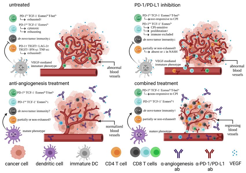
Angiogenic factors such as hypoxia-induced and tumor-microenvironment (TME)-
derived VEGF is capable of downregulating adhesion molecules on endothelial cells.
Expression of intercellular adhesion molecule 1 (ICAM-1) and 2, and vascular cell adhesion
molecule 1 (VCAM-1) inhibit T cell adhesion [25,26]. Consequently, elevated levels of
VEGF in the TME have been correlated with immune exclusion of T cells within the
tumor tissue [27]. Regulatory T cells (Tregs), myeloid-derived suppressor cells (MDSCs),
and tumor-associated macrophages (TAMs) are recruited to HCC tumors, mediated by
VEGF and hypoxia-inducible factor 1 (HIF-1). This links tumor tolerance by failure of
T cell immune surveillance to hypoxia [28,29]. Thus, it is a strong rationale to combine
VEGF inhibition with CPI, as investigated in the phase III IMbrave150 trial. Therapy with
atezolizumab blocks PD-L1 expressed on immune cells and tumor cells. Blocking PD-L1
prohibits interactions with the ligands PD-L1 and CD80. PD-1 is a checkpoint inhibitory
receptor that is expressed on antigen-primed T cells in infection and cancer [30]. This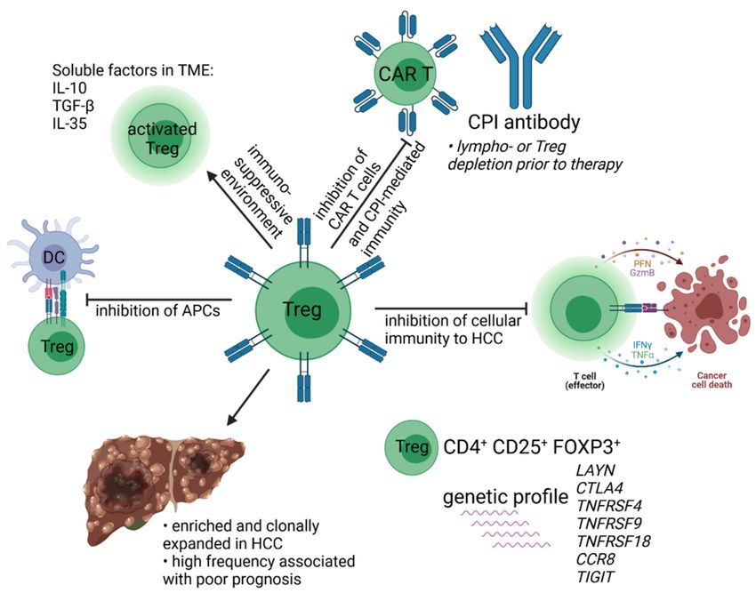
Cells 2021, 10, 1651                                                                                                        3 of 23

                                receptor regulates T cell proliferation and tolerance and is involved in tumor evasion and T
                                cell exhaustion [31].
                                      This effect is reversible by inhibiting VEGF:VEGF receptor-2 interactions [32]. HCC tis-
                                sue is highly neovascularized and accessible for inhibition of angiogenesis, as these tumors
                                also show a high microvessel density [33]. Regarding the combination of atezolizumab
                                and bevacizumab, there is evidence that combined treatment enhances antigen-specific
                                T cell migration, potentially through vascular normalization and endothelial cell activa-
                                tion. The combined regimen leads to increased counts of CD8 T cells, increased Th1- and
                                T-effector markers, intra-tumoral major histocompatibility complex (MHC)-I expression
                                and chemokine levels [34]. This demonstrates that bevacizumab has more effects than
                                merely inhibition of angiogenesis, with a particular benefit for the immune system. Apart
                                from structural remodelling of the tumor blood vessel bed by anti-VEGF therapy that
                                facilitates T cell infiltration, there is also data that it reverses the inhibitory effects of VEGF
                                on dendritic cell (DC) maturation that leads to reduced T cell priming [35]. As single-agent
                                activity in HCC, response rates of atezolizumab and bevacizumab are quite similar. With
                                17% for atezolizumab in a Phase Ib study and 14% for bevacizumab in Phase II studies, it
                                raises the question whether the effects are additive or complementary due to the different
                                modes of action [36–39]. Both compounds mediate their effects on different parts of the
                                immune system that are considered complementary and thus are more likely to elicit
                                tumor immunity and increasing response rates. On the one hand, the IMbrave150 shows
                                a response rate of 27%, which would suggest an additive effect. On the other hand, a
                                majority of patients (88%) maintain their response long term, i.e., for six months or longer.
                                Figure 1 shows the proposed mechanism of combined regimens using CPI and inhibition
                                of angiogenesis.

      Figure 1. Effects of PD-1 or PD-L1 inhibition, anti-angiogenesis treatment, and combined treatment on immune cells and
      tumor tissue in HCC (abbreviations: ↑/↓ high/low; markerhi/lo high/low marker expression).
Cells 2021, 10, 1651 4 of 23
3. Biomarkers and Immunological Classification of HCC
Predicting therapeutic benefit prior to or shortly after therapy starts by biomarkers is
a well sought-after aim in clinical oncology [40]. So far, the most common marker that is
correlated to a therapeutic response in other tumor entities is the expression of PD-L1 in
tumor-tissue and the tumor mutational burden (TMB). However, these parameters have not
been shown to reliably predict treatment responses in HCC patients receiving checkpoint
inhibitors [41–43]. One study even questions the dominant role of neoantigens in HCC for
CPI due to the relatively low mutational burden compared to malignant melanoma [44].
The lack of common markers for the prediction of the treatment response of HCC led to
other assessments as we will see later.
Anti-tumor immunity can appear concomitantly with tumor progression. This ob-
servation is called the “Hellström paradox” according to a study from 1968 that found
humoral and cellular components with tumoricidal activity in patients with growing tu-
mors [45]. These findings suggests that activity of tumor-directed immunity must outpace
tumor cell proliferation to reach a threshold of net reduction in the overall tumor mass.
This view conforms also with the state of equilibrium derived from the hypothesis of
immunoediting [46]. Accordingly, cancer immunotherapy aims at amplification of existing
tumor immunity or de novo generation in order to tip the scale towards favourable out-
comes [47]. With regard to HCC, the stratification of etiologies for the clinical outcome may
help to dissect and understand effects of signaling pathways and immune cell phenotypes
on immunotherapy responses [48]. It is important to distinguish the non-responsiveness
to cancer immunotherapy between the failure of triggering an immune response and the
functional failure of the elicited response. Here, primary, adaptive, and acquired resistance
can be differentiated [49]. Acquired resistance is an important, but often underestimated,
clinical parameter showing that responses are mostly temporary. Thus, the design of future
clinical studies should include strategies to maintain already existing immune responses.
The overall designation of all immune factors within a host that eventually leads to the
killing of cancer cells is briefly called the cancer immune cycle [50]. Any interruption within
that sequence or the functionality of essential networks of the cycle leads to a complete
abortion and eventual failure of tumor rejection. It is a convenient tenet and the ultimate
reason to explain resistance to cancer immunotherapy and treatment failure of CPI. A more
refined view on the clinical effects of checkpoint inhibitors is the cancer immune set point.
This views immunity to cancer as a complex set of tumor, host and environmental factors.
These factors govern the magnitude and timing of the anticancer response [51]. Consistent
with the observation that HCC develops in a complex environment of chronic hepatitis
and fibrosis, likewise the genomic landscape has been described as highly complex and
heterogeneous [52,53]. More suitable for predictions appear multi-omics approaches that
have been proposed for immune profiling of HCC. A study of Sia et al. analysed 956 HCC
samples and found that about 25% of HCC have markers of an inflammatory response
with high expression levels of PD-1 and PD-L1, cytotoxic marker expression, such as an
interferon gamma (IFN-γ) signature, and low levels of chromosomal aberrations. The
immune class correlated with better overall survival [54]. Additionally, the subgroup
of this cohort was either characterized by an adaptive T cell response or an exhausted
immune response that allowed stratification of an active and exhausted immune subclass.
The active immune subclass showed signs of an ongoing cytotoxic response, in which
IFN-γ and granzyme B signatures are present. In contrast, the immune-excluded subclass
was dominated by signature of T cell exhaustion, suppressive myeloid cells, and tumor
growth factor-β (TGF-β). In another study, Zhang et al. performed immune profiling
of HCC and defined three groups that suggest differentiation into immunocompetent,
immunosuppressive, and immunodeficient subtypes [55]. Expression level analysis of
CD45 and Foxp3 in immunohistochemistry (IHC) allowed for correlative classification
of the treatment outcome in this study. The immunocompetent subtype was CD45hi and
FOXP3lo showing infiltration of αβ and γδ T cells. Furthermore, HCCs of the immuno-
suppressive subtype stained CD45hi and FOXP3hi indicating regulatory T and B cells, asCells 2021, 10, 1651 5 of 23
well as tolerogenic macrophages and immunosuppressive molecules, such as PD-1/PD-L1,
TGF-β, VEGF, T cell immunoglobulin and mucin domain containing protein 3 (TIM-3)
and interleukin-10 (IL-10). The immunodeserted subtype showed a CD45lo phenotype
with a significant reduction of immune cell infiltration [55]. A similar classification of the
immune composition of HCC by a study investigating 158 HCC patients was proposed
to distinguish three immune-subtypes: Immune-high, immune-mid, and immune-low.
Increased plasma/B cell and T cell infiltration in the immune-high subtype were identified
as independent positive prognostic factors [56]. These promising studies show that an
in-depth immune profiling potentially combined with genetic approaches may lead to
stratification of HCC for appropriate prediction of the outcome. In addition, these studies
suggest that T cells with distinct properties exists that prevent tumor outgrowth.
4. The Immune Landscape of HCC
An immune landscape of cancer refers to a complex network of the immune cell com-
position of the TME including cytokines and cell ratio patterns, as well as genetic features
of tumor tissue. The latter includes intratumoral heterogeneity, aneuploidy, mutational
burden, and expression of immunomodulatory genes that have an impact on the leukocyte
levels. Mutations in CTNNB1, Nras, and IDH1 are associated with low levels of leukocytes,
whereas BRAF, TP53, and CASP8 are correlated with high levels of leukocytes. All these
factors ultimately contribute to the prognosis [57]. The immune landscape of HCC has re-
cently been investigated by several studies [58–61]. The main driver of HCC development
is cirrhosis of the liver and it has been shown that tumors are inflammation-associated
and generate a tumor microenvironment (TME) that is highly immunosuppressive [62].
The immune cell composition underlies fundamental differences according to healthy
liver tissue, adjacent tissue, and tumor tissue. Here, total B and T cells are significantly
upregulated in tumor tissue, whereas CD8 T cells are abundant in adjacent tissue and
tumors. Interestingly, this study also found that the magnitude of Treg cells is significantly
higher in adjacent tissue than in tumor tissue [60]. Generally, the presence of Tregs in
HCC patients correlates with a poor prognosis [63], whereas tumor infiltrating CD8 T cells
are associated with an improved outcome [54,64]. Single CD45+ immune cell analysis of
the landscape and dynamics in HCC identified lysosomal associated membrane protein
3 positive (LAMP3+ ) dendritic cells that did not correspond to any classical DC subset
in vivo. These cells can migrate from tumors to hepatic lymph nodes and have the potential
to regulate lymphocytes in situ. Moreover, in this study Zhang et al. performed RNA
velocity analysis that indicated a directional flow from proliferative to exhausted CD8
T cells [61]. Exhaustion of CD8 T cells is a central issue for the maintenance of immune
responses in chronic infections and cancer. With regard to cancer, T cell exhaustion is
often associated by interplay with the TME [65]. Exhausted CD8 T cells (Tex) arise as a
distinct cell lineage in mice and man and are characterized by high expression of inhibitory
receptors, such as PD-1, T cell immunoreceptor with Ig and ITIM domains (TIGIT), TIM-3,
and lymphocyte-activation gene 3 (LAG-3). Tex progressively lose effector function and
possess a poor memory recall [66,67]. Interference with regulatory checkpoint targets can
potentially reverse exhaustion by targeting single or multiple receptors in patients with
exhausted HCV-specific CD8 T cells [68], which is also most likely applicable to Tex of
cancer patients. A recent study in rodents concluded that TIGIT is the most reliable marker
to detect and reverse exhausted Tex in liver cancer [69] and also other studies show the
importance of TIGIT as a target for immunotherapy in HCC patients (reviewed in [70]).
Hence, regulatory and inhibitory receptors on Tex cells are important clinical targets of
immunotherapies to inhibit or reverse Tex progression [65]. The HMG-box transcription
factor TOX is a central regulator of Tex. For development of effector (Teff) and memory
(Tmem) CD8 T cells TOX is mostly dispensable. However, when it comes to exhaustion,
without TOX Tex do not form. Conversely, deletion of Tox in tumor-specific T cells re-
siding within the tumor abrogated the exhaustion program. Expression of TOX drives
Tex commitment by a transcriptional and epigenetic developmental program [71,72]. TheCells 2021, 10, 1651 6 of 23
gradual process of exhaustion can be further distinguished by additional markers. Early
exhaustion is marked by expression of PD-1int , TCF-1+ , and Eomeslo . Further chronic
antigen stimulation is then thought to lead to terminal exhaustion that is characterized
by PD-1hi , Tbetlo , Eomeshi , and a loss of TCF-1 [66,73,74]. In contrast to TCF-1− PD-1+
CD8 T cells, TCF-1+ PD-1+ CD8 T cells have been found to exhibit a proliferative response
to CPI and the ability to differentiate into cell lineages of early and terminal exhaustion.
Exhaustion of CD4 T cells has also been investigated in preclinical models [75,76], albeit to
a lesser extent than CD8 T cells. The exhaustion of CD4 T cells leads to an upregulation of
several co-inhibitory receptors, such as PD-1, TIM-3, LAG-3, and TIGIT and is accompanied
by reduced pro-inflammatory effector cytokine secretion. A study investigating exhaustion
of CD4 and CD8 T cells in human HCC specimen in a single cell approach found that both
subsets have distinct profiles when it comes to checkpoint inhibitor molecules, but the
study also identified similar features between CD4 Tex and CD8 Tex cells in several other
pathways [77]. The process of exhaustion in HCC is attributable to the immunosuppressive
microenvironment of the tumor tissue. It has been described that single cell suspensions
of freshly collected specimen of HCC tumors showed CD4 and CD8 T effector cells that
failed to adequately populate tumor tissue, whereas those cells present exhibited a higher
degree of activation compared to their circulating counterparts and occurred with a more
exhausted phenotype [78].
The most prominent T cell sublineages that have been described for conveying cellular
immunity in cancer are cytotoxic CD8 T cells, CD4 T helper cells, and Treg cells [58]. There
is, however, emerging evidence that suggests a more refined view on the T cell landscape
to describe all types of T cells that is involved in complex networks of interactions with
other somatic compartments such as the TME and adjacent tissues. A seminal study
addressing the T cell composition of HCC in detail isolated T cells from peripheral blood
mononuclear cells (PBMCs), tumor, and adjacent normal tissues in HCC patients and
found that these cells can be divided into subsets based on their molecular and functional
characteristics upon single-cell sequencing [79]. Here, Zheng et al. performed deep single-
cell RNA sequencing on over 5000 T cells and found that in the CD8 T cell population
five consensus clusters emerged. The cluster expressing “naïve” marker genes such as
LEF1 and CCR7 is found foremost in peripheral blood. Another cluster of CD8 T cells
were CX3CR1, FCGR3A, and FGFBP2, commonly found in effector T cells. SLC4A10
mostly characterized mucosal-associated invariant T (MAIT) cells prevalent in non-tumor
adjacent liver tissue. Although MAIT cells recognize bacterial B vitamins such as riboflavin
derivatives presented on MR1 [80], it is astonishing that MAIT cell fractions are significantly
reduced in HCC tumors compared with adjacent normal tissues and that lower SLC4A10
expression in HCC correlates with poor prognosis. MAIT cells play an important role as
first line of defence in the liver. However, their role in cancers still remain unclear [81].
Interestingly, the study identified two similar CD8 T cell clusters within tumor-tissue. One
with high levels of exhaustion markers CTLA4, PDCD1, and HAVCR2, representing Tex
and another cluster with shared characteristics to the latter one, but with a GZMK signature
indicating cytotoxicity that was absent in those exhausted cells. How the tumor-derived
CD8 T cell clusters with exhaustion marker genes are intertwined with TOX-driven subsets
of early and terminally exhausted CD8 T cells remain to be determined [79]. Table 1
provides a brief overview of T cell phenotypes in HCC that are discussed in these studies.
Lastly, adenosinergic signaling is an important immuno-metabolic checkpoint in tumors,
comprising HCC. Adenosine is frequently being co-opted by tumors to promote growth
and impair immunity. Despite a complex regulation of extracellular adenosine, pre-clinical
studies have demonstrated significant anti-tumor activity of several agents counteracting
the adenosine axis [82,83]. Interestingly, there is encouraging data that coffee consumption
interferes with adenosine signaling, is supposed to have beneficial effects on the liver, can
prevent liver cirrhosis, and ultimately protect the host from HCC [84–86].Cells 2021, 10, 1651 7 of 23
Table 1. Overview of T cell phenotypes in HCC studies.
T Cell Phenotype T Cell Function Prognosis in HCC Patients Study
FOXP3+ CD45+ including other Suppress CD8-mediated immunity;
poor [55]
lineages expression of TGF-β, VEGF, and IL-10
PD-1hi CD4 Treg and More suppressive and exhausted in poor for Treg
[63]
PD-1hi CD8 Trm HBV-related HCC, reversible by CPI better for Trm
immune defence against tumor
tumor-infiltrating CD8 T cell good [54,64]
progression
CD8 T cell with high expression of
poor (for fully exhausted T
inhibitory receptors: PD-1, TIGIT, exhaustion phenotype (Tex) [66,67]
cells)
TIM-3, LAG-3
early Tex, proliferative capacity,
PD-1int TCF-1+ Eomeslo CD8 T cell good under CPI treatment [71,72]
responsive to CPI
PD-1hi TCF-1− Tbetlo Eomeshi CD8 terminally exhausted Tex, non-responsive
poor [71,72]
T cell to CPI
poor, if frequency is low in
CD8 T cell cluster SLC4A10 MAIT cells HCC [79]
tissue
CD8 T cell cluster
effector T cells n/d [79]
CX3CR1, FCGR3A, FGFBP2
CD8 T cell cluster
terminally exhausted Tex n/d [79]
CTLA4, PDCD1, HAVCR2
CD8 T cell cluster
Early Tex with putative cytotoxicity n/d [79]
CTLA4, PDCD1, HAVCR2, GZMK
CD4 Treg cluster FOXP3, CTLA4, no correlation found in this
T reg [79]
TNFRSF18, TNFRSF4, and CCR8 study
Tex and Treg cluster poor when LAYN expression
suppressive function [79]
LAYN is high
5. HCC Immune Surveillance by T Cells
The immune surveillance of the liver is a well-studied topic that has revealed several
mechanisms throughout different stages of liver cancer development for protection of
the host (reviewed in [87]). During the pre-malignant phase of tumor development it
has been shown that senescence surveillance is the driving force for the elimination of
pre-cancerous and senescent hepatocytes with a secretory phenotype by CD4 T cells
and macrophages [88]. Upon progression to the malignant phase, nascent tumor cells are
primarily under control of CD4 and CD8 T cells [46]. T cell responses directed against tumor-
associated antigens (TAA) in HCC patients are frequently observed and the presence of
responses are correlated with survival [89–91]. Strong T cell responses directed against TAA
are also correlated with suppression of recurring HCC after therapeutic regimens [92]. Well
described TAA-responses are directed against alpha-fetoprotein (AFP), human telomerase
reverse transcriptase (hTERT), glypican-3 (GPC3), melanoma-associated gene-A (MAGE-A),
squamous cell carcinoma antigen recognized by T cells (SART), and New York-esophageal
squamous cell carcinoma-1 (NY-ESO-1) [93–98].
The tumor mutational burden is, as mentioned above, considered as one important
factor for CPI. Mutated neoantigens derive from individual somatic tumor mutations that
are bound and presented on human leukocyte antigen (HLA) molecules and are regarded
as ideal targets for T cells [99]. HCC has a low to intermediate mutational burden of about
2–8 mutations per megabase, depending on the study [43,44,100,101]. In the study of Ang
et al. that analysed 755 patients, only a minority of patients had a TMB-high status (0.8%)
and microsatellite instability (MSI-high) barely existed in HCC (0.2%). The occurrenceCells 2021, 10, 1651 8 of 23
of DNA polymerase alterations (POLE/D) were with 4% more frequent. However, the
mutational burden has been found not to correlate with CPI responses [102]. Interestingly,
neither TMB nor occurrence of neoantigens was associated with the suggested immune
class that predicts favourable responses to CPI [56]. Thus, the exact mechanisms involved
in HCC patient responses to CPI remain for the most part unclear. Having said that,
there are causes that accurately predict treatment failure of HCC patients, which at least
solves a part of the problem. First, activation of the WNT/β-catenin pathway correlates
with immune exclusion across human cancers, including HCC [103]. The WNT/β-catenin
pathway has been mechanistically investigated in a MYC; p53−/− HCC mouse model [104].
The study demonstrated that β-catenin signaling (CTNNB1) mediated immune escape of
tumors by preventing recruitment of CD103+ DCs leading to an early failure of the cancer
immune cycle that inhibited generation of robust tumor-specific T cell activity. This effect
was rescued by expression of CCL5 by CTNNB1+ tumor cells. Most importantly, the study
found that β-catenin-driven tumors were resistant to PD-1 checkpoint inhibition. Hence,
the genetic setup of tumors can inherently influence the immune landscape of HCC that
affects therapeutic outcomes.
The importance of HCC immune surveillance by T cells using TAAs has been inves-
tigated in patients with liver cirrhosis upon HCV clearance by antiviral therapies [105].
Cirrhotic patients had an increased frequency of CD4 and CD8 T cells that secreted IFN-γ
after stimulation with GPC3 peptide pools. Moreover, those patients who developed HCC
after antiviral therapy had CD4 and CD8 T cells with significantly lower cytokine release
and proliferative capacity compared to those patients that remained tumor-free. Higher
magnitudes of GPC3 reactive T cells also delayed diagnosis of HCC developers according
to the time of HCC emergence after initiation of antiviral therapy. This study clearly shows
the link between the importance of tumor-specific T cells not only in relation to delayed
HCC onset, but also for the relevance of immune surveillance for preventing liver can-
cer [105]. The crucial role of T cells for anti-tumor surveillance has also been demonstrated
in a mouse model of liver cancer. Liver tumors were established by transposon-mediated
gene transfer. Transposons coding for oncogenic ras linked to potent CD4 and CD8 T
cell epitopes was used to transform hepatocytes into nascent tumors with tailored tumor
immunogenicity. Potent T cell responses and tumor growth suppression was detected
when both, CD4 and CD8 T cell epitopes were expressed. A lack of CD4 tumor-specific
epitopes led to induction of robust amounts of tumor-specific CD8 T cells that were inca-
pable of tumor surveillance. On the other hand, presence of CD4 tumor-specific epitopes
combined with a lack of CD8 tumor-specific epitopes neither led to CD4, nor to CD8 T
cell responses, showing the mutual dependence that is necessary for efficient liver cancer
immune surveillance [106].
Although HCC immune surveillance can be regarded as a pivotal mechanism in terms
of tumor development, progression, and prognosis, a recent seminal study demonstrated its
limitations in non-alcoholic steatohepatitis (NASH), which is an important driver of HCC.
The authors observed in preclinical models of NASH-induced HCC that CPI treatment
expanded activated PD-1+ CD8 T cells but did not lead to tumor remission. Single cell
sequencing of cells expressing T cell receptor β (TCRβ) showed gene expression profiles of
cytotoxicity and effector-functions together with elevated traits of exhaustion, i.e., Pdcd1
and Tox. PD-1+ CD8 T cells accumulated to high numbers of NASH-HCC mice in the liver
with a resident-like T cell character. At a first glance, it may appear counterintuitive that
accumulation of CD8 T cells within tumor-tissue, that is usually associated with a good
prognosis, leads to a failure of immunotherapies in NASH-HCC. However, depletion of
CD8 T cells in this model with a preventive setup provided a significant protection from
liver damage and HCC development, suggesting that liver CD8 T cells actively promote
HCC in NASH. Moreover, the study found similar results in patients. PD-1+ CD8 T cells
with a residency phenotype were found in two independent NASH cohorts. Interestingly,
the magnitude of hepatic PD-1+ CD8 T cells directly correlated with body-mass index
and the extent of liver damage. Single cell RNA-seq revealed similar gene expressionCells 2021, 10, 1651 9 of 23
signatures that were also found in mice, i.e., PDCD1, GZMB, TOX, CXCR6, RGS1, and
SELL. Furthermore, a meta-analysis of three large randomized controlled phase III trials of
immunotherapies in patients with advanced HCC, namely Checkmate-495, IMbrave150,
and Keynote-240 [15,16,21], showed that anti-PD-1 or anti-PD-L1 treatment in the control
arm led to superior outcome in patients with HBV- and HCV-related HCC, but not in
patients with non-viral HCC. However, this meta-analysis did not differentiate between
different lines of treatment and between alcoholic liver disease and non-alcoholic fatty
liver disease (NAFLD) or NASH. Further investigation revealed that NAFLD was inde-
pendently associated with shortened survival of patients with HCC after CPI. Hence, this
study provides a rationale for stratification of HCC patients according to their etiology of
cancer [107]. In line with these results, Heinrich et al. studied the effect of immunotherapy
on tumors in the liver in the context of steatohepatitis. Here, application of M30-RNA
vaccine or an anti-OX40 antibody led to growth inhibition of intrahepatic B16 melanoma
and CT26 colon cancer cells without steatohepatitis. In the same experimental setup with
additional diet-induced steatohepatitis, however, immunotherapy led to progressive tumor
growth and a loss of CD4 T cells from the liver. The application of reactive oxygen species
(ROS)-reducing N-acetylcysteine rescued the amount of intratumor CD4 T cells in mice
with steatohepatitis and recovered therapeutic efficacy [108]. These results suggest an in
situ mechanism of NASH with regard to failure on immunotherapies and furthermore
identifies a putative strategy to overcome detrimental effects of NASH on CD4 T cell tumor
immunity by protecting these cells from ROS-mediated damage. It will be intriguing
to see whether the application of N-acetylcysteine is sufficient to restore tumor immu-
nity in NASH-HCC patients and if this may even prevent NASH patients from CD8 T
cell mediated liver damage and subsequent tumor development by reintroducing proper
CD4-mediated regulation of CD8 T cell responses [107,108].
In general, a broad genetic analysis of HCC samples could establish a correlative link
between genetic features of the tumor and the prognosis. Such a study was performed by
the research network of The Cancer Genome Atlas (TCGA) in a comprehensive manner
by integrative genomic characterization of HCC [109]. The analysis of 196 HCCs revealed
significantly mutated genes, such as LZTR1, EEF1A1, SF3B1, and SMARCA4. 22% of
samples showed a high to moderate immune cell infiltration. However, overall survival
was not significantly related to immune clustering. Alterations due to mutations or hyper-
methylation of genes that result in downregulation induced a metabolic reprogramming
and, most importantly, the authors defined a genetic cluster that was associated with a
poor prognosis. More data are required, however, to further support statistical significance.
Figure 2 shows an overview of genetic HCC clusters that may have an influence on the
prognosis. Treatment with CPI in combination with inhibition of angiogenesis suggests
better outcomes for the exhausted/excluded subclass and the active immune subclass [109].
In summary, these studies suggest a tumor-specific T cell pool in HCC patients that is
strongly attenuated by the tumor tissue and there appears to be a rather complex link to the
genetic properties of HCC that affects T cell immunity and prognosis. Due to the plasticity
of these T cells, or at least subpopulations of it, cytotoxicity can often be re-established
by prudent selection of therapeutic means. Furthermore, if these cells could be expanded
in vivo or ex vivo and subsequently directed to the tumor, this could provide a promising
basis for T cell-based tumor therapy of HCC.Cells 2021, 10, 1651 10 of 23
Figure 2. Proposed immune-genetic classification after integrated cluster analysis of HCC and influence on the prognosis
established by the Cancer Genome Atlas Research Network [109].
6. Other Immunotherapeutic Approaches of HCC
The liver being an exceptional organ when it comes to tolerance induction, this organ is
mediating the ‘liver tolerance effect’ with regard to local and systemic tolerance to self and
foreign antigens [110]. Liver cancer may exploit multiple mechanisms of this effect to ward
off or silence tumor immunity. As already mentioned, senescence surveillance limits the
outgrowth of pre-malignant hepatocytes [88]. However, if senescent cells are not cleared,
they may give rise to HCCs that block maturation of CCR2+ myeloid cells. This cell type is
required to execute the senescence program, and ablation of CCR2 leads to development of
HCC. Inhibiting the maturation of myeloid precursors leads in turn to inhibition of NK cell
functions and exacerbates HCC progression. Hence, the secretory phenotype of senescent
hepatocytes leads to suppression of liver cancer in early stages of tumor development, but
they may accelerate tumor progression in the late stages of HCC. It appears promising
to investigate immunotherapies combining multiple strategies that include blocking the
CCL2/CCR2 axis thereby enhancing NK cell infiltration and activity [111]. Loco-regional
treatments in HCC are known to stimulate tumor immunity due to massive release of
antigens from dying tumor cells. This may synergize with CPI and other immunotherapies.
One study sought to trigger CD8 T cell immunity by ablative methods and used CPI to
further stimulate T cell immunity. Ablation was performed by a TACE or radiofrequency
ablation (RFA) combined with tremelimumab, a CTLA-4 inhibitor. The authors established
this approach as a putative new treatment approach that leads to the accumulation of CD8 T
cells with a correlation of a positive clinical activity [112]. Similarly, the combination of RFA
with a dendritic cell vaccine based on monocyte-derived DCs stimulated with OK432 was
well tolerated. This treatment combination improved TAA-specific T cell responses and the
5-year recurrence-free survival was significantly higher with 50% in the combined treatment
group compared to 7.7% in patients without combined treatment [113]. Other clinical
studies for HCC, e.g., IMMUTACE (TACE combined with nivolumab, NCT03572582),Cells 2021, 10, 1651 11 of 23
IMMULAB (RFA combined with pembrolizumab, NCT03753659), or IMMUWIN (selective
internal radiation therapy (SIRT) combined with durvalumab (antibody specific for PD-L1),
NCT04522544) are currently active to fathom loco-regional approaches with CPI. These
and other studies (reviewed in [114]) will reveal synergies between established clinical
treatment options with immunotherapies to improve the outcome for HCC patients.
Oncolytic virotherapy (OV) is a promising approach for the treatment of solid tumors.
Viral vectors can be genetically modified to replicate in primarily in tumor tissue [115].
In pre-clinical models OV has shown promising results in combination with checkpoint
inhibitors [116]. Mechanistically, OV appears to broaden the spectrum of tumor-directed T
cell responses when combined with CPI. Viral replication in tumors induces expression of
PD-1 on metastasis and inhibits dissemination, if mice were treated with PD-1 blocking
antibodies in a liver cancer model [117]. In clinical settings, the oncolytic vector talimogene
laherparepvec (T-vec), a herpes simplex virus type-1 armed with an expression cassette of
granulocyte macrophage colony-stimulating factor (GM-CSF) to enhance antitumor immu-
nity, has been used to treat patients with advanced melanoma in a phase III study [118].
This clinical study published in 2015 was the first phase III study with OV that led to
approval of the FDA. Clinical studies investigating OV for the treatment of liver cancer
have also been performed. The oncolytic and immunotherapeutic virus JX-549 (Pexasti-
mogene devacirepvec or Pexa-Vec) based on a vaccinia virus also expresses GM-CSF and
was evaluated in a randomized phase I/II dose-finding study. Pexa-Vec was well tolerated
and showed tumor responses and dose-related survival in individuals with HCC [119].
In a subsequent phase IIb study, Pexa-Vec did not improve the overall survival of HCC
patients as a second line treatment after a sorafenib failure. It was furthermore postulated
that virotherapy has more potential in earlier disease stages [120]. At that time, pre-clinical
studies appear particularly incongruent in comparison to clinical studies with regard to
therapeutic efficiency of oncolytic virotherapy. However, first clinical studies of OV and
CPI have been already performed in melanoma, in part with promising outcomes [121,122]
and now there is also a combinatorial first line phase I/IIa study of oncolytic virotherapy
(Pexa-Vec) with nivolumab in HCC patients ongoing (NCT03071094). Also other tumor
entities such as glioblastoma show promising results for safety and efficacy in recent clinical
trials with OV [123]. In light of these results and the probable high potency of OV especially
in combination with CPI, new clinical trials should be encouraged to further improve the
prognosis and therapeutic options for HCC.
Adoptive transfer of autologous lymphocytes derived from tumor tissue against over-
expressed self-derived differentiation antigens has shown promising results in a subgroup
of melanoma patients almost two decades ago [124]. The transferred cells were proliferat-
ing in vivo after ex vivo expansion, displayed functional activity, and were able to traffic
to tumors. This proof-of-concept study invigorated a new therapeutic field of adoptive
cell therapy (ACT). Since then, ACT of chimeric antigen receptor- (CAR-) T cells such as
lisocabtagene maraleucel for refractory B cell lymphoma induced durable responses and a
manageable long-term safety profile [125,126]. However, CAR T-cells can mediate severe
adverse effects. Treated patients must be monitored closely for cytokine release syndrome
and immune effector cell-associated neurotoxicity syndrome [127]. ACT comprises cells
that mediates cellular immunity, such as CD8 T cells, iNKT cells (invariant NK T cells), γδ
T cells, cytokine-induced immune killer cells, and CAR-T cells. Several clinical studies with
ACT are being conducted. For example, a phase I/II study uses iNKT cells and PD-1+ CD8
T cells, that are assumed to be tumor specific, are used to treat various cancers, including
HCC (NCT03093688). Other clinical studies use highly purified CTLs (cytotoxic lympho-
cytes) in combination with RFA (NCT02678013) or resection (NCT02709070) that have
already reached primary completion. With regard to ACT of CAR-T cells, pre-clinical stud-
ies of patient-derived xenografts or orthotopic liver cancer, ACT of anti-GPC3 CAR-T cells
have delivered positive results [128,129]. There are clinical studies ongoing (NCT04121273,
NCT02905188, NCT03198546) that use GPC3 CAR-T cells. It has been shown that >70% of
HCCs are positive for GPC3 and GPC3 expression is correlated with a poor prognosis [130].Cells 2021, 10, 1651 12 of 23
Shi et al. published results from a first phase I CAR-GPC3 T cell study in 13 patients
and found early signs of anti-tumor activity of these cells in HCC. The described safety
profile included 9 patients with cytokine release syndrome [131]. One phase I study, that is
applicable to HLA-A2+ patients, utilizes autologous genetically modified AFPc332 T cells for
the treatment of HCC (NCT03132792). First promising results have already been presented
(overview for this and other ACT/HCC studies in [132]). In this clinical study, targeting
AFP+ HCC tumors with AFP-specific CAR-T cells resulted in one complete response out of
four patients and one patient had a partial response with 100% reduction of targeted tumors
and only one non-targeted tumor nodule remained at therapy week eight. The application
of CAR-T cells targeting a single antigen is likely to underlie immune escape and thus
leading to treatment failure, especially when non-essential antigens for tumor survival are
selected [46,133]. Hence, selection of multiple targets may lead to a higher success rate.
For instance, study NCT03638206 impeded this putative pitfall and selected DR-5, C-met,
and EGFR V III as CAR-T cell targets for the treatment of HCC. A comprehensive review
including a list of clinical studies for HCC can be found elsewhere [134].
As one of the first vaccination approaches in HCC therapy, peptide immunizations
have been employed to generate de novo cancer-specific T cell responses. Initial vaccination
studies primarily focused on AFP, an oncofetal target which is expressed in approximately
50% of all HCCs. While the initial studies with AFP peptide-pulsed dendritic cells showed
limited therapeutic efficacy [135], a more recent trial with AFP peptides emulsified in
incomplete Freund’s adjuvant demonstrated clinical efficacy with one complete response
and several patients with long-term disease control without severe side effects [136]. Since
increased telomerase expression due to telomerase promoter mutations is a hallmark of
HCC, vaccines targeting the catalytic telomerase subunit hTERT have been employed
in a number of clinical trials. As an example, the peptide vaccine GV1001 targeting the
hTERT epitope 611-626 was tested in a phase 2 trial in combination with GM-CSF and
cyclophosphamide [137]. While the vaccinations were well-tolerated, no clear telomerase-
specific T cells were detected, and clinical responses were limited. In another phase I
study, HLA-A24 specific hTERT-specific peptides were used for adjuvant HCC treatment
following radiofrequency ablation [138]. Side effects were mostly transient and limited
to the skin while a trend towards lower cancer recurrence was noted in patients with
detectable hTERT-specific immune responses. As a third prominent target, GPC3 has
been subject of both preclinical and clinical trials due to its convincing specificity for HCC
and its role as a negative prognostic factor. In an early phase I trial, intradermal peptide
injection induced a partial response in one patient and a correlation between GPC3-specific
immune responses and overall survival was noted [139]. Similar results were obtained
in another phase I study in patients with advanced HCC with one partial response and
several patients reaching stable disease [140]. These clinical trials highlight the potential of
vaccination studies in HCC but reveal yet unsolved limitations regarding the quantity and
quality of cancer-specific T cells induced by current vaccination regimens.
7. Prospects and Challenges for T Cell-Based Therapies
The review of current literature thus far may lead to deduction of five basic require-
ments that can be imagined for successful T cell-based therapies for HCC.
The first requirement is the identification and isolation of tumor-specific T cells. The
source of these cells for the isolation process is usually tumor tissue or peripheral blood.
Both sources face different challenges. Tumor tissue is a restricted source and immune low
or immune excluded HCC may yield insufficient numbers of T lymphocytes. On the other
hand, tumor tissue can also be a suitable source rich in tumor-specific T cells, depending on
the entity and immunological landscape. Peripheral blood challenges the HLA-restricted
identification process of tumor-specific T cells against TAA and neoantigens. The often
much lower frequency in blood compared to tumor tissue is mitigated by the readily and
abundant availability. Additionally, lymphocytes from blood are likely to be less impacted
by the TME and consequently may preserve effector functions.Cells 2021, 10, 1651 13 of 23
Secondly, T cells need to be expanded in vivo or ex vivo. Three approaches can
be followed to expand ideally polyclonal T cells for therapeutic purposes. Prior to the
isolation process, inhibiting the PD-1 pathway combined with anti-angiogenesis or other
appropriate pathways, such as the CTLA-4 pathway already demonstrated good chances
to trigger T cell responses with long term effects on tumor control in vivo. Next, ex vivo
expansion of T cells derived from tumor tissue as proof-of-concept has been established by
Rosenberg and colleagues for cancer therapy, as discussed above. The ex vivo approach
also comprises identification and expansion of tumor-directed T cells from the periphery,
as well as construction of CAR-T cells. In CAR-T cell regimens, selection of suitable tumor-
targets is a crucial step to success. Another approach is vaccination as a third principle to
induce and expand tumor-specific T cells.
The third requirement is trafficking and homing of adoptively transferred cells to the
tumor. Few studies in rodents on how homing of expanded cells and the route of ACT
applications affects outcome in liver cancer are available [141,142]. Homing of T cells on
a molecular level requires correct interplay of cytokines from tumor tissue with cytokine
receptors on T cells, rolling on the endothelium, and efficient extravasation and subsequent
adhesion to the extracellular matrix [143]. Homing implies also proper engraftment within
the host. ACT regimens often include lympho-depletion regimens prior to the cell transfer
that influence the outcome. Hence, the route of application, lympho-depletion, and cellular
features due to culture conditions and genetic manipulation needs to be optimized in
regimens of ACT to reach optimal results.
Safety is the fourth basic requirement for T cell-based therapies and has utmost
priority for the study design. Adverse events do occur in all regimens in which T cells are
involved. Rosenberg reported autoimmunity in treated patients, such as vitiligo and uveitis.
CPI may lead to numerous immune-related adverse events including autoimmunity that
constitute a research field of its own [144]. Safety profiles of immunotherapies have been
studied extensively allowing for precise treatment of severe adverse events that render
these regimens manageable for most patients. (CAR-)ACT may account for cytokine
release syndrome, tumor release syndrome, and, in case of CAR-modified T cells, for
CAR-T cell related encephalopathy syndrome, cytopenia, infections, and immune effector
cell-associated neurotoxicity syndrome [145].
The fifth basic requirement is the management of overcoming tolerance mechanisms
mediated by the TME that abrogate T cell immunity. Immunosuppressive cytokines and
mediators such as IL-10, TGF-β, and adenosine may require inhibition or neutralization.
Cellular components comprise MDSC, M2 macrophages, Tregs, and cancer-associated
fibroblasts (CAFs) frequently produce other immunosuppressive mediators of T cell func-
tions such as reactive oxygen/nitrogen species as well as arginase [146]. Tregs are of
central importance for immunotherapies, as they are not only of prognostic value, but
play a role in several aspects of therapeutic interventions that are addressed in Figure 3.
Tumor-tissue chemo-attract Tregs that are expanded and differentiated locally and can
mediate potent immune suppression in several tumor entities. This is detrimental to most
immunotherapeutic approaches [147]. Hence, selective depletion may be required in these
regimens and can be realized by application of low dose cyclophosphamide to allow for
effective immunotherapy [148].Cells 2021, 10, 1651 14 of 23
Figure 3. Aspects of Treg-functions in immunotherapies of HCC according to [55,79,145,147].
In general, strong inflammatory stimuli have been found to overcome effects of the
TME. By using viral tumor infections, effective DC vaccination led to reduced levels
of MDSC and significantly improved immunotherapeutic efficacies [149]. Additionally,
other studies with OVs suggest a benefit for combinatorial approaches with immunothera-
pies [116,117,150]. The advantage of OV usage may be a ubiquitous inflammation and a
concomitant release of dying tumor cells that dampens persisting tolerance mechanisms.
This can provide a temporal window for induction of adaptive tumor immunity, thereby
maintaining tumor inflammation that could keep immunosuppression further in check.
Due to the heterogeneity of HCC, other approaches to overcome tolerance mechanisms
would require analysis of suppressive pathways on an individual basis for a personalized
therapy. Table 2 summarizes the issues discussed above and provides an overview of
prospects and challenges of T cell-based therapies.
Table 2. This table summarizes the prospects and challenges of T cell-based therapies.
Prospects Challenges
Identification and isolation of tu-specific TAAs well defined for most HLA low frequency of TAA-specific T cells,
T cells: types monoclonal
TAAs and neoantigens neoantigens drive tumor responses, high patient specific, time consuming id process,
frequency of tu-specific T cells not clinically applicable yet, monoclonal
T cell expansion: in vivo by CPI yields polyclonal responses, frequent non-response, temporal response,
no id process necessary, rapid induction of adverse events
in vivo and ex vivo immunity, agents off-the-shelf
ex vivo from blood, well accessible source, ex vivo from blood, requires id or modification
putatively less influenced by the TME by CARs, low initial frequency, monoclonal,
adverse events
ex vivo from tumor-tissue, tumor-spec. T ex vivo from tumor-tissue, time consuming
cells enriched culture/selection, exhausted T cells, limited
availability, still experimental, adverse eventsCells 2021, 10, 1651 15 of 23
Table 2. Cont.
Prospects Challenges
T cell homing local administration: effective in targeted less effective in non-targeted tumor nodules
tumors
systemic transfer requires proper homing,
systemic administration: simple route of putatively less effective than direct targeting in
application CAR-T cell therapies
safety monitoring of adverse events well established adverse events display high diversity of
autoimmune disorders
majority of adverse events manageable adverse events can pose life threatening
complications in some patients
TME management: complementing tumor immune cycle, precise intervention or characterization of
expansion of response time, facilitates tumor attributes required
targeted intervention immunotherapies
oncolytic virotherapy broadening spectrum of T cell responses, accessibility of tumors for OV injection
virus inflammation dampens tolerance required
induction of tumors, facilitates and
promotes immunotherapies
8. Conclusions
The shift in HCC treatment towards immunotherapy demonstrates the potential of
immunity for therapeutic purposes. T cell-based therapies show promising results in
subgroups of HCC patients. Biomarkers, however, that have been shown to be useful in
other tumor entities fail to cover predictions in HCC. Improvement of current regimens
for HCC need to be deduced from features of immunological landscapes and also from
the environment of tumors embedded into cirrhosis or NASH. Different etiologies and the
heterogeneous nature of HCC still need to be investigated to reveal novel immunother-
apeutic targets and individualized approaches. Recent technological progress including
single cell sequencing will continue to provide relevant information to realize these aims.
The prospects and challenges of T cell-based therapies will surely teach important lessons
in the field of immuno-oncology to ameliorate the outcome. An in-depth characterization
of the complex network and interactions within the trinity of immune landscape, genetic
features, and etiology will allow for identification of biomarkers that will guide appropriate
treatment schemes with an improved prognosis for patients with HCC.
Author Contributions: N.W. conceptualized and wrote the manuscript, designed the figures and
edited the final draft; S.A.E. and T.W. wrote the manuscript; H.W. supervised and coordinated this
work and edited the manuscript. All authors have read and agreed to the published version of
the manuscript.
Funding: This work was supported by Center for Research Grants SFB900 “Chronic Infections:
Microbial Persistence and Its Control” and SFB738 of the German Research Foundation, the Integrated
Research and Treatment Centre Transplantation (BMBF 01E01302), the German Centre for Infection
Research, and the Hannover Medical School Transplantation Center.
Institutional Review Board Statement: Not applicable.
Informed Consent Statement: Not applicable.
Data Availability Statement: Not applicable.
Acknowledgments: All figures within this article were created with BioRender.com.
Conflicts of Interest: H.W. is on the speakers’ bureaus of Abbvie, Biotest, Janssen, and Merck/MSD.
He consults for Abbott, Abbvie, Altimmune, Biotest, BMS, BTG, Dicerna, Gilead, Janssen, MYR
GmbH, Novartis, Roche, and Siemens; has received grants from Abbvie, Biotest, BMS, Gilead,
Novartis, Roche; and investigates and has clinical trials with Abbvie, Altimmune, BMS, Gilead,
Janssen, MYR GmbH, Novartis, and Transgene.You can also read