Proteomic Analysis of Human Blister Fluids Following Envenomation by Three Snake Species in India: Differential Markers for Venom Mechanisms of Action
←
→
Page content transcription
If your browser does not render page correctly, please read the page content below
toxins
Article
Proteomic Analysis of Human Blister Fluids
Following Envenomation by Three Snake Species in
India: Differential Markers for Venom Mechanisms
of Action
Jéssica K. A. Macêdo 1,2 , Joseph K. Joseph 3 , Jaideep Menon 4 , Teresa Escalante 5 ,
Alexandra Rucavado 5 , José María Gutiérrez 5 and Jay W. Fox 1, *
 1    Department of Microbiology, Immunology and Cancer Biology, University of Virginia, School of Medicine,
      Charlottesville, VA 22908-734, USA; macedojka@gmail.com
 2    Department of Cell Biology, Brazilian Center of Protein Research, University of Brasilia, Brasilia/DF
      70297-400, Brazil
 3    Little Flower Hospital, Angamaly 683572, India; drjosephkjoseph@gmail.com
 4    Sree Naryana Institute of Medical Science, Kerala 683594, India; menon7jc@gmail.com
 5    Instituto Clodomiro Picado, School of Microbiology, University of Costa Rica, San José 11501-2060,
      Costa Rica; teresa.escalante@ucr.ac.cr (T.E.); alexandra.rucavado@ucr.ac.cr (A.R.);
      jose.gutierrez@ucr.ac.cr (J.M.G.)
 *    Correspondence: jwf8x@virginia.edu; Tel.: +1-434-924-0050

 Received: 11 April 2019; Accepted: 27 April 2019; Published: 30 April 2019

 Abstract: Skin blistering as a result of snakebite envenomation is characteristic of some bites, however
 little is known regarding the mechanism of blister formation or the composition of the blister fluid.
 In order to investigate if blister fluid proteomes from humans suffering snakebite envenomation
 could provide insights on the pathophysiology of these skin alterations, blister fluid was collected
 from six patients upon presentation at a clinic in India bitten by three species of snakes, Daboia
 russelii (3), Hypnale hypnale (2), or Naja naja (1). Standard clinical data were recorded throughout the
 treatment. Approximately 805 proteins were identified in blister fluids using proteomic analyses.
 Informatics analyses of the proteomes identified the top biological response categories as: platelet
 degranulation, innate immune response, receptor-mediated endocytosis, complement activation, and
 blood coagulation. Hierarchical clustering did not show a clear segregation of patients’ proteomes
 being associated with the species of snake involved, suggesting that either the proteomic profiles
 described reflect a general response to venom-induced tissue damage or more patient data sets will
 be required to observe significant differences. Finally, it is of interest that venom proteins were
 also identified in the blister fluids suggesting that this fluid may serve as a reservoir of venom
 biologically active proteins/toxins, and as such, may indicate the clinical value of removing blister
 fluid to attenuate further tissue damage.

 Keywords: proteomics; blister fluid; wound exudate; snake venom; inflammation; extracellular
 matrix; snake venom metalloproteinases (SVMP)

 Key Contribution: The proteomic analysis of blister fluid collected from people suffering snakebite
 envenomation is a valuable tool to understand the pathophysiology of envenomation; to identify
 venom components; and to provide insights into new therapeutic options.

Toxins 2019, 11, 246; doi:10.3390/toxins11050246                                 www.mdpi.com/journal/toxins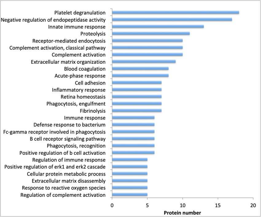
Toxins 2019, 11, 246                                                                                 2 of 14

1. Introduction
      Snakebite envenomations represent a significant public health problem observed on a global basis,
but particularly in sub-Saharan Africa, Asia, Latin America, and parts of Oceania, and have been
included by the World Health Organization (WHO) in the list of neglected tropical diseases [1]. India
is the country that bears the highest number of cases and it has been estimated that 50,000 people die
each year in this country due to snakebites [2].
      Among the many species of venomous snakes that inhabit India, four have been considered
as causing the heaviest toll: the Indian cobra (Naja naja), the common krait (Bungarus caeruleus),
the Russell’s viper (Daboia russelii), and the saw-scaled viper (Echis carinatus); these species comprise
the group known as “the big four” [3]. In addition, it has been observed that, in certain regions,
the hump-nosed pit viper species (Hypnale hypnale) inflicts a high number of bites and is capable of
causing serious accidents, underscoring the need to further study the epidemiology of snakebites
in India [4]. Although polyvalent snake antivenoms (SAV) are produced by several manufacturers
in India against venoms of the “big four” species, envenomations continue to cause fatalities and
pathophysiological sequelae in thousands of people a year. Thus, there is a need to further expand
our knowledge on the various aspects of snakebite envenomation in this country, including a better
understanding of the pathophysiology and mechanisms of action of venom-induced effects. A rapid,
personalized and highly sensitive diagnosis of envenomation and identification of the venom is
one of the essential needs for effective injury management. Thus, efforts have been directed at the
identification of markers of tissue damage, especially at the injury site, since they may provide valuable
information about the mechanisms of disease and the condition and prognosis of the patient, all of
which may lead to a more effective treatment [5–7]
      One of the characteristic manifestations of the local tissue damage inflicted by venoms of snakes
from the family Viperidae, and by some species of the family Elapidae, is the formation of blisters,
resulting in the accumulation of a proteinaceous fluid as a consequence of the collection of wound
exudate [1]. Thereby, the study of wound exudate and blister fluids as a potential source of biomarkers
have emerged as a new approach to study the effects of venoms and may assist in the diagnosis and
treatment of these patients [5,8]. The proteomic analysis of fluids collected from injured tissue, and
from tissue undergoing healing processes, has allowed the identification of markers that differentiate
between healing and non-healing wounds, with mediators characteristic of tissue formation or
mediators characteristic for a persistent inflammatory and tissue damaging response, respectively [7].
Moreover, immunochemical analysis of blister fluid from an envenomated patient has been used to
identify the presence of venom components in the fluid [9].
      Since viperid snake venoms induce the formation of blisters and an inflammatory exudate in the
damaged tissues, a number of experimental studies have addressed the analysis of the proteomes
of exudates collected from mice injected with venoms or isolated toxins. A comparative proteomic
analysis of wound exudates caused by BaP1, a snake venom metalloproteinase (SVMP), and Mtx-I,
a myotoxic phospholipase A2 (PLA2), showed that the SVMP causes degradation of nonfibrillar
collagens, whereas PLA2-mediated tissue damage results in the presence of fibrillar collagen type I
fragments; apolipoproteins A-I, A-IV, and E; and fibronectin in the exudates [5]. Moreover, the use
of inhibitors demonstrated that myotoxic PLA2s and hemorrhagic SVMPs are involved in blister
formation and skin damage induced by Bothrops asper venom [10]. Furthermore, the presence of matrix
metalloproteinases (MMPs) in these exudates was observed, hence suggesting the combined action of
endogenous and exogenous proteinases in these pathological events [11,12].
      In a recent study, the exudates generated by SVMPs of the PI, PII, and PIII classes revealed specific
proteins associated with the mechanism of action of the SVMPs [13]. Exudates from tissue injected
with PI and PII SVMPs contained a higher abundance of keratins as compared to samples from mice
injected with a PIII SVMP. In agreement, a PI SVMP has been shown to induce blistering in the skin
of mice [14]. Hence the study of the composition of exudates and blister fluid has proven to be an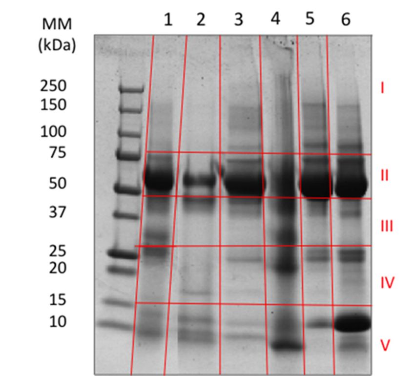
Toxins 2019, 11, 246                                                                                3 of 14

information-rich approach to assess pathological processes taking place in tissues injected with snake
venoms and toxins.
      Despite these advances at the experimental level, to our knowledge, there have been no attempts
to investigate the composition of blister fluid or exudates in humans resulting from snakebite
envenomations. The proteome analysis of fluids collected from affected tissue is therefore an
experimental approach with great potential for biomarker identification by allowing the assessment
of tissue alterations associated with the pathology of envenomation. In this study, utilizing mass
spectrometry, we investigated the composition of blister fluid collected from patients envenomated
by three species of Indian snakes, i.e., Daboia russelii and Hypnale sp. (family Viperidae), or Naja naja
(family Elapidae).

2. Results and Discussion

2.1. Clinical Features of Envenomation
     The basic demographic and clinical information of the six patients included in this study are
shown in Table 1. Three of them were bitten by D. russelii, two by Hypnale sp, and one by Naja naja.
Ages ranged from 2 to 67 years old. The time lapse between the bite and the arrival to the hospital
ranged between 2 and 8.5 h. Envenomation by D. russelii was characterized by local manifestations
(swelling, blisters), and one of them presented evidence of systemic capillary leakage syndrome
(parotid swelling, conjunctival chemosis) and thrombocytopenia. Three patients developed unclottable
blood, as evidenced by prolongation of the 20 min whole blood clotting test, and acute kidney injury
(AKI) was present in two of them, as evidenced by elevated serum creatinine concentration (Table 1).
     The two patients suffering bites by Hypnale sp. presented local manifestations (local pain, swelling
and discoloration in the skin), and one of them indicated abdominal pain. One patient showed
a prolonged 20 min whole blood clotting test, thrombocytopenia, and AKI, with elevated serum
creatinine concentration (Table 1). The patient suffering from a cobra (N. naja) bite presented with local
alterations (swelling and bluish discolouration) and manifestations of systemic neurotoxicity (ptosis
and external ophtalmoplegia).
     Patients suffering envenomations by D. russelii and N. naja received 20 vials (200 mL total) of
polyspecific antivenom, whereas no antivenom was administered to the patients bitten by Hypnale sp.
since this antivenom did not include Hypnale sp. venom in the immunizing mixture. The patient
bitten by N. naja was treated with polyspecific antivenom (200 mL) and neostigmine, and those who
developed acute kidney injury underwent hemodialysis (Table 1). All patients survived.
Toxins 2019, 11, 246 4 of 14
Table 1. Clinical and laboratory parameters of the six patients whose blister fluid was analyzed in this work.
Snake Species Naja naja Hypnale sp. Daboia russelii Hypnale sp. Daboia russelii Daboia russelii
Patient number 1 2 3 4 5 6
Sex Male Male Male Female Male Female
Age 53 years old 62 years old 22 years old 2 years old 23 years old 67 years old
Bite site Left thumb Left big toe Left leg Left index finger Left ankle Right leg
Time between the bite
2h 4 h 20 min 8h 4 h 15 min 8 h 35 min 3 h 20 min
and arrival in the hospital
Ptosis and external Disorientation, left leg
Local pain, swelling,
ophthalmoplegia swelling, and blisters. Local pain, swelling, Local pain and
and bluish
developed 1 h after arrival Under the influence of and discoloration and Local pain and swelling. swelling Bite mark
discoloration of left
to the hospital. Local pain, alcohol at the time of bite. blister on the left Proteinuria. Nausea, present. Nausea and
Symptoms big toe. Nausea,
swelling, and bluish Bilateral parotid swelling index finger. No vomiting, and abdominal vomiting present, and
vomiting, and
discoloration of left thumb. and conjunctival nausea, vomiting, nor pain present. no abdominal pain
abdominal pain
No nausea, vomiting nor chemosis. Nausea, abdominal pain. present.
present.
abdominal pain. vomiting, and pain
Platelet count 177,000/µL 70,000/µL 20,000/µL 300,000/µL 160,000/µL 120,000/µL
Hemoglobin 12.8 g/dL 10 g/dL 15.7 g/dL 11.7 g/dL 17.9 g/dL 14.1 g/dL
Serum creatinine 0.86 mg/dL 3.47 mg/dL 7.94 mg/dL 0.41 mg/dL 2.40 mg/dL 0.80 mg/dL
Serum K+ 4.1 mEq/L 3 mEq/L 4.8 mEq/Ll 4 mEq/L 6.7 mEq/L
Serum Na+ 126 mEq/L 133 mEq/L 130 mEq/L 131 mEq/L
>20 min D dimer 4 µg/mL
Whole blood clotting time Not determined >20 min >20 min 12 min (positive) (normal range >20 minPatients suffering envenomations by D. russelii and N. naja received 20 vials (200 mL total) of
polyspecific antivenom, whereas no antivenom was administered to the patients bitten by Hypnale
sp. since this antivenom did not include Hypnale sp venom in the immunizing mixture. The patient
bitten by N. naja was treated with polyspecific antivenom (200 mL) and neostigmine, and those who
Toxins 2019, 11,
developed 246 kidney injury underwent hemodialysis (Table 1). All patients survived.
acute 5 of 14
2.2. Proteomic Analysis of Blisters
2.2. Proteomic Analysis of Blisters
Blister fluid obtained from these patients were first analyzed using SDS-PAGE gel
Blister fluid obtained from these patients were first analyzed using SDS-PAGE gel electrophoresis.
electrophoresis. For some samples, there appeared to be a correlation between the gel profiles and
For some samples, there appeared to be a correlation between the gel profiles and the species of snakes
the species of snakes causing the bites. For others, however, there were differences even when
causing the bites. For others, however, there were differences even when comparing blisters obtained
comparing blisters obtained from patients bitten by the same species. This was observed in the case
from patients bitten by the same species. This was observed in the case of the two patients bitten by
of the two patients bitten by Hypnale sp (Figure 1). After electrophoresis, bands digestion, and LC-
Hypnale sp. (Figure 1). After electrophoresis, bands digestion, and LC-MS assay, the proteomic data
MS assay, the proteomic data were obtained and various analyses were performed. Mass
were obtained and various analyses were performed. Mass spectrometry results identified a total of 805
spectrometry results identified a total of 805 different proteins in blisters collected from the patients
different proteins in blisters collected from the patients (Supplementary Table S1) Many of the proteins
(Supplementary Table S1) Many of the proteins detected in this analysis have also been described in
detected in this analysis have also been described in laboratory studies collecting exudate fluid from
laboratory studies collecting exudate fluid from the tissue of mice injected with the venom of the
the tissue of mice injected with the venom of the Central American pit viper Bothrops asper or with
Central American pit viper Bothrops asper or with purified hemorrhagic SVMPs or myotoxic PLA2s
purified hemorrhagic SVMPs or myotoxic PLA2 s isolated from this and other viperid venoms [5,13,15].
isolated from this and other viperid venoms [5,13,15]. The individual protein identification was
The individual protein identification was considered significant with a minimum of two peptides
considered significant with a minimum of two peptides detected and protein identification
detected and protein identification probability above 95%.
probability above 95%.
SDS-PAGEprofile
Figure1.1.SDS-PAGE
Figure profileof
ofsamples
samplesof of blister
blister fluid
fluid collected from patients. Samples were were run
run on
on a
12% gel and stained
a 12% gel and stained with Coomassie brilliant blue. Samples from patients bitten by N. naja
brilliant blue. Samples from patients bitten by N. naja (lane 1);1);
(lane
Hypnale sp. (lanes 2 and 4); D. russelii (lanes3,3,5,5,and
Hypnale sp (lanes 2 and 4); D. russelii (lanes and6).6).The
Theleft
leftlane
lanecorresponds
correspondstotomolecular
molecularmass
mass
markers.The
markers. Thered
red lines
lines indicate
indicatethe
thelocations
locationsin inwhich thethe
which bands
bandswerewere
cut for
cutanalysis using using
for analysis LC-MS/MS.
LC-
MS/MS.
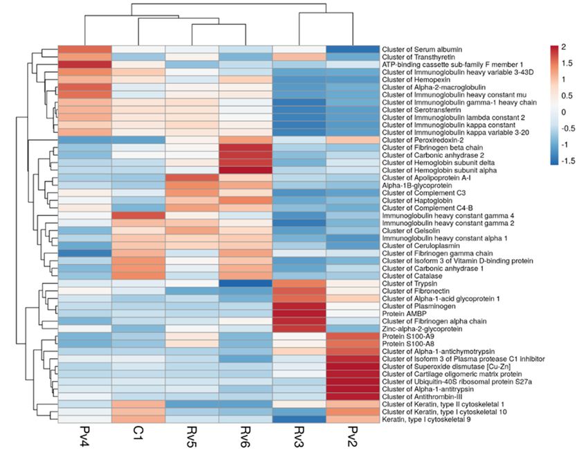
The most abundant proteins based on their quantitative value are presented in Table 2 and the 15
most abundant
The proteins proteins
most abundant in each exudate
based on aretheir
highlighted in gray.
quantitative Informatics
value analysis
are presented of the 2proteomes
in Table and the
identified the top biological response categories as: platelet degranulation,
15 most abundant proteins in each exudate are highlighted in gray. Informatics analysisnegative regulation
of the
of endopeptidase
proteomes activity,
identified the topinnate immune
biological response,
response proteolysis,
categories receptor-mediated
as: platelet degranulation, endocytosis,
negative
complement
regulation of activation, extracellular
endopeptidase activity, matrix
innateorganization, and blood
immune response, coagulation
proteolysis, (Figure 2). Such
receptor-mediated
events illustrate the local and systemic events triggered in the course of envenomation in terms of
change in metabolism, secretion, and synthesis of diverse components as a response to the action of
snake venom in the tissues.Toxins 2019, 11, 246 6 of 14
Table 2. The most abundant proteins identified in each blister’s samples via proteomic analysis. The
top 15 proteins by sample are highlighted in gray. Proteins were identified from the raw data using the
Sequest search algorithm Proteome Discovery 1.4.1 against a nonspecific database and the functional
annotation provided by DAVID Bioinformatics Resources 6.7.
D.russelli Hypnale sp. N.naja
Identified Proteins Accession
3 5 6 2 4 1
Cluster of serum albumin P02768 1892 1837 1620 1142 2525 1891
Cluster of alpha-1-antitrypsin P01009 85 139 37 625 136 102
Cluster of serotransferrin P02787 1 349 267 46 443 366
Cluster of hemoglobin subunit delta P02042 0 341 664 144 86 105
Cluster of keratin, type II cytoskeletal 1 P04264 272 64 65 364 155 371
Cluster of immunoglobulin kappa constant P01834 25 345 282 64 359 306
Cluster of complement C3 P01024 0 355 271 18 220 148
Cluster of hemoglobin subunit alpha P69905 43 221 891 84 46 55
Cluster of alpha-1-antichymotrypsin P01011 349 52 20 521 58 58
Cluster of alpha-2-macroglobulin P01023 1 177 182 24 345 126
Cluster of immunoglobulin gamma-1 heavy chain P0DOX5 29 262 190 81 301 249
Cluster of trypsin P00761 140 100 44 106 88 91
Cluster of immunoglobulin lambda constant 2 P0DOY2 12 151 124 29 193 149
Cluster of carbonic anhydrase 1 P00915 0 108 230 54 22 249
Cluster of keratin, type I cytoskeletal 10 P13645 0 39 24 243 104 205
Cluster of transthyretin P02766 96 77 44 41 103 49
Cluster of haptoglobin Q62558 4 150 186 17 86 31
Cluster of alpha-1-acid glycoprotein 1 P02763 203 59 27 153 65 45
Cluster of antithrombin-III P01008 34 29 25 209 67 31
Cluster of isoform 3 of plasma protease C1 inhibitor P05155-3 37 30 10 119 45 46
Cluster of complement C4-B P0C0L5 23 58 53 21 45 22
Keratin, type I cytoskeletal 9 P35527 0 35 33 83 52 85
Cluster of hemopexin P02790 12 39 73 13 80 65
Cluster of ubiquitin-40S ribosomal protein S27a P68202 0 8 11 139 8 16
Cluster of peroxiredoxin-2 P32119 26 50 81 71 0 1
Cluster of Immunoglobulin heavy constant mu P01871 0 42 45 3 76 25
Cluster of fibronectin P02751 159 7 2 85 78 52
Protein S100-A9 P06702 45 55 6 96 5 14
Cluster of fibrinogen gamma chain P02679 29 24 55 25 5 53
Cluster of fibrinogen beta chain P02675 5 34 90 0 0 23
Immunoglobulin heavy constant alpha 1 P01876 12 70 73 3 24 74
Cluster of fibrinogen alpha chain P02671 114 22 48 21 2 18
Cluster of isoform 3 of vitamin D-binding protein P02774-3 0 32 48 5 13 57
Immunoglobulin heavy constant gamma 2 P01859 0 131 101 30 76 136
Cluster of superoxide dismutase P00441 27 1 1 158 0 14
Protein S100-A8 P05109 32 39 4 61 2 6
Cluster of ceruloplasmin P00450 10 40 41 0 2 42
Cluster of catalase P04040 0 11 73 2 0 80
Cluster of gelsolin P06396 0 37 30 6 4 28
Immunoglobulin constant gamma 4 P01861 8 80 50 31 70 140
Cluster of plasminogen P00747 234 1 8 77 12 4
Cluster of apolipoprotein A-I G3QY98 3 41 27 1 1 0
Protein AMBP P02760 206 8 3 59 3 12
Zinc-alpha-2-glycoprotein P25311 54 26 17 9 25 9
Cluster of immunoglobulin kappa variable 3-20 P01619 0 23 22 3 30 21
Cluster of carbonic anhydrase 2 P00918 0 10 27 3 0 10
Alpha-1B-glycoprotein P04217 3 23 21 2 5 3
Cluster of immunoglobulin heavy variable 3-43D P0DP04 0 24 12 1 49 40
ATP-binding cassette sub-family F member 1 Q8NE71 4 3 5 6 22 11
Cluster of cartilage oligomeric matrix protein P49747 22 3 0 86 3 4Cluster of immunoglobulin kappa variable 3-20 P01619 0 23 22 3 30 21
Cluster of carbonic anhydrase 2 P00918 0 10 27 3 0 10
Alpha-1B-glycoprotein P04217 3 23 21 2 5 3
Cluster of immunoglobulin heavy variable 3-43D P0DP04 0 24 12 1 49 40
ATP-binding
Toxins 2019, 11, 246cassette sub-family F member 1 Q8NE71 4 3 5 6 22 1114
7 of
Cluster of cartilage oligomeric matrix protein P49747 22 3 0 86 3 4
Figure 2. Top biological
biological responses, identified via DAVID
DAVID bionformatic
bionformatic analysis, of the proteomes of
blisters’ samples
samples collected
collectedfrom
fromthe
thepatients
patients[16,17].
[16,17].The
Thenumber
numberofofproteins
proteinsdetected corresponding
detected to
corresponding
each process
to each is indicated.
process is indicated.
Proteins
Proteins were
were classified
classified inin groups
groups according
according to to functional
functional characteristics,
characteristics, using
using Gene
Gene Ontology
Ontology
within
within the DAVID Gene Functional Classification Tool (http://david.abcc.ncifcrf.gov). From the
the DAVID Gene Functional Classification Tool (http://david.abcc.ncifcrf.gov). From the
pathophysiological
pathophysiologicalstandpoint,
standpoint,ititwas
wasofofinterest
interesttotoanalyze
analyzeproteins
proteinsofofthe
thecoagulation
coagulation system
system (Table
(Table3)
and
3) andfrom
fromthethe
extracellular
extracellular matrix
matrix(ECM)
(ECM)(Table
(Table4),4),asascoagulopathies
coagulopathiesand and ECM
ECM degradation
degradation are are
characteristic of viperid snakebite envenomation. Regarding proteins of
characteristic of viperid snakebite envenomation. Regarding proteins of the coagulation system,the coagulation system,
fibrinogen
fibrinogen andandplasminogen
plasminogenwere werethethe
most abundant,
most abundant,withwith
differences notednoted
differences between patients.
between Blister
patients.
fluid
Blisterfrom
fluidpatient 3 (bitten
from patient by D.by
3 (bitten russelii), who who
D. russelii), showedshowedthe most severe
the most clinical
severe manifestations
clinical manifestations of
envenomation (Table 1), presented a higher abundance for fibrinogen
of envenomation (Table 1), presented a higher abundance for fibrinogen alpha-chain and alpha-chain and plasminogen.
Interestingly,
plasminogen.the sample fromthe
Interestingly, thesample
patient bitten
from theby the
patient N. naja,
cobrabitten byalso
thecontained
cobra N. fibrinogen, although
naja, also contained
cobra venoms do not cause significant effects on coagulation. This finding is probably
fibrinogen, although cobra venoms do not cause significant effects on coagulation. This finding is a consequence
of plasma extravasation
probably a consequence following
of plasmacobra venom-induced
extravasation local tissue
following cobradamage.
venom-induced local tissue
The analysis of extracellular matrix (ECM) proteins provided interesting findings (Table 4). Samples
damage.
from patients 3 (D. russelii) and 4 (Hypnale sp.) presented fragments of heparan sulphate proteoglycan
core protein and type IV collagen, two key components of basement membranes. Hydrolysis of these
proteins has been associated, in experimental studies in mice, with the action of hemorrhagic SVMPs
and with the pathogenesis of microvascular damage leading to extravasation [13,18]. The presence of
these proteins in blister fluid is indicative of basement membrane degradation, possibly associated
with local hemorrhage and blistering. Again, in the case of patient 3 bitten by D. russelii, these findings
correlated with the severe manifestations of envenoming. In this patient, tenascin-X and subunit
alpha-5 of laminin were also detected in the blister fluid, further corroborating the ECM damageToxins 2019, 11, 246 8 of 14
induced by D. russelii venom. Fragments of tenascin-X were also found in exudates collected from
mice injected with hemorrhagic SVMPs [5,13,18]. In addition, blister fluid from patients 4 (Hypnale sp.)
and 1 (Naja naja) contained fragments of type VI collagen. Experimental studies in mice injected with
SVMPs have shown similar findings in exudates collected from damaged muscle tissue [13,18]. Type
VI collagen is known to connect the basement membrane with fibrillar collagens, hence integrating
the basement membrane with the surrounding matrix. Hydrolysis of this collagen may also have
implications for the pathogenesis of local tissue damage. Our findings underscore the value of the
proteomic analysis of blister fluid to assess ECM degradation in snakebite envenoming.
Table 3. Proteins in blister fluid in relation to coagulation. Proteins were identified from the raw data
using the Sequest search algorithm Proteome Discovery 1.4.1 against the Swiss-Prot database.
D.russelii Hypnale sp. N.naja
Coagulation Proteins Accession MW
3 5 6 2 4 1
Cluster of fibrinogen alpha chain P02671 95 kDa 114 22 48 21 2 18
Cluster of fibrinogen beta chain P02675 56 kDa 5 34 90 0 0 23
Cluster of fibrinogen gamma chain P02679 52 kDa 29 24 55 25 5 53
Cluster of plasminogen P00747 91 kDa 234 1 8 77 12 4
Cluster of prothrombin P00734 70 kDa 4 0 0 0 0 0
Coagulation factor XII P00748 68 kDa 8 0 0 0 0 0
Coagulation factor XIII B chain P05160 76 kDa 0 0 0 1 0 0
Factor XIIa inhibitor P50448 52 kDa 0 0 0 1 0 0
Heparin cofactor 2 P05546 57 kDa 0 4 2 0 1 0
Isoform LMW of kininogen-1 P01042-2 48 kDa 66 0 0 0 0 0
Kininogen-1 O08677 73 kDa 1 0 0 0 0 0
von Willebrand factor P04275 309 kDa 1 0 0 7 0 0
Table 4. Proteins in blister fluid related to the extracellular matrix. Proteins were identified from the raw
data using the Sequest search algorithm Proteome Discovery 1.4.1 against the human basic database.
D.russelii Hypnale sp. N.naja
Extracellular Matrix Proteins Accession MW
3 5 6 2 4 1
Basement membrane-specific heparan sulfate
P98160 469 kDa 66 0 0 22 0 5
proteoglycan core protein
Cluster of aggrecan core protein P16112 261 kDa 1 0 0 0 0 0
Cluster of cartilage oligomeric matrix protein P49747 83 kDa 22 0 0 0 0 0
Cluster of laminin subunit alpha-3 Q16787 367 kDa 4 0 0 0 0 0
Cluster of laminin subunit gamma-2 Q13753 131 kDa 8 0 0 0 0 0
Cluster of nidogen-2 Q14112 151 kDa 7 0 0 0 0 0
Cluster of thrombospondin-1 P07996 129 kDa 0 2 1 2 0 1
Collagen alpha-1(I) chain P02452 139 kDa 1 0 0 6 0 0
Collagen alpha-1(I) chain (fragments) C0HJN3 88 kDa 0 0 0 1 0 0
Collagen alpha-1(III) chain P04258 94 kDa 0 0 0 5 0 0
Collagen alpha-1(XIV) chain Q05707 194 kDa 21 0 0 0 0 0
Collagen alpha-1(XVI) chain Q07092 158 kDa 0 0 0 2 0 0
Collagen alpha-1(XXVIII) chain Q2UY09 117 kDa 1 0 0 0 0 0
Collagen alpha-2(IV) chain P08572 168 kDa 34 0 0 17 0 1
Collagen alpha-3(VI) chain P12111 344 kDa 3 0 0 23 0 23
Isoform 5 of tenascin-X P22105-4 459 kDa 56 0 0 0 0 0
Isoform C of proteoglycan 4 Q92954-3 141 kDa 8 0 0 0 0 0
Laminin subunit alpha-4 P97927 202 kDa 8 0 0 1 0 0
Laminin subunit alpha-5 O15230 400 kDa 32 0 0 1 0 0
Lumican P51884 38 kDa 0 3 0 0 0 0
Matrix metalloproteinase-9 P14780 78 kDa 0 3 0 0 0 0
Nidogen-1 P14543 136 kDa 3 0 0 6 1 0
Tenascin P24821 241 kDa 0 0 0 1 0 0
Thrombospondin-4 P49744 108 kDa 0 0 0 2 0 0Toxins 2019, 11, 246 9 of 14
2.3. Protein Clustering
Hierarchical clustering created from the proteomic profile did not show a clear segregation on the
basis of the species of snake causing envenomation (Figure 3). This may have a number of possible
explanations including: (a) The set of proteins present in blisters is mostly a consequence of tissue
damage and the inflammatory response to tissue pathology, regardless of the venom responsible
for the damage. The three species involved in this study are known to induce local tissue damage.
(b) The set of proteins found is highly dependent on the severity of the damage and not on the species
causing the bite. (c) The number of patient blisters analyzed by a particular snake was insufficient
to power a significant observation of differences in response. Lastly, (d) the proteomics of blister
fluid likely is also time dependent and thus these samples collected at different times might mask
differences due to the species of snake. As indicated in Table 1, there was a wide variation in the
severity of envenomation within the patients studied. In agreement, patient 3, who suffered a severe
envenomation from D. russelii, presented the highest abundance of some clotting proteins and of
ECM proteins, in contrast with the other two patients affected by this species. Whatever the case, it is
necessary to study the proteomics of blister fluid of a larger number of patients showing a spectrum of
severity and at different times after the bite in order to have a comprehensive view of the changes in
Toxins
the 2019, 11, x FORprofile
proteomic PEER REVIEW
of blister fluid in different circumstances of envenomation. 11 of 16
Figure 3. Hierarchical clusters created from the proteomic profile of blisters’ samples collected from
Figure 3. Hierarchical clusters created from the proteomic profile of blisters’ samples collected from
the patients. Analysis was performed using the Clutvis software [19]. The colors represent a scale on
the patients. Analysis was performed using the Clutvis software [19]. The colors represent a scale on
thethe level
level of expression
of expression thatfrom
that goes goesred,
from red, representing
representing high abundance
high abundance proteins,
proteins, to blue, to blue, representing
representing
low abundant proteins. Pit viper (Pv) corresponds to Hypnale sp., Rv corresponds to
low abundant proteins. Pit viper (Pv) corresponds to Hypnale sp., Rv corresponds to Daboia russelii,Daboia russelii, and
andCCcorresponds Najanaja.
corresponds to Naja naja.
2.4. Identification of Proteins from the Snake Venoms in Blisters
Venom proteins were identified in the blister fluids using proteomics and Western blotting.
Using proteomics, venom proteins of various families were identified in two patients bitten by D.
russelii and N. naja (Table 5). In the case of patient 3 (D. russelii), this is in agreement with the clinical
manifestations that showed a high severity envenomation (Table 1). On the other hand, detection of
venom proteins using immunoblotting performed with Indian polyspecific antivenom was able toToxins 2019, 11, 246 10 of 14
2.4. Identification of Proteins from the Snake Venoms in Blisters
Venom proteins were identified in the blister fluids using proteomics and Western blotting. Using
proteomics, venom proteins of various families were identified in two patients bitten by D. russelii and
N. naja (Table 5). In the case of patient 3 (D. russelii), this is in agreement with the clinical manifestations
that showed a high severity envenomation (Table 1). On the other hand, detection of venom proteins
using immunoblotting performed with Indian polyspecific antivenom was able to identify snake venom
proteins in several samples. The three Russell’s viper samples exhibited proteins imaged by Western
blot in the high molecular mass range, plus some between 40 and 75 kDa and another of around 25 kDa.
The sample from the N. naja case, in turn, presented not only a greater intensity but also a higher
number of bands (Figure 4). Blister fluid from the patient bitten by N. naja was therefore the sample
with a higher abundance of venom detected using both mass spectrometry and Western blot analyses.
The abundance of venom proteins present in the blister fluid in the case of the patient bitten by N. naja
may be related to the fact that cobras usually inject their venoms subcutaneously, whereas viperid
venoms generally inject venom deeper in the tissues. The lower amount of proteins identified by
proteomics in some samples as compared to Western blot may be due to the scarcity of venom sequence
databases for mass spectrometry analysis, which has been a challenge for all venom proteomic studies.
On the other hand, the low reactivity of some samples in the Western blot might be due to variations in
the immunoreactivity of antivenom antibodies against some venom components. Immunochemical
detection of venom of the Taiwanese snake Protobothrops mucrosquamatus was described in the blister
fluid collected from a patient [9].
Table 5. Proteins from snake venoms identified in blister fluids using proteomic analysis. Proteins
were identified from the raw data using the Sequest search algorithm Proteome Discovery 1.4.1 against
a nonspecific database.
D.russelii Hypnale sp. N.naja
Venom Proteins Accession MW
3 5 6 2 4 1
Acidic phospholipase A2 inhibitor chain HPD-1 A4VBF0 15 kDa 3 0 0 0 0 0
Alpha-cobratoxin P01391 8 kDa 0 0 0 0 0 16
Beta-fibrinogenase E0Y419 28 kDa 1 0 0 0 0 0
Cluster of acidic phospholipase A2 P15445 13 kDa 0 1 0 0 0 75
Cluster of acidic phospholipase A2 Drk-a1 A8CG86 15 kDa 27 0 0 0 0 0
Cluster of acidic phospholipase A2 Tbo-E6 Q2YHJ3 16 kDa 0 0 0 2 0 46
Cluster of cytotoxin 2a Q9PST4 9 kDa 0 0 0 0 0 46
Cluster of factor V activator RVV-V alpha P18964 26 kDa 14 0 0 0 0 0
Cluster of Kunitz-type serine protease inhibitor C3 A8Y7N6 9 kDa 10 0 0 0 0 3
Cysteine-rich venom protein kaouthin-1 P84805 27 kDa 0 0 0 0 0 10
Hemorrhagic metalloproteinase-disintegrin-like
P82942 44 kDa 0 0 0 0 0 3
kaouthiagin
Kunitz-type serine protease inhibitor 2 Q2ES49 10 kDa 48 0 0 0 0 0
Kunitz-type serine protease inhibitor 2 P00990 7 kDa 14 0 0 0 0 0
Kunitz-type serine protease inhibitor C6 A8Y7N9 10 kDa 4 0 0 0 0 0
Kunitz-type serine protease inhibitor DrKIn-I H6VC05 10 kDa 8 0 0 0 0 6
Long neurotoxin 1 P25668 8 kDa 0 0 0 0 0 19
Muscarinic toxin-like protein 2 P82463 7 kDa 0 0 0 0 0 2
Muscarinic toxin-like protein 3 P82464 8 kDa 0 0 0 0 0 6
Neutral phospholipase A2 RVV-PFIIc’ (fragment) P0DKX1 2 kDa 10 0 0 0 0 0
Phospholipase A2 1 (fragment) P86529 2 kDa 3 0 0 0 0 0
Serine protease VLSP-3 E0Y420 28 kDa 8 0 0 0 0 0
Snake venom vascular endothelial growth factor
P0DL42 13 kDa 10 0 0 0 0 0
toxin VR-1’
Thrombin-like enzyme 1 A7LAC6 29 kDa 3 0 0 0 0 0
Venom nerve growth factor P30894 13 kDa 23 0 0 0 0 0
Venom nerve growth factor 2 Q5YF89 27 kDa 0 0 0 0 0 11
Zinc metalloproteinase-disintegrin-like
B8K1W0 70 kDa 1 0 0 0 0 0
daborhagin-KPhospholipase A2 1 (fragment) P86529 2 kDa 3 0 0 0 0 0
Serine protease VLSP-3 E0Y420 28 kDa 8 0 0 0 0 0
Snake venom vascular endothelial growth factor toxin VR-1' P0DL42 13 kDa 10 0 0 0 0 0
Thrombin-like enzyme 1 A7LAC6 29 kDa 3 0 0 0 0 0
Venom nerve growth factor P30894 13 kDa 23 0 0 0 0 0
Venom
Toxins 2019, 11, 246 nerve growth factor 2 Q5YF89 27 kDa 0 0 0 0 110of 14 11
Zinc metalloproteinase-disintegrin-like daborhagin-K B8K1W0 70 kDa 1 0 0 0 0 0
KDa
Figure 4. Detection of venom proteins in human blisters using Western blotting. Blister samples
were
Figureseparated under
4. Detection non-reducing
of venom proteinsconditions on 12%using
in human blisters SDS-PAGE
Westerngels and were
blotting. transferred
Blister to a
samples were
nitrocellulose membrane. Immunodetection was performed with polyspecific VINS
separated under non-reducing conditions on 12% SDS-PAGE gels and were transferred to antivenom. Thea
reaction was detected using an anti-horse peroxidase antibody and a chemiluminescent substrate.
Lanes 1 to 6 correspond to samples of blister fluid (see Table 1). Nn: N. naja venom, Dr: D. russelii
venom run in parallel to the blister samples.
3. Conclusions
Previous studies carried out in mice have described the potential of proteomics analysis of wound
exudate fluid to provide a “window” to explore the pathological and inflammatory events occurring
in tissues injected with snake venoms. The present investigation expanded this type of analysis to
the fluid of blisters present in human patients proximal to the anatomical site of venom injection in
bites inflicted by three different snake species present in India. The results corroborate the value of
proteomics analysis for detecting a variety of tissue plasma and inflammatory proteins of different
origin as a consequence of the tissue-damaging action of these venoms and the subsequent tissue
response through inflammation and repair. Although some differences were observed between samples
from patients bitten by different species, overall there was no clear segregation on the basis of the
offending species via hierarchical clustering analysis. Therefore, it would be of value in the future to
expand these analyses to a larger group of patients in order to assess whether specific tissue markers
may characterize different types of envenomation, and whether different proteomic patterns occur
depending on the severity and time-course of envenomation. Notably, venom was detected in the
blister fluid, as observed by both mass spectrometric and immunochemical analyses. Overall, our
findings underscore the use of proteomics analysis of exudate and blister fluids of patients suffering
snakebite envenomations for further advancing the understanding of the pathophysiology of this
neglected tropical disease. Furthermore, the presence of venom in blister fluid suggests that it may
represent a reservoir of venom for further diffusion into the tissues, hence supporting the concept
that aseptic removal of blister fluid may reduce the extent and time-dependence of venom-induced
tissue damage.
4. Materials and Methods
4.1. Clinical Evaluation and Treatment
Upon admission to hospital, routine clinical and laboratory evaluations were carried out for all
patients. The hospital’s routine treatment protocols were followed, including, when appropriate, the
administration of polyspecific Indian antivenom, and various interventions that depended on theToxins 2019, 11, 246 12 of 14
clinical evolution of each case. Identification of the offending snake was done via direct examination of
the specimen, brought in by the patient or his/her relatives.
4.2. Sample Collection
Fluid was aspirated from the blister under sterile conditions using a syringe. Two to five milliliters
of volume was typically collected from a single blister.
4.3. Ethics Statement
The Ethical Committee of the Little Flower Hospital and Research Centre, Algamaly, India,
authorized the project (“Blister fluid of snake bite victims analysis,” IRB session held on May 6, 2012).
An informed consent was obtained from participating patients.
4.4. Proteomic Analysis of the Exudates
4.4.1. SDS-PAGE
Lyophilized blister samples were kept frozen (−18 ◦ C) until time of use. Protein quantification
was performed using a micro BCA protein assay kit (Thermo Scientific, Waltham, WA, USA),.
Twenty micrograms of protein were further resuspended in Laemmli buffer, applied to a 12% precast
electrophoresis gel (Bio-Rad, Hercules, CA, USA), separated, and stained with 0.1% Coomassie Brilliant
Blue in 40% methanol, 10% acetic acid for about 1.5 h, then distained with 40% methanol, 10% acetic
acid, then in 5% acetic acid.
4.4.2. Protein Extraction and Digestion
Gel lanes were cut in ten equal-sized slices, which were destained for 3 h, and the proteins reduced
with 10 mM dithiothreitol (DTT) and alkylated with 50 mM iodoacetamide at room temperature.
Gel slices were washed with 100mM ammonium bicarbonate, dehydrated with acetonitrile, and dried
in a speed vac. A solution of Promega modified trypsin (20 ng/µL) in 50mM ammonium bicarbonate
was used to rehydratate the samples for 30 min on ice. Excess trypsin solution was removed and the
digestion was carried on for an additional 18 h at 37 ◦ C. Tryptic peptides were twice extracted from gel
slices with 30 µL of a 50% acetonitrile/5% formic acid solution. The extracts were dried to a volume of
15 µL for mass spectrometric analysis.
4.4.3. Proteomic Analysis
A Thermo Electron Orbitrap Velos ETD mass spectrometer system was used to perform the
LC/MS/MS experiments. Analytical columns were made by packing 0.5 cm of irregular C18 Beads (YMC
Gel ODS-A, 12 nm, I-10-25 µm) followed by 7.5 cm Jupiter 10 µm C18 packing material (Phenomenex,
Torrance, CA, USA) into 360 × 75 µm fused silica (Polymicro Technologies, Phoenix, AZ, USA) behind
a bottleneck. Aliquots of 7 µL were loaded directly onto these columns and then eluted into the
mass spectrometer at 0.5 µL/min using a 1h gradient consisting of acetonitrile/0.1M acetic acid (2–90%
acetonitrile). The instrument was set to a full MS (m/z 300–1600) resolution of 60,000 and programmed
to acquire a cycle of one mass spectrum followed by collision-induced dissociation (CID) MS/MS
performed in the ion trap on the twenty most abundant ions in a data-dependent mode. Dynamic
exclusion was enabled with an exclusion list of 400 masses, duration of 60 seconds, and repeat count
of 1. The electrospray voltage was set to 2.4 kV, and the capillary temperature was 265 ◦ C. Peak lists
were generated from the raw data against the Uniprot Human and NR database from July 2014 using
the Sequest search algorithm in Proteome Discoverer 1.4.1. Analysis of the spectra generated was
performed using carbamidomethylation on cysteine as a fixed modification, oxidation of methionine as
a variable modification, 10 ppm parent tolerance, and 1 Da fragment tolerance. All hits were required
to be fully tryptic.Toxins 2019, 11, 246 13 of 14
For analysis of the results and validation of peptide and protein identifications, data obtained were
exported to Scaffold (version 4.3.2, Proteome Software Inc., Portland, OR, USA). Protein identifications
were filtered using Xcorr cutoff values dependent on charge state (+1 >1.8, +2 >2.2, +3 >2.5, and +4 >3.5).
Relative quantization of proteins was accomplished by grouping all data from the 10 gel slices for a
particular sample in Scaffold and then displaying the quantitative value. This number gives an average
total of non-grouped spectral counts for a protein divided by the total non-grouping spectral counts
for the 10 mass spectral runs from the gels slices from each lane (http://www.proteomesoftware.com/)
and allows a relative quantitative comparison between specific protein from different samples.
Hierarchical clustering analysis was performed using ClustVis (http://biit.cs.ut.ee/clustvis/) to
determine any clusters of samples or proteins. To create heat maps, the list of proteins obtained using
proteomic analysis was log transformed and both rows and columns were clustered using Euclidean
distance and average linkage. DAVID (https://david.ncifcrf.gov/) was applied to the list of the most
abundant proteins in order to identify gene ontology terms and KEGG pathways or over-represented
biological processes.
4.5. Western Blot
Detection of venom proteins in human wound exudate was done using Western blotting. Exudate
samples were separated under non-reducing conditions on 12% SDS-PAGE gels and were transferred
to a nitrocellulose membrane. Immunodetection was performed with polyvalent VINS polyspecific
antivenom (VINS Bioproducts Limited, India, batch No. 01AS13100). The reaction was detected using
an anti-horse peroxidase antibody and a chemiluminiscent substrate. This antivenom is an F(ab’)2
preparation, produced from the plasma of horses immunized with venoms of the Indian species Naja
naja, Bungarus caeruleus, Daboia russelii, and Echis carinatus.
Supplementary Materials: The following are available online at http://www.mdpi.com/2072-6651/11/5/246/s1,
Table S1: Total proteins identified by proteomic analysis. Proteins were identified from the raw data using the
Sequest search algorithm Proteome Discovery 1.4.1 against a non-specific database and the functional annotation
provided using DAVID Bioinformatics Resources 6.7.
Author Contributions: Conceptualization, J.K.J., J.W.F.; methodology, J.K.A.M., J.K.J., J.M., T.E., A.R., J.M.G.,
J.W.F.; data analysis, J.K.A.M., J.K.J., J.M., T.E., A.R., J.M.G., J.W.F.; data curation, J.K.A.M., T.E., A.R., J.M.G., J.W.F.;
writing—original draft preparation, J.K.A.M., A.R., J.M.G., J.W.F.; writing—review and editing, J.K.A.M., J.K.J.,
J.M., T.E., A.R., J.M.G., J.W.F.; funding acquisition, A.R., J.W.F.
Funding: This research was funded by Vicerrectoría de Insvestigación (grant number 741-B6-125) and the
University of Virginia, School of Medicine.
Acknowledgments: The authors thank the Biomolecular Analysis Facility of the University of Virginia, USA, for
the proteomic analyses.
Conflicts of Interest: The authors declare no conflict of interest. The funders had no role in the design of the
study; in the collection, analyses, or interpretation of data; in the writing of the manuscript, or in the decision to
publish the results.
References
1. Gutiérrez, J.M.; Calvete, J.J.; Habib, A.G.; Harrison, R.A.; Williams, D.J.; Warrell, D.A. Snakebite envenoming.
Nat. Rev. Dis. Prim. 2017, 3, 17063. [CrossRef] [PubMed]
2. Mohapatra, B.; Warrell, D.A.; Suraweera, W.; Bhatia, P.; Dhingra, N.; Jotkar, R.M.; Rodriguez, P.S.; Mishra, K.;
Whitaker, R.; Jha, P. Snakebite mortality in India: A nationally representative mortality survey. PLoS Negl.
Trop. Dis. 2011, 5, 1–8. [CrossRef] [PubMed]
3. Warrell, D.A.; Gutiérrez, J.M.; Calvete, J.J.; Williams, D. New approaches & technologies of venomics to meet
the challenge of human envenoming by snakebites in India. Indian J. Med. Res. 2013, 138, 38–59. [PubMed]
4. Simpson, I.D.; Norris, R.L. Snakes of medical importance in India: Is the concept of the “Big 4” still relevant
and useful? Wilderness Environ. Med. 2007, 18, 2–9. [CrossRef] [PubMed]Toxins 2019, 11, 246 14 of 14
5. Escalante, T.; Rucavado, A.; Pinto, A.F.M.; Terra, R.M.S.; Gutiérrez, J.M.; Fox, J.W. Wound exudate as a
proteomic window to reveal different mechanisms of tissue damage by snake venom toxins. J. Proteome Res.
2009, 8, 5120–5131. [CrossRef] [PubMed]
6. Kool, J.; Reubsaet, L.; Wesseldijk, F.; Maravilha, R.T.; Pinkse, M.W.; D’Santos, C.S.; Van Hilten, J.J.; Zijlstra, F.J.;
Heck, A.J.R. Suction blister fluid as potential body fluid for biomarker proteins. Proteomics 2007, 7, 3638–3650.
[CrossRef] [PubMed]
7. Eming, S.A.; Koch, M.; Krieger, A.; Brachvogel, B.; Kreft, S.; Bruckner-Tuderman, L.; Krieg, T.; Shannon, J.D.;
Fox, J.W. Differential Proteomic Analysis Distinguishes Tissue Repair Biomarker Signatures in Wound
Exudates Obtained from Normal Healing and Chronic Wounds. J. Proteome Res. 2010, 9, 4758–4766.
[CrossRef] [PubMed]
8. Herrera, C.; Escalante, T.; Rucavado, A.; Fox, J.W.; Gutiérrez, J.M. Metalloproteinases in disease: Identification
of biomarkers of tissue damage through proteomics. Expert Rev. Proteomics 2018, 15, 967–982. [CrossRef]
[PubMed]
9. Lin, C.-C.; Wang, P.-J.; Liu, C.-C. Venom concentrations in blisters and hemorrhagic bullae in a patient
bitten by a Taiwan habu (Protobothrops mucrosquamatus). Rev. Soc. Bras. Med. Trop. 2019, 52, e20180160.
[CrossRef] [PubMed]
10. Rucavado, A.; Escalante, T.; Shannon, J.; Gutiérrez, J.M.; Fox, J.W. Proteomics of Wound Exudate in Snake
Venom-Induced Pathology: Search for Biomarkers To Assess Tissue Damage and Therapeutic Success.
J. Proteome Res. 2011, 10, 1987–2005. [CrossRef] [PubMed]
11. Rucavado, A.; Núñez, J.; Gutiérrez, J.M. Blister formation and skin damage induced by BaP1, a haemorrhagic
metalloproteinase from the venom of the snake Bothrops asper. Int. J. Exp. Pathol. 1998, 79, 245–254.
[PubMed]
12. Herrera, C.; Macêdo, J.K.A.; Feoli, A.; Escalante, T.; Rucavado, A.; Gutiérrez, J.M.; Fox, J.W. Muscle Tissue
Damage Induced by the Venom of Bothrops asper: Identification of Early and Late Pathological Events
through Proteomic Analysis. PLoS Negl. Trop. Dis. 2016, 10, e0004599. [CrossRef] [PubMed]
13. Herrera, C.; Escalante, T.; Voisin, M.B.; Rucavado, A.; Morazán, D.; Macêdo, J.K.A.; Calvete, J.J.; Sanz, L.;
Nourshargh, S.; Gutiérrez, J.M.; Fox, J.W. Tissue Localization and Extracellular Matrix Degradation by PI,
PII and PIII Snake Venom Metalloproteinases: Clues on the Mechanisms of Venom-Induced Hemorrhage.
PLoS Negl. Trop. Dis. 2015, 9, 1–20. [CrossRef] [PubMed]
14. Jiménez, N.; Escalante, T.; Gutiérrez, J.M.; Rucavado, A. Skin Pathology Induced by Snake Venom
Metalloproteinase: Acute Damage, Revascularization, and Re-epithelization in a Mouse Ear Model. J. Invest.
Dermatol. 2008, 128, 2421–2428. [CrossRef] [PubMed]
15. Rucavado, A.; Escalante, T.; Shannon, J.D.; Ayala-Castro, C.N.; Villalta, M.; Gutiérrez, J.M.; Fox, J.W. Efficacy
of IgG and F(ab0 ) 2 antivenoms to neutralize snake venom-induced local tissue damage as assessed by the
proteomic analysis of wound exudate. J. Proteome Res. 2012, 11, 292–305. [CrossRef] [PubMed]
16. Huang, D.W.; Sherman, B.T.; Lempicki, R.A. Bioinformatics enrichment tools: Paths toward the comprehensive
functional analysis of large gene lists. Nucleic Acids Res. 2009, 37, 1–13. [CrossRef] [PubMed]
17. Huang, D.W.; Sherman, B.T.; Lempicki, R.A. Systematic and integrative analysis of large gene lists using
DAVID bioinformatics resources. Nat. Protoc. 2009, 4, 44–57. [CrossRef] [PubMed]
18. Escalante, T.; Ortiz, N.; Rucavado, A.; Sanchez, E.F.; Richardson, M.; Fox, J.W.; Gutiérrez, J.M. Role of
collagens and perlecan in microvascular stability: Exploring the mechanism of capillary vessel damage by
snake venom metalloproteinases. PLoS ONE 2011, 6, e28017. [CrossRef] [PubMed]
19. Metsalu, T.; Vilo, J. ClustVis: A web tool for visualizing clustering of multivariate data using Principal
Component Analysis and heatmap. Nucleic Acids Res. 2015, 43, W566–W570. [CrossRef] [PubMed]
© 2019 by the authors. Licensee MDPI, Basel, Switzerland. This article is an open access
article distributed under the terms and conditions of the Creative Commons Attribution
(CC BY) license (http://creativecommons.org/licenses/by/4.0/).You can also read