Pulmonary Edema in COVID-19 Patients: Mechanisms and Treatment Potential - Frontiers
←
→
Page content transcription
If your browser does not render page correctly, please read the page content below
REVIEW
                                                                                                                                                published: 07 June 2021
                                                                                                                                        doi: 10.3389/fphar.2021.664349

                                           Pulmonary Edema in COVID-19
                                           Patients: Mechanisms and Treatment
                                           Potential
                                           Xinyu Cui 1, Wuyue Chen 1, Haoyan Zhou 1, Yuan Gong 1, Bowen Zhu 1, Xiang Lv 1,
                                           Hongbo Guo 2, Jinao Duan 1, Jing Zhou 1*, Edyta Marcon 2* and Hongyue Ma 1*
                                           1
                                             Jiangsu Collaborative Innovation Center of Chinese Medicinal Resources Industrialization, and Jiangsu Key Laboratory for High
                                           Technology Research of TCM Formulae, College of Pharmacy, Nanjing University of Chinese Medicine, Nanjing, China, 2Donnelly
                                           Centre for Cellular and Biomolecular Research, University of Toronto, Toronto, ON, Canada

                                           COVID-19 mortality is primarily driven by abnormal alveolar fluid metabolism of the lung,
                                           leading to fluid accumulation in the alveolar airspace. This condition is generally referred to
                                           as pulmonary edema and is a direct consequence of severe acute respiratory syndrome
                                           coronavirus 2 (SARS-CoV-2) infection. There are multiple potential mechanisms leading to
                                           pulmonary edema in severe Coronavirus Disease (COVID-19) patients and understanding
                         Edited by:        of those mechanisms may enable proper management of this condition. Here, we provide
               Timothy E. Albertson,
            UC Davis Medical Center,       a perspective on abnormal lung humoral metabolism of pulmonary edema in COVID-19
                      United States        patients, review the mechanisms by which pulmonary edema may be induced in COVID-
                       Reviewed by:        19 patients, and propose putative drug targets that may be of use in treating COVID-19.
                      Antonio Molino,
 University of Naples Federico II, Italy
                                           Among the currently pursued therapeutic strategies against COVID-19, little attention has
                  Salvatore Fuschillo,     been paid to abnormal lung humoral metabolism. Perplexingly, successful balance of lung
       Fondazione Salvatore Maugeri        humoral metabolism may lead to the reduction of the number of COVID-19 death limiting
                        (IRCCS), Italy
                                           the possibility of healthcare services with insufficient capacity to provide ventilator-assisted
                  *Correspondence:
                         Hongyue Ma        respiration.
          hongyuema@njutcm.edu.cn
                                           Keywords: COVID-19, pulmonary edema, abnormal lung humoral metabolism, syndrome coronavirus 2, drug,
                        Edyta Marcon
                                           traditional Chinese medicine
           edyta.marcon@utoronto.ca
                           Jing Zhou
                zhoujing_nj@126.com
                                           INTRODUCTION
                   Specialty section:
        This article was submitted to      COVID-19, an infectious disease caused by a severe acute respiratory syndrome coronavirus 2
          Respiratory Pharmacology,        (SARS-CoV-2), has a global reach. By March 31st of 2021, a total of 128.54 million cases of severe
              a section of the journal     Coronavirus Disease (COVID-19) have been diagnosed globally, and a total of 2.81 million people
           Frontiers in Pharmacology       have died from the disease (WHO, 2021). Currently, no specific drug has been developed to against
        Received: 05 February 2021         COVID-19, although some existing drugs have been repurposed and approved for treating
           Accepted: 25 May 2021           hospitalized patients (Ferner and Aronson, 2020; Schlagenhauf et al., 2020). Recently, several
          Published: 07 June 2021
                                           companies came out with vaccines against COVID-19 which have been approved for use. Some
                           Citation:       others will likely be approved soon (Kaur and Gupta, 2020; Thanh Le et al., 2020). However, it is still
   Cui X, Chen W, Zhou H, Gong Y,          unclear how the vaccination will proceed and how fast can vaccinations be done. In the meantime, we
Zhu B, Lv X, Guo H, Duan J, Zhou J,
                                           urgently need potent reduction in fatality of COVID-19.
Marcon E and Ma H (2021) Pulmonary
       Edema in COVID-19 Patients:
                                               Pulmonary edema is the disequilibrium between formation and reflux of lung tissue fluid leading
Mechanisms and Treatment Potential.        to the absorption of massive tissue fluid by lung lymph and vein failure. The fluid transudes into and
       Front. Pharmacol. 12:664349.        accumulates in the interstitium of lungs and finally alveolars from lung capillary, leading to severe
    doi: 10.3389/fphar.2021.664349         disorder of pulmonary ventilation and gas exchange (Staub, 1974). In COVID-19 patients,

Frontiers in Pharmacology | www.frontiersin.org                                    1                                             June 2021 | Volume 12 | Article 664349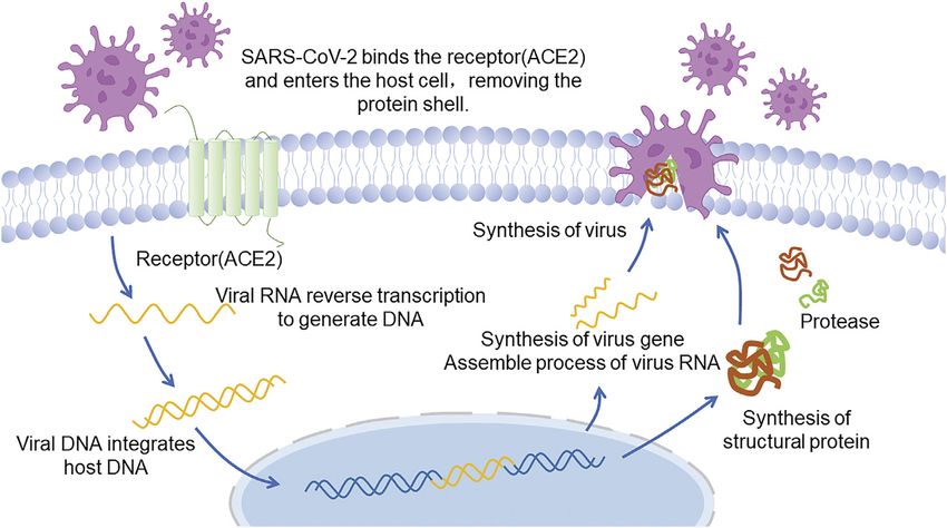
Cui et al.                                                                                                           Pulmonary Edema in COVID-19 Patients

pulmonary edema is diagnosed by lung ultrasound and a
computerized tomography (CT) scan (Udugama et al., 2020).
The condition presents itself as a slowly evolving pneumonia with
insidious early onset interstitial pulmonary edema that undergoes
acute exacerbation in the late stages and alveolar edema (Xu et al.,
2020; Wiersinga et al., 2020). Currently, these symptoms are the
primary consequences of pulmonary virus infection. It is known
that SARS-CoV-2 invades human cells by binding angiotensin-
converting enzyme-2 (ACE-2) receptor and other membrane
ectopeptidases (Xu et al., 2020). When there, the virus itself
and virus-mediated protein-protein interactions lead to the
lung inflammatory storm responsible for the observed
increasing vascular permeability in lung and pulmonary edema
(Tang et al., 2020). It is likely that alveolar fluid clearance (AFC)
failure plays a major role in the pathogenesis of pulmonary
edema. The imbalance of fluid metabolism, pulmonary fluid
clearance (PFC) and rich-protein fluid entrance, may be a key
reason for the acute exacerbation of pulmonary edema in
COVID-19 patients.
    Here, we describe molecular mechanisms of PFC and propose
that proteins functioning in this process might serve as an                    FIGURE 1 | A vision of coronavirus with the minimal set of structural
underappreciated, but yet promising targets for reducing lung                  proteins.
edema in severe COVID-19 patients. These proteins include ion
channels (Na channel, K channel and TRPV4), aquaporins
(AQP), renin angiotensin system (RAS) proteins, and                           attachment and fusion with the host cell membrane, and
bradykinin/hyaluronic acid-related enzymes. Drugs targeting at                promotes cellular invasion. As such, the S protein is essential
least some of these proteins have already existed and could be                for viral infection (Heald-Sargent and Gallagher, 2012; Li, 2016;
repurposed to manage pulmonary edema seen in SARS-COV-2                       Walls et al., 2020).
infections. Chinese Medicine (TCMs), already widely used in                      It has been shown that SARS-CoV-2 infects human cells via
China, may also be beneficial in addressing pulmonary edema in                 specific binding of S-protein to angiotensin-converting enzyme 2
COVID-19 patients (Zhang et al., 2020; Yang et al., 2020d). There             (ACE2) (Figure 2 Infection and replication process of SARS-
are also several natural compounds which were previously shown                CoV-2) (Xu et al., 2020) and the binding affinity between these
to have positive effects on the lung edema-associated targets                 two proteins is 10–20 times greater than that of SARS-CoV and
described in this paper (Zhou et al., 2006; Ho et al., 2007; Ji et al.,       ACE2 (Yan et al., 2020; Wrapp et al., 2020). ACE2 is most
2014; Wang et al., 2015; Qu, 2019; Fan et al., 2020; Lung et al.,             abundantly expressed in human vascular endothelial cells as
2020). In this review, we discuss the clinical characteristics of             well as alveolar and intestinal epithelial cells. It is also highly
COVID-19, current as well as potential new treatments based on                expressed in cardiomyocytes, epithelial cells of renal proximal
the reduction of lung edema through various means, drugs,                     convoluted tubule, urothelial cells, esophagus, and ileum
TCMs or natural compounds. We speculate that treatment of                     (Harmer et al., 2002; Zhang et al., 2020), facilitating a quick
lung-edema will lead to a lower mortality in COVID-19 patients                invasion of the human body by SARS-COV-2 and causing
with severe infections.                                                       complications. The latest research revealed that alveolar
                                                                              macrophages, which normally play a protective role, may also
                                                                              be infected by SARS-CoV-2 and release T cell chemokines,
VIROLOGICAL CHARACTERISTICS OF                                                resulting large amounts of T cells gathering in lung and
COVID-19                                                                      generating IFNγ. IFNγ will continually induce inflammatory
                                                                              cytokines released by alveolar macrophages, promoting the
SARS-CoV-2 is an enveloped RNA coronavirus of the genus β,                    activation of T cells and forming a positive feedback loop that
and is the seventh coronavirus which can infect human (Xu et al.,             drives persistent alveolar inflammation (Grant et al., 2021). It is
2020). The structure of coronavirus (Figure 1 A vision of                     worth mentioning that, except alveoli, which is widely known as
coronavirus with the minimal set of structural proteins.)                     the target tissue, cardiomyocyte can be infected as well. Researches
includes glycoproteins, membranes and nucleic acids. The                      proved that SARS-CoV-2 can directly infect human induced
spike (S) protein of coronavirus, one of the surface                          pluripotent stem cell-derived cardiomyocytes (hiPSC-CMs) as
glycoproteins, is divided into two functional units, S1 and S2.               well as an engineered heart tissue (EHT), in cellular and organ
S1 facilitates virus infection by binding to host receptors, and S2           level respectively, suggesting that the virus can replicate rapidly in
regulates the membranes fusion to enable viral RNA entering into              the cardiomyocytes, infecting other cardiomyocytes, contributing
host cells for further replication. Therefore, the S protein                  to cardiomyocyte cell death, myocardial inflammation and even
determines the host cell of the virus, regulates the viral                    heart failure (Sharma et al., 2020; Bailey et al., 2021).

Frontiers in Pharmacology | www.frontiersin.org                           2                                           June 2021 | Volume 12 | Article 664349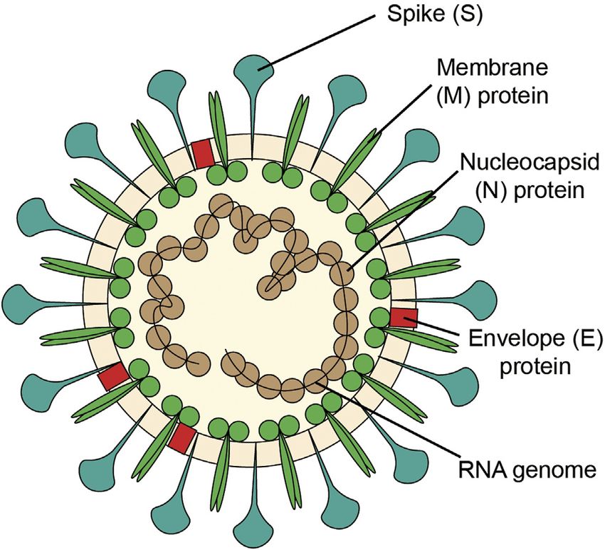
Cui et al. Pulmonary Edema in COVID-19 Patients
FIGURE 2 | Infection and replication process of SARS-CoV-2.
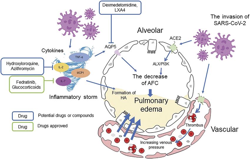
FIGURE 3 | Cause of COVID-19 pulmonary edema.
ABNORMAL LUNG HUMORAL humoral metabolism which can influence the AFC and PFC,
METABOLISM IN COVID-19 resulting the manifestation of pulmonary edema (Figure 3 Cause
of COVID-19 pulmonary edema).
SARS-CoV-2 invasion leads to alveolar and vascular epithelial Abnormal humoral metabolism is mainly manifested as
cells damage impelling the formation of minimal thrombus, imbalances of water and electrolytes. Water and sodium
increasing pulmonary venous pressure and vascular disturbances along with the unusual serum potassium levels
permeability and leading to massive loss of tissue fluid. are most common. It has been shown that the incidence and
Besides those direct causes of COVID-19 pulmonary edema, severity of COVID-19 are closely related to abnormal metabolism
there are other factors that can be described as abnormal of inorganic salts. Serum sodium shows a decrease trend in
Frontiers in Pharmacology | www.frontiersin.org 3 June 2021 | Volume 12 | Article 664349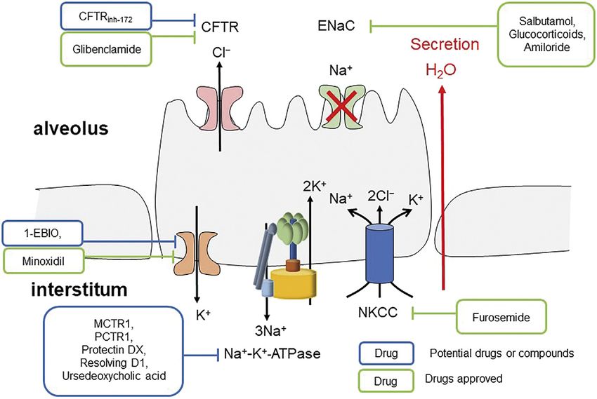
Cui et al.                                                                                                  Pulmonary Edema in COVID-19 Patients

  FIGURE 4 | Mechanism of inhibiting ENaC inducing pulmonary edema.

CODIV-19 development (Krolicka et al., 2020). Hyponatremia                additional complications, and eventually death (Yang et al.,
(low blood sodium concentration) as well as low                           2020).
concentrations of potassium and calcium in the blood
serum are also associated with COVID-19 (Lippi et al.,                    Sodium Channels and Sodium Pumps
2020). The functional inhibition of relevant lung enzymes                 Sodium transport is the main ion transport involved in the AFC.
and ion channels may disturb AFC, thus resulting                          Epithelial sodium channel (ENaC), present in human lungs,
pulmonary edema in COVID-19 patients. Na+/K+-ATPase                       kidneys and other organs, plays a vital role in lung fluid
and ion channels (sodium, potassium, AQPs, and TRPs) are                  clearance (Figure 4 Mechanism of inhibiting ENaC inducing
all involved in the regulation of AFC.                                    pulmonary edema) (Matthay et al., 2002). Its active absorption of
                                                                          Na+ is the main driving force of fluid clearance at birth and
                                                                          alveolar fluid absorption at adult stage (Bardou et al., 2012). The
Clinical Characteristics and Pathological                                 cystic fibrosis transmembrane conductance regulator (CFTR) and
Mechanism of COVID-19                                                     the ENaC located in the airway apical membranes and alveolar
COVID-19 patients often have pathological features such as                epithelial cells are essential in regulating lung fluid balance across
pulmonary interstitial or alveolar edema, diffuse tracheal                airway as the chloride (Cl−) and bicarbonate (HCO3−) secretion
phlegm thrombus and pulmonary inflammatory lymphoid                        conduits, and alveolar epithelia by sodium (Na+) ion absorption
infiltration, and are prone to acute respiratory distress                  (Matalon, 1999; Birket et al., 2016; Londino et al., 2017). These
syndrome (ARDS), causing lung injury (Cutts et al., 2017).                channels are important in maintaining the optimum volume and
The clinical manifestations and CT scans show the presence of             ion constitution of bronchial periciliary fluid and alveolar lining
ARDS in critical COVID-19 patients (Ai et al., 2020; Guan et al.,         fluid layers, which are necessary in appropriate pathogens
2020; Huang et al., 2020). Some patients also have leucopenia and         mucociliary clearance and optimum gas exchange, respectively
lymphopenia, suggesting a weak immune function, and high                  (Londino et al., 2017). Therefore, severe infections, which are
prothrombin time and D-dimer level, indicating abnormal                   induced by influenza virus, target the distal lung epithelial cells,
blood clotting function (Wang et al., 2020), which can all lead           inhibit the ENaC via activating protein kinase C (Kunzelmann
to lung damage and the severer pulmonary edema. Patients in               et al., 2000), and damage the pulmonary surfactant (Hofer et al.,
intensive care units (ICU) have higher plasma levels of IL2, IL7,         2015; Ito et al., 2015; Woods et al., 2015). SARS-CoV-2 may act in
IL10, MCP1, MIP1A, GSCF, IP10, and TNFα (Huang et al.,                    a similar way. With the presence of active basolateral Na+-K+-
2020). In these patients, white blood cells count, neutrophil count       ATPase, inhibition of the entry of apical Na+ can generate a
and D-dimer level keep rising while lymphocyte count keeps                concentration gradient inducing the uptake of basolateral Na+
decreasing as the disease progresses. Therefore, the infection            with Cl− by Na+-K+-2Cl− cotransporter (NKCC) and thus induce
along with the rapid replication of SARS-CoV-2 causes a large             the apical secretion of Cl− (Solymosi et al., 2013). The secretion of
amount of body fluid permeating through pulmonary alveoli,                 alveolar fluid, caused by inhibiting Na+ entry, is sensitive to
leading to ADRS. As the infection progresses, the immune                  inhibition of CFTR, NKCC, or Na+-K+-ATPase (Solymosi et al.,
function is impaired, causing damage to multiple organs,                  2013), suggesting that CFTR, NKCC or Na+-K+-ATPase

Frontiers in Pharmacology | www.frontiersin.org                       4                                     June 2021 | Volume 12 | Article 664349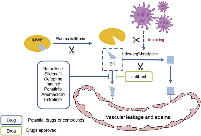
Cui et al.                                                                                                  Pulmonary Edema in COVID-19 Patients

inhibitors may have potential in treating pulmonary edema                 emerging from studies on various inhalational chemical threats
caused by low expression of ENaC. As in lipopolysaccharide-               (Achanta and Jordt, 2020). TRPs regulate their functions through
induced acute lung injury, docosahexaenoic acid and its                   sensory neuronal and nonneuronal pathways and play an
derivatives stimulate AFC through alveolar ENaC, Na,K-                    important role in complicated pulmonary pathophysiologic
ATPase via ALX/cAMP/PI3K pathway (Wang et al., 2014;                      events, such as increased intracellular calcium levels,
Zhang et al., 2017). Moreover, lower expression of alveolar               recruitment of pro-inflammatory cells, cough reflex, blocked
Na-K-ATPase promotes pulmonary edema, and when the                        mucus clearance, epithelia integrity disruption, pulmonary
expression of Na-K-ATPase α1- and α2-subunits decreases,                  edema, fibrosis and so on (De Logu et al., 2016).
maximal alveolar epithelial fluid clearance is reduced (Looney                TRPA1 distributes on C-fibers throughout the respiratory
et al., 2005).                                                            system (De Logu et al., 2016). The stimulation of TRPA1 can
                                                                          cause coughing, hypersecretion of mucus, rapid shallow
Potassium Channels                                                        breathing as well as bronchoconstriction (Bessac and Jordt,
Potassium channels are usually involved in maintaining the                2008; Birrell et al., 2009), which, if persistent, may cause
reabsorption of Na+ and the steady state of electrochemical               ARDS and other chronic diseases. TRPV1, expressed in
gradient, ions and body fluids in airway epithelial cells                  C-fibers of the vagus nerves innervating airways (Cui et al.,
specifically. Potassium channels up-regulate ENaC expression               2016), has been considered to play a key role in cough reflex
via activating KvLQT1 pathway so as to control AFC (Bardou                and increased airway sensitivity caused by various diseases
et al., 2012). In addition, potassium channels can act as oxygen          (Andrè et al., 2009; Couto et al., 2013). It has been reported
sensors in alveolar epithelium and thus adjust lung function to           that infection with a respiratory-associated virus can significantly
environmental changes in O2 levels (Bartoszewski et al., 2017). As        increase the expression and activity of TRPV1 (Abdullah et al.,
reported, large-conductance calcium-activated potassium                   2014). TRPV4, expressed in alveolar type I, type II cells and
channels (BKCa) in alveoli can reduce alveoli opening during              alveolar capillary endothelial cells, has been considered as a
hypoxia, detect O2 variation, and adjust ion transport and fluid           crucial regulator of alveolo-capillary barrier integrity (Alvarez
clearance (Jovanović et al., 2003).                                      et al., 2006; Yin et al., 2008; Goldenberg et al., 2015; Yin et al.,
                                                                          2016). Studies have confirmed that selective TRPV4 activation
Aquaporins                                                                induces rapid loss of alveolo-capillary barrier function and
The abnormal expression of AQP is closely related to the                  consequent alveolar edema formation (Alvarez et al., 2006).
abnormal alveolar fluid metabolism and the subsequent                      Indeed, in several preclinical studies, selective TRPV4
pulmonary fibrosis of COVID-19 patients. AQP-5 protein,                    inhibition showed efficacy in preventing or attenuating lung
present in the apical membranes of AT-I cell of alveolar                  edema (Thorneloe et al., 2012). Moreover, it has been revealed
epithelium, can regulate the transport of water molecules. It             that exosomes derived from human adipocyte can inhibit
promotes the clearance of surplus fluid in alveoli and keeps               TRPV4-mediated calcium influx and thus protect mice against
alveolar space dry (Wittekindt and Dietl, 2019). The                      ventilator-induced lung injury (Yu et al., 2020). As such, TRPV4
expression of AQP-5 is regulated by inflammatory cytokines,                inhibition likely has protective and beneficial effect on mucus
like TNF-α, elevated in the plasma of critical COVID-19 patients          clearance and pulmonary edema.
(Hui and Zumla, 2019). As Towne et al., (2001) reported, AQP-5
expression     significantly    declined      during    pulmonary          Renin Angiotensin System and Bradykinin
inflammation and edema, and TNF-α decreased AQP5 mRNA                      During the infection of SARS-CoV-2, RAS, BK and hyaluronic
and protein expression levels via TNFR1 and NF-κB pathway                 acid (HA) are all involved in the regulation of AFC and the
(Towne et al., 2001).                                                     formation of pulmonary edema (Garvin et al., 2020). RAS,
    Idiopathic pulmonary fibrosis is also a risk factor for severe         especially several cleavage products of the peptide angiotensin
COVID-19 which can be observed in the CT scans of COVID-19                (AGT) along with their receptors, maintains a balance of fluid
patients (Xu et al., 2020; George et al., 2020). As Gabazza et al.,       volume and pressure. For instance, angiotensin II (Ang II) can
(2004) reported, lung fibrosis is linked to decreased mRNA and             typically generate vasoconstriction and sodium retention when
protein expression of AQP-5 in the lung. This is supported by the         binding to the AGTR1 receptor and vice versa via the AGTR2
studies of AQP-5 deficient mice where a fibrotic phenotype with             receptor (Garvin et al., 2020). According to previous studies,
increased deposition of extracellular collagen type I was observed        activation of AT1 receptor inhibits AFC by down-regulating
in thickened alveolar walls (Gabazza et al., 2004). Therefore,            cAMP and dysregulating ENaC expression, leading to Ang II-
AQP-5 may be a promising drug target for treating abnormal                dependent pulmonary edema and alveolar filling increase (Deng
humoral metabolism as well as lung injury caused by COVID-19.             et al., 2012).
                                                                             Bradykinin (BK) is an important cellular mediator that causes
Transient Receptor Potential Ion Channels                                 vasodilatation and leaky blood vessels, leading to vascular leakage
TRP channels are nonspecific cationic channels located                     and edema (de Maat et al., 2020). BK is generated by cleavage of
throughout the respiratory system (Clapham et al., 2001),                 high-molecular-weight kininogen (HMWK) from plasma-
where TRPA1, TRPV1 and TRPV4 are the most abundant                        kallikrein and binds to the BKB2 receptor, and thus results
TRP subtypes (Kaneko and Szallasi, 2014; Steinritz et al.,                vascular hemorrhage (Garvin et al., 2020). Inhibition of ACE2
2018). Evidences for TRPs as medium of lung injury are                    by SARS-CoV-2 impairs the hydrolysis of des-arg9-bradykinin.

Frontiers in Pharmacology | www.frontiersin.org                       5                                     June 2021 | Volume 12 | Article 664349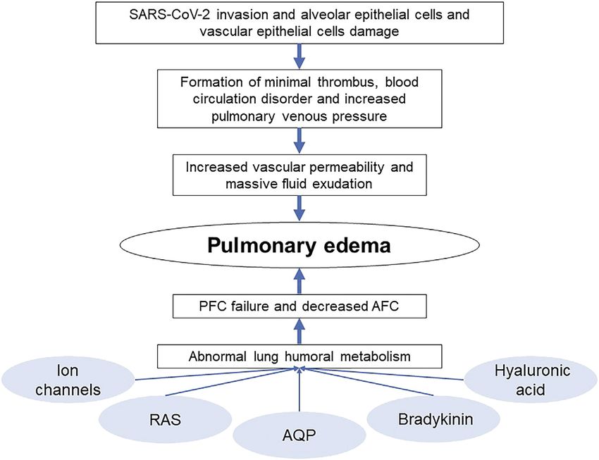
Cui et al.                                                                                                           Pulmonary Edema in COVID-19 Patients

  FIGURE 5 | Mechanism of BK inducing pulmonary edema and potential drugs.

Therefore, the excessive release and decreased hydrolysis of BK                  MMPs may become a potential target for pulmonary edema
through activating BKB1 and BKB2 receptors result in extra                       treatment.
vascular leakage and pulmonary edema (Figure 5 Mechanism                            Overall, the decreased expression of alveolar Na-K-ATPase,
of BK inducing pulmonary edema and potential drugs)                              misregulation of sodium, potassium, AQP, and RAS channels and
(Zwaveling et al., 2020).                                                        abnormal metabolism of BK and HA can all lead to lung liquid
                                                                                 clearance failure and pulmonary edema, resulting in severe lung
Hyaluronic Acid and Proteoglycans                                                damage and ARDS in COVID-19 patients (Figure 6 The general
CT images of COVID-19 patients revealed fluid and clear liquid                    regulation approaches of AFC).
jelly in their lungs, both closely linked to HA (Xu et al., 2020;
Wang et al., 2020). HA is a polysaccharide existing in most
connective tissues which can trap approximately 1,000 times its                  CONVENTIONAL TREATMENT OF
weight in water and form hydrogel. HA-related hydrogel has been                  PULMONARY EDEMA IN COVID-19
found in both, ARDS and SARS. Inflammatory cytokines and
inflammatory storm in COVID-19 patients can strongly induce
                                                                                 PATIENTS
the expression of HA-synthase-2 (HAS2), while hyaluronidase                      Currently, many clinical trials are in progress to test coronavirus
level decreases, resulting in the accumulation of HA and inducing                treatment, including new drugs and drug repurposing or
ARDS and pulmonary edema (Shi. et al., 2020).                                    repositioning. Immune-modulatory agents, supportive cares,
    In addition, HA is a part of a three-dimensional matrix in                   and antiviral drugs have been tested as COVID-19 treatments
pulmonary interstitial, which consists of HA, PGs and fibrillar                   in patients with severe infections (Pascarella et al., 2020; Ren et al.,
macromolecules providing resistance to tissue compression and                    2020).
interstitial fluid expansion (Negrini et al., 2008).When PGs and                     Immune-modulatory agents for COVID-19 include
HA interact with collagen IV, a fibrillar macromolecule                           tocilizumab, human immunoglobulin and the convalescent
modulating capillary permeability in the vascular basement                       plasma. IL6 monoclonal antibody or tocilizumab was thought
membrane, the compound substance limit fluid influx into the                       to work by interrupting inflammatory storm after the infection,
interstitium. Thus, PGs play a key role in the formation of                      but the latest clinical study published in NEJM showed that
pulmonary edema. The integrity of PG molecules in the                            Tocilizumab was not effective in preventing intubation or death
vascular capillary basement membrane can make sure that                          in mild hospitalized COVID-19 patients (Stone et al., 2020).
endothelial permeability to fluid and solutes in a low level.                     Convalescent plasma have been initially shown to be beneficial
However, the activation of matrix metalloproteinases (MMPs),                     for COVID-19 patients with severe infection stabilizing the
which may be triggered by inflammatory factors (Shapiro, 2001),                   immune system (Shen et al., 2020), but the subsequent
brings PG degradation, inducing pulmonary edema. It has been                     randomized controlled trial did not show significant
observed that MMP-2 and MMP-9, two most crucial MMPs in                          improvement within 28 days (Li et al., 2020).
the lung are over-expressed in pulmonary edema (Negrini et al.,                     Supportive cares for COVID-19 include respiratory support
1996; Negrini et al., 1998; Passi et al., 1998), suggesting that                 and circulatory support. Patients receive high-flow nasal cannula

Frontiers in Pharmacology | www.frontiersin.org                              6                                       June 2021 | Volume 12 | Article 664349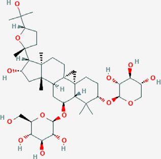
Cui et al.                                                                                                    Pulmonary Edema in COVID-19 Patients

  FIGURE 6 | The general regulation approaches of AFC.

(HFNC), non-invasive ventilation (NIV), mechanical ventilation            metabolism in lungs and the resulting pulmonary edema have
or ECMO as respiratory supports (Guo et al., 2020; Pascarella             become the main life-threatening factors in COVID-19 patients.
et al., 2020), a crystalloid fluid to ensure body fluid equilibrium         Thus, the relieve of pulmonary edema should be one of the critical
(Christ-Crain et al., 2020), and anticoagulants for restraining the       concerns in terms of the treatment of COVID-19 patients.
thrombus formation to aid in circulatory support (Connors and             Consequently, drugs that can normalize humoral metabolism
Levy, 2020). These supportive approaches have been shown to be            should be clinically evaluated for their use in the treatment of
beneficial as adjuvant therapies in COVID-19 patients.                     COVID-19 patients (Table 1).
    Repurposed drugs with proofs of antiviral effects for other
viral infections have been tried. However, the efficacy and safety         Targeting Sodium Channels and Pumps
of these drugs in COVID-19 patients are unclear. So far, only             Since the inhibition of ENaC induces pulmonary edema
remdesivir was approved by FDA for the compassionate use in               formation, targeting ENaC is rational in order to enhance
severe infected COVID-19 patients (Beigel et al., 2020). Other            fluid clearance from the alveoli. Studies showed that ENaC
anti-viral drugs, such as arbidol, chloroquine phosphate and              activators or stimulators can regulate ENaC-dependent fluid
ritonavir, did not exhibit efficacy in randomized, placebo-                absorption in alveolar and pulmonary edema (Fronius, 2013).
controlled trials in COVID-19 patients. Regardless, it seems              The activation of β-adrenergic receptor, especially β2 (Mutlu
reasonable that antiviral therapy might be adopted to patients            et al., 2004), was found to stimulate Na+ and fluid
with high risk factors as early as possible rather than wait for          reabsorption. It was observed that the expression of ENaC and
severe manifestation of the disease.                                      Na+/K+-ATPase in primary alveolar type II cells from rat lungs
    Other therapeutic options including organ support,                    increased responding to terbutaline (Minakata et al., 1998).
glucocorticoid therapy, nutritional support have been applied             Inhalation or infusion of salbutamol, a β2-adrenergic agonist,
to COVID-19 clinical treatment without much knowledge of                  reduced the incidences of pulmonary edema (Sartori et al., 2002)
their efficacy. However, among all current treatments mentioned            and was found to be beneficial in ARDS patients (Perkins et al.,
above, little attention has been paid to the abnormal humoral             2006). Glucocorticoids were shown to have the ability of inducing
metabolism and pulmonary edema, which is a key factor                     de novo synthesis of ENaC (Chow et al., 1999; Otulakowski et al.,
threatening patients’ lives.                                              1999; Sayegh et al., 1999) and affecting ENaC regulatory pathway
                                                                          via serum and glucocorticoids-inducible kinase-1 (SGK-1) (Chen
                                                                          et al., 1999; de la Rosa et al., 1999; Itani et al., 2001; Zhang et al.,
PUTATIVE DRUG TARGETS FOR                                                 2007). However, clinical evidence showing that glucocorticoids
PULMONARY EDEMA IN COVID-19                                               can reduce pulmonary edema by regulating fluid absorption in
                                                                          alveolar and glucocorticoids is missing. Potentially,
PATIENTS                                                                  glucocorticoids can be used as anti-inflammatory drugs in
Ion channels, AQPs, RAS, bradykinin and hyaluronic acid are               ARDS and pulmonary edema as well as in COVID-19
factors influencing the pulmonary edema. The abnormal humoral              patients. Amiloride, a prototypic inhibitor of ENaC, might also

Frontiers in Pharmacology | www.frontiersin.org                       7                                       June 2021 | Volume 12 | Article 664349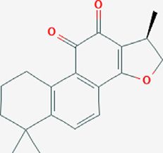
Cui et al.                                                                                                                    Pulmonary Edema in COVID-19 Patients

TABLE 1 | Potential drugs for normalizing humoral metabolism.

Drugs                            Targets                               Functions                                                  References

Terbutaline               ENaC                    β2-adrenergic agonist                               Minakata et al. (1998)
Salbutamol                                                                                            Sartori et al. (2002)
Amiloride                                         Prototypic inhibitor of ENaC                        Bull and Laragh (1968), Maronde et al. (1983), Adil et al. (2020),
                                                                                                      Cure and Cumhur Cure (2020)
Furosemide                NKCC                    NKCC inhibitor                                      Pickkers et al, (1997), Solymosi et al. (2013)
Glibenclamide             CFTR                    CFTR inhibitor                                      Solymosi et al. (2013)
CFTRinh-172 inhibitor
MCTR1                     Na+ channel and         Activate the sodium channel and Na-K-ATPase         Han et al, (2020)
PCTR1                     Na-K-ATPase                                                                 Zhang et al. (2020c)
protectin DX                                                                                          Zhuo et al. (2018)
Resolving D1                                      Stimulate AFC through alveolar epithelial sodium    Wang et al. 2014
                                                  channel, Na-K-ATPase via ALX/cAMP/PI3K pathway
KCa3.1 (1-EBIO)           K+ channel              K+ channel openers                                  Fleckenstein (1977); Nayler and Dillon (1986)
KATP (minoxidil)
Dexmedetomidine           AQP                     Regulate AQP expression                             Jiang et al. (2015)
Lipoxin A4 (LXA4)                                                                                     Shi et al. (2018)
Losartan                  AT1                     AT1 receptor blockers                               Yang et al. (2020a); Liu et al. (2020); Richardson et al. (2020)
Valsartan
Icatibant                 Bradykinin              Bradykinin antagonist                               Rasaeifar et al. (2020)
Raloxifene
Sildenafil
Cefepime
Cefpirome
Imatinib
Ponatinib
Abemaciclib
Entrectinib
Glucocorticoids           ENaC, cytokines         Regulate ENaC expression and impact cytokines       Chen et al. (1999); Chow et al. (1999); de la Rosa et al. (1999),
                                                                                                      Otulakowski et al. (1999); Sayegh et al. (1999); Itani et al. (2001);
                                                                                                      Zhang et al. (2007); Ahmed and Hassan (2020)

have a potential in treating COVID-19 patients. Amiloride was                          that MCTR1 (Han et al., 2020), PCTR1 (Zhang et al., 2020) and
shown to induce the reduction of ACE2 expression in bronchial                          protectin DX (Zhuo et al., 2018), endogenously produced lipid
and alveolar epithelial cells (Adil et al., 2020) and can counteract                   mediators, can effectively improve PFC, ameliorate
the low cytosolic pH which has been observed in COVID-19                               morphological damage, reduce lung inflammation, and
patients by acting on Na+/H+ exchanger (Cure and Cumhur                                increase sodium channel and Na-K-ATPase expression and
Cure, 2020). Since hypokalemia is a major issue in severe                              activity in vivo and in vitro in lipopolysaccharide (LPS)-
COVID-19 patients (Lippi et al., 2020), amiloride with its                             induced ARDS rats model. Resolving D1 (Wang et al., 2014),
potassium-sparing diuretic activity (Bull and Laragh, 1968) can                        generated from ω-3 fatty docosahexaenoic acids, and
potentially be used to restore normal serum potassium                                  ursodeoxycholic acid (Niu et al., 2019) can stimulate AFC and
concentrations (Maronde et al., 1983). However, as we                                  Na-K-ATPase in LPS-induced pulmonary edema via alveolar
discussed above, the inhibition of ENaC can also induce                                epithelial sodium channel and ALX/cAMP/PI3K pathway,
pulmonary edema and further studies are required to determine                          respectively.
the potential use of ENaC inhibitors for COVID-19 treatment.
   CFTR and NKCC inhibitors show promise in treating                                   Targeting Potassium Channels
pulmonary edema. Furosemide, a NKCC inhibitor, has been                                Potassium channels modulate the expression of ENaC. It has been
acknowledged as first-line therapeutic drug for pulmonary                               reported that transepithelial ion transport in alveolar monolayers
edema all the time (Pickkers et al., 1997; Solymosi et al.,                            can be activated by K+ channel openers in vitro under
2013). CFTR inhibitors, like glibenclamide and CFTRinh-172                             physiological conditions (Leroy et al., 2006). KCa3.1 (1-EBIO)
inhibitor, distinctly reduced absorptive alveolar fluid transport                       and KATP (minoxidil) channel openers can greatly recover AFC in
(Solymosi et al., 2013).                                                               mice intratracheally administrated verapamil, which is the first
   Since pulmonary edema can be promoted by decreased                                  generation of the phenylalkylamine class of calcium channel
expression of alveolar Na-K-ATPase (Woods et al., 2015),                               antagonists (Fleckenstein, 1977; Nayler and Dillon, 1986),
drugs or compounds which activate the sodium channel and                               suggesting that K+ channel openers might be potential drugs
Na-K-ATPase may be putative therapeutics. Studies have shown                           for treating pulmonary edema (Han et al., 2010).

Frontiers in Pharmacology | www.frontiersin.org                                    8                                            June 2021 | Volume 12 | Article 664349
Cui et al. Pulmonary Edema in COVID-19 Patients
Targeting Aquaporins and Transient Targeting Hyaluronic Acid and
Receptor Potential Ion Channels Proteoglycans
AQP-5 plays a significant role in pulmonary edema and Accumulation of HA can directly induce ARDS and pulmonary
decreased expression of AQP-5 has been observed during the edema. Thus, promoting the degradation of HA may be
disease process. Dexmedetomidine can upregulate AQP-1 and significant in the recovery process. HA is synthesized by
AQP-5 expression in rats with acute lung injury induced by LPS HAS2. However, so far, no effective inhibitors have been
and thus induce pulmonary edema (Jiang et al., 2015). Lipoxin A4 developed against HAS2. HA is degraded by hyaluronidases
(LXA4) can stabilize the permeability of pulmonary encoded by HYAL1 and HYAL2, whose activity depends on
microvascular endothelial cell by regulating the expression of CD44, an HA receptor (Harada and Takahashi, 2007). CD44
AQP-5 and MMP-9, and reduce alveolar fluid exudation (Shi inhibition reduces the IL-2 induced vascular leakage syndrome,
et al., 2018). revealing that CD44 may act as a potential target in COVID-19
TRPs are essential for the respiratory system and pulmonary treatment. Nonetheless, little attention has been paid to CD44 in
edema, in which TRPA1, TRPV1 and TRPV4 are the most COVID-19 treatment. Further studies may determine if CD44
important. Inhibiting these TRPs may benefit the treatment of inhibitors can be of use to in COVID-19.
pulmonary edema. A recent review has summarized the effects of It has already been shown that the dysregulated release of
TRPs in pulmonary chemical injuries, which includes the cytokines is one of the key factors behind poor outcomes in
representative TRPA1, TRPV1 and TRPV4 antagonists which COVID-19 patients. These cytokine storms can be treated with
have participated in preclinical and clinical studies (Achanta and steroids, IL-1 antagonists, TNF inhibitors, and Janus kinase
Jordt, 2020) (Supplementary Tables S1–S3). inhibitor (JAK) inhibitors (McCreary and Pogue, 2020).
Fedratinib, an FDA approved JAK2 inhibitor, may be used to
reduce the mortality associated with hyperinflammation by
Targeting Renin Angiotensin System and suppressing the production of several Th17 cytokines
Bradykinin (i.e., IL1b and TNF-alpha, IL21, IL22, IL17) and the
RAS plays a significant role in ARDS as well as pulmonary formation of pulmonary edema in combination with anti-
edema processes. The activities of the molecules in RAS are viral drugs (Yang et al., 2020e). Glucocorticoids, especially
ruled by dynamic changes responding to an injury. As the dexamethasone, have already been applied in the clinical
activation of AT1 receptor promotes the pulmonary edema, treatment of COVID-19. It not only can induce de novo
AT1 receptor blockers (ARBs) like losartan, valsartan may be synthesis of ENaC and affect ENaC regulatory pathway, but
effective in decreasing pulmonary edema. However, also has an impact on cytokines. A recent review suggested that
preliminary reports showed that some ACE inhibitors and low-to-moderate doses of dexamethasone may lower the
ARBs have no significant clinical benefits in treating COVID- mortality rate in patients with severe infections (Ahmed and
19 (Richardson et al., 2020), while others showed protective Hassan, 2020) and the latest finding indicated that the
effects among patients with pre-existing hypertension (Yang neutrophil-to-lymphocyte ratio determines the clinical
et al., 2020; Liu et al., 2020). The likelihood of hypertensive efficacy of corticosteroid therapy in COVID-19 patients, as a
patients developing COVID-19, who were treated by ARBs, neutrophil-to-lymphocyte ratio >6.11 associating with lower
was reported to decrease by 76% (Yan et al., 2020). Moreover, mortality in patients on corticosteroids (Cai et al., 2021).
exogenous delivery of Ang (1–7) was shown to play a part in Moreover, other drugs which can regulate immune system
reducing inflammation and improving pulmonary function in like hydroxychloroquine and azithromycin may also show
ARDS models (Wosten-van Asperen et al., 2011). effects in treating SARS-COV-2-induced pulmonary edema
Recombinant ACE2 was also reported to be a potential and their effectiveness in treating SARS-COV-2 infections
therapy in the clinical study of ARDS, which can lead to should be further investigated.
rapid decrease of plasma Ang II level and IL-6 expression.
(Imai et al., 2007; Zhang and Baker, 2017).
Raising evidences suggest that the effect of kinins on
bradykinin receptor triggers the inflammatory responses,
NATURAL COMPOUNDS AND
which have been observed in patients with COVID-19. TRADITIONAL CHINESE MEDICINES FOR
Consequently, the use of bradykinin antagonists is supposed to THE TREATMENT OF PULMONARY EDEMA
be regarded as a strategy for COVID-19 treatment interventions. IN COVID-19
Currently, only icatibant has been approved as bradykinin
antagonist for clinical treatment, and relevant studies have Besides chemical drugs and compounds we discussed above,
revealed that cefepime, cefpirome, imatinib, raloxifene, natural compounds and TCMs also possess promising antiviral
sildenafil, ponatinib, abemaciclib and entrectinib may also act effects against SARS-CoV-2 and had notably contribution in
as prospective non-selective bradykinin antagonists and have curing COVID-19, especially in alleviating pulmonary edema
potential for treatment of COVID-19 (Rasaeifar et al., 2020). and preventing the disease development from mild to severe. In
However, further researches into the mode of action, efficacy and TCM theory, evils of COVID-19 are derived from cold-dampness,
safety of these drugs are required. whose core pathogenesis are “toxin” and “dampness”. “Toxin”
Frontiers in Pharmacology | www.frontiersin.org 9 June 2021 | Volume 12 | Article 664349Cui et al. Pulmonary Edema in COVID-19 Patients
TABLE 2 | Summary of potential natural compounds against COVID-19.
Plant Compound Structure Antiviral and reducing References
pulmonary edema mechanisms
Stephania Japonica Cepharanthine ACE inhibitor Fan et al. (2020), Rogosnitzky
et al. (2020)
Rheum palmatum Emodin Blocks the binding of S protein to ACE2 Ho et al. (2007)
Black tea Theaflavin Inhibits RdRp activity Lung et al. (2020)
Atractylodes Atractylonolide-I Inhibits the formation of IL-6 and TNF-α Ji et al. (2014)
macrocephala
Astragalus propinquus Astragaloside-IV Activates ACE2-Ang-(l–7)-Mas pathway Wang (2015), Qu (2019)
Salvia miltiorrhiz Cryptotanshinone Induces the synthesis of cGMP and NO in cells and Zhou et al. (2006)
activating NO/cGMP pathway
means various pathogenic microorganism and inflammatory their antiviral and reducing pulmonary edema mechanisms are
storm presenting in infected patients, and “dampness” means shown in Table 2.
the abnormal humoral metabolism like inflammatory exudation. Natural compounds can inhibit the binding between the virus
Here, we summarized several natural compounds and TCM and the ACE2 receptor of host cells. Cepharanthine, a
formulas which are very likely to be potential drugs or have bisbenzylisoquinoline alkaloid derived from tubers of
already shown predominant efficacy of COVID-19 patients. Stephania Japonica, was shown to have a wide-spectrum
inhibitor of pan-β-coronavirus (Fan et al., 2020; Rogosnitzky
et al., 2020). Emodin, an anthraquinone compound from genus
Natural Compounds and Their Effects on Rheum and Polygonum, can markedly prevent the binding of S
Syndrome Coronavirus 2 Infections protein and ACE2 in the study of SARS-CoV (Ho et al., 2007).
Extensive studies have been conducted to identify the antiviral RNA-dependent RNA polymerase (RdRp) is a crucial protease
and pulmonary edema reducing efficacy of natural compounds, that catalyzes RNA replication from RNA templates and is an
some of which have already been tested specifically against SARS- appealing therapeutic target. Theaflavin from black tea was found
CoV and SARS-COV-2. Some natural compounds along with to present a lower binding energy when it docks in the catalytic
Frontiers in Pharmacology | www.frontiersin.org 10 June 2021 | Volume 12 | Article 664349Cui et al. Pulmonary Edema in COVID-19 Patients
TABLE 3 | Summary of potential TCM formulae against COVID-19.
TCM Constituent Antiviral and reducing Clinical efficacy References
formulae pulmonary edema mechanisms
Lian-Hua- Forsythiae Fructus, Lonicerae Japonicae Inhibits the replication of SARS-CoV-2, Combined treatment had higher Zhang et al. (2018), Luo
Qing-Wen Flos, Ephedrae Herba, Armeniacae affects virus morphology, exert anti- recovery rate (91.5% vs. 82.4%, p et al. (2020), Runfeng
Capsule Semen, Amarum, Isatidis Radix, inflammatory activity and triggers 0.022), a dramatically shorter median et al. (2020)
Dryopteridis Crassirhizomatis Rhizoma, bronchodilation time to symptom recovery (7 vs.
Houttuyniae 10 days, p < 0.001), as well as a
Herba, Pogostemonis Herba, Rhei Radix remarkably shorter time to recovery of
et Rhizoma, Rhodiolae Crenulatae, Radix fever (2 vs. 3 days), fatigue (3 vs.
et Rhizoma, Glycyrrhizae Radix et 6 days) and coughing (7 vs. 10 days)
Rhizoma and Gypsum Fibrosum (p < 0.001 for all)
Qing-Fei-Pai- Astragali Radix, Bupleuri Radix, Ephedrae Intervenes the inflammatory storm and Has an effective rate higher than 90% Shi et al. (2020a), Yang
Du Decoction Herba, Armeniacae Semen Amarum, triggers bronchodilation and early treatment with Qing-Fei-Pai- et al. (2020b)
Gypsum Fibrosum, Coicis Semen, Du Decoction can result better
Trichosanthis Pericarpium, Platycodonis outcomes, faster recovery, and a
Radix, Menthae Haplocalycis Herba, shorter duration of hospital stay
Scutellariae Radix, Glycyrrhizae Radix et
Rhizoma, Lonicerae Japonicae Flos, and
Artemisiae Annuae Herba
Toad venom Toad venom Improving PaO2/FiO2 and ROX index Improves the PaO2/FiO2 and ROX Hu et al. (2020)
Injection index (p < 0.001, 95% CI, −111.30 to
−35.90 for PaO2/FiO2; p < 0.001,
95% CI, −7.56 to −2.94 for ROX)
by 95.2%
Liu Shen Bezoar, Musk, venom toad, pearl, realgar, Inhibiting the replication of SARS-CoV-2, Improves respiratory function and Ma et al. (2020a)
Capsule and borneol reducing inflammatory cytokines lymphocyte count (similar to the Toad
production at the mRNA levels and venom Injection)
suppressing the NF-κB signaling
pathway to downregulate the expression
of cytokines in vitro
Xue-Bi-Jing Carthami Flos, Paeoniae Radix Rubra, Anti-inflammatory, anti-coagulation, The 28-day mortality of patients with Ma et al. (2020b), Li
Injection Chuanxiong Rhizoma, Salviae immune regulation, vascular endothelial severe pneumonia could be reduced et al. (2021)
Miltiorrhizae, Radix et Rhizoma, Angelicae protection, anti-oxidative stress and by 8.8%, significantly improving
Sinensis Radix other mechanisms pneumonia severity index (from
93.18 ± 23.17 to 52.18 ± 30.53)
pocket of SARS-CoV-2 RdRp. Thus, it could be a potential RdRp cells, activate NO/cGMP pathway and improve the blood
inhibitor for SARS-CoV-2 (Lung et al., 2020). circulation.
Inhibiting the inflammatory storm can effectively alleviate
pulmonary edema. Atractylenolide-I, the active component of
atractylodes, could minimize the formation of IL-6 and TNF-α (Ji Traditional Chinese Medicines and
et al., 2014), inhibiting the generation of inflammatory cytokines COVID-19 Infection
causing inflammatory response, thus lowering the possibility of Multiple TCMs have been already clinical applied for COVID-19
developing pulmonary edema. in China and achieved high recovery rate. Some of these TCMs
As RAS plays an important role in PFC and AFC, astragaloside along with their constituent and antiviral and reducing
IV from Astragalus propinquus is able to protect kidney and pulmonary edema mechanisms are shown in Table 3. Lian-
respiratory by activating the ACE2-Ang-(1–7)-Mas pathway in Hua-Qing-Wen capsules significantly affect virus morphology,
RAS and improving ACE2, Ang-(1–7), Mas level (Wang et l., inhibit the SARS-CoV-2 replication with the IC50 value of
2015; Qu, 2019). 411.2 μg/ml, reduce pro-inflammatory cytokines production at
When SARS-COV-2 invades, vascular endothelial cells are the mRNA level, and show anti-inflammatory effect in vitro
damaged, causing insufficient arterial flow and minimal (Runfeng et al., 2020). It has been revealed that COVID-19
thrombus. Elevated pulmonary venous pressure leads to faster patients treated with Lian-Hua-Qing-Wen capsules for 14 days
fluid infiltration into the interstitial lung than the ability of the resulted in a considerably higher recovery rate of 91.5%, a
pulmonary lymphatic vessel to drain away fluid, resulting in dramatically shorter median time to symptom recovery of
pulmonary edema. Improving the blood circulation can advance 7 days than the control group, which applied conventional
the oxygen supply for organs, accelerate the absorption of fluid treatment (Hu et al., 2021). Moreover, the constituent Ephedra
and eventually improve pulmonary edema. Zhou et al. (2006) can trigger bronchodilation, relieve breathing disorders and
reported that the cryptotanshinone in Salvia miltiorrhiz can alleviate pulmonary edema (Zhang et al., 2018). The toad
inhibit the synthesis of cGMP and NO in vein endothelial venom injection can significantly improve the pulmonary
Frontiers in Pharmacology | www.frontiersin.org 11 June 2021 | Volume 12 | Article 664349Cui et al. Pulmonary Edema in COVID-19 Patients
function of COVID-19 patients by regulating PaO2/FiO2 and and renin angiotensin system, and abnormal metabolism of
ROX index and thus alleviate pulmonary edema. As reported, bradykinin and hyaluronic acid as well as cytokine inflammatory
PaO2/FiO2 and ROX index of patients receiving conventional storm all lead to ARDS and pulmonary edema. These in turn, lead to
treatment combined with toad venom injection (20 ml/day) severe lung damage in COVID-19 patients. Existing drugs and
improved significantly (−111.30 to −35.90 for PaO2/FiO2 and inhibitors targeting the components of humoral metabolism may
−7.56 to −2.94 for ROX). Meanwhile, the number of patients in serve as potential treatments for COVID-19 and should be further
the treatment group presenting improved PaO2/FiO2 and ROX investigated. In addition, natural compounds and TCMs which
index was higher than that of the control group (95.2% vs. 68.4% generally have multiple targets should also be investigated, both in
and 73.7%). Moreover, the peripheral blood mononuclear terms of their efficacy and safety. Focusing on decreasing the
lymphocyte of COVID-19 patients was also greatly improved, formation of body fluid in lung or promoting the absorption of
from 0.91 ± 0.54 to 1.24 ± 0.67 after being treated for a week, body fluid can contribute to a decrease in lung damage and decreased
while there was no obvious change in control group (Hu et al., mortality in COVID-19 patients. Therefore, drugs targeted at the
2020). The Liu Shen capsule, of which pharmacodynamic humoral mechanisms might turn out to be highly effective against
component is also toad venom, was shown to have antiviral SARS-COV-2 infections.
and anti-inflammatory activity against SARS-CoV-2 in vitro, as it
can inhibit the replication of SARS-CoV-2 in Vero E6 cells,
reduce inflammatory cytokines production at the mRNA levels AUTHOR CONTRIBUTIONS
and suppress the NF-κB signaling pathway to downregulate the
expression of cytokines (Ma et al., 2020a). Qing-Fei-Pai-Du XC analyzed and reviewed the research articles as well as drafting
Decoction, which is officially recommended for the treatment of the manuscript. HM designed the framework of the review. EM
COVID-19 patients as mentioned in the guideline issued by NHC did the language polishing. All authors contributed to manuscript
(Trial 7th edition) (PRC, 2020), has an effective rate higher than revision, read, and approved the submitted version.
90% (2020) and can mediate the inflammatory storm induced by
COVID-19 (Yang et al., 2020b), regulate the innate immune,
cytokine activities (IL-17, NF-κB, TNF etc.), cell growth and FUNDING
death, as well as the degradation of damaged cells (Zhao et al.,
2020). Moreover, as a retrospective multicenter cohort study The project supported by Natural Science Foundation of China
reported, early treatment with Qing-Fei-Pai-Du Decoction (grant number 81673563, 81102762, and 81274199); Open
associated with better outcomes, faster recovery, and a shorter Project Program of Jiangsu Collaborative Innovation Center of
duration of hospital stay (Shi et al., 2020a). Xue-Bi-Jing injection is Chinese Medicinal Resources Industrialization (ZDXM-1-14,
also widely applicated in treating COVID-19 patients and by FJGJS-2015-15); Fund of Quality Standardization of Liu-
adding it based on the routine anti-infective therapy, the 28-day Shen-Wan (BA2016104, ZYBZH-C-JS-30). Jiangsu Qing Lan
mortality of patients with severe pneumonia could be reduced by Project for Young Academic Leaders of China.
8.8%, greatly improving pneumonia severity index (from 93.18 ±
23.17 to 52.18 ± 30.53) (Ma et al., 2020b). Xue-Bi-Jing injection
may act in COVID-19 by anti-inflammatory, anticoagulation, ACKNOWLEDGMENTS
immune regulation, vascular endothelial protection, anti-
oxidative stress and other mechanisms (Li et al., 2021). We are grateful to all of the colleagues who have given critical
comments on this work.
CONCLUSION
SUPPLEMENTARY MATERIAL
The abnormal humoral metabolism and pulmonary edema
contribute to the severity of symptoms and fatality of COVID- The Supplementary Material for this article can be found online at:
19 patients. Decreased expression of alveolar Na-K-ATPase, https://www.frontiersin.org/articles/10.3389/fphar.2021.664349/
dysregulation of sodium and potassium channels, aquaporins, full#supplementary-material
Adil, M. S., Narayanan, S. P., and Somanath, P. R. (2020). Is Amiloride a Promising
REFERENCES Cardiovascular Medication to Persist in the COVID-19 Crisis?. DD&T 14 (5),
256–258. doi:10.5582/ddt.2020.03070
Abdullah, H., Heaney, L. G., Cosby, S. L., and McGarvey, L. P. A. (2014). Ahmed, M. H., and Hassan, A. (2020). Dexamethasone for the Treatment of
Rhinovirus Upregulates Transient Receptor Potential Channels in a Coronavirus Disease (COVID-19): a Review. SN Compr. Clin. Med. 2,
Human Neuronal Cell Line: Implications for Respiratory Virus-Induced 2637–2646. doi:10.1007/s42399-020-00610-8
Cough Reflex Sensitivity. Thorax 69 (1), 46–54. doi:10.1136/thoraxjnl- Ai, J.-W., Zhang, H.-C., Xu, T., Wu, J., Zhu, M., Yu, Y.-Q., et al. (2020). Optimizing
2013-203894 Diagnostic Strategy for Novel Coronavirus Pneumonia, a Multi-center Study in
Achanta, S., and Jordt, S. E. (2020). Transient Receptor Potential Channels in Eastern China. medRxiv. doi:10.1101/2020.02.13.20022673
Pulmonary Chemical Injuries and as Countermeasure Targets. Ann. N.Y. Acad. Alvarez, D. F., King, J. A., Weber, D., Addison, E., Liedtke, W., and Townsley, M. I.
Sci. 1480 (1), 73–103. doi:10.1111/nyas.14472 (2006). Transient Receptor Potential Vanilloid 4-Mediated Disruption of the
Frontiers in Pharmacology | www.frontiersin.org 12 June 2021 | Volume 12 | Article 664349Cui et al. Pulmonary Edema in COVID-19 Patients Alveolar Septal Barrier. Circ. Res. 99 (9), 988–995. doi:10.1161/01.RES. Cutts, S., Talboys, R., Paspula, C., Prempeh, E., Fanous, R., and Ail, D. (2017). 0000247065.11756.19 Adult Respiratory Distress Syndrome. annals 99 (1), 12–16. doi:10.1308/rcsann. Andrè, E., Gatti, R., Trevisani, M., Preti, D., Baraldi, P., Patacchini, R., et al. (2009). 2016.0238 Transient Receptor Potential Ankyrin Receptor 1 Is a Novel Target for Pro- de la Rosa, D. A., Zhang, P., Náray-Fejes-Tóth, A., Fejes-Tóth, G., and Canessa, C. tussive Agents. Br. J. Pharmacol. 158 (6), 1621–1628. doi:10.1111/j.1476-5381. M. (1999). The Serum and Glucocorticoid Kinase Sgk Increases the Abundance 2009.00438.x of Epithelial Sodium Channels in the Plasma Membrane of Xenopus Oocytes. Bailey, A. L., Dmytrenko, O., Greenberg, L., Bredemeyer, A. L., Ma, P., Liu, J., et al. J. Biol. Chem. 274 (53), 37834–37839. doi:10.1074/jbc.274.53.37834 (2021). SARS-CoV-2 Infects Human Engineered Heart Tissues and Models De Logu, F., Patacchini, R., Fontana, G., and Geppetti, P. (2016). TRP Functions in COVID-19 Myocarditis. JACC: Basic Translational Sci. 6, 331–345. doi:10. the Broncho-Pulmonary System. Semin. Immunopathol 38 (3), 321–329. doi:10. 1016/j.jacbts.2021.01.002 1007/s00281-016-0557-1 Bardou, O., Privé, A., Migneault, F., Roy-Camille, K., Dagenais, A., Berthiaume, Y., de Maat, S., de Mast, Q., Danser, A. H. J., van de Veerdonk, F. L., and Maas, C. et al. (2012). K+ Channels Regulate ENaC Expression via Changes in Promoter (2020). Impaired Breakdown of Bradykinin and its Metabolites as a Possible Activity and Control Fluid Clearance in Alveolar Epithelial Cells. Biochim. Cause for Pulmonary Edema in COVID-19 Infection. Semin. Thromb. Hemost. Biophys. Acta (Bba) - Biomembranes 1818 (7), 1682–1690. doi:10.1016/j. 46 (7), 835–837. doi:10.1055/s-0040-1712960 bbamem.2012.02.025 Deng, J., Wang, D.-x., Deng, W., Li, C.-y., Tong, J., and Ma, H. (2012). Regulation Bartoszewski, R., Matalon, S., and Collawn, J. F. (2017). Ion Channels of Alveolar Fluid Clearance and ENaC Expression in Lung by Exogenous of the Lung and Their Role in Disease Pathogenesis. Am. Angiotensin II. Respir. Physiol. Neurobiol. 181 (1), 53–61. doi:10.1016/j.resp. J. Physiology-Lung Cell Mol. Physiol. 313 (5), L859–L872. doi:10. 2011.11.009 1152/ajplung.00285.2017 Fan, H.-H., Wang, L.-Q., Liu, W.-L., An, X.-P., Liu, Z.-D., He, X.-Q., et al. Beigel, J. H., Tomashek, K. M., Dodd, L. E., Mehta, A. K., Zingman, B. S., Kalil, (2020). Repurposing of Clinically Approved Drugs for Treatment of A. C., et al. (2020). Remdesivir for the Treatment of Covid-19 - Final Coronavirus Disease 2019 in a 2019-novel Coronavirus-Related Report. N. Engl. J. Med. 383 (19), 1813–1826. doi:10.1056/ Coronavirus Model. Chin. Med. J. (Engl) 133 (9), 1051–1056. doi:10. NEJMoa2007764 1097/CM9.0000000000000797 Bessac, B. F., and Jordt, S.-E. (2008). Breathtaking TRP Channels: TRPA1 and Ferner, R. E., and Aronson, J. K. (2020). Chloroquine and Hydroxychloroquine in TRPV1 in Airway Chemosensation and Reflex Control. Physiology 23, 360–370. Covid-19. BMJ 369, m1432. doi:10.1136/bmj.m1432 doi:10.1152/physiol.00026.2008 Fleckenstein, A. (1977). Specific Pharmacology of Calcium in Myocardium, Birket, S. E., Chu, K. K., Houser, G. H., Liu, L., Fernandez, C. M., Solomon, G. M., Cardiac Pacemakers, and Vascular Smooth Muscle. Annu. Rev. Pharmacol. et al. (2016). Combination Therapy with Cystic Fibrosis Transmembrane Toxicol. 17, 149–166. doi:10.1146/annurev.pa.17.040177.001053 Conductance Regulator Modulators Augment the Airway Functional Fronius, M. (2013). Treatment of Pulmonary Edema by ENaC Activators/ Microanatomy. Am. J. Physiology-Lung Cell Mol. Physiol. 310 (10), Stimulators. Cmp 6 (1), 13–27. doi:10.2174/1874467211306010003 L928–L939. doi:10.1152/ajplung.00395.2015 Gabazza, E. C., Kasper, M., Ohta, K., Keane, M., D’Alessandro-Gabazza, C., Birrell, M. A., Belvisi, M. G., Grace, M., Sadofsky, L., Faruqi, S., Hele, D. J., et al. Fujimoto, H., et al. (2004). Decreased Expression of Aquaporin-5 in (2009). TRPA1 Agonists Evoke Coughing in guinea Pig and Human Bleomycin-Induced Lung Fibrosis in the Mouse fibrosis in the Mouse. Volunteers. Am. J. Respir. Crit. Care Med. 180 (11), 1042–1047. doi:10.1164/ Pathol. Int. 54 (10), 774–780. doi:10.1111/j.1440-1827.2004.01754.x rccm.200905-0665OC Garvin, M. R., Alvarez, C., Miller, J. I., Prates, E. T., Walker, A. M., Amos, B. K., Bull, M. B., and Laragh, J. H. (1968). Amiloride. Circulation 37 (1), 45–53. doi:10. et al. (2020). A Mechanistic Model and Therapeutic Interventions for COVID- 1161/01.cir.37.1.45 19 Involving a RAS-Mediated Bradykinin Storm. Elife 9, e59177. doi:10.7554/ Cai, J., Li, H., Zhang, C., Chen, Z., Liu, H., Lei, F., et al. (2021). The Neutrophil-To- eLife.59177 Lymphocyte Ratio Determines Clinical Efficacy of Corticosteroid Therapy in George, P. M., Wells, A. U., and Jenkins, R. G. (2020). Pulmonary Fibrosis and Patients with COVID-19. Cel Metab. 33, 258–269. doi:10.1016/j.cmet.2021. COVID-19: the Potential Role for Antifibrotic Therapy. Lancet Respir. Med. 8 01.002 (8), 807–815. doi:10.1016/s2213-2600(20)30225-3 Chen, S.-y., Bhargava, A., Mastroberardino, L., Meijer, O. C., Wang, J., Buse, P., Goldenberg, N. M., Ravindran, K., and Kuebler, W. M. (2015). TRPV4: et al. (1999). Epithelial Sodium Channel Regulated by Aldosterone-Induced Physiological Role and Therapeutic Potential in Respiratory Diseases. Protein Sgk. Proc. Natl. Acad. Sci. 96 (5), 2514–2519. doi:10.1073/pnas.96.5. Naunyn-schmiedeberg’s Arch. Pharmacol. 388 (4), 421–436. doi:10.1007/ 2514 s00210-014-1058-1 Chow, Y.-H., Wang, Y., Plumb, J., O’Brodovich, H., and Hu, J. (1999). Hormonal Grant, R. A., Morales-Nebreda, L., Morales-Nebreda, L., Markov, N. S., Regulation and Genomic Organization of the Human Amiloride-Sensitive Swaminathan, S., Querrey, M., et al. (2021). Circuits between Infected Epithelial Sodium Channel α Subunit Gene. Pediatr. Res. 46 (2), 208–214. Macrophages and T Cells in SARS-CoV-2 Pneumonia. Nature 590, doi:10.1203/00006450-199908000-00014 635–641. doi:10.1038/s41586-020-03148-w Christ-Crain, M., Hoorn, E. J., Sherlock, M., Thompson, C. J., and Wass, J. A. H. Guan, W.-j., Ni, Z.-y., Hu, Y., Liang, W.-h., Ou, C.-q., He, J.-x., et al. (2020). Clinical (2020). Endocrinology in the Time of COVID-19: Management of Diabetes Characteristics of Coronavirus Disease 2019 in China. N. Engl. J. Med. 382 (18), Insipidus and Hyponatraemia. Eur. J. Endocrinol. 183 (1), G9–G15. doi:10. 1708–1720. doi:10.1056/NEJMoa2002032 1530/EJE-20-0338 Guo, Y.-R., Cao, Q.-D., Hong, Z.-S., Tan, Y.-Y., Chen, S.-D., Jin, H.-J., et al. (2020). Clapham, D. E., Runnels, L. W., and Strübing, C. (2001). The TRP Ion Channel The Origin, Transmission and Clinical Therapies on Coronavirus Disease 2019 Family. Nat. Rev. Neurosci. 2 (6), 387–396.doi:10.1038/35077544 (COVID-19) Outbreak - an Update on the Status. Mil. Med Res 7 (1), 11. doi:10. Connors, J. M., and Levy, J. H. (2020). COVID-19 and its Implications for 1186/s40779-020-00240-0 Thrombosis and Anticoagulation. Blood 135 (23), 2033–2040. doi:10.1182/ Han, D.-Y., Nie, H.-G., Gu, X., Nayak, R. C., Su, X.-F., Fu, J., et al. (2010). K+ blood.2020006000 Channel Openers Restore Verapamil-Inhibited Lung Fluid Resolution and Couto, M., de Diego, A., Perpiñi, M., Delgado, L., and Moreira, A. (2013). Cough Transepithelial Ion Transport. Respir. Res. 11 (1), 65. doi:10.1186/1465- Reflex Testing with Inhaled Capsaicin and TRPV1 Activation in Asthma and 9921-11-65 Comorbid Conditions. J. Investig. Allergol. Clin. Immunol. 23 (5), 289–301. Han, J., Li, H., Bhandari, S., Cao, F., Wang, X. Y., Tian, C., et al. (2020). doi:10.5114/pdia.2013.37040 Maresin Conjugates in Tissue Regeneration 1 Improves Alveolar Fluid Cui, M., Gosu, V., Basith, S., Hong, S., and Choi, S. (2016). Polymodal Transient Clearance by Up-regulating Alveolar ENaC, Na, K-ATPase in Receptor Potential Vanilloid Type 1 Nocisensor. Adv. Protein Chem. Struct. Lipopolysaccharide-induced Acute Lung Injury. J. Cel Mol Med 24 (8), Biol. 104, 81–125. doi:10.1016/bs.apcsb.2015.11.005 4736–4747. doi:10.1111/jcmm.15146 Cure, E., and Cumhur Cure, M. (2020). Comment on "Organ-protective Effect of Harada, H., and Takahashi, M. (2007). CD44-dependent Intracellular and Angiotensin-converting Enzyme 2 and its Effect on the Prognosis of COVID- Extracellular Catabolism of Hyaluronic Acid by Hyaluronidase-1 and -2. 19". J. Med. Virol. 92 (9), 1423–1424. doi:10.1002/jmv.25848 J. Biol. Chem. 282 (8), 5597–5607. doi:10.1074/jbc.M608358200 Frontiers in Pharmacology | www.frontiersin.org 13 June 2021 | Volume 12 | Article 664349
You can also read