RADIOLOGY AND IMAGING 2021 - The Offi cial HOPE Reference Book hospitalhealthcare.com - Hospital Healthcare Europe
←
→
Page content transcription
If your browser does not render page correctly, please read the page content below

The Official HOPE
           Reference Book
           hospitalhealthcare.com

RADIOLOGY AND
IMAGING 2021
2 | HHE 2021 | hospitalhealthcare.com                                              RADIOLOGY AND IMAGING

                           Contents

                       03 High-level dIsinfection of ultrasound probes
                          Sponsored by Nanosonics

                       05 The world of medical ultrasound:
                          A President’s perspective
                          Pamela Parker

                       07 Meet the Expert: Paul Sidhu

                       10 Meet the Expert: Neelam Dugar

                       13 Guideline summary: Integrating artificial intelligence
                          with the radiology reporting workflows

                       15 Guideline summary: Vetting (triaging) and cancellation
                          of inappropriate radiology requests

                       16 News roundup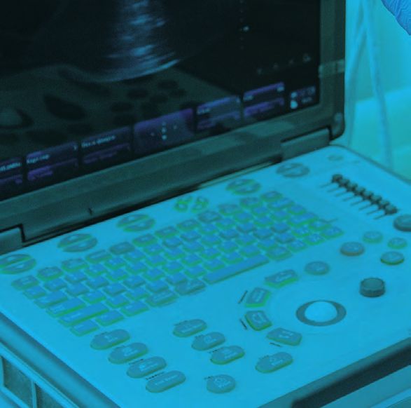
3 | HHE 2021 | hospitalhealthcare.com                                                                                 SPONSORED

High-level disinfection
of ultrasound probes
A large population-level study has revealed an unacceptable risk of infection following endocavitary
ultrasound procedures. Nanosonics is intent on ensuring that vulnerable patients are protected from
the risk of cross-contamination.

Support for the            Patients can be at risk from ultrasound               patients were found to be at a 41% greater risk
development of             associated infections when low-level                  of infection and a 26% greater risk of needing
this advertorial has       disinfection (LLD) is the standard of care.           an antibiotic prescription in the 30 days
been provided by           In order to quantify this risk, Scotland’s National   following their transvaginal ultrasound procedure
Nanosonics                 Health Service undertook a retrospective              when compared to gynaecological patients who
                           analysis of microbiological and prescription data     had not undergone a transvaginal ultrasound.
                           through linked national health databases.                During the study period, 90.5% of facilities
                           Patient records were examined in the 30-day           reported that they were performing low level
                           period following semi-invasive ultrasound probe       disinfection for transvaginal ultrasound probes.
                           (SIUP) procedures.                                    These patients were at a greater risk of infection
                              The study analysed almost one million patient      due to inadequate reprocessing and the study
                           journeys that occurred during a six-year period       concluded that: “Hence failure to comply with
                           from 2010.1                                           existing guidance on [high-level disinfection] of
                              Of the 982,911 patients followed, 330,500          SIUPs will continue to result in an unacceptable
                           were gynaecological patients; and 60,698 of           risk of harm to patients .”1
                           these gynaecological patients had undergone              The diverse use of ultrasound probes is now
                           a transvaginal (TV) ultrasound procedure. These       prompting a renewed focus on correct probe
                                                                                 reprocessing to ensure patient safety. To ensure
                                                                                 best practice standards, decontamination
                                                                                 experts and ultrasound users need to work
                                                                                 together to reduce the risk of infection that
                                                                                 is associated with using ultrasound probes.
                                                                                    Ultrasound procedures are performed in
                                                                                 various inpatient and outpatient settings by
                                                                                 a wide range of health professionals. This has
                                                                                 increased the use of surface probes to guide
                                                                                 procedures such as biopsies, cell retrieval,
                                                                                 cannulation, catheterisation, injections,
                                                                                 ablations, surgical aspirations, and drainages.
                                                                                 Across these procedures, the probe has the
                                                                                 potential to contact various patient sites
                                                                                 – including intact skin, non-intact skin, mucous
                                                                                 membranes and sterile tissue. This presents
                                                                                 a complex challenge, as contact with these
                                                                                 various body sites requires differing levels of
                                                                                 disinfection or sterilisation between patient
                                                                                 uses. Failure to adequately clean and disinfect
                                                                                 medical devices like ultrasound probes between
                                                                                 patients poses a serious risk to patient safety.
                                                                                    In 2012, a patient in Wales died from
                                                                                 a hepatitis B infection – most likely caused by
                                                                                 a failure to appropriately decontaminate
                                                                                 a transoesophageal echocardiography probe
                                                                                 between patients. As a result of this fatality,
                                                                                 a Medical Device Alert was issued by the
                                                                                 Medicines and Healthcare Products Regulatory
                                                                                 Agency (UK) advising users to appropriately
                                                                                 decontaminate all types of reusable ultrasound
                                                                                 probes.2
                                                                                    The UK and European guidelines require
                                                                                 ultrasound probes that come into contact with
                                                                                 mucous membranes and non-intact or broken
                                                                                 skin to be high-level disinfected. In particular,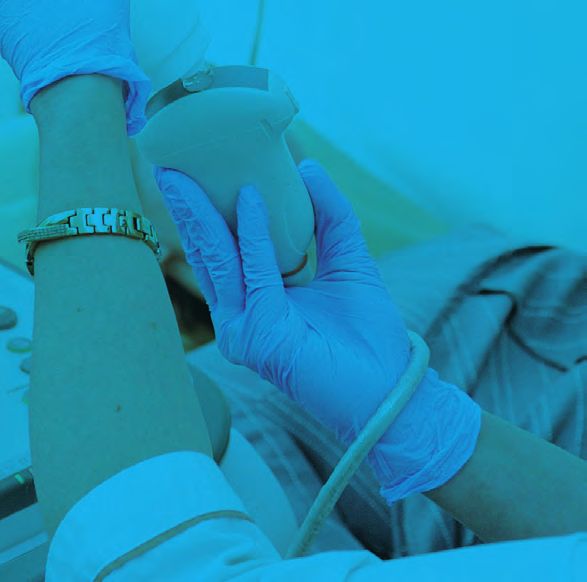
4 | HHE 2021 | hospitalhealthcare.com                                                                                    SPONSORED

                                Automated high-level disinfection

                                The trophon® system is designed to reduce           properties enabling the disinfectant to kill
                                the risks of infection transmission through         bacteria, fungi and viruses. The mist particles
                                automated high-level disinfection of                are so small that they reach crevices,
                                transvaginal, transrectal and surface probes.       grooves and imperfections on the probe
                                With over 25,000 units operating                    surface. Nanosonics works collaboratively
                                worldwide, 80,000 people each day are               with probe manufacturers to carry out
                                protected from the risk of cross-                   extensive probe compatibility testing. More
                                contamination with trophon devices. As a            than 1000 surface and intracavity probes
                                fully enclosed system, trophon2 can be              from all major and many specialist probe
                                placed at the point of care to integrate with       manufacturers are approved for use with
                                clinical workflows and maintain patient             trophon devices.
                                throughput. trophon technology# uses                   # The trophon family includes the trophon
                                proprietary hydrogen peroxide disinfectant          EPR and trophon2 devices which share the
                                that is sonically activated to create a mist.       same core technology of sonically-activated
                                Free radicals in the mist have oxidative            hydrogen peroxide.

                               automated and validated processes for                This category also includes contact surfaces
                               ultrasound reprocessing are preferred. This is       that are not intended for patient contact in
                               supported by a study relating to manual              health settings. These devices and surfaces
                               disinfection methods, which found that only          should be cleaned and low level disinfected.
References                     1.4% of reprocessing systems were fully                  It is important to note the difference between
1 Scott D et al. Risk          compliant when using manual methods,                 cleaning and low-level disinfection. Cleaning is
of infection following
semi-invasive ultrasound       compared to 75.4% when using semi-automated          the removal of soil and visible material until the
procedures in Scotland,        disinfection methods.3                               item is clean by visual inspection. Low level
2010 to 2016: A
retrospective cohort study
                                                                                    disinfection is the elimination of most bacteria,
using linked national          The Spaulding classification system                  some fungi and some viruses.
datasets. Ultrasound           The Spaulding classification system4 must be              A final and important point for consideration
2018;26(3):168–77.
2 Medicines and Healthcare     applied before a procedure commences so that         is the use of probe covers.
products Regulatory            information about what tissues or body sites             While many ultrasound users and
Agency (MHRA). Medical
Device Alert. Reusable
                               may be contacted is taken into account.              sonographers believe that their transvaginal
transoesophageal                  This classification system is a widely adopted    ultrasound patients are protected from infection
echocardiography,              disinfection framework for classifying medical       risk by using barrier shields and/or condoms,
transvaginal and transrectal
ultrasound probes              devices, based on the degree of infection            research has shown that up to 13% of condoms
(transducers) Document:        transmission risk, and requires the following        fail and up to 5% of commercial covers fail.
MDA/2012/037. 2012.
3 Ofstead CL et al.
                               approaches:                                          Probe covers may have microscopic tears or
Endoscope reprocessing         • Critical devices are defined as those that         breakages which can allow microorganisms to
methods: Prospective study     come into contact with sterile tissue or the         pass through.5
on the impact of human
factors and automation.        bloodstream. Probes in this category should
Gastroenterol Nurs             generally be cleaned and sterilised. Where           Conclusion
2010;33(4):304–11.
4 Spaulding EH. Chemical
                               sterilisation is not possible, high-level            Ultrasound users should work with their
disinfection of medical        disinfection is acceptable with the use of           decontamination colleagues to understand the
and surgical materials.        a sterile cover for ultrasound probes.               current UK and European guidelines for
In: Lawrence C, Block
SS, editor. Disinfection,      • Semi-critical devices contact intact mucous        reprocessing ultrasound probes. There are
sterilization, and             membranes and do not ordinarily penetrate            patient risks associated with ultrasound usage
preservation. Lea &
Febiger Philadelphia (PA);
                               sterile tissue. Ultrasound probes scanning over      when proper disinfection procedures are not
1968:517–31.                   non-intact skin are also considered semi-critical.   followed, as well as from ancillary products such
5 Basseal JM, Westerway        Semi-critical ultrasound probes include              as contaminated ultrasound gel. While the
SC, Hyett JA. Analysis of
the integrity of ultrasound    endocavitary probes, which should be used with       increased use of ultrasound has brought many
probe covers used for          a cover in addition to being high-level              benefits for patients, effective education and
transvaginal examinations.
Infect Dis Health 2020
                               disinfected.                                         disinfection protocols are required to minimise
Mar;25(2):77–81.               • Non-critical devices only contact intact skin.     the risk of infection.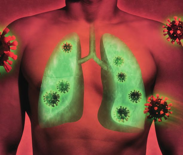
5 | HHE 2021 | hospitalhealthcare.com                                                                            EXPERT OPINION

The world of medical
ultrasound – A President’s
perspective

Pamela Parker              There can be no one truly unaffected by the          pandemic, and difficult to consider planning in
President, British         COVID-19 pandemic. This ongoing international        the immediate future. This, too, has led to the
Medical Ultrasound         health crisis has impacted on lives around the       organisation pausing, reflecting, and
Society; Department        world; arguably, none more so than those of          redesigning what can be delivered. The need for
of Radiology, Hull         front-line health care workers. Speaking as a        support for ultrasound professionals has not
& East Yorkshire           sonographer based in a large teaching hospital       diminished; indeed, with increasing role
Hospitals NHS Trust,       in the north of England, I have experienced          development, it can be argued the need is
UK                         first-hand the significant impact the pandemic       greater than before. A virtual webinar
                           has had on service delivery, my colleagues and,      programme was developed by BMUS during
                           most of all, our patients. However, as an            2020 to provide relevant, and pertinent,
                           optimist, I like to believe that with every dark     education and standard setting in line with the
                           cloud a silver lining can be sought. Therefore,      objectives of the organisation; a journal club via
                           as an optimist, one opportunity the pandemic         Twitter established, and now plans are well
                           presents is the opportunity to reset normal          underway for a full virtual online conference.
                           within our clinical and professional landscapes.     None of these are likely to have developed
                           In some instances, of course, this happened          without the dark cloud of the COVID-19
                           rapidly and without time to pause and reflect.       pandemic causing much disruption.
                           However, there has been an opportunity to               Webinars and e-learning, in the world of
                           review those services that were stopped and an       medical ultrasound at least, have developed at
                           opportunity to redesign as we move to restart        a pace in the last 18 months. Having a creative
                           planned care. Indeed, in my own institution,         and innovative team supporting an online
                           entire pathways have been remodelled, placing        training programme has been essential and has
                           ultrasound imaging at the very core of where it      unearthed hidden talents within the BMUS
                           was once an afterthought and poorly resourced        administrative team as well as the multitude of
                           or acknowledged. This has provided an ideal          professionals who have supported and
                           opportunity for modern ultrasound imaging to         volunteered to provide the education
                           be showcased; those technological                    programme. As with many professional
                           developments that have gradually become              charitable organisations, BMUS is reliant on
                           integral in diagnostic ultrasound imaging are        volunteers who are willing and able to share
                           now being recognised as essential to providing       their time and knowledge with peers. Despite
                           a powerful first-line core test for many patients,   the difficulties we have all faced, the willingness
                           particularly those with delayed presentation and     of our clinical colleagues to support the
                           for whom swift assessments and management            ongoing education and professional guidance
                           plans are required.                                  output of BMUS has been phenomenal, and yet,
                                                                                despite this wealth of readily available material,
                           The British Medical Ultrasound Society               attracting new members and minimising
                           The British Medical Ultrasound Society (BMUS)        attrition of existing members remains as very
                           has the core objectives: “to promote the             real challenge. It is pleasing to note that the
                           advancement of the science and technology of         sonographer membership of BMUS has
                           ultrasonics as applied to medicine” and “to          marginally increased recently, but attracting
                           ensure the highest standards in practice are         medical colleagues, in particular radiologists,
                           maintained”. Coupled with the changing role of       to support the organisation remains difficult.
                           ultrasound in patient pathways, BMUS has             BMUS prides itself on being a multi-disciplinary
                           developed and provided education and                 organisation and there to support any
                           guidance to all professionals working in the field   professional using medical ultrasound within
                           of medical ultrasound. For BMUS, as the premier      their scope of practice. In the UK, by far the
                           ultrasound society in the UK, the annual             largest professional group is sonographers who
                           scientific meeting has been the stage to present     primarily use ultrasound in their everyday
                           developing best practice, new and emerging           clinical practice. Essentially, a sonographer is
                           technology, and provide an opportunity to            medical ultrasound! Radiologists learn
                           discuss research and development with                ultrasound as a core skill during their training
                           like-minded professionals. Face to face events       but, and as is common in larger establishments,
                           have clearly been impossible to hold during the      radiologists are specialty-focused and many use
6 | HHE 2021 | hospitalhealthcare.com                                                                                EXPERT OPINION

                                                                               been established to promote career
                                                                               development for non-medical ultrasound
                                                                               practitioners and show case the very best in
                                                                               ultrasound innovation. However, membership
                                                                               of the society and an engaged workforce, from
                                                                               whatever background, is essential to the future
                                                                               success of these committees and the wider
                                                                               BMUS family. BMUS upholds the highest
                                                                               standards of practice and, it is with this aim,
                                                                               that the society engaged with Health Education
                                                                               England (HEE) in a project to increase the
                                                                               sonography workforce. BMUS, in collaboration
                                                                               with the Consortium for Accreditation of
                                                                               Sonographic Education, the Royal College of
                                                                               Radiologists and the Society of Radiographers
                                                                               has been integral member of this HEE
                                                                               sonographer workforce project group and
                                                                               progress is slowly being made. Every arm of
                                                                               health care is under pressure to increase activity,
                                                                               improve turn-around-times, and deliver safe and
                                                                               effective care. There is no profession that is not
                                                                               struggling with workforce capacity, with
                                                                               vacancy rates well documented; radiology and
                                                                               sonography are no different, and solutions have
                                                                               needed. While BMUS has no remit to act as a
                                                                               trade union or provide workplace advice, it does
                           cross-sectional imaging modalities or               have a remit to ensure standards of practice are
                           interventional suites that are now far removed      optimised, safe and of high standards. The
                           from the hands-on image acquisition,                multi-disciplinary nature of its membership
                           interpretation and reporting of ultrasound          ensures BMUS is well-placed to offer opinion,
                           imaging. Increasingly, there are numerous           advice and guidance as part of this collaborative
                           non-radiology professions using medical             approach to HEE as the path of defining and
                           ultrasound as a tool to enhance their clinical      developing an ultrasound / sonography career
                           practice and to guide patient management.           framework emerges.
                           Indeed, many Royal Colleges now have specific          Whilst there have been significant challenges,
                           ultrasound training programmes and                  in the workplace, professionally and personally
                           competency assessments that their members           over the last 18 months there has been
                           are expected to complete to be able to deliver      opportunity to reflect and set a new or revised
                           this point-of-care ultrasound (PoCUS). The          course for the future. The publication of the
                           importance of PoCUS cannot be overstated. As        Richards Report in 20201 and NHS Long Term
                           such, publications produced by our emergency        Plan in 20192 has provided an opportunity for
                           medicine, chest, and intensive care colleagues      the role of sonography, and the role of medical
About the author           ensured shared learning of the signs of COVID-      ultrasound within healthcare, to be established
Pamela Parker              19 on lung imaging, which became critical as        as essential in first line investigations in critical,
is a consultant            hospital admissions rose in the first and second    acute and planned care, the improvement of
sonographer working        waves. The aim of BMUS is to bring this             obstetric outcomes, and in guiding interventions
within radiology of        multi-disciplinary family together and recognise    within an out-patient setting. Professionals
the Hull University        the benefits of sharing knowledge and skills but,   working with medical ultrasound, as a career
Teaching Hospitals         at the same time, not alienating those for whom     path or using it as a tool, have the opportunity
NHS Trust. Pamela          medical ultrasound is a professional career         to embrace new ways of working and new
has over 25 years’         choice; sonographers, physicists, and               pathways. Supporting national organisations,
experience in the          radiologists alike.                                 such as the BMUS, provides the opportunity for
field of medical              BMUS has had, as do many societies,              professional societies to have a voice around
ultrasound. Her            sub-committees whose roles are to ensure the        the table at national discussions and highlight
specialist interests are   objectives of the organisation are met.             the benefits of a highly trained, well-motivated,
uro-genital ultrasound,    Longstanding and hardworking committees             recognised workforce. There are challenges
contrast and fusion-       such as the physics and safety, publications and    ahead but none that cannot be faced together.
guided imaging. She        education committees have been the                     I am proud to represent BMUS and my
has established the        workhorses of the society since its inception.      multi-disciplinary ultrasound colleagues and will
first sonographer-         Latterly, new committees have been set up to        work hard in my tenure as President to turn
delivered fusion and       reflect the changing professional landscape and,    those challenges into golden opportunities as
transperineal prostate     the now more diverse, users of medical              we head into the new normal of 2021 and
biopsy service within      ultrasound. PoCUS and obstetric clinical            beyond.
radiology in the UK.       imaging groups have been established to
Pamela is studying         support improvements in these critical fields of    References
part-time for a PhD                                                            1 Diagnostics: Recovery and Renewal – Report of the
                           medical ultrasound. The professional standards
                                                                               Independent Review of Diagnostic Services for NHS
investigating the role     group was established to better nurture and         England. www.england.nhs.uk/publication/diagnostics-
of ultrasound within       enhance working relationships with like-minded      recovery-and-renewal-report-of-the-independent-review-of-
                                                                               diagnostic-services-for-nhs-england/ (accessed June 2021).
the active surveillance    professional bodies and organisations.              2 NHS Long Term Plan. www.longtermplan.nhs.uk/
of prostate cancer.        A consultant sonographer interest group has         (accessed June 2021).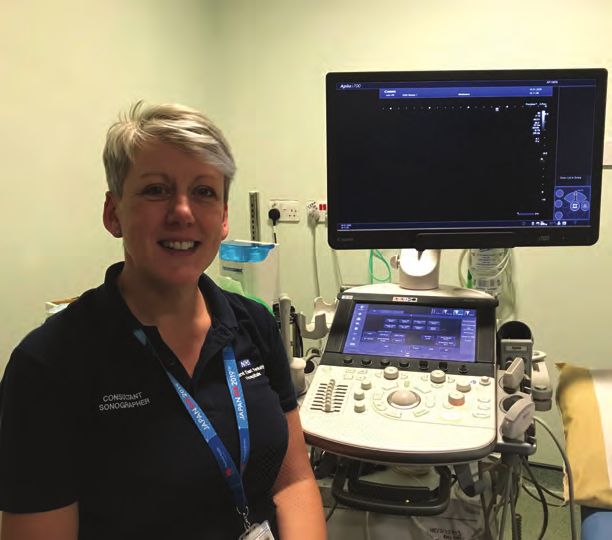
7 | HHE 2021 | hospitalhealthcare.com                                                                                 INTERVIEW

Meet the Expert: Paul Sidhu
Paul Sidhu is Professor of Imaging Sciences at King’s College London and consultant radiologist
in the Department of Radiology at King’s College Hospital. His research interests have focused on
ultrasound and interventional radiology. Here he shares his thoughts with Hospital Healthcare Europe
on ultrasound and how this technique has evolved and will continue to develop in the future.

                           On how radiology services are organised              identify pleural effusions that need to be
                           at King’s                                            drained, and rheumatologists will make use of
                           Professor Sidhu described how King’s is a large      ultrasound to examine the small joints of the
                           tertiary, general hospital in southeast London       hands. The evolution of ultrasound in other
                           serving a population of over 2.2 million people      specialties has been considerable and
                           in a largely deprived area. King’s has several       radiologists no longer have either the time or
                           different sites that cover various services,         perhaps inclination to perform such scans.
                           including paediatric, neonatal, cardiothoracic,      Despite these developments, as Professor Sidhu
                           neurosurgery, breast cancer screening, and liver     described, it is safety regulations, focused on
                           transplantation. There is one centralised, very      the patient, that keep radiology departments
                           large, radiology department employing around         together. However, while the use of ultrasound
                           80 consultant radiologists and up to 700 staff.      has now extended beyond the realms of the
                           The department is also involved in the provision     radiology department, he felt that the radiology
                           of training for radiologists and radiographer        community was not overly concerned about this
                           staff. Although radiologists are qualified           direction of travel, especially given that in the
                           doctors, their path towards becoming a               UK, there is a shortage of radiologists, and this
                           radiologist is a long one that can take up to        delegation has to some extent been welcomed.
                           12 years. In contrast, radiographers, who are the    In fact, he now believes that no single speciality
                           individuals responsible for taking images, still     effectively “owns” ultrasound and in many
                           need to undergo degree level training although       radiology departments, the ultrasound scanning
                           their role has greatly expanded in recent years.     is performed by sonographers and the
                           At King’s for example, radiographers manage all      radiologists themselves have moved on to
                           aspects of imaging for CT and MRI scans              becoming more focused on the interpretation of
                           imaging in interventional radiology and some         specialised scans. As a passing thought, he felt
                           radiographers have become sonographers               that while radiologists were likely to be the most
                           involved in the scanning and issuing of medical      competent individuals to perform and interpret
                           reports. All aspects require several years of        scans, if an ultrasound was being used as
                           training to become established within these          a point-of-care service for a specific indication,
                           roles.                                               provided that an individual healthcare
                              Ultrasound is available and employed in many      professional appropriately interpret a scan, he
                           different specialities by experienced and            had no objection to these developments.
                           competent individuals. Professor Sidhu
                           explained how there remains strict controls on       On the ESR subcommittee on ultrasound, his
                           who can perform scans that involve exposure to       work as Chair, and future plans
                           radiation and particularly in nuclear medicine,      Professor Sidhu mentioned how he had been
                           with the administration of radioactive isotopes.     a member of the European Society of
                           Administrators are required to have a specific       Radiologists (ESR) ultrasound subcommittee
                           ARSAC licence. All hospitals that undertake          for several years during which time, it had
                           examinations with radiation exposure will have       produced a number of different position papers.
                           a radiation protection officer. In contrast, while   He cited what has become a very successful
                           there are no specific controls on who can            position paper on infection control and
                           perform either an MRI and ultrasound scan, he        prevention from 2017,1 highlighting the
                           highlighted how MRI equipment is prohibitively       importance of good hygiene measures,
                           expensive and ultimately is best reserved for the    especially with transducers and how these
                           radiology department. Nevertheless, according        should be cleaned after each use. More recently
                           to Professor Sidhu, the position is rather           in 2020, the group have published best practice
                           different with respect to ultrasound. While in       recommendations and imaging use.2
                           the past, ultrasound has been the responsibility        This latest paper was an update of an earlier
                           of the radiology department, as Professor Sidhu      2009 position paper on ultrasound.3
                           explained, things are rapidly changing with          As Professor Sidhu clarified, the newer version
                           much ultrasound, particularly point-of-care,         provides a series of recommendations on
                           becoming performed outside of radiology. For         appropriate standards for the use of ultrasound
                           instance, chest physicians will use ultrasound to    in radiology from the perspective of the ESR.
8 | HHE 2021 | hospitalhealthcare.com                                                                                  INTERVIEW

                           For instance, the position paper, defines the
                           essential requirements for equipment, practice
                           aspects of use, infection control, requirements
                           for training, certification and competence. He
                           noted that while not all ultrasound operators
                           would necessarily conform to the standards
                           delineated in the position paper, it did define
                           the professional standards which would be
                           expected if the service were delivered by
                           a radiologist. Professor Sidhu explained that an
                           important aim of the position paper was to
                           hopefully clarify for any non-radiologists, the
                           anticipated standards which should be followed,
                           in a sense, a standard operating procedure for
                           undertaking ultrasound, which had the support
                           of the ESR and therefore credibility. Professor
                           Sidhu described how it was important that the
                           position paper provided the necessary guidance
                           because when using ultrasound, it is not the
                           machine which does the job but the operator.
                           If the operator is not competent, neither is the
                           output from the machine! In short, it is vital that
                           operators will need to practice, learn and
                           develop all the time to perfect the technique
                           to ensure that they get the best use from the
                           device.
                               In terms of ensuring continued best practice,
                           Professor Sidhu outlined documents produced
                           by the ESR were designed to support non-
                           radiologist operators. This advice encouraged         plans for three speakers at the meeting and
                           participation in audits of their service but          who will discuss different models of ultrasound
                           additionally and equally important, was that          practice. Firstly, there is the German model, in
                           non-specialists needed to maintain their skills       which many GPs perform ultrasound as well as
                           and competence through an examination of the          having a centralised hospital department run by
                           practice all the time, together will ensuring         radiologists, but with other medical specialists
                           equipment maintenance, the environment used           effectively dipping into the service. Second is
                           to perform imaging and how best to manage             the Russian model, whereby both radiologists
                           patient throughput, all of which were essential       and other physicians only do ultrasound and no
                           for adherence to professional standards.              other imaging modality. The third, and often
                           Professor Sidhu hinted that with a rapid pace of      perceived as a controversial model, is the one
                           development in ultrasound technology, it is           deployed in the UK, where it is the radiologists
                           highly likely that the professional standards of      who performs less scanning, taking on more
                           today would require updating in the near future.      specialised examinations (e.g. MSK) and
                           He pointed to the fact that equipment was             delegating the sonographers to effectively
                           becoming smaller, so that handheld devices are        undertake most of the scanning, and providing
                           able to do a reasonable job and ultrasound            diagnostic reports. He felt that a discussion of
                           technology is also available for use on               the different models would undoubtedly
                           smartphones and an iPad for scanning.                 provoke a lively debate. Professor Sidhu
                                                                                 mentioned that tied in with this debate will be
                           Future plans                                          a position paper on where the ESR believes
                           Professor Sidhu indicated that one of his aims        ultrasound should sit within radiology and how
                           for the ESR over the next few years was to            the speciality should evolve over the coming
                           define the place of ultrasound within radiology       years. Professor Sidhu, though not the final
                           more clearly. He remarked on how the position         arbiter on any decisions, felt that in the future,
                           of ultrasound within radiology is far less clear      perhaps radiologists should not see themselves
                           cut than say 20 years ago and that is often seen      as guardians of the ultrasound world. He added
                           as the ‘Cinderella’ modality in radiology. He         that anyone from whatever subspecialty and
                           noted for instance how today, a lot of younger        who has an interest and can demonstrate
                           radiologists were more interested in CT and MRI       competency and safety in their practice should
                           scanning because this was perceived as being          be encouraged to use ultrasound. Professor
                           more cutting edge and that ultrasound is often        Sidhu believed that there was nothing inherently
                           seen as harder work than the other modalities.        wrong with, for example, a rheumatologist
                           After all, clinicians must operate the device, sit    upskilled in the use of ultrasound, if they saw
                           with the patient, and scan them, whereas there        the benefit of the imaging modality in their
                           is no direct patient contact with MRI and CT,         assessment of a patient. He also thought it
                           with the hard work coming from the ability to         possible that radiologists could retain
                           form a diagnostic image, readily interpretable.       ultrasound within their departments but
                           He revealed how for the next ESR meeting in           allowing access to physicians from different
                           2022,4 the committee is preparing a session           specialities with an overarching goal of
                           dealing with where they believe ultrasound will       improvements in patient care.
                           sit in 20 years’ time. He stated that there are          A further advantage of ultrasound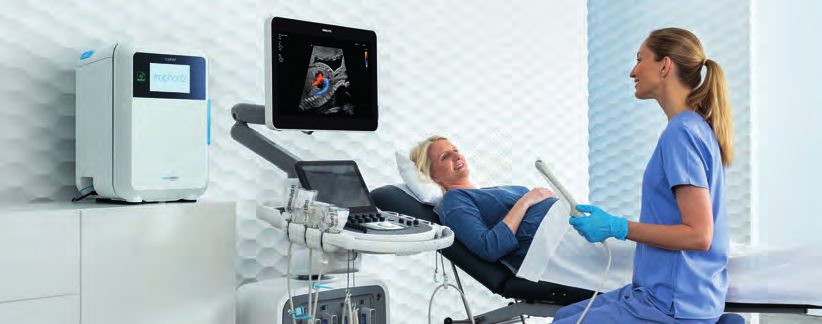
9 | HHE 2021 | hospitalhealthcare.com                                                                                       INTERVIEW

                                highlighted by Professor Sidhu was the                enormous developments in ultrasound
                                flexibility of the modality. In contrast to the       technology, and which were of huge benefit.
                                static imagery of an MRI or CT scan, ultrasound       He mentioned that a difficulty was that many
                                was performed in real-time. Consequently, it          people still see ultrasound as only black and
                                was possible during the scan to enquire as to         white, but that this is no longer the case and
                                whether the patient experienced any pain or           ultrasound has a far greater capability for
                                discomfort, particularly if the imaging indicated     imaging that all other modalities. He cited how
                                a potential cause for the pain. Alternatively, the    multiparametric ultrasound imaging can be
                                patient may offer a snippet of history during the     used to assess patients with steatotic livers6 and
                                scanning, and which helps to confirm the              that other innovations such as colour Doppler
                                diagnosis, neither of which are available to          ultrasound, contrast ultrasound and
                                radiologists when interpreting other imaging          elastography ultrasound,7 which looks at the
                                modalities.                                           stiffness of the liver, the amount of fibrosis and
                                                                                      scarring have all proved to be of value in patient
                                On the impact of the pandemic on imaging              care. He added that a further advantage of the
                                Professor Sidhu described how King’s was very         developments in ultrasound was that the
                                busy during the first and second waves of the         technology was less expensive than other
                                COVID-19 pandemic.5 With the global                   modalities and safe. He sensed that in the next
                                cancellation of routine imaging during the first      couple of years there would be many further
                                wave, the radiology department was quiet with         innovations with ultrasound-based technology
                                ultrasound becoming extremely useful within           and that these would continue to be patient-
                                the intensive care unit as a point-of-care for        friendly. Using the example of scanning the liver
                                imaging of patients’ lungs; this was done             of a two- or three-year-old child, Professor
                                predominately by the pulmonary physicians             Sidhu described how for an MRI scan, the child
                                although radiologists did perform some                needed to be sedated perhaps, given the
                                abdominal scans. He added that during the             contrast agent gadolinium, and kept still.
                                second wave, the department was a lot more            However, for an ultrasound scan, the child
                                prepared and continued with as much routine           remains awake, and the parents can also be
                                scanning, if patients could safely attend the         present to help with any possible anxiety.
References                      hospital, but suspects that this has resulted in      He thought that as an imaging modality,
1 Nyhsen C et al. Infection     a huge backlog of imaging awaiting to be              radiologists would be ultimately unwise to give
prevention and control in       undertaken. While the magnitude of this               up on ultrasound, adding that the technology
ultrasound – best practice
recommendations from            backlog remains uncertain, Professor Sidhu            is now at a level where the device does almost
the European Society of         remarked that there are still a lot of patients       everything for the operator, adjusting the
Radiology Ultrasound
Working Group. Insights         waiting to be scanned.                                parameters automatically to produce the
Imaging 2017;8(6):523–35.                                                             best image.
https://pubmed.ncbi.nlm.        On the key learnings since the pandemic                   Another development that had made
nih.gov/29181694/
2 European Society              Professor Sidhu thinks that an important              a considerable impact on ultrasound mentioned
of Radiology. Position          learning from the pandemic is that services           by Professor Sidhu was artificial intelligence.
statement and best practice
recommendations on the          need to become more patient centric and that          The technology can recognise the organs
imaging use of ultrasound       the provision of imaging services should              under investigation, identifies any abnormalities
from the European Society       become easier and more accessible for the             as well as providing a differential diagnosis.
of Radiology ultrasound
subcommittee. Insights          patient. With this idea in mind, there is likely to   He added that it even writes the report for the
Imaging 2020;11:115.            be a wholesale shift of routine outpatient            operator although currently, it still needs
https://insightsimaging.
springeropen.com/               scanning out of the acute hospital and make           a clinician to interpret the results of the scan.
articles/10.1186/s13244-020-    greater use of community-based imaging,
00919-x                         a move he says which has been supported by            On the skills that radiologists will require
3 ESR Executive Council
2009; European Society          government. He mentioned that although this           in the future
of Radiology. ESR position      change had been under discussion for several          Professor Sidhu thinks that will all the emerging
paper on ultrasound.
Insights Imaging                years, it was really brought into sharper focus as    technologies, some radiologists have been left
2010;1(1):27–9. https://        a consequence of the pandemic. After all, he          behind simply because the developments in
pubmed.ncbi.nlm.nih.            reflected on how it has been ludicrous to bring       ultrasound are largely driven by physicians in
gov/22347899/
4 ESR Congress. www.            large numbers of patients into a busy hospital        other specialties; in particular, hepatologists.
myesr.org/congress/             for routine/GP imaging. Consequently, there is        He explained how a key driver is not so much
ecr2022.
5 Panayiotou A et               now a move in progress to establish diagnostic        that other specialists embrace the technology
al. Escalation and              hubs, adjacent or close to the hospital and           but more that unlike radiologists, hepatologists
De-escalation of the            introducing agreed patient pathways to ensure         and other specialties do not have routine access
Radiology Response to
COVID-19 in a Tertiary          that only those patients who need further             to CT and MRI, ensuing the best aspects of
Hospital in South London:       management must visit the hospital. Professor         ultrasound are utilised constantly, before
The King’s College Hospital
Experience. Br J Radiol         Sidhu felt over the next few years, elective          reverting to another imaging modality.
2020;93:20201034.               imaging could be undertaken within the hub               Even though in the future ultrasound might
6 Basavarajappa L et al.        and that this would release capacity with the         well move outside of the sphere of radiology,
Multiparametric ultrasound
imaging for the assessment      hospital, allowing time to see acute patients and     Professor Sidhu still believes that there will
of normal versus steatotic      those who required other forms of                     always be a need for radiologists to be skilful in
livers. Sci Rep 2021;11:2655.
https://www.nature.com/         interventional or complex imaging, with the           ultrasound. As a profession, they possess the
articles/s41598-021-82153-z     important caveat, that the hospital service is        necessary skills to match up the results from all
7 Sigrist R et al. Ultrasound   accessible for patients when needed.                  the other scans and images and in doing, so will
Elastography: Review of
Techniques and Clinical                                                               continue to make an important contribution to
Applications. Theranostics      On the current exciting technological                 patient care.
2017;7(5):1303–29. https://
www.ncbi.nlm.nih.gov/pmc/       developments
articles/PMC5399595/            Professor Sidhu noted how there were
10 | HHE 2021 | hospitalhealthcare.com                                                                                INTERVIEW

Meet the Expert: Neelam Dugar
After specialising in oncology imaging at Manchester, Neelam Dugar is now a consultant radiologist
at Doncaster and Bassetlaw NHS Trust and also Chair of the Radiology Informatics Committee at the
Royal College of Radiologists. Hospital Healthcare Europe had the pleasure of speaking with her about
her career to date and to discuss recent documents produced by the College on the use of artificial
intelligence in radiology and the development of a vetting procedure for inappropriate scan requests.

                           Organisation of services at the Trust               supplement for a summary of guidance). The
                           Dr Dugar’s department has approximately             guidance defined the standards for how AI
                           16 radiologists and, with a newly purchased third   should be incorporated into the radiology
                           CT scanner, the department is extremely busy,       information (RIS) and picture archiving and
                           operating on an almost conveyor belt-like basis.    communication systems (PACS). Dr Dugar
                           In a typical day, radiographers will perform        highlighted how, in some respects, AI is
                           around 1000 imaging investigations including        considered a very broad term and can be
                           those for elective, acute and ward patients.        interpreted differently depending on the
                           As there is a requirement to have A&E CT scans      context. For the present document, the RCR
                           reported on within an hour, Dr Dugar                informatics group considered AI in the narrow
                           emphasised the importance of prioritising the       context of ‘computer vision’ used for radiology
                           workload within the team. Hence, one radiologist    image pre-analysis. As Dr Dugar explained,
                           is always allocated to report emergency scans,      because AI systems have already been
                           and while others do elective work. During the       developed for facial recognition, given that the
                           evening and weekends only one radiologist is        role of a radiologist is to visualise images and to
                           available for undertaking emergency CT and MRI      make interpretations to inform the ongoing care
                           scan reporting. Night-time emergency radiology      of patients, it seemed only right that this should
                           has now been outsourced to Australia. Since         be the area to focus upon.
                           starting her role over 17 years ago, the               However, a primary focus was to ensure that
                           workload had expanded considerably due to           IT vendors could develop the necessary
                           a combination of increased expectations and         infrastructure to incorporate AI systems with
                           national guidelines that often recommend the        different hospitals. A further consideration for
                           use of imaging. For example, she described how      the implementation of AI was the apparent
                           17 years ago, during a typical Sunday, she might    national shortage of radiologists in the UK.
                           be called upon to report one emergency scan         For instance, in a 2018 report,1 it was noted how
                           but on a recent weekend shift, her hospital         in the UK, only one in five UK Trusts and health
                           performed 100 emergency CT and MRI scans.           boards had enough interventional radiologists
                           She feels that no other specialty has               to provide safe 24/7 services to perform urgent
                           experienced that kind of explosion in workload.     procedures.
                                                                                  Dr Dugar defined how the AI workflow
                           On the work of the RCR Informatics Committee        guidance should work in practice, explaining that
                           and her role as Chair                               the AI system would initially review the image
                           Dr Dugar emphasises that technology is the          before it was seen by a radiologist. The AI
                           backbone of radiology and had a desire to make      system algorithms were such that it was able
                           best use of technological advances as a means       to highlight any relevant features, which is also
                           of enhancing patient care. She explained that       within the remit of radiologist. Nevertheless,
                           she was appointed as informatics advisor in         whereas the AI systems are capable of detecting
                           2015, because of an interest in the topic coupled   some abnormalities, the radiologist would then
                           with the fact that she had led the development      combine these findings with other test/imaging
                           of digitisation in her own department. She          results, and any other relevant clinical findings,
                           suggested that the Royal College of Radiologists    to create a more personalised report for the
                           (RCR) felt that it was necessary to have            patient.
                           a committee that was able to set the standards
                           of what should be achieved by all radiology         Will AI replace radiologists in the future?
                           departments implementing informatics. In other      If the AI system can do the essential job of
                           words, the RCR Informatics Committee was            a radiologist, surely these individuals can be
                           tasked with defining the standards and hence,       easily replaced? Dr Dugar disagrees. She
                           best practice, which should be achievable           revealed how in 2016 AI pioneer, Geoffrey
                           through hospitals’ information technology (IT)      Hinton, had said “we should stop training
                           systems.                                            radiologists now. It’s just completely obvious
                              Out of this work came the publication            that within five years, deep learning is going to
                           Integrating artificial intelligence (AI) with the   do better than radiologists.”2 At the time, she
                           radiology reporting workflows (see later in the     said this created a major staffing crisis,
11 | HHE 2021 | hospitalhealthcare.com INTERVIEW
especially in the US, which saw a downturn in AI system simply identifies any abnormality and
doctors choosing radiology as a career, fearing is unable to make a subjective judgement within
that they would be deemed superfluous in the the context of any other clinical findings.
near future. But, as Dr Dugar added, medicine Dr Dugar labelled the AI system as a ‘junior
is not maths – if it were then there would be no radiologist’, i.e., it was able to provide a limited
need for a radiologist! She feels that it is virtually role as a preliminary reviewer on images.
impossible to create an ‘artificial’ radiologist Nonetheless, she did believe that in the future,
simply because of the huge number of with improvements in AI occurring, the input
algorithms that would be required to emulate from a radiologist might be unnecessary,
the thought-processing of a radiologist. especially for simple imaging, e.g., reporting on
Dr Dugar believes that having an AI system the presence/absence of a fracture. However,
evaluating a scan goes some way towards the radiologists would still be required to interpret
need for two independent reviewers of a scan more complex imaging from MRI or CT scans.
result. This is considered as best practice, She added that while an AI system could identify
lending support to the metaphor that “two a filling defect in the lungs and report the most
heads are always better than one”. As she said, likely cause to be a pulmonary embolism, as
this is the current recommendation for breast a radiologist, you are always thinking laterally
cancer screening. She stressed that having two about other differential diagnoses.
independent reviews was necessary because
even though individual radiologists are highly On the vetting and cancellation of
trained, they are fallible. Thus, from a safety inappropriate scan guidelines
perspective, dual review is the ideal standard. As Dr Dugar described, being both medically
The integration of an AI system was also qualified and trained in radiology allowed her
important but for a very different reason. and her colleagues to assess whether or not
Dr Dugar highlighted that, in reality and with a particular imaging request was appropriate.
a national shortage of radiologists, it becomes She emphasised how often both junior doctors
impossible to achieve the two-reporter standard and those from other specialities, may not be
for images. One of the key reasons for a second completely clear on which imaging tests were
reporter was to the minimise the phenomenon correct. The vetting (triaging) and cancellation
of satisfaction of search, which describes the of inappropriate radiology requests document
situation where some lesions remain undetected was introduced simply to help manage the
after an initial lesion. As Dr Dugar illustrated, workload within radiology departments.
when a radiologist finds an abnormality, they An important part of Dr Dugar’s role is to always
become fixated on that particular problem and vet or triage any requests for imaging that reach
start to process this finding within the context of the department. This vetting process, she added,
other clinical information and sometimes ignore was crucial because of the high workload of the
other findings. With an AI system able to review department, which makes it impossible to
the image prior to the radiologist, it effectively perform every exam request.
becomes that second reporter and While the vetting process amounts to
a helper, alerting the radiologists to the full a clinical assessment task in itself, Dr Dugar
range of abnormalities present on the image. highlighted how within her department over
In discussion with colleagues, a barrier to greater 90% of the vetting process was undertaken by
use of AI is the perception among some the radiographers rather than the radiologists.
radiologists that the system is very sensitive This had been made possible through the
but not specific. Using the example of the introduction of a protocolised approach for the
assessment of a lung scan, Dr Dugar explained radiographers. Moreover, Dr Dugar believes that
that while the AI system would report on the radiographers can quickly acquire the necessary
presence of tiny nodules, the focus of the vetting skills and then approve and book a scan
radiologist was in looking for metastases. or reject the request. A further advantage to
With greater knowledge of the patient’s clinical radiographer-based vetting, is that, as these
history than the AI system, the radiologist can individuals are involved in performing the
potentially discount the relevance of these imaging, they are able to quickly assess, and
nodules and advise accordingly. In contrast, the then cancel, any duplicate requests and in some12 | HHE 2021 | hospitalhealthcare.com INTERVIEW
cases, even determine if the request would be of transformation within the NHS. She thinks this
additional value, i.e., if the request is for was of enormous benefit, enabling more virtual
a broadly similar scan. A difficulty for meetings which were a great advance compared
radiographers, however, is that without the to teleconferences. Another important
necessary medical training, they may feel development for work–life balance was allowing
uncomfortable cancelling an imaging request radiologists to have workstations at home.
that was requested on clinical grounds and in Dr Dugar says that having an interest in digital
such instances, the protocol would dictate that technology, she had tried to implement greater
the request is forwarded to the radiologist. home working for some time, but her request
As a consequence of introducing the vetting was always denied due to lack of funding.
process in her department, Dr Dugar felt that on However, she also thinks in the future, this
a typical day, she might be asked to vet up to innovation of homeworking and virtual meetings
30 requests and allocates up to 30 minutes of will not revert to pre-pandemic times but there
her day to this task. She thinks that such vetting will still be a balanced need for office working.
is a key task given that the department performs
around 1000 scans each day. Although some of On the evolution of the imaging landscape over
the requests passed to her from radiographers the next few years
can be challenging, in many cases, it can Dr Dugar believes that AI algorithms will develop
sometimes be very straightforward and require in the next two to five years and become a much
simply altering the request to a more better preliminary reporter on many more things
appropriate imaging modality. For more such as fracture detection, lung nodule
complex cases, she will need to review the detection etc. She mentioned how AI is already
patient’s medical history or initiate a discussion being used in brain imaging for strokes.
with the referring clinician to discuss the best She worries, however, that future innovations in
option. In cases where the request is rejected, AI by computer scientists will require additional
Dr Dugar ensures that the requesting clinician is funding, and that this should not be at the
informed of her decision and the rationale expense of a radiologist training.
behind the cancellation. She thinks that the Another potential growth area she feels is
departmental system, being fully electronic has in the evolution of enterprise imaging3 and
streamlined the whole request/cancellation revealed how all her own radiology department’s
process. images have already been archived and made
Dr Dugar expressed the view that the vetting available throughout the enterprise. An
document was desperately needed because in important current problem, she explained, was
some radiology departments, no vetting process how various medical images from other
was in place. She realised that part of the reason specialties/departments have been generated
behind this lack of vetting was largely due to but are stored in different locations and formats
a lack of functionality within the hospital’s without the correct patient identifiers etc. and
internal IT system. The purpose of the vetting are not indexed properly (e.g., endoscopy
guidance was thus to ensure that while not all images, ECG, audiometry, sleep studies etc).
NHS Trusts employed the same vendors, these Incorporation of all the images and graphs in
vendors would modify the IT infrastructure to a single and accessible location will be of
enable electronic communication for the vetting enormous value, not only to radiologists but also
process. As Dr Dugar said, in discussion with all treating physicians. With the ability to review
radiologists from outside of her own all images, and together with other pieces of
department, the cancellation process was often clinical data, it will allow radiologists to create
not communicated to the original requesting a much more personalised report for the patient.
clinician and this led to internal friction and, in Although improvements in smartphone
some cases, the radiologists in an attempt to technology allow for image review, Dr Dugar
appease the requesting clinicians, decided to no thinks that at the present time, the quality of the
longer triage requests, with a resultant increase images is not of sufficient quality for diagnostic
in their workload. Although it seems unusual purposes. Furthermore, from a medico-legal
that the whole of the NHS must deal with perspective, she would not use the images
different IT vendors, Dr Dugar is against the reviewed on a smartphone for reporting. She
idea of a national vendor. She thinks that with also felt that while mobile scanning units for MRI
such a huge monopoly, there would be little and CT scans were available and could be
incentive to innovate. What is more important utilised for elective work, these imaging
she feels, is that the same workflow processes modalities would still need to remain within the
References should be adopted in the different NHS hospital premises, where the equipment was
1 Bassett M. Radiologist Trusts, to improve the efficiency of radiology needed for emergency scans.
shortage deepens in the UK.
www.rsna.org/news/2019/ departments, even if this occurs through Image acquisition and interpretation were
May/uk-radiology-shortage dissimilar IT systems. separate, and Dr Dugar believes that she does
(accessed June 2021). not need to be present at a mobile scanning unit
2 Marcus G, Little M.
Advancing AI in health care: On the impact of the pandemic on imaging and can remain either in the office or at home to
It’s all about trust. www. services undertake her interpretive role for the images.
statnews.com/2019/10/23/
advancing-ai-health-care- Dr Dugar said that for her, radiology services did However, radiologists (whether remote or
trust/ (accessed June 2021). not stop during the pandemic although the on-site) must continue to work closely with the
3 Petersilge C. The focus shifted to COVID patients. She felt that her radiographers operating the scanners to provide
evolution of enterprise
imaging and the role of the own work, which is either cancer or emergency- support and advise on appropriateness and
radiologist in the new world. based, did not slow during the pandemic. She vetting/triaging support. Radiologists and
www.ajronline.org/doi/
full/10.2214/AJR.17.17949 believed that one of the greatest changes radiographers must always work as teams to
(accessed June 2021). because of the pandemic was the digital improve patient care.13 | HHE 2021 | hospitalhealthcare.com GUIDELINE SUMMARY
Integrating artificial
intelligence with the radiology
reporting workflows
This guidance from the Royal College of Radiologists sets out the standards that a department
should meet when integrating artificial intelligence into already established systems, producing a safe
seamless system with the patients at the centre
The fast pace of developments in artificial the system will query and retrieve a prior similar
intelligence (AI) means that the technology will image from the picture archiving and
have an important role to play in many clinical communication system (PACS) for comparative
specialities, including radiology and will change, analytical purposes. A further advantage of
hopefully in a positive direction, the way in using an AI system, is ‘computer-assisted triage’,
which patient care is delivered. which helps with the prioritisation of reporting
AI platforms and algorithms are designed to worklists once an abnormality has been
work in collaboration with existing technologies detected.
and have a wide range of potential uses in Nevertheless, the RCR report emphasises
radiology. For example, in magnetic resonance the importance of radiologists acknowledging
imaging (MRI), an AI algorithm can detect the limitations of an AI system report, i.e., its
multiple sclerosis, strokes, brain bleeds etc. sensitivity and specificity, and what these
Within the arena of computed tomography figures mean in the context of the specific
(CT), AI systems are designed to detect skull pathology. In other words, the AI system is
fractures, brain haemorrhages, infarcts, and simply a supportive tool and radiologists should
tumours. In body CT, the introduction of AI has not become overly reliant upon on the AI
a role in mammography, allowing for the findings and assume that these findings will be
detection of both suspicious lesions and 100% accurate all the time.
calcification. The overarching aim of the RCR report is
Once an image has been captured by the firstly to ensure that any innovations in AI are
radiographer, the AI will perform a ‘pre-analysis’ fully integrated into existing reporting systems
of the image, and, if an abnormality is detected, and secondly, to define the necessary standards14 | HHE 2021 | hospitalhealthcare.com GUIDELINE SUMMARY
required to enable radiology service providers General standards for data output
to facilitate this integration without creating Any AI platform adopted should have standard
additional burden for staff. output, and which must include:
The report does not make any specific Graphical representation of the region of
recommendations about which AI system interest (of the detected abnormalities) or
should be purchased, or any ethical mark-up/pointers using global technical
considerations related to the use of AI, and standards (DICOM) so that images can be
finally discusses the issue of AI solutions for viewed in the PACS viewers.
workflow and radiology management efficiency. • AI detected abnormalities should be output as
The report is directed more towards defining text e.g., fracture, infarct etc.
the parameters within which an AI platform • A notification that the image analysis has
should operate. been completed.
• Some AI alerts may be defined as critical
Standards within the system and should be pre-specified
The report begins with a series of standards by the radiologist.
for the use of AI systems. • A declaration or disclaimer should be sent out
1 AI must be integrated seamlessly with existing including the list of any abnormalities which
radiology information systems (RIS) and PACS were evaluated by the AI system. This might, for
without creating an additional burden for example, include a CT scan that detected a
radiologists. brain haemorrhage. It is also necessary to
2 The accuracy of the AI algorithms must be include the sensitivity and specificity (or true/
clearly declared to both the radiologist and false positives or negatives) of the applied
others involved in patient management. algorithm for each of the abnormality being
3 The AI finding should be communicated to evaluated.
the RIS and PACS through existing and global With the RIS, it will be necessary to
technical standards. incorporate additional data fields which capture
4 The department workflow should be AI abnormalities and any alerts.
sufficiently robust to ensure that the AI analysis The report concludes on a positive note
is complete and available on PACS before being saying that: “AI image pre-analysis is likely to
viewed and interpreted by a human. have a very positive impact on radiologists’
An important element of the report is the future working lives if properly integrated into
necessity to ensure that all instrumentation, the reporting workflow”.
e.g., scanners, RIS, PACS and the AI platform,
all work cooperatively within the radiology A copy of the full guidance can be viewed here.
department. It is also necessary that the AI www.rcr.ac.uk/publication/integrating-artificial-
platform only begins the analysis once the intelligence-radiology-reporting-workflows-ris-
radiographer has completed the examination and-pacs
and has sent the information to the AI system,
i.e., that the imaging should be ‘pre-analysed’
before reaching the PACS for displayed.You can also read