Recommended composition of influenza virus vaccines for use in the 2020- 2021 northern hemisphere influenza season February 2020
←
→
Page content transcription
If your browser does not render page correctly, please read the page content below
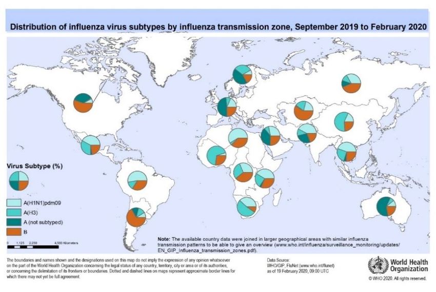
Recommended composition of influenza virus vaccines for use in the 2020-
2021 northern hemisphere influenza season

February 2020

WHO convenes technical consultations1 in February and September each year to recommend
viruses for inclusion in influenza vaccines2 for the northern and southern hemisphere influenza
seasons, respectively. This recommendation relates to influenza vaccines for use in the
forthcoming northern hemisphere 2020-2021 influenza season. A recommendation will be
made in September 2020 for vaccines that will be used for the southern hemisphere 2021
influenza season. For countries in tropical and subtropical regions, WHO guidance for
choosing between the northern and southern hemisphere formulations is available on the WHO
Global Influenza Programme website3.

Seasonal influenza activity

Between September 2019 and January 2020, influenza activity was reported in all regions,
with influenza A(H1N1)pdm09, A(H3N2) and influenza type B viruses co-circulating.

In the temperate zone of the northern hemisphere, influenza activity remained at inter-
seasonal levels until late October, when it started to increase. In Europe, influenza activity
commenced earlier than in recent years and influenza A(H1N1)pdm09, A(H3N2) and type B
viruses were reported, although the distribution was not homogeneous. Virus predominance
varied between countries. In most countries, influenza activity increased sharply by late
January. Countries in North America reported high influenza activity from December, with
influenza A and B viruses cocirculating in Canada and the United States of America. In both
countries, influenza A(H1N1)pdm09 viruses circulated in higher proportions than A(H3N2)
viruses. Generally, countries in East Asia experienced increased influenza activity from late
December, with influenza A(H3N2) predominant in China and Mongolia and A(H1N1)pdm09
viruses predominant in Japan and Republic of Korea. In some countries in North Africa, West
and Central Asia, influenza activity increased between October and January with co-circulation
of influenza A and B viruses in different proportions. In North Africa and West Asia,
predominance of influenza A(H1N1)pdm09 viruses was reported, while in Central Asia
A(H1N1)pdm09 and A(H3N2) viruses circulated equally.

1 http://www.who.int/influenza/vaccines/virus/en/
2 Description of the process of influenza vaccine virus selection and development available at:
 http://www.who.int/gb/pip/pdf_files/Fluvaccvirusselection.pdf
3 Influenza in the tropics and sub-tropics: http://www.who.int/influenza/vaccines/tropics/en/

28 February 2020                                                                                  Page 1 of 12
Influenza activity in the tropical and subtropical regions of Asia was high with A(H3N2)
viruses predominant in Lao People’s Democratic Republic, while in other countries influenza
A and B viruses cocirculated. Influenza A(H1N1)pdm09 viruses predominated in some
countries including Iran (Islamic Republic of), Malaysia, Oman and Qatar. Influenza activity
in most tropical countries of Central America, the Caribbean and South America was
generally low, with A(H1N1)pdm09, A(H3N2) and type B viruses co-circulating. High
influenza A(H1N1)pdm09 virus activity was reported in El Salvador and Mexico and
A(H3N2) virus activity was reported in Nicaragua. For countries in the tropical and
subtropical zone of Africa, influenza A(H1N1)pdm09 viruses were predominant in Central
African Republic, Ethiopia, Kenya and South Sudan, while A(H3N2) virus activity was
predominant in Burkina Faso, Cameroon, Côte d'Ivoire, Ghana, Guinea, Niger, Nigeria,
Senegal and Togo.

In the temperate zone of the southern hemisphere, influenza activity was generally low and
remained at inter-seasonal levels between October and January. In the temperate zone of South
America there was co-circulation of influenza A and B viruses in most countries. In South
Africa there was co-circulation of influenza A and B viruses with A(H3N2) viruses
predominating. Influenza activity was reduced in Australia and was below the seasonal
threshold in New Zealand throughout this period.

Influenza A

Influenza A viruses were predominant in most countries and accounted for 59% of all
influenza viruses detected. Globally, co-circulation of both A(H1N1)pdm09 and A(H3N2)
viruses was evident in most reporting countries, areas and territories. Influenza A(H3N2)
viruses circulated in higher proportions than A(H1N1)pdm09 viruses in several countries in
West and South Africa, East Asia, Northern Europe and temperate South America. Influenza
A(H1N1)pdm09 viruses were predominant in other regions (North and East Africa, North
America, Central America and Caribbean, tropical South America, Central, West, South and
South-East Asia).

Influenza B

Influenza B viruses circulated in higher proportion than influenza A viruses in some countries
in the Americas, Europe and Central and West Asia. Viruses of the B/Victoria/2/87 lineage
circulated in higher proportion than the B/Yamagata/16/88 lineage viruses worldwide, with
the exception of a few countries in Central and South America (Chile, Colombia, Dominican
Republic and Haiti), where B/Yamagata/16/88 lineage viruses were predominant.

28 February 2020                                                                  Page 2 of 12
Detailed information by country of the extent and type of seasonal influenza activity
worldwide is available on the WHO website:
http://www.who.int/influenza/resources/charts/en/

Zoonotic influenza infections

In the period between 24 September 2019 to 24 February 2020, five human cases of A(H9N2)
virus infections were detected. Two cases, one each from India and Senegal, had illness onset
dates prior to September 2019. These were the first reports of human infections with A(H9N2)
viruses from these countries. The other cases were reported by China (2) and Hong Kong
Special Administrative Region of China (1). All cases recovered.

One new human case of an A(H1N1)v virus infection was identified in this period in China, in
addition to a previously unreported A(H1N1)v case with an onset date of December 2018. Both
cases were caused by viruses from the 1C (Eurasian avian-like) swine influenza virus lineage.

During this period, no human infections with A/goose/Guangdong/1/96-lineage A(H5)
viruses or A/Anhui/1/2013-lineage A(H7N9) viruses were reported. A(H5) viruses were
detected in domestic and wild birds in several countries.

Antigenic and genetic characteristics of recent seasonal influenza viruses,
serology and antiviral susceptibility

Influenza A(H1N1)pdm09 viruses

28 February 2020                                                                  Page 3 of 12
Currently circulating A(H1N1)pdm09 viruses have haemagglutinin (HA) genes that belong to
phylogenetic clade 6B.1A, with subclades 5A, 5B and 7 most frequently detected. The majority
belong to subclade 5A and encode HA1 amino acid substitutions N129D, S183P, T185I and
N260D, with continued diversification observed. The HA proteins of the majority of 5A
subclade viruses globally have acquired additional amino acid substitutions D187A and
Q189E, such as A/Guangdong-Maonan/SWL1536/2019 and A/Hawaii/70/2019. Some other
recently emerged viruses within the 5A subclade have acquired an N156K substitution.

Antigenic characterization by haemagglutination inhibition (HI) assay using post-infection
ferret antisera indicated that all recent A(H1N1)pdm09 viruses, except those with HA1 amino
acid substitutions at residues 155 or 156, were antigenically similar to egg- and cell culture-
propagated A/Brisbane/02/2018-like viruses. However, assays using post-vaccination human
antisera showed reduced HI titres against recent viruses compared with titres against cell
culture-propagated A/Brisbane/02/2018-like reference viruses (Table 1).

Table 1. Antigenic analysis of A(H1N1)pdm09 viruses by haemagglutination inhibition
assay
REFERENCE VIRUSES                             FERRET SERA      INDIVIDUAL ADULT HUMAN SERA
                                                               CA/07     CA/07     MI/45     MI/45
STRAIN DESIGNATION          HA CLADE          ID/07   ME/38   2010-11   2010-11   2017-18   2017-18
A/California/07/2009                          2560     160     1280      2560       640       160
A/Michigan/45/2015   6B.1                     2560     640      320      5120      1280       320
A/Idaho/07/2018      6B.1A-3                  2560     320      320      2560       640        80
A/Maine/38/2018      6B.1A-2+156K              160    5120      640      1280       640        80
TEST VIRUSES
A/Nebraska/15/2018     6B.1A-5A               2560      320     320      2560      640        80
A/Nebraska/14/2019     6B.1A-5A+187A, 189E    2560      160     320      1280      320        40
A/Wisconsin/588/2019   6B.1A-5A+156K           160     2560     320       640      320        40
A/Iowa/33/2019         6B.1A-5B               2560     1280    1280      2560      640        80
A/Arkansas/28/2019     6B.1A-5B+137S          2560     1280     320      1280      320        40
A/Virginia/41/2019     6B.1A-5B+138Q          2560      640     320      2560      640        40
A/Minnesota/60/2019    6B.1A-7                5120      320     320      2560      320        40
A/Alabama/27/2019      6B.1A-7                2560      160     320      2560      320        40

Human serology studies used serum panels from children (6 months to 17 years), adults (18-
64 years) and elderly adults (≥65 years) who had received quadrivalent inactivated egg- or cell-
based vaccines or recombinant HA vaccine with the composition recommended for the
northern hemisphere 2019-2020 season (A/Brisbane/02/2018 (H1N1)pdm09-like,
A/Kansas/14/2017 (H3N2)-like, B/Colorado/06/2017-like and B/Phuket/3073/2013-like
viruses), or trivalent high-dose vaccine (without the inclusion of B/Phuket/3073/2013-like
viruses). Geometric mean HI titres against many recent representative cell culture-propagated
A(H1N1)pdm09 viruses from 6B.1A subclades, including those in 5A with additional
substitutions D187A and Q189E, were reduced compared to HI titres to cell culture-propagated
A/Brisbane/02/2018-like vaccine viruses; reductions were more pronounced when measured
against the egg-propagated vaccine virus.

28 February 2020                                                                     Page 4 of 12
Of 2455 influenza A(H1N1)pdm09 viruses tested for neuraminidase inhibitor (NAI) susceptibility, 23 showed reductions in susceptibility to one or more of the inhibitors. All but two viruses showed highly reduced inhibition by oseltamivir and peramivir and had an H275Y amino acid substitution in the neuraminidase (NA). Two other A(H1N1)pdm09 viruses carried a Q136K amino acid substitution in the NA and had reduced inhibition by zanamivir. Only one of 1355 A(H1N1)pdm09 viruses analysed for susceptibility to the endonuclease inhibitor baloxavir showed reduced susceptibility to the antiviral drug. The virus was isolated from a patient in Japan with no prior treatment and had an E23K amino acid substitution in the PA protein, which is known to confer reduced susceptibility to this inhibitor. Influenza A(H3N2) viruses HA phylogenetic analysis of A(H3N2) viruses collected from September 2019 to February 2020 showed regional heterogeneity, with clade 3C.3a viruses predominating in some countries in Europe and group 3C.2a1b viruses predominating in many countries globally. The majority of HA genes of genetic group 3C.2a1b viruses included viruses having either T135K or T131K amino acid substitutions in HA1. Over this period there has been an increase in detection of 3C.2a1b+T135K subgroup viruses, which divide into two main clusters: one with additional substitutions at S137F, A138S and F193S, and the other defined by an S198P substitution. Antigenic characterisation of clade 3C.2a viruses continued to be technically difficult because a proportion of viruses did not agglutinate red blood cells, preventing HI analysis. Virus neutralisation (VN) assays have become the preferred methods for determining the antigenic characteristics of current A(H3N2) viruses. In VN or HI assays, ferret antisera raised against cell culture-propagated A/Kansas/14/2017 (3C.3a) virus inhibited 3C.3a viruses well, but viruses from the predominant 3C.2a1b group were less well inhibited. Ferret antisera raised against egg-propagated A/Kansas/14/2017 (3C.3a) or A/South Australia/34/2019-like (subgroup 3C.2a1b+T131K) viruses did not inhibit many recently circulating viruses well. The majority of subgroup 3C.2a1b+T131K and both clusters of 3C.2a1b+T135K viruses were well inhibited by ferret antisera raised against the cell culture-propagated reference virus A/Hong Kong/45/2019 and less well by egg-propagated A/Hong Kong/2671/2019, both 3C.2a1b+T135K viruses with S137F, A138S and F193S HA substitutions (Table 2). Human serology studies, using a subset of the human serum panels described above, showed that geometric mean HI titres against most recent representative A(H3N2) viruses from genetic group 3C.2a1b were somewhat reduced compared to HI titres against the egg-propagated and cell-propagated A/Kansas/14/2017-like vaccine viruses. In VN assays, titres against these genetic group 3C.2a1b viruses were reduced to varying degrees when compared to the titres against egg- and cell-propagated A/Kansas/14/2017-like vaccine viruses; notably titres were significantly reduced in sera obtained from vaccinated children. Recently circulating clade 3C.3a viruses were recognised well in both HI and VN assays. 28 February 2020 Page 5 of 12
One of 1875 influenza A(H3N2) viruses tested showed reduced inhibition by zanamivir, but
all were sensitive to oseltamivir. Of 1012 A(H3N2) viruses assessed for susceptibility
to baloxavir by genetic and/or phenotypic analysis, a single virus contained the amino acid
substitution I38M in the PA gene, which is known to confer reduced baloxavir susceptibility.

Table 2. A(H3N2) focus reduction assay
                                                                                REFERENCE FERRET ANTISERA
                                                                         2a1b+131K         2a1b+135K+137F       3a
                                                                     SIAT        EGG       SIAT      EGG      SIAT
REFERENCE VIRUSES               Passage            3C Clade          IA/60      AUS/34    HK/45     HK/2671   KS/14
  1 A/IOWA/60/2018                S1              2a1b+131K           5120        640       320       320      320
  2 A/SOUTH AUSTRALIA/34/2019    E5/E2            2a1b+131K           1280       1280
Influenza B viruses Influenza B viruses of the B/Victoria/2/87 and the B/Yamagata/16/88 lineages accounted for 41% of typed viruses. Among them, the vast majority of the viruses were from the B/Victoria lineage in all regions except for South America. All available HA gene sequences of B/Yamagata lineage viruses belonged to genetic clade 3. In HI assays the vast majority of recently circulating B/Yamagata lineage viruses were well inhibited by post-infection ferret antisera raised against cell culture-propagated B/Phuket/3073/2013 virus. However, a proportion of viruses showed reduced inhibition by post-infection ferret antisera raised against the egg-propagated B/Phuket/3073/2013 virus. The HA gene sequences of the B/Victoria lineage viruses characterized belonged to genetic clade 1A, but significant genetic diversity continued to be observed. Of the B/Victoria lineage viruses genetically characterized, the vast majority were viruses with a three amino acid deletion in HA1 (positions 162-164); a small minority were viruses with a two amino acid deletion (positions 162-163), most of which were detected in Central and South America. The vast majority of viruses with the three amino acid deletion possessed additional HA1 substitutions G133R and K136E. A small proportion of the viruses with the three amino acid deletion with K136E had additional substitutions N150K, G184E, N197D and R279K. A majority of viruses with the three amino acid deletion were inhibited well by post-infection ferret antisera raised against both cell culture- and egg-propagated three amino acid deletion viruses, such as B/Washington/02/2019. The majority of viruses with the three amino acid deletion were poorly inhibited by post-infection ferret antisera raised against both egg- and cell culture-propagated B/Colorado/06/2017-like viruses. Human serology studies, using the serum panels described above, showed minor reductions in post-vaccination HI geometric mean titres against the majority of recent representative B/Yamagata lineage viruses when compared to the cell culture-propagated B/Phuket/3073/2013 vaccine virus. Post-vaccination HI geometric mean titres against recent viruses of the B/Victoria lineage representing the dominant HA three amino acid deletion genetic group were reduced when compared to the egg- or cell culture-propagated B/Colorado/06/2017-like vaccine viruses. Of the 1734 influenza B viruses screened for NAI susceptibility, 9 showed reduced susceptibility to one or more NAIs. A total of 930 viruses were screened for susceptibility to baloxavir by genetic and/or phenotypic analysis; all retained normal susceptibility. 28 February 2020 Page 7 of 12
Recommended composition of influenza virus vaccines for use in the 2020-
2021 northern hemisphere influenza season

From September 2019 to January 2020, influenza activity was reported globally, with influenza
A(H1N1)pdm09, A(H3N2) and both lineages of influenza B viruses co-circulating. Influenza
A viruses predominated in most countries and accounted for 59% of all influenza viruses
detected. Influenza B viruses circulated in higher proportion in some countries in the Americas,
Central and West Asia. Viruses of the B/Victoria/2/87 lineage circulated in higher proportion
than the B/Yamagata/16/88 lineage viruses worldwide, except for a few countries in Central
and South America where B/Yamagata/16/88 lineage viruses were the predominant lineage.

All currently circulating A(H1N1)pdm09 viruses belong to phylogenetic clade 6B.1A with
subclades 5A, 5B and 7 detected most frequently. Of these, viruses from subclade 5A were
predominant and encoded additional HA1 amino acid substitutions N129D, S183P, T185I and
N260D. Globally, the majority of viruses in subclade 5A contained amino acid substitutions
D187A and Q189E (e.g. A/Guangdong-Maonan/SWL1536/2019 and A/Hawaii/70/2019).
Although antigenic analyses using post-infection ferret antisera do not detect all antigenic
differences in these current A(H1N1)pdm09 viruses, the amino acid substitutions acquired in
recent viruses are within antigenic epitopes in the HA. Furthermore, serological assays with
human antisera showed reduced HI titres against recent subclade 5A and 5B viruses compared
with titres against cell culture-propagated A/Brisbane/02/2018-like viruses. Geometric mean
HI titres against many recent cell culture-propagated A(H1N1)pdm09 viruses, including those
in the globally prevalent 5A subgroup with additional substitutions at positions 187 and 189,
were reduced compared to HI titres to the cell culture-propagated vaccine virus
A/Brisbane/02/2018. These reductions were more pronounced when measured against the egg-
propagated vaccine virus. Taken together, the results with human antisera and location of
amino acid substitutions are indicative of significant antigenic drift.

A(H3N2) viruses collected in this period showed regional heterogeneity, with group 3C.2a1b
viruses predominating in many countries globally. The majority of 3C.2a1b viruses included
either T135K or T131K amino acid substitutions in HA1. There was an increase in
3C.2a1b+T135K subgroup viruses with two main clusters: one with additional substitutions
S137F, A138S and F193S, and the other defined by an S198P substitution. The majority
of 3C.2a1b+T131K viruses and both clusters of 3C.2a1b+T135K viruses were well inhibited
by ferret antisera raised against the cell reference virus A/Hong Kong/45/2019 although less
well by ferret antisera raised against egg-propagated A/Hong Kong/2671/2019.

Influenza B viruses of the B/Victoria/2/87 and the B/Yamagata/16/88 lineages co-circulated
globally and accounted for 41% of typed viruses, with the B/Victoria lineage being dominant
in all regions except South America. The vast majority of recently circulating B/Yamagata
lineage viruses were antigenically and genetically closely related to the cell culture-propagated
vaccine virus B/Phuket/3073/2013.

28 February 2020                                                                   Page 8 of 12
A vast majority of the B/Victoria lineage viruses contained the three amino acid deletion in
HA1 (position 162-164). A majority of viruses were not inhibited well by post-infection ferret
antisera raised against both egg- and cell culture-propagated B/Colorado/06/2017-like viruses,
but were inhibited well by post-infection ferret antisera raised against both cell culture- and
egg-propagated three amino acid deletion viruses, such as B/Washington/02/2019.

The WHO recommends that quadrivalent vaccines for use in the 2020-2021 northern
hemisphere influenza season contain the following:

Egg-based Vaccines
   • an A/Guangdong-Maonan/SWL1536/2019 (H1N1)pdm09-like virus;
   • an A/Hong Kong/2671/2019 (H3N2)-like virus;
   • a B/Washington/02/2019 (B/Victoria lineage)-like virus; and
   • a B/Phuket/3073/2013 (B/Yamagata lineage)-like virus.

Cell- or recombinant-based Vaccines
   • an A/Hawaii/70/2019 (H1N1)pdm09-like virus;
   • an A/Hong Kong/45/2019 (H3N2)-like virus;
   • a B/Washington/02/2019 (B/Victoria lineage)-like virus; and
   • a B/Phuket/3073/2013 (B/Yamagata lineage)-like virus.

The WHO recommends that trivalent influenza vaccines for use in the 2020-2021 northern
hemisphere influenza season contain the following:

Egg-based Vaccines
   • an A/Guangdong-Maonan/SWL1536/2019 (H1N1)pdm09-like virus;
   • an A/Hong Kong/2671/2019 (H3N2)-like virus; and
   • a B/Washington/02/2019 (B/Victoria lineage)-like virus.

Cell- or recombinant-based Vaccines
   • an A/Hawaii/70/2019 (H1N1)pdm09-like virus;
   • an A/Hong Kong/45/2019 (H3N2)-like virus; and
   • a B/Washington/02/2019 (B/Victoria lineage)-like virus.

Lists of egg- or cell culture-propagated candidate vaccine viruses (CVVs) suitable for use in
human vaccine production are available on the WHO website4. A list of reagents for vaccine
standardization, including those for this recommendation, can also be found on the WHO
website. CVVs for zoonotic influenza viruses are listed on the same website.

As in previous years, national or regional authorities approve the composition and formulation
of vaccines used in each country. National public health authorities are responsible for making

4   http://www.who.int/influenza/vaccines/virus/candidates_reagents/home

28 February 2020                                                                  Page 9 of 12
recommendations regarding the use of the vaccine. WHO has published recommendations on
the prevention of influenza5.

CVVs (including reassortants) and reagents for use in the laboratory standardisation of
inactivated vaccines may be obtained from:

•      Biomedicines and Influenza Vaccines Section, Laboratories Branch, Medical Devices and
       Product Quality Division, Therapeutic Goods Administration, P.O. Box 100, Woden,
       ACT, 2606, Australia (fax: +61262328564, email: influenza.reagents@health.gov.au;
       web site: http://www.tga.gov.au)
•      Division of Virology, National Institute for Biological Standards and Control, a centre of
       the Medicines and Healthcare products Regulatory Agency (MHRA), Blanche Lane,
       South Mimms, Potters Bar, Hertfordshire, EN6 3QG, UK (fax: +441707641050, e-mail:
       enquiries@nibsc.org, web site:
       http://www.nibsc.org/science_and_research/virology/influenza_resource_.aspx
•      Division of Biological Standards and Quality Control, Center for Biologics Evaluation
       and Research, Food and Drug Administration, 10903 New Hampshire Avenue, Silver
       Spring, Maryland, 20993, USA (fax: +1 301 480 9748), email:
       cbershippingrequests@fda.hhs.gov)
•      Influenza Virus Research Center, National Institute of Infectious Diseases, 4-7-1 Gakuen,
       Musashi-Murayama, Tokyo 208-0011, Japan (fax: +81425616156, email: flu-
       vaccine@nih.go.jp)

Requests for reference viruses should be addressed to:

•      WHO Collaborating Centre for Reference and Research on Influenza, VIDRL, Peter
       Doherty Institute, 792 Elizabeth Street, Melbourne, Victoria 3000, Australia (fax:
       +61393429329, web site: http://www.influenzacentre.org, email:
       whoflu@influenzacentre.org)
•      WHO Collaborating Centre for Reference and Research on Influenza, National Institute
       of Infectious Diseases, 4-7-1 Gakuen, Musashi-Murayama, Tokyo 208-0011, Japan (fax:
       +81425616149 or +81425652498, email: whocc-flu@nih.go.jp
•      WHO Collaborating Centre for Surveillance, Epidemiology and Control of Influenza,
       Centers for Disease Control and Prevention, 1600 Clifton Road, Mail Stop H17-5,
       Atlanta, GA 30329, United States (fax: +14046390080, web site:
       http://www.cdc.gov/flu/, email: influenzavirussurveillance@cdc.gov)
•      WHO Collaborating Centre for Reference and Research on Influenza, The Francis Crick
       Institute, 1 Midland Road, London NW1 1AT, UK (Tel: +44 203 796 1520 or +44 203
       796 2444) (website: http://www.crick.ac.uk/research/worldwide-influenza-centre email:
       whocc@crick.ac.uk)
•      WHO Collaborating Centre for Reference and Research on Influenza, National Institute
       for Viral Disease Control and Prevention, China CDC, 155 Changbai Road, Changping

5   http://www.who.int/wer/2012/wer8747.pdf

28 February 2020                                                                   Page 10 of 12
District, 102206, Beijing, P.R. China. (tel: +86 10 5890 0851, fax: +86 10 5890 0851,
email: whocc-china@cnic.org.cn, website: http://www.chinaivdc.cn/cnic/en).
WHO provides fortnightly updates6 of global influenza activity. Other information about
influenza surveillance can be found on the WHO Global Influenza Programme website7.
Acknowledgements
The WHO recommendation on vaccine composition is based on the year-round work of the
WHO Global Influenza Surveillance and Response System (GISRS). We thank the National
Influenza Centres (NICs) of GISRS, and non-GISRS laboratories including the OIE/FAO
Network of Expertise on Animal Influenza (OFFLU), who contributed information, clinical
specimens, viruses and associated data; WHO Collaborating Centres of GISRS for their in-
depth characterization and comprehensive analysis of viruses; University of Cambridge for
performing antigenic cartography and phylogenetic analysis; WHO Essential Regulatory
Laboratories of GISRS for their complementary virus analyses and contributions from a
regulatory perspective; and laboratories involved in the production of high growth/yield
reassortants as candidate vaccine viruses. We also acknowledge the Global Initiative for
Sharing All Influenza Data (GISAID) for the EpiFlu database and other sequence databases
which were used to share gene sequences and associated information; modelling groups for
virus fitness forecasting; and the Global Influenza Vaccine Effectiveness (GIVE) Collaboration
for sharing estimates of influenza vaccine effectiveness on a confidential basis.
6 http://www.who.int/influenza/surveillance_monitoring/updates/en/
7 http://www.who.int/influenza/gip
28 February 2020 Page 11 of 12Annex 1
Declarations of interest
The WHO recommendation on the composition of influenza vaccines for use in the northern
hemisphere influenza season 2020-2021 was made through a WHO Consultation with
relevant WHO Collaborating Centres on Influenza (CCs) and Essential Regulatory
Laboratories (ERLs).
In accordance with WHO policy, Directors and experts of the relevant WHO CCs and ERLs,
in their capacity as representatives of their respective institutions ("Advisers"), completed the
WHO form for Declaration of Interests for WHO experts before being invited to the
Consultation. At the start of the Consultation, the interests declared by the Advisers were
disclosed to all participants.
The Advisers declared the following personal current or recent (within the past 4 years)
financial or other interests relevant to the subject of work:
Institution Representative Personal interest
WHO CC Atlanta Dr David Wentworth Co-inventor with others and employers
of US patents
• Influenza reassortment
• Modified bat influenza viruses
and their uses.
Both are current, not licensed and zero
income.
WHO CC Beijing Dr Dayan Wang None
WHO CC London Dr John McCauley • Served on an organizing
committee for an educational flu
awareness meeting/symposium
organized by Seqirus (May and
November 2019. No payment
received.
• Attended a meeting on new
influenza inhibitors organized
by ROCHE. No payment
received.
WHO CC Melbourne Dr Kanta Subbarao None
WHO CC Memphis Dr Richard Webby None
WHO CC and ERL Dr Hideki Hasegawa None
NIID Tokyo
WHO ERL CBER Dr Zhiping Ye None
Bethesda
WHO ERL NIBSC Dr Othmar Engelhardt None
London
WHO ERL TGA Dr Mandvi Bharadwaj None
Canberra
The interests declared by Drs Wentworth and McCauley were reviewed by WHO and
determined not to present a conflict of interest with the objectives of the WHO consultation.
Therefore, Drs Wentworth and McCauley participated in the consultation as an Adviser.
28 February 2020 Page 12 of 12You can also read