COUPLET MEDICINES OF LEECH AND CENTIPEDE GRANULES IMPROVE ERECTILE DYSFUNCTION VIA INACTIVATION OF THE CASR/PLC/PKC SIGNALING IN ...
←
→
Page content transcription
If your browser does not render page correctly, please read the page content below
Bioscience Reports (2020) 40 BSR20193845
https://doi.org/10.1042/BSR20193845

Research Article

Couplet medicines of leech and centipede granules
improve erectile dysfunction via inactivation of the
CaSR/PLC/PKC signaling in streptozotocin-induced
diabetic rats

                                                                                                                                                                                           Downloaded from https://portlandpress.com/bioscirep/article-pdf/40/2/BSR20193845/867248/bsr-2019-3845.pdf by guest on 15 April 2020
Jian Xiong Ma1,* , Bin Wang2,* , Cai Fei Ding1 , Hai Song Li2 , Xue Juan Jiang1 , Chen Ye Wang1 , Jia Yu1 and
   Wang Qiang Chen1
1 Department of Reproductive Medicine, Zhejiang Integrated Traditional and Western Medicine Hospital, Zhejiang, China; 2 Department of Andrology, Beijing University of Chinese
Medicine Affiliated Dongzhimen Hospital, Beijing, China
Correspondence: Wang Qiang Chen (reproductivedcf@sina.com)

                                           Erectile dysfunction (ED) is one of the significant complications of diabetes mellitus (DM),
                                           and CASR plays an important role in cellular antiapoptosis and NO production in the vascu-
                                           lar endothelium by activating PKC. The present study was aimed to investigate the efficacy
                                           of Leech and Centipede Granules (LCG) through the CaSR/PLC/PKC signaling. Fifty male
                                           Sprague-Dawley rats were treated with streptozotocin to induce the DM model. After 10
                                           weeks, an apomorphine test was used to confirm DMED. Rats with DMED were admin-
                                           istrated with LCG and U73122 for 4 weeks. Fasting blood glucose, body weight, insulin
                                           and glucagon levels were measured. Erectile function in rats was assessed by apomor-
                                           phine. Serums were measured using enzyme-linked immunosorbent assay and flow cytom-
                                           etry, and penile tissues were harvested for histologic and the expression of related targets
                                           analyses. After treatment, fasting blood glucose, body weight, insulin, glucagon levels, and
                                           erectile function were significantly ameliorated in the LCG groups. The LOX-1, NOX, and
                                           EMPs concentrations were significantly decreased with LCG treatment. LCG also contin-
                                           uously increased NO and decreased ET-1 content in penile tissues. LCG and U73122 ad-
                                           ministration also improved penile fibrosis by significantly decreasing VCAM-1, ICAM-1, and
                                           CD62P. The data also showed that LCG reduced the apoptosis level in the penis. Further-
                                           more, the inhibited activation of the CaSR/PLC/PKC pathway was observed in DMED rats
                                           with LCG treatment. Collectively, LCG significantly ameliorated erectile function of DMED
                                           rats via increased NO generation, inhibiting endothelial cells apoptosis and penile fibrosis,
                                           which might benefit from the suppression of CaSR/PLC/PKC pathway in DMED rats.

                                         Introduction
* These authors contributed              Erectile dysfunction (ED) is characterized by the distinctive inability to achieve or maintain a sufficient
equally to this work.                    erectile function for satisfaction during sexual activity [1], which was also accompanied by hypertension,
Received: 04 November 2019
                                         coronary atherosclerosis, and diabetes mellitus [2]. ED is also known as impotency in men, estimated on
Revised: 05 January 2020                 322 million men would suffer from ED global by the year 2025 [3], threatening human health, spousal
Accepted: 08 January 2020                relationship, and family life. Among diseases, diabetes mellitus (DM) is a chronic metabolic disease char-
                                         acterized by hyperglycemia, and which is showed to be significantly associated with ED. It has been noted
Accepted Manuscript online:
10 January 2020                          that the incidence of ED in diabetic patients has risen nearly 3-fold when compared with non-diabetic men
Version of Record published:             [2,4]. In addition, approximately 50% of men with diabetes noted ED within 10 years of diagnose [5,6].
04 February 2020                         The pathophysiology of diabetes mellitus-induced erectile dysfunction (DMED) is multifactorial and not

© 2020 The Author(s). This is an open access article published by Portland Press Limited on behalf of the Biochemical Society and distributed under the Creative Commons Attribution   1
License 4.0 (CC BY).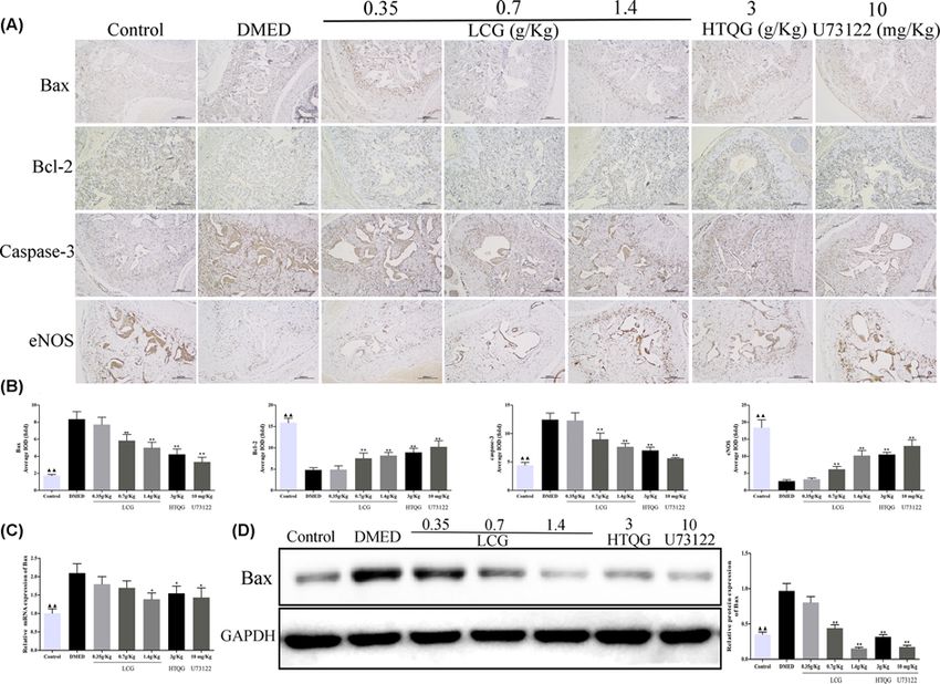
Bioscience Reports (2020) 40 BSR20193845
                                                                                                                                         https://doi.org/10.1042/BSR20193845

           fully enucleated. Obviously, it is believed that a high glucose environment caused damage to the central nervous
           system plays a crucial role in the development of DMED [7]. In brief, hypogonadism, advanced glycation end product
           (AGE) formation, vascular endothelial dysfunction, and decreased production of nitric oxide (NO) [8] are considered
           the main functional impairments in cavernoursal tissues. Cavernous endothelium involved in the cavernosal smooth
           muscle cell relaxation, which is a basic physiological phenomena contributes to the initiation and maintenance of
           erectile function [9]. Moreover, endothelial dysfunction and fibrosis were identified as a common pathogenic factor
           under high glucose levels in DMED [9–11]. Unfortunately, the exact mechanism of this effect of DMED still remains
           a mystery.
              Protein kinase C (PKC) is serine/threonine kinase that belongs to a protein family and consisted of three subfami-
           lies: conventional PKCs, novel PKCs, and atypical PKCs. Several studies reported that PKC activation can strengthen
           the NADPH-oxidases (NOX) activity, and further promote reactive oxygen species (ROS) production [12]. Stud-
           ies have demonstrated that PKCδ, one of the novel PKCs activated by diacylglycerol or 12-O-tetradecanoylphorbol

                                                                                                                                                                                           Downloaded from https://portlandpress.com/bioscirep/article-pdf/40/2/BSR20193845/867248/bsr-2019-3845.pdf by guest on 15 April 2020
           13-acetate under oxidative stress, whose activation is associated with various biological events, including endothe-
           lial cells dysfunction, cell proliferation, and apoptosis, and vascular smooth muscle cells contractility [13]. As the
           study of Durpes et al. [14] demonstrated that PKC activation promotes endothelial cells dysfunction by inhibiting
           IL-18/IL-18BP pathway for exacerbated the progress of atherosclerotic in diabetes. Additionally, over-generation of
           ROS can lead to diminished NO bioavailability [15]. NO is the primary neurotransmitter of corporal smooth muscle
           relaxation, which plays a significant role in penile erection [2]. A unifying opinion suggests that the initiation and de-
           velopment of DMED may be involved in impaired NO synthesis [16]. Besides, NO is synthesized by endothelial NOS
           (eNOS), inducible NOS (iNOS), and nNOS. Additionally, the iNOS contributes to the synthesis NO incessantly de-
           pend on Ca2+ [17]. The Ca2+ concentration was detected in the blood by using the calcium-sensing receptor (CASR),
           which is a class C G-protein coupled receptor (GPCR), and typified by 1078-amino acid as a disulphide-linked
           homodimer [18]. Recent evidence indicates that CASR combined with multiple G-protein subtypes for activating
           predominantly signals. For instance, Conigrave [19] and Brennan et al. [20] reported that the CaSR coupled to the
           Gq/11-phospholipase C (PLC)-mediated pathway, to generate inositol 1,4,5-trisphosphate (IP3) and diacylglycerol
           (DAG) for activating PKC and Ca2+ mobilization as well as mitogen-activated protein kinase (MAPK) cascades in-
           cluding JNK and p38. Further, the JNK and p38 also mediated apoptosis that accompanied by increased Bax levels in
           the vascular endothelium cells [21]. Therefore, the CaSR/PLC/PKC signaling pathway may play a crucial role in the
           erectile dysfunction associated with streptozotocin-induced diabetic rats.
              Currently, phosphodiesterase type 5 (PDE5) inhibitors that help augment the NO/cyclic guanosine monophosphate
           (cGMP) pathway are used as the first-line therapeutic strategy for ED. However, studies have reported that these
           drugs are not effective in some diabetic male patients with severe ED [22] and result in several side effects, including
           facial redness, headache, and gastrointestinal reactions [4]. Thus, exploring novel therapeutic strategies and targets
           for DMED is still urgent. ED is classified as “Yangshuo” syndrome in traditional Chinese medicine (TCM), and TCM
           scholars believe that the “qi stagnation and blood stasis” is the main pathological features of DMED. Previously, we
           reported that the “Huoxue Tongluo Qiwei soup” granules (HTQGs) was achieved a significant therapeutic efficiency
           of 76% in clinical trials [23], and composed of Leech (10 g), Centipede (5 g), Radix Paeoniae Rubra (20 g), Vaccariae
           Semen (10 g), Angelicae Sinensis Radix (15 g), Radix Bupleuri (15 g), Cyathulae Radix (15 g), Tribulifructus (20
           g), Citri Reticulatae Pericarpium Viride (10 g), Curcumae Radix (10 g), Morindae Officinalis Radix (15 g), and
           Epimrdii Herba (10 g). Among them, the couplet medicines of Leech and Centipede is the key medicines for the
           purpose of promoting blood circulation and removing blood stasis in TCM, and also the core herbs in HTQs.
              Given the beneficial effects of couplet medicines of Leech and Centipede in the treatment of DMED and rare studies
           correlating the CaSR/PLC/PKC signaling axis with DMED, the present study aims to establish DMED rat models to
           investigate the effect of Leech and Centipede Granules (LCG) and the possible underlying mechanism of DMED in
           penis tissue, which might be a beneficial treatment strategy of DMED in human.

           Materials and methods
           Experimental animals
           Fifty-six 12-week-old male Sprague-Dawley (SD) rats (weight, 260–280 g) were purchased from Shanghai SLAC Lab-
           oratory Animal Co.,Ltd (Shanghai, China). All rats were raised in the Animal Center of Zhejiang Chinese medical
           university (Zhejiang, China) with a 12/12 light-dark cycle at 24◦ C +  ◦
                                                                               − 1 C, water and food available ad libitum. The
           current experimental protocols were approved by the Animal Care and Use Committee of Zhejiang Chinese medical
           university (Zhejiang, China). The mating test was conducted and showed that all rats possessed the normal erec-
           tile function. Diabetes was induced by sustaining a high-fat diet (HFD) feeding routine for a month. Then, after

2   © 2020 The Author(s). This is an open access article published by Portland Press Limited on behalf of the Biochemical Society and distributed under the Creative Commons Attribution
    License 4.0 (CC BY).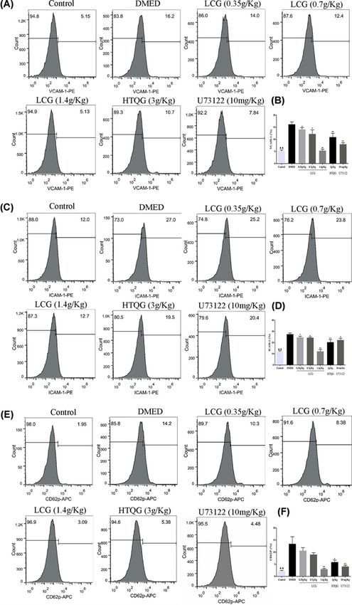
Bioscience Reports (2020) 40 BSR20193845
https://doi.org/10.1042/BSR20193845

an overnight fast, 50 SD rats were injected with a single intraperitoneal injection of 60 mg/kg streptozotocin (STZ,
Sigma-Aldrich Chemical Co, St. Louis, MO, U.S.A.). Six age-matched rats only got an intraperitoneal injection of 0.1
mol/l citrate–phosphate buffer (pH 4.5) and selected as a control group. Rats with a constant non-fasted blood glucose
concentration ≥16.7 mmol/l were considered diabetic after 72 h. The diabetic rats were fed for 10 weeks to develop
ED. Next, apomorphine (APO)-induced erection test was performed to evaluate the erectile function. The rats were
moved to a quiet and dimly laboratory to adapt to the environment for 15 min in a transparent observation kit. Then,
the rat soft skin of the neck region was injected with a one-off injection with 100 μg/kg of APO (Shenyang, Liaoning,
China). The status and frequency of penile erection in rats were observed for 30 min, and the penis was enlarged, the
prepuce was receded or the glans was exposed represented one erection [24]. Rats with abnormal erectile function
were defined as having DMED.
   Finally, 36 DMED rats were identified for the subsequent experiments. The DMED rats were divided randomly into
six treatment groups (n = 6): the DMED model group, low-dose group, middle-dose group, high-dose group, HTQG

                                                                                                                                                                                           Downloaded from https://portlandpress.com/bioscirep/article-pdf/40/2/BSR20193845/867248/bsr-2019-3845.pdf by guest on 15 April 2020
group, and the phospholipase C (PLC) inhibitor U73122 group. The low-, middle- and high-dose group rats have
received a daily gavage of LCG (Huisong Pharmaceuticals Co., Ltd, Zhejiang, China) at a dose of 0.35 g/kg, 0.7 g/kg,
and 1.4 g/kg for 4 weeks, respectively. Besides, the HTQG group rats were administered daily with the prescription
of “Huoxue Tongluo Qiwei soup” granules at a dose of 3 g/kg. For the U73122 group rats, there was 10 mg/kg PLC
inhibitor was injected in the tail vein of rats every one day for 4 weeks. The control and DMED model group received
physiological saline only. At the end of the study, all rats were fasted for 10 h, then the tail vein blood glucose levels,
body weights and erectile function of all rats were measured. After that, all rats were anesthetized with pentobarbital
sodium (50 mg/kg, i.p. Sigma), then blood sample was collected from the abdominal aorta and centrifuged at 3000
rpm/min for 15 min to acquire the sera. Subsequently, the rats were killed by decapitation, then the penile tissues
were harvested stored at −80◦ C until further analysis.

Erectile function assessment
Four weeks after treatment, rats in each group were placed in a transparent observation cage with dimmed lights and
a quiet environment that allowed observation. Then, the rats were injected with 100 μg/kg APO in the neck. The
number of penile erections in rats was observed and recorded for 30 min.

Measurement of insulin, glucagon, lectin-like oxidized low-density
lipoprotein receptor-1, NADPH oxidase, NO, and endogenous ET-1
Levels of insulin, glucagon, lectin-like oxidized low-density lipoprotein receptor-1 (LOX-1), and NADPH oxidase
(NOX) in rats sera were measured using commercial insulin, glucagon, LOX-1, and NOX ELISA Kit, respectively
(Solarbio, Beijing, China). Furthermore, NO and ET-1 concentrations in penile tissues were estimated with a total
NO assay kit (Beyotime, China) and ET-1 Elisa kit (Jianglai Biotechnology Co., Ltd., Shanghai, China) according to
the manufacturer’s procedure.

Flow cytometry
Blood samples were collected from all rats in each group by clean arteriopuncture into 2 ml vacutainer tubes under
strict sterile conditions. The samples were centrifuged a 1000 rpm for 20 min to obtain platelet-rich plasma (PRP).
Then, the PRP was centrifuged at 12000 rpm for 5 min to prepare platelet-poor plasma (PPP). Phenylethylamine (PE)
anti-Rat CD31-PECy7 (eBioscience, U.S.A.) and allophycocyanin (APC) labeled anti-mouse/rat CD42b (Biolegend,
Germany) were used to detect endothelial microparticles (EMPs). About 50 μl PPP were incubated with 8 ml of each
labeled antibodies for 30 min at 4◦ C in the dark. The stained samples were resuspended in 0.2 ml of PBS and then
kept for flow cytometry analysis.
   For flow cytometric detection of vascular cell adhesion molecule (VCAM)-1, intracellular adhesion molecule
(ICAM)-1, and selectin P (CD62P) in cavernous tissue. The cavernous tissue was digested to prepare 1 × 10⁶ /ml
single-cell suspension. Cells were incubated for 30 min in PBS containing anti-rat CD106-PE (VCAM-1), anti-rat
CD54-PE (ICAM-1), and anti-mouse/rat CD62P-APC (P-selectin). The stained cells were then detected by the MAC-
SQUANT Q10 cytometer (Miltenyi Biotec) and analyzed using FlowJo v7.6.5 software (Tree Star).

Hematoxylin–eosin (HE) and immunohistochemical staining
Freshly dissected central parts of penile tissues were fixed in 4% paraformaldehyde. The sample tissues were then
dehydrated by an ethanol gradient and embedded in paraffin. About 4 μm thick sections were prepared for HE stain-
ing as well as immunochemistry (IHC) according to standard protocols for histological examinations. Sections were

© 2020 The Author(s). This is an open access article published by Portland Press Limited on behalf of the Biochemical Society and distributed under the Creative Commons Attribution   3
License 4.0 (CC BY).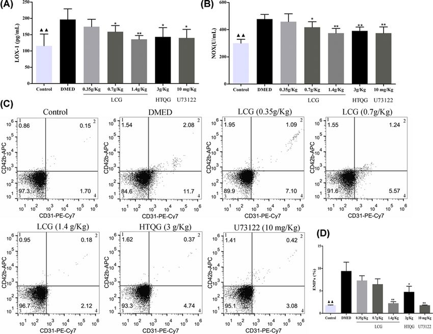
Bioscience Reports (2020) 40 BSR20193845
                                                                                                                                                https://doi.org/10.1042/BSR20193845

Table 1 The primer sequences for qRT-PCR
 Gene                                                      Forward Primer (5 -3 )                                       Reverse Primer (5 -3 )

 JNK                                                       GCCGGAGGTGATTTTGGGTA                                           GTAACGGGGCGATAACGGAT
 P38                                                       CGGTGTGTGCTGCTTTTGAT                                           CAGACGCAACTCTCGGTAGG
 BAX                                                       GAGCTGCAGAGGATGATTGCT                                          TGATCAGCTCGGGCACTTTA
 CASR                                                      CACCCAAGTGGAGAAAACGC                                           CTGAACTATTGGCAACGCCG
 PLC                                                       CCCACCACCCAGGAAATTGT                                           GGTTTTGCTTGCTCAGGTGG
 PKCδ                                                      GAAAGCCACACTGAATCCCG                                           ACTTGTACCATCCATCCACAGG
 GADPH                                                     GCGGGAGCGGATCCTAATA                                            TGGTGCATCCATGGGCTAC

 JNK: c-Jun N-terminal kinase JNK; MAPK14 (Also known as P38): mitogen-activated protein kinase 14; BAX: BCL2-associated X protein; CASR:
 Calcium-sensing receptor; PLC: phospholipase C; PKCδ: Protein kinase C δ; GADPH: glyceraldehyde-3-phosphate dehydrogenase; qRT-PCR, quan-

                                                                                                                                                                                                  Downloaded from https://portlandpress.com/bioscirep/article-pdf/40/2/BSR20193845/867248/bsr-2019-3845.pdf by guest on 15 April 2020
 titative real-time polymerase chain reaction.

                  cut at 4 μm and incubated at room temperature for 4 h with primary antibodies against Bax (1:250, Abcam), Bcl-2
                  (1:500, Abcam), Caspase-3 (1:500, Abcam) and eNOS (1:100, Abcam). Next, the sections were washed and incu-
                  bated with appropriate secondary antibodies for a 15 min incubation. Afterward, the sections were incubated with
                  3,3-diaminobenzidine (DAB), and the cell nuclei were stained with hematoxylin. Finally, sections were examined
                  under a light microscope. The semi-quantitative analysis of the expression of Bax, Bcl-2, Caspase-3, and eNOS in
                  penile tissue in the images was measured by using Image-Pro Plus version 6.0 software (Media Cybernetics Inc., MD,
                  U.S.A.).

                  Real-time PCR
                  The total RNA from rat penile tissues was extracted using Trizol reagent (Life Technologies, Grand Island, NY, U.S.A.)
                  according to the manufacturer’s protocol. The cDNA was synthesized utilizing the reverse transcription kit (Fer-
                  mentas, Waltham, MA, U.S.A.). qRT-PCR was performed using a LightCycler® 96 Real-Time PCR System (Roche,
                  Switzerland) with the SYBR Green PCR kit (Takara, Japan). The program was comprised of denaturation at 95◦ C for
                  10 min, trailed by 40 cycles of liquefying at 95◦ C for 15 s and prolongation at 60◦ C for the 60 s. Primer sequences are
                  shown in Table 1. The level of GAPDH mRNA expression was measured as an endogenous reference and used for
                  normalization. The relative quantification of the expression levels of the target genes was calculated using the 2−Ct
                  method.

                  Western blotting
                  Total protein samples were extracted from penile tissues. The penile tissues were homogenized by precooled tissue
                  lysates and centrifuged at 12,000 rpm for 15 min at 4◦ C, and the supernatants were obtained. Protein concentra-
                  tions were determined using a BCA protein assay kit (Solarbio, Beijing, China). Protein lysate of equal concentration
                  was loaded on 10% sodium dodecyl sulfate/polyacrylamide gel electrophoresis (SDS-PAGE) at 80 V for 30 min and
                  120 V for 60 min, and then electrotransferred to a polyvinylidene difluoride membrane (Bio-Rad, CA, U.S.A.). The
                  membrane was blocked for 1 h at room temperature with 5% skimmed milk powder. Then, membranes were in-
                  cubated with primary antibodies against CaSR (1:1000; ab18200, Abcam), PLC (1:5000; ab76155, Abcam), PKCδ
                  (1:5000; ab182126, Abcam), JNK (1:2000; ab208035, Abcam), p38 (1:1000, ab170099, Abcam), Bax (1:2000, ab32503,
                  Abcam), GAPDH (1:5000, 60004-1-1g, HuaBio, Hangzhou, China) at 4◦ C overnight, followed the membranes were
                  washed and incubated with the appropriate horseradish peroxidase-conjugated secondary antibodies at room tem-
                  perature for 1 h. Finally, the protein bands were visualized using an enhanced chemiluminescence detection system
                  (Clinx Science Instruments, U.S.A.).

                  Statistical analysis
                  All data are expressed as mean +   − standard deviation. Groups were compared using Student’s t-test or one-way
                  ANOVA. All data were analyzed using SPSS 22.0 statistical software (SPSS Inc., Chicago, IL, U.S.A.) and P < 0.05
                  was considered to be statistically significant.

                  Results
                  LCG improves the erectile functioning of rats with DMED
                  Fasting blood glucose level and body weight changes are shown in Figure 1. After diabetes induction, the fasting blood

4          © 2020 The Author(s). This is an open access article published by Portland Press Limited on behalf of the Biochemical Society and distributed under the Creative Commons Attribution
           License 4.0 (CC BY).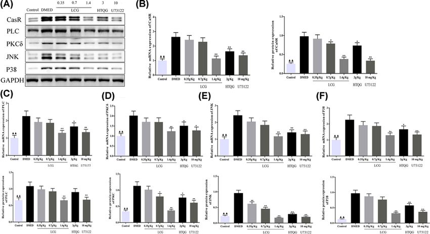
Bioscience Reports (2020) 40 BSR20193845
https://doi.org/10.1042/BSR20193845

                                                                                                                                                                                           Downloaded from https://portlandpress.com/bioscirep/article-pdf/40/2/BSR20193845/867248/bsr-2019-3845.pdf by guest on 15 April 2020
Figure 1. Comparisons of fasting blood glucose, body weight, insulin, glucagon, and the times of erection in rats with DMED
(A) The fasting blood glucose level is decreased in LCG and HTQG groups. (B) The body weight is increased in LCG and HTQG
groups. (C) The insulin levels of rats are expressed at a high level with LCG treatment. (D) The glucagon level is expressed at a low
level in treated groups, compared with the DMED group. (E) APO experiments are performed to evaluate erectile function in each
group. Data are expressed as mean + − standard deviation from N = 6 per group.
                                                                               䉱䉱 P < 0.01 the Control group versus the DMED

group; *P < 0.05, **P < 0.01 the LCG, HTQG, and U73122 groups versus the DMED group.

glucose level was found to be significantly elevated of rats in the DMED group (P < 0.01, Figure 1A) but significantly
lower body weights (P < 0.01, Figure 1B). LCG treatment improves these differences. In comparison with the DMED
group, the insulin levels of in rats in the LCG groups was found to dramatically increased after LCG administration
(P < 0.01, Figure 1C), whereas the glucagon levels of rats in LCG, HTQG, and U73122 groups were decreased (P <
0.01, Figure 1D). The result of APO-induced erection testing is shown in Figure 1E. The number of erection for 30
min of rats in LCG and HTQG groups was found to be significantly increased with a dosage dependence. Compared
with the DMED group, there were no significant differences in the fasting blood glucose level, body weight, insulin
levels, and the times of erection in the U73122 group. These findings suggested that the LCG has a defensive effect
on ED in the diabetic penis.

LCG inhibits endothelial cells injury and ameliorates vascular endothelial
relaxation of rats with DMED
Some reports indicate that the LOX-1 and NOX are the important marker evaluation of vascular endothelial cell
injury. In the present study, results of LOX-1 (Figure 2A) and NOX (Figure 2B) detection displayed that compared
with the control group, the LOX-1 and NOX concentrations were markedly elevated in the DMED group (all P <
0.01), while it was partly reduced in the middle-dose group, high-dose group, HTQG group, and the U73122 group.
Besides, there were no significant differences in the levels of the low-dose group compared with the DMED group.
EMPs, the cell plasma fragments, directly reflecting the physiological process of the endothelial cell, including cell
activation, proliferation, and apoptosis [25]. In the present study, four quadrants histogram was used to differentiate
EMPs, being negative for CD42b and positive for CD31. The flow cytometry analyses showed that the EMPs counts
were significantly decreased in the 1.4 g/kg LCG, HTQG and U73222 groups compared with the DMED group (Figure
2C,D). Also, NO concentration in penile tissues of the LCG-treated group was significantly elevated compared with
the DMED group (Figure 3A). Furthermore, the ET-1 concentration of penile tissues in treated rats was significantly
lower than that in DMED group (P < 0.05; P < 0.01, Figure 3B). Taken together, the results show that LCG could
inhibit the pathological process of endothelial cell impairments and ameliorate vascular endothelial relaxation of
penile tissue in DMED.

LCG reduced vascular fibrosis markers expression in the sera of DMED
rats
Previous studies have revealed that proposed biomarkers, VCAM-1, ICAM-1, and CD62P with higher concentra-
tions are independently associated with fibrosis [26,27]. To investigate the role of LCG on the vascular fibrosis in

© 2020 The Author(s). This is an open access article published by Portland Press Limited on behalf of the Biochemical Society and distributed under the Creative Commons Attribution   5
License 4.0 (CC BY).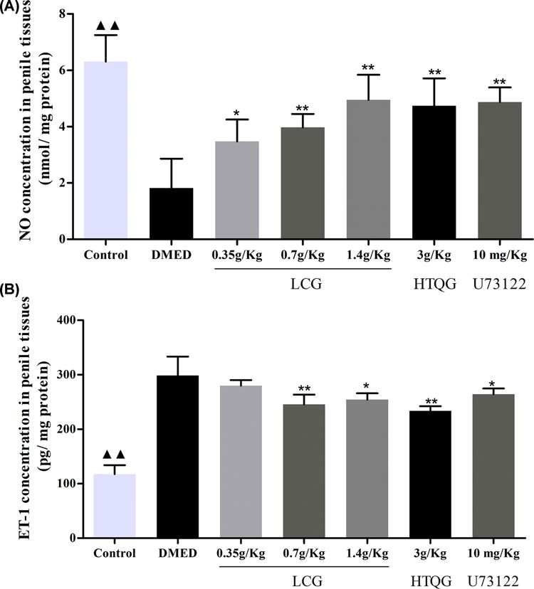
Bioscience Reports (2020) 40 BSR20193845
                                                                                                                                         https://doi.org/10.1042/BSR20193845

                                                                                                                                                                                           Downloaded from https://portlandpress.com/bioscirep/article-pdf/40/2/BSR20193845/867248/bsr-2019-3845.pdf by guest on 15 April 2020
           Figure 2. Effect of LCG on endothelial cells injury of rats with DMED
           (A,B) Evaluation of LOX-1 and NOX concentration in DMED rats sera by Elisa. (C), four-quadrant histogram to detect markers with
           negative for CD42b and positive for CD31 in quadrant 4. (D) Quantification of CD42b- CD31+ EMPs by flow cytometry in DMED
           rats sera. Measurement data were expressed as mean +− SD,
                                                                       䉱䉱 P < 0.01 the Control group versis the DMED group; *P < 0.05, **P

           < 0.01 the LCG, HTQG, and U73122 groups versus the DMED group.

           DMED rats, the VCAM-1, ICAM-1, and CD62P levels in serum samples of rats were assayed with flow cytometry. As
           shown, compared with the control group, the count of VCAM-1 (Figure 4A,B), ICAM-1 (Figure 4C,D), and CD62P
           (Figure 4E,F) were increased in the DMED group (P < 0.01). Intriguingly, flow cytometry analysis showed that the
           serum of rats in the LCG, HTQG, and U73122 group notably decreased levels of VCAM-1, ICAM-1, and CD62P
           in a dose-dependent manner compared with DMED rats group. These results suggest that LCG and HTQG were
           determined to have exerted a greater effect on the amelioration of vascular fibrosis in DMED rats.

           LCG alleviates the pathology of cavernous bodies
           The present study assessed the effects of LCG and HTQG on the pathology of cavernous bodies. A large number of the
           cavernous sinus and endothelial cells were observed in the corpus cavernosum tissues of rats of control, LCG, HTQG,
           and U73122 groups but not in the DMED group (Figure 5). In comparison with the DMED group, the number of the
           cavernous sinus and endothelial cells in the cavernosum tissues of the treated group was observed to be significantly
           improved, while the rats of DMED group with obviously decreased numbers. In the concern of the above results,
           we can demonstrate that LCG and HTQG can improve the pathological changes of cavernous bodies of the penis by
           depressing the endothelial cell lesion.

6   © 2020 The Author(s). This is an open access article published by Portland Press Limited on behalf of the Biochemical Society and distributed under the Creative Commons Attribution
    License 4.0 (CC BY).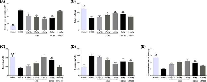
Bioscience Reports (2020) 40 BSR20193845
https://doi.org/10.1042/BSR20193845

                                                                                                                                                                                           Downloaded from https://portlandpress.com/bioscirep/article-pdf/40/2/BSR20193845/867248/bsr-2019-3845.pdf by guest on 15 April 2020
Figure 3. Effect of LCG on vascular endothelial relaxation of penile tissue in rats with DMED
(A) The NO concentration and (B) ET-1 of penile tissue measured by Elisa, and represented by mean +
                                                                                                  − standard deviation; the
one-way ANOVA was performed to analyze data in each group. 䉱䉱 P < 0.01 the Control group versus the DMED group; *P < 0.05,
**P < 0.01 the LCG, HTQG, and U73122 groups versus the DMED group.

Immunohistochemical analysis of Bax, Bcl-2, Caspase-3, and eNOS
expression
The protein expression levels of Bax, Bcl-2, Caspase-3, and eNOS in rat penile tissue endothelial cells were estimated
by immunohistochemistry. As shown in Figure 6A,B. Immunohistochemical analysis revealed that the protein ex-
pression of Bax and Caspase-3 were significantly higher in the rats from the DMED group compared with those from
the control group (P < 0.01). However, the protein expression of Bcl-2 and eNOS was significantly lower than that in
control rats. After treatment, the protein expression of Bax and Caspase-3 was significantly decreased in the treated
group rats than that in the DMED group, and significantly increased the protein levels of Bcl-2 and eNOS in the
treated groups with dose-dependence. Of note, the protein expression levels of Bax, Bcl-2, Caspase-3, and eNOS in
the low-dose LCG group did not present with a statistical difference. Additionally, the results of RT-qPCR and West-
ern blot analysis showed that the mRNA and protein expression of Bax was significantly decreased in the high-dose
group, HTQG, and U73122 group (Figure 6C,D).

© 2020 The Author(s). This is an open access article published by Portland Press Limited on behalf of the Biochemical Society and distributed under the Creative Commons Attribution   7
License 4.0 (CC BY).
Bioscience Reports (2020) 40 BSR20193845
https://doi.org/10.1042/BSR20193845
Downloaded from https://portlandpress.com/bioscirep/article-pdf/40/2/BSR20193845/867248/bsr-2019-3845.pdf by guest on 15 April 2020
Figure 4. Effect of LCG on vascular fibrosis in rats with DMED
(A,C,E) Flow cytometry measured the VCAM-1, ICAM-1, and CD62P level after LCG treatment for 4 weeks. (B,D,F) Bar graph
demonstrated comparison of VCAM-1, ICAM-1, and CD62P concentration. Data presented as mean + − SD; the comparison of
multiple groups was performed by ANOVA; 䉱䉱 P < 0.01 the Control group versus the DMED group; *P < 0.05, **P < 0.01 the LCG,
HTQG, and U73122 groups versus the DMED group.
8 © 2020 The Author(s). This is an open access article published by Portland Press Limited on behalf of the Biochemical Society and distributed under the Creative Commons Attribution
License 4.0 (CC BY).Bioscience Reports (2020) 40 BSR20193845
https://doi.org/10.1042/BSR20193845
Downloaded from https://portlandpress.com/bioscirep/article-pdf/40/2/BSR20193845/867248/bsr-2019-3845.pdf by guest on 15 April 2020
Figure 5. Effect of LCG on the pathological progression of DMED
Pathological changes of cavernous bodies of rats treated with LCG at the dosage of 0.35 g/kg, 0.7 g/kg, and 1.4 g/kg were observed
by hematoxylin and eosin staining (200× and 400×).
LCG inhibited the CaSR/PLC/PKC signaling pathway and the expression
of JNK, P38, and Bax are decreased in DMED
To investigate the expressions of CaSR, PLC, PKCδ, JNK, and P3 in DMED rats penile tissue obtained with LCG
treatment, we also performed RT-qPCR and Western blot analysis. In comparison with the control group, the mRNA
expression of CaSR, PLC, PKCδ, JNK, and P38 was significantly increased, yet significantly decreased, in the middle-
or high-dose group, HTQG, and U73122 group (Figure 7B–F). While no obvious change was found in the low-dose
LCG group (P > 0.05). Furthermore, the results of Western blot analysis suggesting that the protein expression of
CaSR, PLC, PKCδ, JNK, and P38 were significantly elevated in the DMED group (Figure 7A–F). Simultaneously, the
gray values of CaSR, PLC, PKCδ, JNK, and P38 expression were significantly diminished in the middle- or high-dose
group, HTQG, and U73122 group but not in the low-dose LCG group, which are consistent with the result of mRNA
expression in the present study.
Discussion
In recent years, diabetic patients are also suffering from ED with increased susceptibility, affecting seriously male
health and psychology. Although efforts have been made to investigate novel therapeutic avenues for DMED, includ-
ing gene therapy, transplantation of stem cell, anti-PDF5 therapy and low energy shock wave therapy [28]. However,
the strategy contributes to ameliorate DM as well as ED simultaneously is still under investigation and the mechanism
of DMED is largely unknown. TCM believes that the etiology and pathogenesis of ED are associated with qi and blood
block. Modern research shows that Chinese medicine can promote blood circulation and improve the blood supply
to aid in the cavernous congestion [29]. The Leech is a blood-sucking annelid worms, which was first recorded in
Shen Nong’s Herbal Classic. It has effects on promoting blood circulation, alleviating blood coagulation, activating
meridians and relieving stasis by using alone or with other medicines with the dried whole body that used as a tra-
ditional TCM [30]. Chinese red-headed centipede, predatory terrestrial arthropods, belongs to the class Chilopoda
and is characterized by a head and an externally segmented body with a pair of articulated legs for each segment that
© 2020 The Author(s). This is an open access article published by Portland Press Limited on behalf of the Biochemical Society and distributed under the Creative Commons Attribution 9
License 4.0 (CC BY).Bioscience Reports (2020) 40 BSR20193845
https://doi.org/10.1042/BSR20193845
Downloaded from https://portlandpress.com/bioscirep/article-pdf/40/2/BSR20193845/867248/bsr-2019-3845.pdf by guest on 15 April 2020
Figure 6. Evaluation of Bax, Bcl-2, Caspase-3, and eNOS protein expression by immunohistochemistry
(A) Representative results of IHC staining for Bax, Bcl-2, Caspase-3, and eNOS in penile tissue (original magnification, 100×). (B)
Representative statistical analysis of Bax, Bcl-2, Caspase-3, and eNOS in the penile tissue among the groups. (C) RT-qPCR results
show that Bax was expressed at a high level in DMED rats, while decreased by LCG, HTQG, and U73122 treatment. (D) Protein
bands of Bax, and GAPDH, and representative statistical analysis of Bax. Each bar depicts the mean values (mean + − s.d.);
䉱䉱 P <
0.01 the Control group versus the DMED group; **P < 0.01 the LCG, HTQG, and U73122 groups versus the DMED group.
feeds with reptiles, bats, rats, and so on [31]. Centipede, an ancient antipyretic medicine used for centuries in tradi-
tional Chinese with the dried whole body that is processed using a special technique called PaoZhi, which is salted in
flavor, warm in nature and attributives to the liver, spleen and lung meridians according to the Pharmacopoeia of the
People’s Republic of China. It also can improve blood microcirculation and relieve blood stasis with calm the wind
and activating collaterals, attacking toxin and regulating Qi [32].
In the present study, we attempted to demonstrate the therapeutic effect and the molecular mechanism associated
with Leech and Centipede Granules in the penis tissues of DMED rats. The DMED rats generally underwent a higher
fasting blood glucose, severe body weight and insulin loss, and glucagon increase. Initially, the present study revealed
that compared with DMED rats, rats with LCG administration significantly improved the aforementioned serious
symptoms. Additionally, the numbers of erection measurements were utilized for the evaluation of erectile function
preliminarily. We found that LCG as a therapeutic agent for DMED can significantly aid in the erectile function
restoration. Moreover, the results of our study revealed that impairment of the vascular endothelial of the penis was
noted to be alleviated with the inhibition of the LOX-1, NOX, and EMP.
As is known, oxidative stress is well recognized as a major pathogenic factor in the pathogenesis of DMED, which
characterized by overproduction of ROS [33] and induces endothelial cell injury with the reduction of NO [34]. Nu-
merous reports have indicated that NO plays a crucial role in the induction and maintenance of normal vascular
physiology, is released from noncholinergic nerves and endothelium [16,35], and has been clarified that its bioavail-
ability is decreased in the penis of human DMED patients [10,36]. The erectile response and maintain erection relies
10 © 2020 The Author(s). This is an open access article published by Portland Press Limited on behalf of the Biochemical Society and distributed under the Creative Commons Attribution
License 4.0 (CC BY).Bioscience Reports (2020) 40 BSR20193845
https://doi.org/10.1042/BSR20193845
Downloaded from https://portlandpress.com/bioscirep/article-pdf/40/2/BSR20193845/867248/bsr-2019-3845.pdf by guest on 15 April 2020
Figure 7. Expressions of CaSR, PLC, PKC, JNK, and P38 detected by RT-qPCR and Western blot analysis
(A) Protein bands of CaSR, PLC, PKC, JNK, P38, and GAPDH. (B–F) RT-qPCR and Western blot analysis of CaSR, PLC, PKC,
JNK, and P38 were expressed at a high level in DMED rats, while reversed by LCG, HTQG, and U73122 treatment, respectively.
Statistical data are presented as mean value +
− SD.
䉱䉱 P < 0.01 the Control group versus the DMED group; **P < 0.01 the LCG,
HTQG, and U73122 groups versus the DMED group.
on the production of NO via nNOS initiates and eNOS, respectively. Further, eNOS is the key enzyme for NO syn-
thesis, which is expressed in the vascular endothelium widely. Moreover, NO plays an important role in ameliorat-
ing tissue fibrosis. In addition, Ferrini et al. found that iNOS inactivation caused exacerbated penile fibrosis in DM
mice by increasing oxidative stress under hyperglycemic conditions [37]. Besides, ET-1, a potent vasoconstrictor, and
proinflammatory peptide is the predominant isoform expressed in the vasculature, which released mostly by vascular
endothelial cells and is another major regulator of vascular tone [38]. A previous report of Mao et al. has highlighted
a correlation for the imbalance between ET-1 and NO systems in the pathogenesis of ED under cardiac hypertro-
phy [39]. Consistent with the reported previously, our current study showed that rats in DMED group elucidated by
decreased NO and eNOS levels, and increased the potent vasoconstrictors, ET-1 content in penile tissues, as well as
excessive concentration of early vascular fibrosis biomarkers such as VCAM-1, ICAM-1, and CD62P in the corpus
cavernosum. In contrast, a significant increase of NO and eNOS, and markedly decrease of ET-1, VCAM-1, ICAM-1,
and CD62P were observed in the LCG and U73122 treatment groups. In the present study, histological changes also
observed by staining results reflected the protective effect of LCG on penile tissue endothelial cells. Taken together,
these findings elucidated that the therapeutic impact of LCG on DMED in the diabetic penis may be due to improve
vascular endothelial injury, the ability of vasodilation and decrease the penile fibrosis.
Evidence has been presented verifying that DM also has a tight correlation with the apoptosis of various cell types,
including the cavernous smooth muscle cells [40], corpus cavernosal endothelial cell [10]. Apoptosis is a basic pro-
grammed cell death process to maintain the dynamic balance of the cell environment. The Bcl-2 family consists of
the anti-apoptotic protein Bcl-2 and the pro-apoptotic protein Bax. Bcl-2 is a famous representative suppressor of
apoptosis, while Bax can combine with Bcl-2 to form a heterodimer for antagonize the inhibitory effect of Bcl-2 on
apoptosis and promote apoptosis [41]. On the other hand, Bax also activates caspase-3 with promoting the Ca2+ re-
lease and then causes cell apoptosis [42]. Caspase-3 is as a representative executor plays a pivotal role in cell apoptosis.
Therefore, the Bax, Bcl-2, and caspase-3 levels are considered to be an indicator for evaluation of apoptosis. In our
study, we also found that compared with that in the control group, the protein expression levels of BAX and caspase-3
were up-regulated and that Bcl-2 was down-regulated in the DMED group via immunohistochemical analysis, which
agrees with previous studies [10,11]. Encouragingly, the protein level of Bcl-2 in the penile tissue of the LCG group
© 2020 The Author(s). This is an open access article published by Portland Press Limited on behalf of the Biochemical Society and distributed under the Creative Commons Attribution 11
License 4.0 (CC BY).Bioscience Reports (2020) 40 BSR20193845
https://doi.org/10.1042/BSR20193845
significantly increased, and the protein expression of ax and the Caspase-3 decreased significantly in the penile tissue,
indicating that LCG can effectively reduce the degree of apoptosis in the cavernous tissues of DMED rats.
It is established that the cytoplasmic free calcium concentration associated with various functions of vascular en-
dothelium, and has significant roles in regulating vascular tone and the generation of NO [43]. The CaSR, a PLC
sensitive G protein-coupled receptor, can increase the release of [Ca2+ ]i from the endoplasmic reticulum via the inos-
itol triphosphate receptor (IP3R). Notably, Liu et al. demonstrated that CaSR activated PKCδ to induce cardiomyocyte
apoptosis during ischemia/reperfusion (I/R) injury [44]. However, the associations between CaSR, PLC, PKCδ, and
DMED are not clear. Our qPCR and Western blot results suggest that LCG might attain its effects via inhibition of
the CaSR/PLC/PKC pathway, as well as the mRNA and protein expression of JNK, P38, and BAX. Consistent with the
results of LCG, the present study showed the U73122, a PLC inhibitor helps to eliminate the deterioration of ED in the
present study. Thus, we concluded that LCG may enhance the NO level to ameliorate erectile function via decreasing
cell apoptosis by inactivation of the CaSR/PLC/PKC pathway. There are many defects of our study, such as the only
Downloaded from https://portlandpress.com/bioscirep/article-pdf/40/2/BSR20193845/867248/bsr-2019-3845.pdf by guest on 15 April 2020
Type 2 diabetic ED rats were investigated, and the Ca2+ concentration should be measured in penile tissue. Besides,
further the gain-of-function and loss-of-function cell assays are needed. Therefore, we will also reconfirm the cellular
effects and accurate mechanism of LCG in our future studies.
In the present study, we demonstrated that the couplet medicines of Leech and Centipede Granules ameliorate
erectile function in DMED rats, and the inactivation of CaSR/PLC/PKC signaling pathway by the couplet medicines
of Leech and Centipede Granules were responsible for the reduction of endothelial cells apoptotic death.
Competing Interests
The authors declare that there are no competing interests associated with the manuscript.
Funding
This work was supported by grants from the National Natural Sciences Foundation of China [grant number 81804092].
Author Contribution
J.X.M., B.W., C.F.D., H.S.L., and W.Q.C. participated in the design of the study and performed the statistical analyses. J.X.M.
and W.Q.C. conceived the study, participated in its design, helped draft the manuscript and gave final approval of the version to
be published. X.J.J. and C.Y.W. participated in PCR, Western blot analysis and drafted the manuscript. JY conducted the ELISA
analysis. All authors read and approved the final manuscript.
Abbreviations
AGE, advanced glycation end; APC, allophycocyanin; APO, apomorphine; CD62P, selectin P; cGMP, cyclic guanosine
monophosphate; DAB, diaminobenzidine; DAG, diacylglycerol; DM, diabetes mellitus; DMED, diabetes mellitus-induced erec-
tile dysfunction; ED, erectile dysfunction; EMPs, endothelial microparticles; eNOS, endothelial NOS; GPCR, G-protein coupled
receptor; HTQGs, Huoxue Tongluo Qiwei soup granules; I/R, ischemia/reperfusion; ICAM-1, intracellular adhesion molecule-1;
IHC, immunochemistry; IP3R, inositol triphosphate receptor; LCG, Leech and Centipede Granules; LOX-1, lectin-like oxidized
low-density lipoprotein receptor-1; MAPK, mitogen-activated protein kinase; NO, nitric oxide; NOX, NADPH-oxidases; PDE5,
phosphodiesterase type 5; PE, Phenylethylamine; PKC, Protein kinase C; PLC, phospholipase C; PPP, prepare platelet-poor
plasma; PRP, platelet-rich plasma; ROS, reactive oxygen species; SD, Sprague-Dawley; SDS-PAGE, sodium dodecyl sul-
fate/polyacrylamide gel electrophoresis; TCM, traditional Chinese medicine; VCAM-1, vascular cell adhesion molecule-1.
References
1 Wen, Y., Liu, G., Zhang, Y. et al. (2019) MicroRNA-205 is associated with diabetes mellitus-induced erectile dysfunction via down-regulating the
androgen receptor. J. Cell. Mol. Med. 23, 3257–3270, https://doi.org/10.1111/jcmm.14212
2 Qiao, H., Zhang, Y., Lin, W. et al. (2018) Decreased expression of pigment epithelium-derived factor within the penile tissues contributes to erectile
dysfunction in diabetic rats. Clin. Sci. 132, 2175–2188, https://doi.org/10.1042/CS20180192
3 Shamloul, R. and Ghanem, H. (2013) Erectile dysfunction. Lancet 381, 153–165, https://doi.org/10.1016/S0140-6736(12)60520-0
4 Yu, X.D., Wang, J.S., Zuo, G. et al. (2019) Traditional Chinese medicine on treating diabetic mellitus erectile dysfunction: Protocol for a systematic
review and meta-analysis. Medicine (Baltimore). 98, e14928, https://doi.org/10.1097/MD.0000000000014928
5 Li, H., Chen, L.P., Wang, T., Wang, S.G. and Liu, J.H. (2018) Calpain inhibition improves erectile function in diabetic mice via upregulating endothelial
nitric oxide synthase expression and reducing apoptosis. Asian J. Androl. 20, 342–348, https://doi.org/10.4103/aja.aja˙63˙17
6 Li, W.J., Xu, M., Gu, M., Zheng, D.C., Guo, J., Cai, Z. et al. (2017) Losartan preserves erectile function by suppression of apoptosis and fibrosis of
corpus cavernosum and corporal veno-occlusive dysfunction in diabetic rats. Cell. Physiol. Biochem. 42, 333–345, https://doi.org/10.1159/000477388
7 Tian, R., Ding, Y., Peng, Y.-Y. et al. (2017) Myeloperoxidase amplified high glucose-induced endothelial dysfunction in vasculature: Role of NADPH
oxidase and hypochlorous acid. Biochem. Biophys. Res. Commun. 484, 572–578, https://doi.org/10.1016/j.bbrc.2017.01.132
12 © 2020 The Author(s). This is an open access article published by Portland Press Limited on behalf of the Biochemical Society and distributed under the Creative Commons
Attribution License 4.0 (CC BY).Bioscience Reports (2020) 40 BSR20193845
https://doi.org/10.1042/BSR20193845
8 Redrow, G.P., Thompson, C.M. and Wang, R. (2014) Treatment strategies for diabetic patients suffering from erectile dysfunction: an update. Expert
Opin. Pharmacother. 1–10
9 Castela, Â. and Costa, C. (2016) Molecular mechanisms associated with diabetic endothelial-erectile dysfunction. Nat. Rev. Urol. 13, 266–274,
https://doi.org/10.1038/nrurol.2016.23
10 Zhang, Z., Zhang, H.-Y., Zhang, Y. et al. (2019) Inactivation of the Ras/MAPK/PPARγ signaling axis alleviates diabetic mellitus-induced erectile
dysfunction through suppression of corpus cavernosal endothelial cell apoptosis by inhibiting HMGCS2 expression. Endocrine 63, 615–631,
https://doi.org/10.1007/s12020-018-1810-2
11 Lin, H., Wang, T., Ruan, Y. et al. (2018) Rapamycin Supplementation May Ameliorate Erectile Function in Rats With Streptozotocin-Induced Type 1
Diabetes by Inducing Autophagy and Inhibiting Apoptosis, Endothelial Dysfunction, and Corporal Fibrosis. J. Sex Med. 15, 1246–1259,
https://doi.org/10.1016/j.jsxm.2018.07.013
12 Thallas-Bonke, V., Jha, J.C., Gray, S.P. et al. (2014) Nox-4 deletion reduces oxidative stress and injury by PKC-α-associated mechanisms in diabetic
nephropathy. Physiol. Rep. 2, e12192, https://doi.org/10.14814/phy2.12192
13 Gao, X. and Schottker, B. (2017) Reduction-oxidation pathways involved in cancer development: A systematic review of literature reviews. Oncotarget
Downloaded from https://portlandpress.com/bioscirep/article-pdf/40/2/BSR20193845/867248/bsr-2019-3845.pdf by guest on 15 April 2020
8, 51888–51906
14 Durpes, M.C., Morin, C., Paquin-Veillet, J. et al. (2015) PKC-β activation inhibits IL-18-binding protein causing endothelial dysfunction and diabetic
atherosclerosis. Cardiovasc. Res. 106, 303–313, https://doi.org/10.1093/cvr/cvv107
15 Kan, R.J., Joong, K.D., Taek, L. et al. (2003) The Role of Free Radical in the Pathogenesis of Impotence in Streptozotocin-Induced Diabetic Rats. Yonsei
Med. J. 44, 236–241, https://doi.org/10.3349/ymj.2003.44.2.236
16 Zhang, Y., Yang, J., Zhuan, L., Zang, G., Wang, T. and Liu, J. (2019) Transplantation of adipose-derived stem cells overexpressing inducible nitric oxide
synthase ameliorates diabetes mellitus-induced erectile dysfunction in rats. Peer J. 7, e7507, https://doi.org/10.7717/peerj.7507
17 Eissa, N.T., Yuan, J.W., Haggerty, C.M. et al. (1998) Cloning and characterization of human inducible nitric oxide synthase splice variants: a domain,
encoded by exons 8 and 9, is critical for dimerization. Proc. Natl. Acad. Sci. U. S. A. 95, 7625–7630, https://doi.org/10.1073/pnas.95.13.7625
18 Ward, D.T., Brown, E.M. and Harris, H.W. (1998) Disulfide Bonds in the Extracellular Calcium-Polyvalent Cation-sensing Receptor Correlate with Dimer
Formation and Its Response to Divalent Cations in Vitro. J. Biol. Chem. 273, 14476–14483, https://doi.org/10.1074/jbc.273.23.14476
19 Conigrave, A.D. and Ward, D.T. (2013) Calcium-sensing receptor (CaSR): pharmacological properties and signaling pathways. Best Pract. Res. Clin.
Endocrinol. Metab. 27, 315–331, https://doi.org/10.1016/j.beem.2013.05.010
20 Brennan, S.C. and Conigrave, A.D. (2009) Regulation of cellular signal transduction pathways by the extracellular calcium-sensing receptor. Curr.
Pharm. Biotechnol. 10, 270–281, https://doi.org/10.2174/138920109787847484
21 Soetikno, V., Sari, F.R., Sukumaran, V. et al. (2012) Curcumin prevents diabetic cardiomyopathy in streptozotocin-induced diabetic rats: Possible
involvement of PKC–MAPK signaling pathway. Eur. J. Pharm. Sci. 47, 604–614, https://doi.org/10.1016/j.ejps.2012.04.018
22 Assaly-Kaddoum, R., Giuliano, F., Laurin, M., Gorny, D., Kergoat, M., Bernabe, J. et al. (2016) Low Intensity Extracorporeal Shock Wave Therapy
Improves Erectile Function in a Model of Type II Diabetes Independently of NO/cGMP Pathway. J. Urol. 196, 950–956,
https://doi.org/10.1016/j.juro.2016.03.147
23 Huangfu, M.A., Liu, Y., Wang, B., Dang, J., Jianxiong, M.A. et al. (2017) Clinical randomized controlled study for the treatment of erectile dysfunction by
Tong Luo Xi Feng Qi Wei soup. Chin. J. Human Sexuality 26, 78–81
24 Ouyang, B., Sun, X., Han, D. et al. (2014) Human urine-derived stem cells alone or genetically-modified with FGF2 Improve type 2 diabetic erectile
dysfunction in a rat model. PLoS One 9, e92825, https://doi.org/10.1371/journal.pone.0092825
25 Horstman, L.L., Jy, W., Jimenez, J.J. et al. (2004) Endothelial microparticles as markers of endothelial dysfunction. Front. Biosci. 9, 1118–1135,
https://doi.org/10.2741/1270
26 Nowak, J.K., Wojsyk-Banaszak, I., Ma¸dry, E. et al. (2017) Increased Soluble VCAM-1 and Normal P-Selectin in Cystic Fibrosis: a Cross-Sectional Study.
Lung 195, 445–453, https://doi.org/10.1007/s00408-017-0029-y
27 Song, Y.J., Cao, J.Y., Jin, Z. et al. (2019) Inhibition of microRNA-132 attenuates inflammatory response and detrusor fibrosis in rats with interstitial
cystitis via the JAK-STAT signaling pathway. J. Cell. Biochem. 120, 9147–9158, https://doi.org/10.1002/jcb.28190
28 Zhu, G.Q., Jeon, S.H., Bae, W.J., Choi, S.W., Jeong, H.C., Kim, K.S. et al. (2018) Efficient promotion of autophagy and angiogenesis using mesenchymal
stem cell therapy enhanced by the low-energy shock waves in the treatment of erectile dysfunction. Stem. Cells Int. 2018, 1–14
29 Dou, F., Xi, M., Wang, J. et al. (2013) (-Glucosidase and -amylase inhibitory activities of saponins from traditional Chinese medicines in the treatment of
diabetes mellitus. Pharmazie 68, 300–304
30 Chu, F., Wang, X., Sun, Q. et al. (2016) Purification and characterization of a novel fibrinolytic enzyme from Whitmania pigra Whitman. Clin. Exp.
Hypertens. 38, 594–601, https://doi.org/10.3109/10641963.2016.1174254
31 Chen, M., Li, J., Zhang, F. et al. (2014) Isolation and characterization of SsmTx-I, a Specific Kv2.1 blocker from the venom of the centipede Scolopendra
Subspinipes Mutilans L. Koch. J. Pept. Sci. 20, 159–164, https://doi.org/10.1002/psc.2588
32 Liu, Q. and Wang, X. (2017) Experience of Professor WANG Xiangli by Applying Centipedes in Treating Erectile Dysfunction. Guangming J. Chin. Med.
32, 29–32
33 Hu, L.L., Zhang, K.Q., Tian, T. et al. (2018) Probucol improves erectile function via Activation of Nrf2 and coordinates the HO-1 / DDAH / PPAR-γ/ eNOS
pathways in streptozotocin-induced diabetic rats. Biochem. Biophys. Res. Commun. 507, 9, https://doi.org/10.1016/j.bbrc.2018.10.036
34 Chen, Y.Y. (2012) The effects of long-term administration of tadalafil on STZ-induced diabetic rats with erectile dysfunction via a local antioxidative
mechanism. Asian J. Androl. 14, 616, https://doi.org/10.1038/aja.2012.22
35 Pan, C., Lian, F.-Z., Wang, X.-Y. et al. (2019) Xin-Ji-Er-Kang Alleviates Myocardial Infarction-Induced Cardiovascular Remodeling in Rats by Inhibiting
Endothelial Dysfunction. Biomed. Res. Int. 2019, 4794082
© 2020 The Author(s). This is an open access article published by Portland Press Limited on behalf of the Biochemical Society and distributed under the Creative Commons 13
Attribution License 4.0 (CC BY).Bioscience Reports (2020) 40 BSR20193845
https://doi.org/10.1042/BSR20193845
36 Bernier, S.G., Haldar, S. and Michel, T. (2000) Bradykinin-regulated interactions of the mitogen-activated protein kinase pathway with the endothelial
nitric-oxide synthase. J. Biol. Chem. 275, 30707–30715, https://doi.org/10.1074/jbc.M005116200
37 Ferrini, M.G., Rivera, S., Moon, J. et al. (2010) The genetic inactivation of inducible nitric oxide synthase (iNOS) intensifies fibrosis and oxidative stress
in the penile corpora cavernosa in type 1 diabetes. J. Sex. Med. 7, 3033–3044, https://doi.org/10.1111/j.1743-6109.2010.01884.x
38 Dushpanova, A., Agostini, S., Ciofini, E. et al. (2016) Gene silencing of endothelial von Willebrand Factor attenuates angiotensin II-induced endothelin-1
expression in porcine aortic endothelial cells. Sci. Rep. 6, 30048, https://doi.org/10.1038/srep30048
39 Mao, G., Cao, Y., Wang, B. et al. (2017) The Salutary Influence of Forest Bathing on Elderly Patients with Chronic Heart Failure. Int. J. Environ. Res.
Public Health 14, 368, https://doi.org/10.3390/ijerph14040368
40 Hirata, H., Kawamoto, K., Kikuno, N., Kawakami, T., Kawakami, K., Saini, S. et al. (2009) Restoring erectile function by antioxidant therapy in diabetic
rats. J. Urol. 182, 2518–2525, https://doi.org/10.1016/j.juro.2009.07.009
41 Zhang, K.-Q., Tian, T., Hu, L.-L. et al. (2019) Effect of probucol on autophagy and apoptosis in the penile tissue of streptozotocin-induced diabetic rats.
Asian J. Androl. 21, 1–5
42 Sun, T., Liu, H., Cheng, Y., Yan, L., Krittanawong, C. et al. (2019) 2,3,5,4 -Tetrahydroxystilbene-2-O-β-D-glucosideeliminates ischemia/reperfusion
Downloaded from https://portlandpress.com/bioscirep/article-pdf/40/2/BSR20193845/867248/bsr-2019-3845.pdf by guest on 15 April 2020
injury-induced H9c2 cardiomyocytes apoptosis involving in Bcl-2, Bax, caspase-3, and Akt activation. J. Cell. Biochem. 7, 10922–10977,
https://doi.org/10.1002/jcb.27949
43 Qu, Y., Wang, L., Zhong, H. et al. (2017) TRPC1 stimulates calcium-sensing receptor-induced store-operated Ca2+ entry and nitric oxide production in
endothelial cells. Mol. Med. Rep. 16, 4613–4619, https://doi.org/10.3892/mmr.2017.7164
44 Liu, C., Li, H., Zheng, H. et al. (2019) CaSR activates PKCδ to induce cardiomyocyte apoptosis via ER stress-associated apoptotic pathways during
ischemia/reperfusion. Int. J. Mol. Med. 44, 1117–1126
14 © 2020 The Author(s). This is an open access article published by Portland Press Limited on behalf of the Biochemical Society and distributed under the Creative Commons Attribution
License 4.0 (CC BY).You can also read