REST is a major negative regulator of endocrine differentiation during pancreas organogenesis
←
→
Page content transcription
If your browser does not render page correctly, please read the page content below
Downloaded from genesdev.cshlp.org on September 20, 2021 - Published by Cold Spring Harbor Laboratory Press

REST is a major negative regulator
of endocrine differentiation during
pancreas organogenesis
Meritxell Rovira,1,2,3,4 Goutham Atla,5 Miguel Angel Maestro,5,6 Vane Grau,5,6 Javier García-Hurtado,5,6
Maria Maqueda,7 Jose Luis Mosquera,7 Yasuhiro Yamada,8 Julie Kerr-Conte,9 Francois Pattou,9
and Jorge Ferrer5,6,10
1
 Department of Physiological Science, School of Medicine, Universitat de Barcelona (UB), L’Hospitalet de Llobregat, Barcelona
08907, Spain; 2Pancreas Regeneration: Pancreatic Progenitors and Their Niche Group, Regenerative Medicine Program, Institut
d’Investigació Biomèdica de Bellvitge (IDIBELL), L’Hospitalet de Llobregat, Barcelona 08908, Spain; 3Program for Advancing the
Clinical Translation of Regenerative Medicine of Catalonia (P-CMR[C]), L’Hospitalet de Llobregat, Barcelona 08908, Spain; 4Center
for Networked Biomedical Research on Bioengineering, Biomaterials, and Nanomedicine (CIBER-BBN), Madrid 28029, Spain;
5
 Regulatory Genomics and Diabetes, Centre for Genomic Regulation, Barcelona Institute of Science and Technology, Barcelona
08003, Spain; 6Centro de Investigación Biomédica en Red Diabetes y Enfermedades Metabólicas Asociadas (CIBERDEM), Madrid
28029, Spain; 7Bioinformatics Unit, Bellvitge Biomedical Research Institute, IDIBELL, L’Hospitalet del Llobregat, Barcelona 08908,
Spain; 8Division of Stem Cell Pathology, Center for Experimental Medicine and Systems Biology, Institute of Medical Science,
University of Tokyo, Tokyo 108-8639, Japan; 9Institute Pasteur Lille, University of Lille, Institut National de la Santé et de la
Recherche Médicale (INSERM), Centre Hospitalier Universitaire de Lille (CHU Lille), U1190, European Genomic Institute for
Diabetes (EGID), Lille F-59000, France; 10Department of Metabolism, Digestion, and Reproduction, Section of Genetics and
Genomics, Imperial College London, London W12 0NN, United Kingdom

Multiple transcription factors have been shown to promote pancreatic β-cell differentiation, yet much less is known
about negative regulators. Earlier epigenomic studies suggested that the transcriptional repressor REST could be a
suppressor of endocrinogenesis in the embryonic pancreas. However, pancreatic Rest knockout mice failed to show
abnormal numbers of endocrine cells, suggesting that REST is not a major regulator of endocrine differentiation.
Using a different conditional allele that enables profound REST inactivation, we observed a marked increase in
pancreatic endocrine cell formation. REST inhibition also promoted endocrinogenesis in zebrafish and mouse early
postnatal ducts and induced β-cell-specific genes in human adult duct-derived organoids. We also defined genomic
sites that are bound and repressed by REST in the embryonic pancreas. Our findings show that REST-dependent
inhibition ensures a balanced production of endocrine cells from embryonic pancreatic progenitors.
[Keywords: REST; β cells; bipotent progenitors; endocrine differentiation; pancreas; pancreas development;
transcriptional repressors]
Supplemental material is available for this article.
Received March 24, 2021; revised version accepted July 15, 2021.

Progress in our understanding of the transcriptional                          Cellular programming and differentiation result from
mechanisms underlying pancreatic β-cell differentiation                     an interplay of positive and negative transcriptional regu-
has been crucial for recent advances in the development                     latory mechanisms (Crews and Pearson 2009; Graf and En-
of regenerative therapy strategies for type 1 diabetes mel-                 ver 2009). Several DNA binding transcription factors are
litus, including efforts to generate functional β cells from                known to promote endocrine differentiation during pan-
stem cells, organoids, or in vivo reprograming (Rezania                     creas development (Sussel et al. 1998; Gradwohl et al.
et al. 2014; Huch and Koo 2015; Zhou and Melton 2018).                      2000; Osipovich et al. 2014; De Vas et al. 2015). A more
Pancreatic islet cell transcription is also central to the                  limited number of transcriptional regulators, such as
mechanisms that underlie various forms of diabetes (Ser-                    HES1 and TEAD-YAP, have been shown to exert negative
vitja and Ferrer 2004; Guo et al. 2013; Miguel-Escalada                     endocrine regulation (Jensen et al. 2000; Cebola et al.
et al. 2019).                                                               2015; Mamidi et al. 2018). Some lines of evidence have
                                                                            also suggested that the RE-1 silencing transcription factor
                                                                            (REST; also known as NRSF, for neural-restrictive
Corresponding authors: jorge.ferrer@crg.eu, mrovira@idibell.cat
Article published online ahead of print. Article and publication date are
online at http://www.genesdev.org/cgi/doi/10.1101/gad.348501.121. Free-     © 2021 Rovira et al. This article, published in Genes & Development, is
ly available online through the Genes & Development Open Access             available under a Creative Commons License (Attribution 4.0 Internation-
option.                                                                     al), as described at http://creativecommons.org/licenses/by/4.0/.

                  GENES & DEVELOPMENT 35:1–14 Published by Cold Spring Harbor Laboratory Press; ISSN 0890-9369/21; www.genesdev.org               1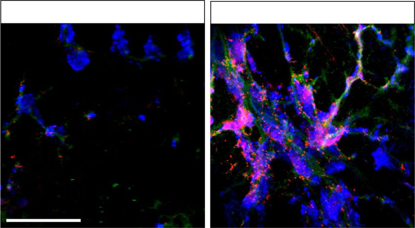
Downloaded from genesdev.cshlp.org on September 20, 2021 - Published by Cold Spring Harbor Laboratory Press

Rovira et al.

silencing factor) could be a negative regulator of endocrine          dent program during pancreas organogenesis. Our results,
differentiation during pancreas development (van Are-                 therefore, show an essential role of REST as a major nega-
nsbergen et al. 2010).                                                tive regulator of pancreatic endocrine differentiation.
   REST is best known for its role as a suppressor of neuro-
nal genes in nonneuronal cell types (Schoenherr and An-
derson 1995). It binds a 21-bp DNA recognition sequence               Results
and has two repressor domains that recruit corepressor
                                                                      REST expression in pancreas is largely restricted
complexes. Consistent with its function to inhibit neuro-
                                                                      to progenitors and duct cells
nal genes, REST is largely expressed in nonneuronal cell
types. However, REST is not expressed in endocrine cell               The expression of REST in pancreatic cell types has been
lines, and several genes that are repressed by REST are ac-           difficult to resolve unequivocally owing to low expression
tive in islet cells (Atouf et al. 1997; Martin et al. 2008,           levels and lack of robust antibodies (van Arensbergen et al.
2012). Furthermore, genome-wide studies in embryonic                  2010; Martin et al. 2015). We found nuclear REST immu-
stem cells and nonneuronal cell types have shown direct               noreactivity in most nonendocrine epithelial and mesen-
binding of REST near β-cell-enriched genes (Johnson                   chymal cells of the mouse E12.5 pancreas, whereas from
et al. 2008; van Arensbergen et al. 2010; Mukherjee et al.            E14.5 onward it was largely restricted to duct-like clus-
2016). REST binding sites in embryonic stem cells overlap             ters, and absent from acinar and endocrine cell clusters
with genomic regions that carry Polycomb-repressed chro-              (Supplemental Fig. S1A). Purified duct cells from adult
matin in FACS-purified multipotent progenitors of the ear-            and E18.5 Sox9-eGFP transgenic mice (Gong et al. 2003)
ly embryonic pancreas (van Arensbergen et al. 2010). Many             confirmed Rest mRNA expression in duct cells (Supple-
of these Polycomb-repressed regions are β-cell regulatory             mental Fig. S1B,C). Finally, single-cell RNA-seq data (Ta-
genes that are subsequently derepressed during pancreatic             bula Muris et al. 2018) showed Rest mRNA in adult duct
endocrine differentiation, in parallel with the concomi-              and nonepithelial cells but not in acinar or endocrine cells
tant loss of REST expression (van Arensbergen et al.                  (Supplemental Fig. S1D). These results reinforce the no-
2010). These correlations suggested that REST could be                tion that REST is expressed in embryonic bipotent progen-
an important negative regulator of the endocrine differen-            itors and adult pancreatic ductal cells but is not detected
tiation program of the developing pancreas. A recent report           in endocrine cells, consistent with a potential function
exploited this notion by inhibiting REST to enhance                   of REST as a negative regulator of pancreatic endocrine
PDX1-mediated activation of endocrine genes in adult                  differentiation.
pancreatic exocrine cells (Elhanani et al. 2020).
   Genetic loss-of-function studies, however, failed to
                                                                      REST inactivation in pancreatic progenitors induces
support a significant role of REST in pancreatic endocrine
                                                                      Neurog3
differentiation. Cre/LoxP-based excision of Rest in pancre-
atic progenitors led to changes in the expression of some             Previous genetic studies concluded that genetic ablation
endocrine genes but did not affect the number of endocrine            of Rest in pancreatic multipotent progenitors has no im-
cells, suggesting it was not an essential modulator of endo-          pact on the formation of NEUROG3+ endocrine precur-
crine differentiation (Martin et al. 2015). Another pancreas          sors or hormone-producing cells (Martin et al. 2015),
deletion study reported that REST tempers pancreatic tis-             although this was examined in a mouse model that cre-
sue damage and prevents acinoductal metaplasia, but the               ates a deletion of Rest exon 2, which produces a functional
study did not explicitly assess endocrine differentiation             isoform that can still bind DNA and recruit corepressors
(Bray et al. 2020). These studies, however, used an allele            (Nechiporuk et al. 2016). We thus used an allele that en-
that removes Rest exon 2 (Gao et al. 2011). Recent work               ables conditional excision of exon 4, which encodes
using a gene trap that disrupts transcription from all Rest           >75% of REST protein residues (Yamada et al. 2010).
promoters revealed dramatic effects on embryonic neuro-               Breeding this line with a Pdx1-Cre transgene (Gu et al.
genesis that were not observed when targeting Rest exon               2002) enabled the excision of Rest and a severe depletion
2 (Nechiporuk et al. 2016). The same study showed that ex-            of REST protein in most embryonic pancreatic epithelial
cision of Rest exon 2 does not prevent translation of a C-            cells (hereafter referred to as Rest pKO mice) (Supplemental
terminal REST peptide that is able to bind DNA, recruit               Fig. S2A–C).
corepressors, and repress target genes (Nechiporuk et al.                Previous work showed that REST binds to pancreatic
2016). Existing data, therefore, warrant a need to explore            endocrine regulatory genes in mouse ES cells and that dur-
the true impact of REST in pancreatic endocrine differen-             ing embryonic pancreas differentiation, REST target genes
tiation using alternative genetic tools.                              loose Polycomb-repressed chromatin and undergo tran-
   We have now inactivated Rest in the embryonic pancre-              scriptional activation (van Arensbergen et al. 2010). To
as using a conditional allele that led to a marked increase           directly test whether this means that REST truly acts as
in endocrine differentiation, proliferation, and cell mass.           a repressor of endocrine differentiation during pancreas
Inactivation of Rest in adult mature duct cells, however,             development, we examined expression of the endocrine
failed to elicit this effect. We used chemical inhibitors             lineage-determinant NEUROG3 in Rest pKO embryos.
to show that REST function is conserved in zebrafish                  This showed a 3.0-fold ± 0.03-fold increase of Neurog3
and represses endocrine genes in human pancreas organo-               mRNA in pancreas from Rest pKO versus control E13.5 em-
ids. Finally, we defined key properties of the REST-depen-            bryos (SEM, Student’s t-test P < 0.01) and 1.7-fold

2       GENES & DEVELOPMENT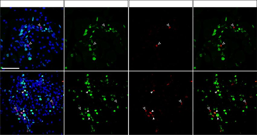
Downloaded from genesdev.cshlp.org on September 20, 2021 - Published by Cold Spring Harbor Laboratory Press
REST inhibits pancreas endocrine differentiation
increased NEUROG3 protein (P < 0.05) (Fig. 1A,B). At ROG3 protein (Villasenor et al. 2008). We therefore inves-
E18.5, a time point in which the wave of NEUROG3+ cells tigated if REST inactivation affected the proliferation of
have normally begun to wane, Neurog3 mRNA was discrete NEUROG3-expressing cells. Immunofluores-
10.7-fold ± 1.2-fold higher in Rest pKO embryos (P < 0.01) cence analysis of E18.5 Rest pKO pancreas showed that
(Fig. 1A). Furthermore, E18.5 mutant pancreas showed 48.3% ± 6.8% of NEUROG3+ cells coexpressed Ki67 ver-
an approximately sixfold increase in NEUROG3+ cells sus 4.6% ± 1.5% in control embryos (P < 0.01) (Fig. 1D).
normalized by the total number of CK19+ cells (P < 0.01) This suggests that REST acts as a negative regulator of
(Fig. 1C). Several NEUROG3+ cells in mutant pancreas cell cycle exit in NEUROG3+ cells, which could contrib-
appeared to line the epithelium of large ducts (Fig. 1C). ute in part to the increased number of NEUROG3+ cells in
Therefore, REST inactivation in pancreatic progenitors Rest pKO embryonic pancreas.
led to an increased yield of NEUROG3+ cells throughout During embryogenesis, NEUROG3+ cells arise from
embryogenesis. pancreatic progenitors that form a tubular plexus that pro-
During normal pancreas development, the activation of gressively evolves into a ductal tree (Gu et al. 2002; Solar
Neurog3 is followed by cell cycle exit of most progenitor et al. 2009; Bankaitis et al. 2015). We tested whether Rest
cells (Miyatsuka et al. 2011; Krentz et al. 2017). This chan- deficiency could not only increase the yield of NEUROG3
ge in cell cycle activity has been shown to occur in dis- + cells during embryonic development but also cause per-
crete, strongly expressing NEUROG3+ cells, in contrast sistent Neurog3 activation in the duct epithelium
to remaining bipotent progenitor cells, which also express throughout postnatal life. We therefore examined postna-
Neurog3 mRNA but very low, often undetectable NEU- tal (2-wk-old and 12-wk-old) Rest pKO mice yet found no
Figure 1. Rest inactivation in pancreatic
Rest pKO
Rest pKO
A B
Control
Control
4 progenitors induces NEUROG3. (A) Neurog3
expression normalized to Hprt
Control
** Rest
pKO
mRNA increases in E13 and E18 Rest pKO pan-
Neurog3 mRNA relative
** NEUROG3 23 kDa
3 creas. Normalization by Hprt mRNA; n = 3–4
LAMINB1 66 kDa
mice per group. (B) Western blot and quantifi-
NEUROG3/LAMINB1
2 * cations of NEUROG3 in nuclear extracts
2
1.5 from E13.5 control and Rest pKO pancreas.
1 1 LamininB1 was used as loading control. n = 2
0.5
samples per group with a pull of three E13.5
0 0
C E13.5 E18.5 Control Rest
pKO
pancreas per sample. (C) Immunofluores-
cence for NEUROG3 (red), cytokeratin 19
NEUROG3+CK19+/CK19+ (%)
NEUROG3/CK19/DAPI NEUROG3/CK19/DAPI
(CK19; green), and DAPI (blue) in E18.5 con-
35 **
30 trol and Rest pKO pancreas. Arrowheads indi-
25 cate NEUROG3+ cells. Bars show
20 NEUROG3+ CK19+ cells in E18.5 pancreas.
15 Scale bar, 100 µm. (D) Immunofluorescence
10
for NEUROG3 (red), Ki67 (green), and DAPI
5
0 (gray) in E18.5 pancreas. Empty arrowheads
pKO
E18.5 Control E18.5 Rest Control Rest
pKO
indicate NEUROG3+Ki67− cells, and white
D arrowheads indicate NEUROG3+Ki67+ cells.
DAPI Ki67 NEUROG3 NEUROG3/Ki67 n = 4–6 mice per group. Scale bar, 50 µm. (E)
Representative whole mounts for NEU-
ROG3+ (red), DBA (green), and insulin (blue)
of the tail of E18.5 control and Rest pKO pan-
NEUROG3+KI67+/NEUROG3+ (%)
creas. Scale bars, 100 µm. Error bars are
SEM. (∗ ) P ≤ 0.05, (∗∗ ) P ≤ 0.01.
E18.5 Control 60 **
50
40
30
20
10
pKO 0
E18.5 Rest Control Rest
pKO
E
NEUROG3/DBA/INSULIN NEUROG3/DBA/INSULIN
Whole-mount
pKO
E18.5 Control E18.5 Rest
GENES & DEVELOPMENT 3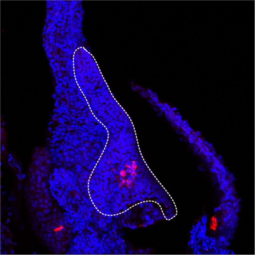
Downloaded from genesdev.cshlp.org on September 20, 2021 - Published by Cold Spring Harbor Laboratory Press

Rovira et al.

NEUROG3+ cells (Supplemental Fig. S3). This suggests                     The binding properties of repressors are poorly under-
that additional REST-independent mechanisms are re-                   stood. Integration with ATAC-seq profiles from E13.5 pan-
quired for persistent Neurog3 activation during postnatal             creas revealed that REST-bound regions were accessible
life.                                                                 to transposase cleavage yet had an accessibility footprint
   Our studies, therefore, show that REST tempers the                 that was narrower than recognition sites of activating pan-
yield of NEUROG3+ cells in the embryonic pancreas, in                 creatic transcription factors, plausibly because active reg-
part by suppressing NEUROG3+ cell proliferation. The                  ulatory elements are occupied by multiple DNA binding
lack of NEUROG3+ cells in the adult Rest pKO pancreas                 factors (Supplemental Fig. S4).
suggests that REST function is not required to prevent                   DNA binding transcription factors that are expressed in
the continuous formation of endocrine progenitor cells                multiple cell types often bind to different genomic regions
from duct cells during postnatal life.                                across cell types (e.g., see Servitja et al. 2009), and this has
                                                                      also been observed for REST (Bruce et al. 2009; Hwang and
                                                                      Zukin 2018). We found that 1806 (∼94%) of REST-bound
Increased β-cell mass in mice lacking REST in pancreatic              sites in embryonic pancreas were shared with embryonic
progenitors                                                           stem cells but only 1030 (∼53%) with neuronal stem cells
                                                                      (Johnson et al. 2008; Whyte et al. 2012), whereas 91
Given that the inactivation of REST in pancreatic progen-
                                                                      (∼4.7%) were exclusively detected in mouse embryonic
itors led to the expansion of endocrine-committed NEU-
                                                                      pancreas (Fig. 3B).
ROG3+ progenitors, we investigated how this influenced
                                                                         These findings, therefore, defined direct REST-bound
the formation of endocrine cells. Whole-mount stainings
                                                                      regions in early embryonic pancreas. They confirmed
for NEUROG3, CK19, and insulin in E18.5 pancreas not
                                                                      that numerous bound regions vary across cell types, al-
only confirmed increased NEUROG3+ cells but also
                                                                      though the vast majority are shared with embryonic
showed a marked increase of the number of insulin-ex-
                                                                      stem cells (Supplemental Table S3).
pressing cells (Fig. 1E).
                                                                         To further assess REST function in pancreatic pro-
   Because the increase in NEUROG3+ cells observed in
                                                                      genitors, we compared RNA-seq profiles of E18.5 Rest pKO
Rest pKO pancreas was transient, we investigated whether
                                                                      versus control pancreas, a stage at which many progeni-
increased β-cell mass was maintained in the adult pancre-
                                                                      tors have already been allocated to distinct cellular lineag-
as. Young (12- to 16-wk-old) male Rest pKO mice had nor-
                                                                      es. We identified 484 down-regulated and 259 up-
mal weight (24.39 g ± 0.5 vs. 24.36 g ± 0.6 control mice)
                                                                      regulated genes in Rest pKO E18.5 embryonic pancreas (ad-
and fasting glycemia (49.71 mg/dL ± 1.83 vs. 54.26 mg/
                                                                      justed P < 0.05) (Supplemental Table S4). We found that
dL ± 3.44 control mice). Morphometric analysis, however,
                                                                      29.8% of up-regulated genes were bound by REST (Fisher
showed an approximately twofold increase of β-cell mass
                                                                      P < 10−4, relative to 15,548 expressed genes), whereas only
in 12-wk-old Rest pKO versus control mice, which was pri-
                                                                      9.8% of down-regulated genes were bound, a similar fre-
marily owing to an increase of large islets (Student’s t-test,
                                                                      quency as all expressed genes (11.2%; Fisher P = 0.81)
P < 0.01), as well as an apparent increase in glucagon-ex-
                                                                      (Fig. 3C; Supplemental Table S5). This was consistent
pressing cells (Fig. 2A–D). These findings, therefore,
                                                                      with the notion that the Rest pKO phenotype reflects a
showed that inactivation of Rest in pancreatic progenitors
                                                                      transcriptional repressor function of REST in the develop-
results in a transient expansion of NEUROG3+ cells and a
                                                                      ing pancreas.
sustained increase of pancreatic endocrine cell mass.
                                                                         REST binds to distal and proximal genomic sites, both
                                                                      of which are likely to harbor functional relevance. Howev-
                                                                      er, up-regulated genes in Rest pKO were most strongly en-
REST is a direct regulator of pancreatic endocrine
                                                                      riched among genes with promoter-proximal REST
differentiation
                                                                      binding (Fisher P < 104) (Fig. 3D), which was also observed
Several genome-wide studies show that REST binds and                  in early experiments that examined dominant negative in-
regulates different genes in different cell types (Bruce              hibition of REST in mouse ESCs (Johnson et al. 2008) Fur-
et al. 2009; Hwang and Zukin 2018), although REST bind-               thermore, REST-bound up-regulated genes were enriched
ing sites have not yet been mapped in pancreas. To study              in Polycomb-repressed chromatin in pancreatic progeni-
how REST controls pancreatic endocrine differentiation,               tors (odds ratio = 3.61, Fisher P = 7 × 10⁻⁴ for up-regulated
we identified REST genomic binding sites and REST-de-                 REST-bound genes; odds ratio = 1.12, P = 0.75 for down-
pendent transcriptional changes in embryonic pancreatic               regulated REST-bound genes; both calculated relative to
progenitors. We performed ChIP-seq analysis using a                   H3K27me3-enriched genes in PDX1+ E10.5 pancreatic
monoclonal antibody (12C11) directed to the REST C-ter-               progenitors as defined by van Arensbergen et al. 2010)
minal region (Chen et al. 1998), and used chromatin from              (Fig. 3E).
E13.5 wild-type pancreas, a stage at which REST expres-                  Consistent with the increase in endocrine cells in
sion is largely confined to progenitor cells and inter-               Rest pKO pancreas, genes that were up-regulated, as well
spersed mesenchymal cells (Supplemental Fig. S1A). We                 as pancreas REST-bound genes at large, showed a strong
detected 1968 REST-bound regions (Supplemental Table                  enrichment in pancreatic endocrine annotations, includ-
S1). These were highly enriched in canonical REST recog-              ing insulin secretion and processing, glucose homeosta-
nition motifs, confirming the specificity of REST binding             sis, and endocrine pancreas development (Fig. 3F,G;
(Supplemental Table S2; Fig. 3A).                                     Supplemental Tables S6, S7). Closer inspection of

4       GENES & DEVELOPMENT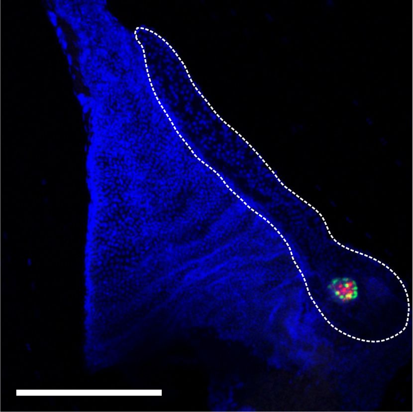
Downloaded from genesdev.cshlp.org on September 20, 2021 - Published by Cold Spring Harbor Laboratory Press

                                                                                                 REST inhibits pancreas endocrine differentiation

               A

                   B                                                        C

               D

Figure 2. Increased β-cell mass in Rest pKO mice. (A) Representative images of 10 × 10 frame reconstructions used for β-cell morphometry
of insulin (red) and DAPI (green) stainings in pancreas from 12-wk-old control and Rest pKO mice. Scale bar, 2 mm. (B) Morphometry of β-
cell mass estimated from insulin surface area/total DAPI surface area (percentage). Rest pKO mice have an approximately twofold increase
in β-cell mass. n = 12 sections from five to six mice in each group. (C ) Islet size in control and Rest pKO adult pancreas. (D) Representative
immunofluorescence for glucagon (red), insulin (green), and DAPI (blue) in whole-pancreas from 12-wk-old control and Rest pKO pancreas.
Scale bar, 200 µm. Error bars indicate SEM. (∗∗ ) P ≤ 0.01.

individual loci disclosed the location of REST-bound re-                  through which REST controls pancreatic endocrine differ-
gions at important regulators of pancreatic differentiation               entiation programs.
(Neurog3, Neurod1, Insm1, Hnf4a, Onecut1, Pax4, Glis3,
and Hnf1a), insulin biosynthesis or exocytosis (Pcsk1,
                                                                          Postnatal inactivation of REST
Pcsk2, Scg3, Snap25, and Syt7), as well as endocrine cell
growth (Bid, Mapk8IP1, Mapk10, and Mapk11) (Fig. 3H;                      Embryonic duct-like bipotent progenitors express many
Supplemental Table S3). Regions bound by REST in em-                      duct cell markers, as well as progressively lose their pro-
bryonic pancreas but not in neural or even embryonic                      genitor capacity as they mature to differentiated duct cells
stem cells included genes previously associated with pan-                 (Solar et al. 2009; Kopinke et al. 2011; Kopp et al. 2011;
creatic differentiation and function such as Isl1 (Ahlgren                Bankaitis et al. 2015) REST expression, however, is main-
et al. 1997), Prox1 (Paul et al. 2016), or Cdh13 (Supplemen-              tained as embryonic progenitors transition to adult differ-
tal Fig. S5; Tyrberg et al. 2011). Given the increased prolif-            entiated ductal cells (Supplemental Fig. S1). We therefore
eration of NEUROG3+ cells, it was also interesting to note                asked whether REST inactivation immediately after birth
up-regulation of REST-bound positive cell cycle regulators                could increase the capacity for de novo generation of endo-
in Rest pKO, including Cdk5r2, Cdk2ap1, Ccnd1, Mapk3,                     crine cells. To this end, we used the Hnf1b-CreERT2trans-
and Ret (Supplemental Fig. S5; Supplemental Table S5).                    genic line (Solar et al. 2009) to excise the Rest LoxP allele in
These studies, therefore, identified direct target genes                  HNF1B+ cells (most of which are duct cells), and also used

                                                                                                             GENES & DEVELOPMENT               5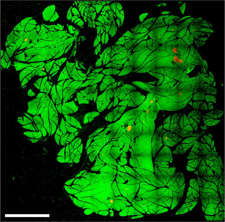
Downloaded from genesdev.cshlp.org on September 20, 2021 - Published by Cold Spring Harbor Laboratory Press

Rovira et al.

                 A                                                         B

                 C                                D                                      E

                 F                                                             G

                 H

Figure 3. Functional direct REST targets in the embryonic pancreas. (A) Top de novo and known motif enrichments in REST-bound re-
gions. (B) REST-bound regions in E13.5 pancreas, mESCs, and mNSCs. (C ) Percentage of up-regulated and down-regulated genes in Rest pKO
mice, or all genes, that were bound by REST. Results indicate that REST predominantly acts as a repressor. P-values are Fisher exact test. (D)
REST binds preferentially to promoter-proximal (0- to 5-kb) regions of genes that were up-regulated in Rest pKO mice. (E) Percentage of differ-
entially expressed genes that were bound by REST, broken down by H3K27me3 enrichment in purified pancreatic progenitors in (van Are-
nsbergen et al. 2010). REST binding was enriched in genes that were up-regulated in Rest pKO and showed H3K27me3 in progenitors. P-
values from Fisher exact test. (F ) Up-regulated genes were functionally annotated using Gorilla (Eden et al. 2009), and REVIGO (Supek
et al. 2011) was used to visualize annotation clusters. The most significant terms are highlighted according to a P-value color scale. (G) Sig-
nificant GSEA terms for up-regulated genes. (H) REST binding associated to pancreatic endocrine development (Neurog3 and NeuroD1),
insulin secretion (Snap25 and Pcsk2), and β-cell survival (Mapk11 and Mapk8ip2) genes in E13.5 pancreas. Y-axes are −log10 P-values.
The vert. cons. track depicts vertebrate conservation.

6       GENES & DEVELOPMENT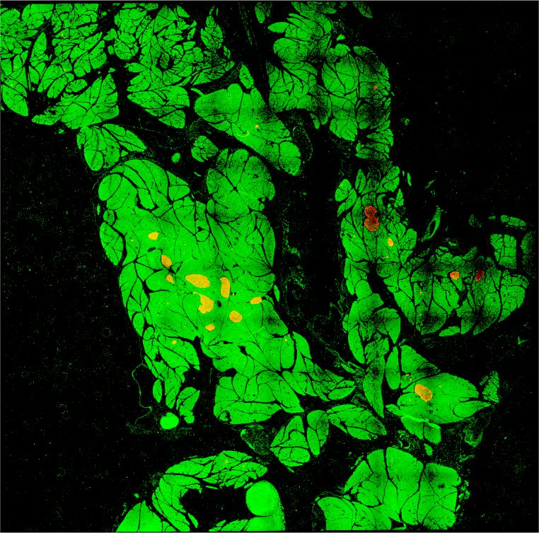
Downloaded from genesdev.cshlp.org on September 20, 2021 - Published by Cold Spring Harbor Laboratory Press
REST inhibits pancreas endocrine differentiation
a Rosa26-LSL-RFP reporter (Luche et al. 2007) to trace In contrast, REST inactivation in pancreatic duct
the progeny of cells that have undergone Rest excision cells from 12 wk olds using the same lineage tracing
(Fig. 4A). We treated dams of triple transgenic newborns model did not lead to significantly increased number of in-
(hereafter called Rest dKO Rosa26RFP) or Hnf1b-CreERT2; sulin+/RFP+ cells 4 wk after induction (2.54% ± 0.33% in
Rosa26RFPcontrols with tamoxifen at postnatal days 1 Rest dKO Rosa26RFP vs. 1.96% ± 0.49% in control mice; Stu-
and 3 and then analyzed mice after weaning (Fig. 4B). dent’s t-test, P = 0.177) (Supplemental Fig. S6). Thus, al-
This showed that the number of insulin+/RFP+ and gluca- though REST retains an essential function to suppress
gon+/RFP+ cells was increased 5.3-fold ± 0.5-fold and 6.4- endocrine cell formation in early postnatal periods, this
fold ± 1.6-fold (Student’s t-test P < 0.01), respectively, in role subsides in adult mice, consistent with a more limited
Rest dKO mice relative to Hnf1b-CreERT2;Rosa26RFP con- differentiation potency of mature duct cells.
trol mice (Fig. 4C). These results indicate that although
the inactivation of REST in embryonic pancreatic progen-
Chemical inhibition of REST in zebrafish
itors did not result in persistent activation of endocrine
progenitor markers throughout adult life, induced inacti- Encouraged by the observation that inactivation of REST
vation of REST in neonatal pancreas did transiently in- in early postnatal duct cells increased the de novo genera-
crease endocrine cell formation. tion of endocrine cells, we explored whether similar
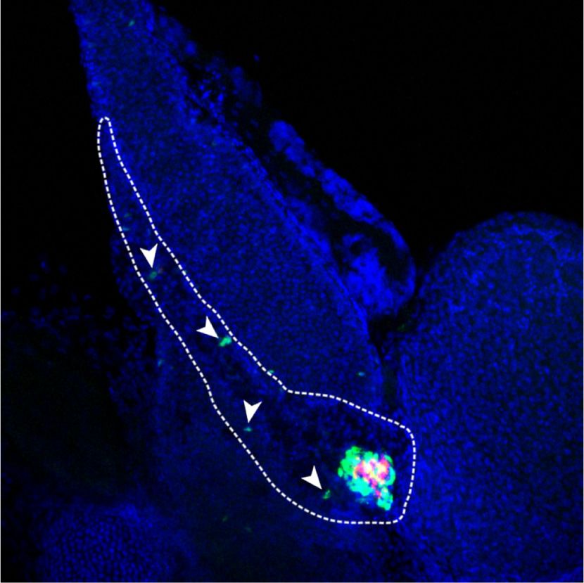
Figure 4. Pancreas-specific inactivation of
A B Rest in neonatal ducts. (A) Schematic of ge-
netic models used to inactivate Rest and ac-
tivate RFP expression in duct cells and
progeny. Hnf1b-CreERT2 is a BAC trans-
genic that specifically marks duct cells
(Solar et al. 2009), as well as non-Rest-ex-
pressing ∂ cells in reporters that are excised
with high efficiency (Rovira et al. 2021), but
not other endocrine or acinar cells. (B) Sche-
C matic of the lineage tracing experiment. Ta-
moxifen was given to mothers at day 1 (P1)
and day 3 (P3) after delivery, and mice
were analyzed at P30. Hnf1b-CreERT2;
Rosa26RFP control mice were also treated.
(C) Representative images of double-posi-
tive RFP (red) and insulin (green) cells and
of double-positive RFP (red) and glucagon
(green) cells in Rest dKO and control mice.
The graph shows RFP-expressing glucagon
and insulin cells in Rest dKO versus control
mice. n = 5–6 mice per each group. Arrow-
heads indicate double-positive cells, and
an asterisk marks examples of cells in a
duct, which were very efficiently labelled.
Scale bar, 100 µm. Error bars are SEM. Stu-
dent’s t-test; (∗∗ ) P < 0.01.
GENES & DEVELOPMENT 7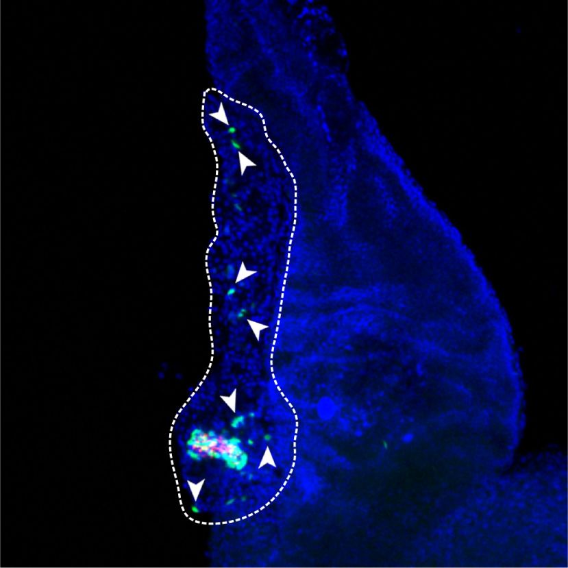
Downloaded from genesdev.cshlp.org on September 20, 2021 - Published by Cold Spring Harbor Laboratory Press

Rovira et al.

effects could be extended to other model systems using                Secondary islets are normally apparent in ∼10% of control
chemical inhibition of REST. We used X5050, recently                  larvae at 5 dpf, and this percentage increases gradually
identified in a high-throughput screen to inhibit REST                thereafter (Parsons et al. 2009). Compared with DMSO
by protein destabilization (Charbord et al. 2013).                    controls, X5050-treated 6-dpf embryos displayed a dose-
   To study REST function in zebrafish, we used a double              dependent increase in secondary islet formation (5 µM
transgenic line in which glucagon- and insulin-expressing             and 50 µM: 2.2-fold ± 0.4-fold and 3.5-fold ± 0.1-fold in-
cells show green and red fluorescence, respectively (Ins:             crease; SD, Student’s t-test P < 0.05 and P < 0.01, respec-
mcherry/Gcga:gfp). We treated zebrafish embryos at 3                  tively), which was comparable with 50 µM DAPT (4.2-
dpf with X5050 for 3 d or with the Notch inhibitor                    fold ± 0.4-fold, P < 0.01) (Fig. 5A). These results suggest
DAPT as a positive control (Parsons et al. 2009), and at 6            that REST regulation of pancreatic endocrine differentia-
dpf, we dissected the pancreas to quantify secondary islet            tion is conserved in zebrafish and that this process can
formation as a readout for endocrine cell differentiation.            be manipulated through chemical inhibition.

A                                                                                        Figure 5. REST regulation of endocrine dif-
                                                                                         ferentiation is conserved in zebrafish. (A)
                                                                                         Embryos of Ins:mCherry/Gcga:GFP double-
                                                                                         transgenic zebrafish line treated with 50
                                                                                         µM DAPT (Notch inhibitor used as positive
                                                                                         control), 0.5 or 5 µM X5050, or vehicle
                                                                                         (DMSO, negative control) from 3 dpf until 6
                                                                                         dpf. After drug treatment at 6 dpf, zebrafish
                                                                                         pancreas was dissected, and the presence or
                                                                                         absence of secondary islets was quantified.
                                                                                         Arrows show representative secondary islets
                                                                                         of double-transgenic zebrafish embryos (Ins:
                                                                                         mCherry/Gcga:GFP; [blue] DAPI). The graph
                                                                                         shows the percentage of zebrafish with
                                                                                         detectable secondary islets in each condi-
                                                                                         tion. n = 20–25 fish per each condition. (B)
                                                                                         An Ins:NTR-mCherry line was used to selec-
                                                                                         tively ablate β cells upon treatment with 5
                                                                                         µM nifurpirinol (NFP) (Pisharath et al.
                                                                                         2007; Bergemann et al. 2018) in 3-dpf embry-
                                                                                         os; 24 h later after complete β-cell ablation of
                                                                                         the principal islet, embryos were exposed to
                                                                                         5 µM ×5050 or vehicle, and β cells were ana-
                                                                                         lyzed 36 h later. Representative images of β-
                                                                                         cell regeneration in Ins:NTR-mCherry em-
B                                                                                        bryos treated with vehicle (DMSO), with
                                                                                         NFP only, or with NFP and X5050. (Red) In-
                                                                                         sulin, (blue) DAPI. The graph shows the per-
                                                                                         centage of pancreas showing >10 insulin-
                                                                                         expressing cells in every condition. n = 32–
                                                                                         36 fish per each condition. Scale bars, 200
                                                                                         µm. Error bars are SEM. (∗∗ ) P < 0.01, (∗ ) P <
                                                                                         0.05, χ2 test.

8       GENES & DEVELOPMENT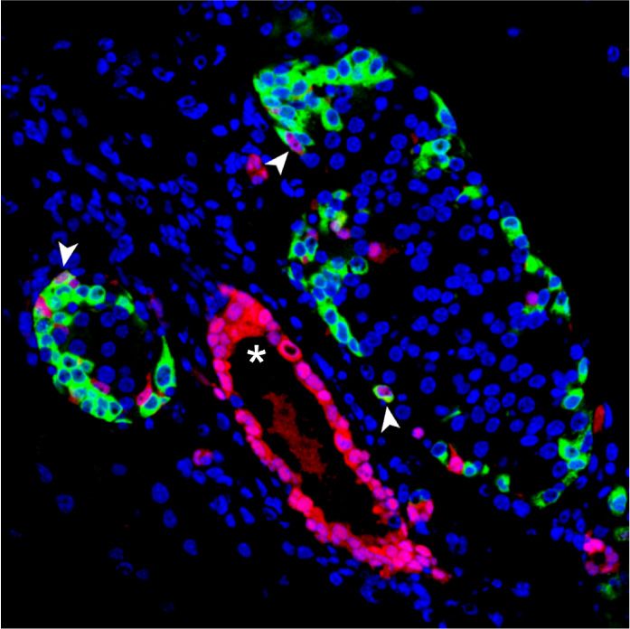
Downloaded from genesdev.cshlp.org on September 20, 2021 - Published by Cold Spring Harbor Laboratory Press

                                                                                       REST inhibits pancreas endocrine differentiation

   We next investigated whether REST inhibition could ac-          ex vivo organoid cultures from human adult ducts isolated
celerate β-cell neogenesis after β ablation with nifurpirinol      from the exocrine fraction of the pancreas of cadaveric do-
(NFP), using Ins:NTR-mcherry transgenic zebrafish (Berge-          nors. Organoids were generated and expanded as previous-
mann et al. 2018). In this model, 3-dpf embryos are treated        ly described (Boj et al. 2015), and experiments were
with NFP, causing ablation of >95% of β cells in 24 h and          performed at passages 3–4 (Fig. 6B). Currently, the efficien-
full recovery of β-cell mass in 48–72 h (Bergemann et al.          cy of endocrine differentiation from published ex vivo pan-
2018). We note that at 3 dpf, all β cells form part of principal   creas organoid protocols is still limited (Huch et al. 2013;
islets. After washing NFP, we ascertained complete β-cell          Boj et al. 2015; Loomans et al. 2018). We thus investigated
ablation with a stereomicroscope and then treated embryos          if REST inhibition in pancreatic human organoids could
with X5050 or vehicle, and 36 h later, we examined which           promote the activation of pancreatic endocrine lineage
embryos had recovered >10 β cells, a threshold that enables        genes. We treated human organoids with X5050 for 48 h
unequivocal distinction from complete ablation. We ob-             and observed only rare endocrine cells in treated and non-
served that 73.4% ± 3.3% of X5050-treated embryos showed           treated organoids. However, X5050-treated organoids
>10 insulin-positive cells in the principal islet, whereas this    showed induction of INS, NEUROG3, and PDX1 mRNA
was only seen in 34.7% ± 6.1% of controls (mean and SD of          levels (2.27-fold ± 0.43-fold, 3.18-fold ± 1.06-fold, and
34–38 embryos in each group, χ2 test, P < 0.01) (Fig. 5B). Thus,   2.18-fold ± 0.54-fold increased vs. DMSO, respectively;
REST inhibition promoted β-cell formation in a zebrafish           SD, Student’s t-test, P < 0.01), whereas the duct cell marker
embryo regeneration model.                                         SOX9 mRNA did not change (Fig. 6B). These results there-
                                                                   fore show that chemical interference of REST in adult hu-
                                                                   man pancreas organoids did not lead to β-cell formation,
REST inhibition in human organoids                                 consistent with genetic findings in adult mice, although
                                                                   it induced the transcription of endocrine genes.
We next explored the impact of REST manipulation in hu-
man cells. We first validated that 24-h treatment of an im-
mortalized duct cell line (PANC-1) with X5050 caused an            Discussion
∼50% reduction of REST full-length protein, as well as a
relative increase in the REST4 isoform, as previously de-          Despite early suggestions that REST could be important
scribed (Fig. 6A; Charbord et al. 2013). Next, we studied          for pancreatic endocrine differentiation during embryonic

A                                                                                     Figure 6. REST chemical inhibition in hu-
                                                                                      man pancreatic organoids. (A) Western blot
                                                                                      analysis of REST FL (full-length) and
                                                                                      REST4 protein levels in PANC1 cells treated
                                                                                      with X5050 50 µM or DMSO (control).
                                                                                      (Lamin B1) Loading control. Bar plot shows
                                                                                      the quantification of the Western blot for
                                                                                      REST. (B) Human organoids generated from
                                                                                      pancreatic exocrine fractions from two ca-
                                                                                      daveric donors were treated at passage 3 for
                                                                                      48 h with 5 µM X5050 or DMSO (control ve-
B                                                                                     hicle). qPCR analysis of mRNA for indicated
                                                                                      genes, relative to TBP. Scale bars, 200 µm. Er-
                                                                                      ror bars are SD. Student’s t-test, (∗∗ ) P < 0.01.

                                                                                                    GENES & DEVELOPMENT               9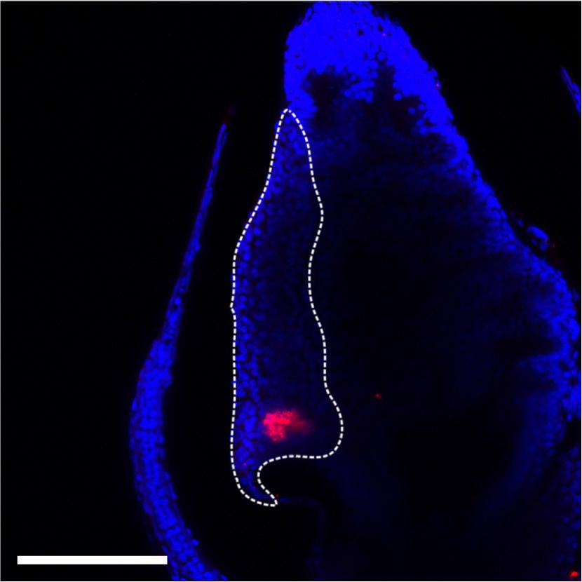
Downloaded from genesdev.cshlp.org on September 20, 2021 - Published by Cold Spring Harbor Laboratory Press

Rovira et al.

development (Martin et al. 2008, 2012; van Arensbergen                endocrinogenesis in embryonic progenitors and even early
et al. 2010), conditional ablation of Rest in the mouse pan-          postnatal pancreas but was clearly less efficient in the
creas unexpectedly showed modest gene expression differ-              adult differentiated pancreas. This may mean that REST
ences and no quantitative changes in endocrine cell                   restrains endocrine differentiation in progenitors that ex-
formation (Martin et al. 2015). This result clearly did               press positive endocrine regulators and have the appropri-
not suggest a major regulatory role in pancreas endocrino-            ate epigenetic competence, whereas mature duct
genesis. We have now combined genetic and chemical                    epithelial cells may lack these properties. Nonetheless,
perturbations to show that REST plays a key evolutionary              the observation that REST inhibition did elicit increased
conserved role to modulate the generation of endocrine                expression of islet endocrine genes in human exocrine
cells during pancreas organogenesis. We define for the first          organoids, together with the recent observation that it
time a blueprint of direct REST target genes in the embry-            can enhance transcription factor-mediated reprogram-
onic pancreas that underpin this regulatory function. We              ming of mouse adult exocrine cells (Elhanani et al.
further show that the capacity to increase endocrinogene-             2020), suggests that REST modulators may form part of
sis upon REST inactivation in duct cells decreases during             an arsenal for future manipulations to promote endocrino-
postnatal life, whereas REST inhibition in human adult                genesis in experimental model systems or replacement
pancreas organoids influenced expression of endocrine                 therapies.
genes but did not trigger endocrine cell formation.
These findings therefore establish REST as an important
regulator of endocrinogenesis during embryonic pancreas               Materials and methods

development.
                                                                      Mouse models
   During pancreas development, a subset of HNF1B+
duct–endocrine bipotent progenitors that form a tubular               All experiments were approved by the Institutional Animal Care
plexus trigger an endocrine gene program, whereas others              Committee of the University of Barcelona. Mice with Rest exon 4
give rise to mature ductal cells (Solar et al. 2009; Bankaitis        floxed allele (RestLSL) (Yamada et al. 2010) were crossed to Pdx1-
et al. 2015). The mechanisms that underlie this binary lin-           Cre (Gu et al. 2002) or Hnf1b-CreERT2 (Solar et al. 2009) and
                                                                      Rosa26-LSL-RFP transgenic lines (Luche et al. 2007). We also gen-
eage choice in a seemingly uniform pool of progenitors is
                                                                      erated RestLSL mice carrying a different Pdx1-Cre transgene (Hin-
unclear. Our results suggest that REST restrains the fre-             gorani et al. 2003) and confirmed increased endocrine cell mass in
quency with which bipotent progenitors consolidate an                 adult mice as well as increased NEUROG3+ cells at E18.5. To in-
endocrine vs. duct fate. On the other hand, the fact that             duce recombination in triple transgenics (Hnf1b-CreERT2;
REST deficiency did not cause an en masse conversion                  Rosa26RFP;RestLSL or Hnf1b-CreERT2;Rosa26RFP control
of bipotent progenitors into endocrine cells is consistent            mice), 20 mg of tamoxifen (Sigma T5648) was administered by ga-
with the notion that REST is not the sole guardian of en-             vage to the mother at days 1 and 3 after delivery. Mice were then
docrine differentiation but instead acts in concert with              sacrificed at 30 d of age. For adults, tamoxifen was given by gavage
other positive and negative regulators to define a differen-          in three doses (20 mg, 20 mg, and 10 mg) over 1 wk to 8- to 12-wk-
                                                                      old mice, and mice were analyzed 4 wk later. Oligonucleotides
tiation probability.
                                                                      used for genotyping are in Supplemental Table S8.
   Genetic experiments have indeed revealed numerous
transcriptional regulators that promote pancreatic endo-
crine cell formation, including NKX6-1, NKX2-2, NEU-                  Dissociation and FACS analysis of pancreatic cells
ROG3, HNF1B, and INMS1, among others (Sussel et al.
                                                                      Adult and E18.5 mouse pancreas from Sox9-eGFP transgenics
1998; Gradwohl et al. 2000; Osipovich et al. 2014; De                 (Gong et al. 2003) were digested in 1.4 mg/mL collagenase-P
Vas et al. 2015). Among DNA binding factors that sup-                 (Roche) for 20–30 min at 37°C. Peripheral acinar-ductal units, de-
press endocrinogenesis, Hippo-responsive TEAD-YAP                     pleted of endocrine islets, were prepared as previously described
complexes are an integral component of pancreatic multi-              (Wang et al. 2013). Tissue was filtered through 600-μm and 100-
potent progenitor enhancers and plausibly counteract en-              μm polypropylene meshes (BD), and peripheral acinar-ductal
docrine differentiation by promoting a progenitor                     units were further dissociated in diluted TrypLE (Invitrogen) for
transcriptional state (Cebola et al. 2015; Mamidi et al.              5 min at 37°C. Dispersed cells were filtered through a 40-μm poly-
2018; Rosado-Olivieri et al. 2019). Notch-responsive tran-            propylene mesh (BD) before FACS sorting.
scriptional repressors, notably HES1, bind near endocrine
genes, where they are likely to exert direct transcriptional          Immunoblots
repression (Jensen et al. 2000; de Lichtenberg et al. 2018).
An important unsolved question is how REST interplays                 Nuclear extracts were prepared as previously described (Maestro
                                                                      et al. 2003) and separated on a 7% SDS-PAGE gel and transferred
with such inhibitory and positive regulators at different
                                                                      to an Immobilon polyvinylidene difluoride membrane (Milli-
stages to ensure a timely and balanced generation of endo-            pore). Immunodetection was performed with mouse 12C11
crine cells.                                                          anti-REST (1:1000) or rabbit anti-LaminB1 (1:2000; Cell Signal-
   The inhibitory function of REST has potential implica-             ing). Quantification was performed with ImageJ-Fiji.
tions for efforts to enhance endocrinogenesis in various in
vivo or in vitro settings. Current pancreas organoid mod-
els are limited because existing protocols largely recapitu-          RNA analysis
late exocrine cell expansion. On the other hand, our                  RNA was isolated using the RNeasy minikit (Qiagen) or TRIzol
experiments showed that REST derepression enhanced                    followed by DNase I treatment (Invitrogen). RNA was reverse-

10       GENES & DEVELOPMENT
Downloaded from genesdev.cshlp.org on September 20, 2021 - Published by Cold Spring Harbor Laboratory Press
REST inhibits pancreas endocrine differentiation
transcribed with SuperScript III reverse transcriptase (Roche) and Motif analysis
random hexamers, and qPCR was performed on a 7900 real-time
De novo and known motifs of REST-bound regions were analyzed
PCR system (Applied Biosystems) using Power SYBR green (Ap-
with HOMER, using a 500-bp window centered on the REST
plied Biosystems). Oligonucleotides are shown in Supplemental
peak.
Table S8.
Immunolocalization methods
RNA-seq
Paraffin-embedded pancreas were processed for immunolocaliza-
DNase-treated RNA (RIN > 8) was generated from three E18.5 tion as described by Maestro et al. (2003). Whole-mount staining
pancreas for each genotype and used for 100-bp paired-end read of E18.5 pancreas was performed as previously described (Ahn-
Illumina sequencing. Reads were aligned to the NCBI36/mm9 ge- felt-Rønne et al. 2007) without TSA amplification. For β-cell
nome using STAR (v2.3.0) (Dobin et al. 2013) with default param- mass measurements in 3-mo-old mice, 4-µm sections were ob-
eters, allowing only uniquely mapped reads. The resulting bam tained at 150-µm intervals, and 21–24 sections per pancreas
files were used to quantify gene expression using FeatureCounts were analyzed by immunofluorescence for insulin and DAPI. Im-
(v1.5) using UCSC mm9 reference gene annotations. Differential ages were taken by automated capturing and reconstruction of 10
expression analysis was performed using DESeq2 (Love et al. × 10 frames using a Leica SP confocal microscope. Insulin-posi-
2014) using an adjusted P-value < 0.05 cutoff. tive and total tissue areas (measured by DAPI saturation) were de-
termined by ImageJ. Islet size distribution was quantified with an
automated ImageJ plugin. Antibodies are shown in Supplemental
ChIP-seq Table S9.
ChIPs were from dissected E13.5 pancreatic buds and were per-
formed as described by van Arensbergen et al. (2010, 2013). Human organoid culture
ChIP DNA (1–2 ng) from two independent pools of E13.5 pan-
creas were used for ChIP sequencing of single-end 50-bp reads. Human exocrine tissue, obtained from the discarded fraction af-
Reads were aligned to NCBI36/mm9 genome using Bowtie2 ter human islet purifications from cadaveric organ donors
(v2.2.5) allowing for one mismatch. Bam files were filtered to re- (Gmyr et al. 2000), was used only if islets were insufficient for
tain reads with a MAPQ ≥ 10. Bam files from biological replicates clinical transplantation and if scientific research was granted ac-
were pooled using samtools, and peaks were called using MACS2 cording to national French regulations. Ethical approval for pro-
(v2.1.0) using default parameters. Input DNA was used to define cessing pancreatic samples from deidentified organ donors was
significant peaks at a false detection rate of 95% of
TFBSs of interest were generated with HINT for a 500-bp window
the β cells are ablated and β-cell mass recovered in 48–72 h (Par-
size, enabling ATAC-seq bias correction.
sons et al. 2009; Bergemann et al. 2018). NFP-treated embryos
were washed and treated with 5 µM X5050 or DMSO. β-Cell re-
generation was analyzed after 36 h. After drug treatment, 5-dpf
REST binding enrichment in differentially expressed genes
zebrafish were fixed overnight in 4% paraformaldehyde, and pan-
Genomic regions were associated with genes using GREAT v3.0, creas was dissected for confocal image analysis.
applying default parameters to the basal plus extension associa- To microdissect pancreas, fixed embryos were placed in PBS on
tion rule (McLean et al. 2010). Proximal and distal REST-bound an agarose-lined plate. Then, using pulled capillaries as tools, first
regions were defined as 5 kb from the transcriptional the yolk and then the whole foregut were pried away from the em-
start sites, respectively. REST-bound genes were annotated based bryo. The pancreas was placed islet-down on a coverslip and dried
on H3K27me3 enrichment (van Arensbergen et al. 2010). by removing all excess. This coverslip was then mounted onto a
GENES & DEVELOPMENT 11Downloaded from genesdev.cshlp.org on September 20, 2021 - Published by Cold Spring Harbor Laboratory Press
Rovira et al.
microscope slide. Water was introduced under the cover slip to re- RNA isolation. J.K.-C. and F.P. provided purified human tissues.
hydrate the sample. M.R. and J.F. wrote the manuscript with input from the remain-
Confocal Z-series stacks were acquired on a Leica SP5 confocal ing authors.
microscope. Maximum projections were obtained by LAS AF
software. To count endocrine cells, we used a double-transgenic
line. Tg(gcga:GFP) and Tg(T2KIns:hmgb1-mCherry), where glu-
References
cagon-positive cells were green and insulin-positive cells were
red. Upon maximum projections of Z-series of the entire pancre- Ahlgren U, Pfaff SL, Jessell TM, Edlund T, Edlund H. 1997. Inde-
as, the presence or absence of secondary islets was computed. pendent requirement for ISL1 in formation of pancreatic mes-
enchyme and islet cells. Nature 385: 257–260. doi:10.1038/
385257a0
Statistics
Ahnfelt-Rønne J, Jørgensen MC, Hald J, Madsen OD, Serup P,
Statistical analyses were performed using either R or GraphPad Hecksher-Sørensen J. 2007. An improved method for three-di-
Prism 6. Statistical significance was calculated with a Fisher ex- mensional reconstruction of protein expression patterns in in-
act test or an unpaired, two-tailed Student’s t-test with data ex- tact mouse and chicken embryos and organs. J Histochem
pressed as mean ± SEM unless otherwise specified. P-values < Cytochem 55: 925–930. doi:10.1369/jhc.7A7226.2007
0.05 were considered significant. Atouf F, Czernichow P, Scharfmann R. 1997. Expression of neuro-
nal traits in pancreatic β cells: implication of neuron-restric-
tive silencing factor/repressor element silencing
Data availability
transcription factor, a neuron-restrictive silencer. J Biol
ChIP-seq and RNA-seq data sets are available in GSE179120. Chem 272: 1929–1934. doi:10.1074/jbc.272.3.1929
Bankaitis ED, Bechard ME, Wright CV. 2015. Feedback control of
growth, differentiation, and morphogenesis of pancreatic en-
Competing interest statement docrine progenitors in an epithelial plexus niche. Genes Dev
29: 2203–2216. doi:10.1101/gad.267914.115
The authors declare no competing interests. Bergemann D, Massoz L, Bourdouxhe J, Carril Pardo CA, Voz ML,
Peers B, Manfroid I. 2018. Nifurpirinol: a more potent and re-
liable substrate compared to metronidazole for nitroreduc-
Acknowledgments tase-mediated cell ablations. Wound Repair Regen 26: 238–
244. doi:10.1111/wrr.12633
We thank the University of Barcelona School of Medicine animal
Boj SF, Hwang CI, Baker LA, Chio II, Engle DD, Corbo V, Jager M,
facility and National Institute for Health Research Imperial Bio-
Ponz-Sarvise M, Tiriac H, Spector MS, et al. 2015. Organoid
medical Research Centre Genomics Unit and Cristina García
models of human and mouse ductal pancreatic cancer. Cell
for zebrafish support and maintenance. We thank Isabelle Manf-
160: 324–338. doi:10.1016/j.cell.2014.12.021
roid (University of Liège) for generous sharing of the zebrafish
Bray JK, Elgamal OA, Jiang J, Wright LS, Sutaria DS, Badawi M,
lines and Irene Miguel-Escalada, Anthony Beucher, and Inês
Borcyk MG, Liu X, Fredenburg KM, Campbell-Thompson
Cebola for critical comments on the manuscript. This research
ML, et al. 2020. Loss of RE-1 silencing transcription factor ac-
was supported by Ministerio de Ciencia, Innovación y Universi-
celerates exocrine damage from pancreatic injury. Cell Death
dades (SAF2015-73226-JIN [Agencia Estatal de Investigación
{AEI}/European Regional Development Fund, European Union Dis 11: 138. doi:10.1038/s41419-020-2269-7
{UE}] and RYC-2017-21950 [AEI/European Social Fund, UE] to Bruce AW, López-Contreras AJ, Flicek P, Down TA, Dhami P, Dil-
M.R., and BFU2014-54284-R and RTI2018-095666-B-I00 to J.F.); lon SC, Koch CM, Langford CF, Dunham I, Andrews RM, et al.
the Medical Research Council (MR/L02036X/1), Wellcome Trust 2009. Functional diversity for REST (NRSF) is defined by in
(WT101033), and European Research Council Advanced Grant vivo binding affinity hierarchies at the DNA sequence level.
(789055) (to J.F.); the Instituto de Salud Carlos III (CA18/00045 Genome Res 19: 994–1005. doi:10.1101/gr.089086.108
to J.L.M.); and a Spanish Ministry of Science, Innovation, and Cebola I, Rodríguez-Seguí SA, Cho CH, Bessa J, Rovira M, Luengo
Universities (MCIU) fellowship (PTA2018-016371-I to M.M.). M, Chhatriwala M, Berry A, Ponsa-Cobas J, Maestro MA, et al.
J.K.-C.’s and F.P.’s research was supported by L’Agence Nationale 2015. TEAD and YAP regulate the enhancer network of hu-
de la Recherche (ANR) grants, L’Institut Européen de Génomique man embryonic pancreatic progenitors. Nat Cell Biol 17:
du Diabète (EGID), ANR-10-LABX-0046, a French state fund 615–626. doi:10.1038/ncb3160
managed by ANR under the frame program Investissements Charbord J, Poydenot P, Bonnefond C, Feyeux M, Casagrande F,
d’Avenir (I-SITE ULNE/ANR-16-IDEX-0004 ULNE to F.P.), and Brinon B, Francelle L, Aurégan G, Guillermier M, Cailleret
the European Consortium for Islet Transplantation funded by M, et al. 2013. High throughput screening for inhibitors of
the Juvenile Diabetes Research Foundation International. Work REST in neural derivatives of human embryonic stem cells re-
in the Centre for Genomic Regulation was supported by the Cen- veals a chemical compound that promotes expression of neu-
tres de Recerca de Catalunya (CERCA) Programme, Generalitat ronal genes. Stem Cells 31: 1816–1828. doi:10.1002/stem
de Catalunya, and Centro de Excelencia Severo Ochoa (SEV- .1430
2015-0510). Work at Institut d’Investigació Biomèdica de Bell- Chen ZF, Paquette AJ, Anderson DJ. 1998. NRSF/REST is re-
vitge was supported by the CERCA Programme and Generalitat quired in vivo for repression of multiple neuronal target genes
de Catalunya. during embryogenesis. Nat Genet 20: 136–142. doi:10.1038/
Author contributions: M.R. and J.F. conceived and coordinated 2431
the study. M.R. performed mouse, organoid, zebrafish, and com- Crews ST, Pearson JC. 2009. Transcriptional autoregulation in
putational studies. M.R. and M.A.M. performed image analysis of development. Curr Biol 19: R241–R246. doi:10.1016/j.cub
mouse mutants. G.A, M.M., and J.L.M performed computational .2009.01.015
analysis. V.G. maintained mouse colonies and performed mor- de Lichtenberg KH, Seymour PA, Jørgensen MC, Kim Y-H, Gra-
phometry studies. J.G.-H. performed ChIP experiments and pin-Botton A, Magnuson MA, Nakic N, Ferrer J, Serup P.
12 GENES & DEVELOPMENTDownloaded from genesdev.cshlp.org on September 20, 2021 - Published by Cold Spring Harbor Laboratory Press
REST inhibits pancreas endocrine differentiation
2018. Notch controls multiple pancreatic cell fate regulators Hwang JY, Zukin RS. 2018. REST, a master transcriptional regu-
through direct hes1-mediated repression. bioRxiv doi:10 lator in neurodegenerative disease. Curr Opin Neurobiol 48:
.1101/336305 193–200. doi:10.1016/j.conb.2017.12.008
De Vas MG, Kopp JL, Heliot C, Sander M, Cereghini S, Haumaitre Jensen J, Pedersen EE, Galante P, Hald J, Heller RS, Ishibashi M,
C. 2015. Hnf1b controls pancreas morphogenesis and the gen- Kageyama R, Guillemot F, Serup P, Madsen OD. 2000. Con-
eration of Ngn3+ endocrine progenitors. Development 142: trol of endodermal endocrine development by Hes-1. Nat Ge-
871–882. doi:10.1242/dev.110759 net 24: 36–44. doi:10.1038/71657
Dobin A, Davis CA, Schlesinger F, Drenkow J, Zaleski C, Jha S, Johnson R, Teh CH, Kunarso G, Wong KY, Srinivasan G, Cooper
Batut P, Chaisson M, Gingeras TR. 2013. STAR: ultrafast uni- ML, Volta M, Chan SS, Lipovich L, Pollard SM, et al. 2008.
versal RNA-seq aligner. Bioinformatics 29: 15–21. doi:10 REST regulates distinct transcriptional networks in embryon-
.1093/bioinformatics/bts635 ic and neural stem cells. PLoS Biol 6: e256. doi:10.1371/jour
Eden E, Navon R, Steinfeld I, Lipson D, Yakhini Z. 2009. Gorilla: a nal.pbio.0060256
tool for discovery and visualization of enriched go terms in Kerr-Conte J, Pattou F, Lecomte-Houcke M, Xia Y, Boilly B, Proye
C, Lefebvre J. 1996. Ductal cyst formation in collagen-embed-
ranked gene lists. BMC Bioinformatics 10: 48. doi:10.1186/
ded adult human islet preparations. A means to the reproduc-
1471-2105-10-48
tion of nesidioblastosis in vitro. Diabetes 45: 1108–1114.
Elhanani O, Salame TM, Sobel J, Leshkowitz D, Povodovski L,
doi:10.2337/diab.45.8.1108
Vaknin I, Kolodkin-Gal D, Walker MD. 2020. REST inhibits
Kopinke D, Brailsford M, Shea JE, Leavitt R, Scaife CL, Murtaugh
direct reprogramming of pancreatic exocrine to endocrine
LC. 2011. Lineage tracing reveals the dynamic contribution of
cells by preventing PDX1-mediated activation of endocrine
Hes1 + cells to the developing and adult pancreas. Develop-
genes. Cell Rep 31: 107591. doi:10.1016/j.celrep.2020.107591 ment 138: 431–441. doi:10.1242/dev.053843
Gao Z, Ure K, Ding P, Nashaat M, Yuan L, Ma J, Hammer RE, Kopp JL, Dubois CL, Schaffer AE, Hao E, Shih HP, Seymour PA,
Hsieh J. 2011. The master negative regulator REST/NRSF con- Ma J, Sander M. 2011. Sox9+ ductal cells are multipotent pro-
trols adult neurogenesis by restraining the neurogenic pro- genitors throughout development but do not produce new en-
gram in quiescent stem cells. J Neurosci 31: 9772–9786. docrine cells in the normal or injured adult pancreas.
doi:10.1523/JNEUROSCI.1604-11.2011 Development 138: 653–665. doi:10.1242/dev.056499
Gmyr V, Kerr-Conte J, Belaich S, Vandewalle B, Leteurtre E, Van- Krentz NAJ, van Hoof D, Li Z, Watanabe A, Tang M, Nian C, Ger-
tyghem MC, Lecomte-Houcke M, Proye C, Lefebvre J, Pattou man MS, Lynn FC. 2017. Phosphorylation of neurog3 links en-
F. 2000. Adult human cytokeratin 19-positive cells reexpress docrine differentiation to the cell cycle in pancreatic
insulin promoter factor 1 in vitro: further evidence for plurip- progenitors. Dev Cell 41: 129–142.e6. doi:10.1016/j.devcel
otent pancreatic stem cells in humans. Diabetes 49: 1671– .2017.02.006
1680. doi:10.2337/diabetes.49.10.1671 Li Z, Schulz MH, Look T, Begemann M, Zenke M, Costa IG. 2019.
Gong S, Zheng C, Doughty ML, Losos K, Didkovsky N, Schambra Identification of transcription factor binding sites using
UB, Nowak NJ, Joyner A, Leblanc G, Hatten ME, et al. 2003. A ATAC-seq. Genome Biol 20: 45. doi:10.1186/s13059-019-
gene expression atlas of the central nervous system based on 1642-2
bacterial artificial chromosomes. Nature 425: 917–925. Loomans CJM, Williams Giuliani N, Balak J, Ringnalda F, van
doi:10.1038/nature02033 Gurp L, Huch M, Boj SF, Sato T, Kester L, de Sousa Lopes
Gradwohl G, Dierich A, LeMeur M, Guillemot F. 2000. Neuroge- SMC, et al. 2018. Expansion of adult human pancreatic tissue
nin3 is required for the development of the four endocrine cell yields organoids harboring progenitor cells with endocrine dif-
lineages of the pancreas. Proc Natl Acad Sci 97: 1607–1611. ferentiation potential. Stem Cell Reports 10: 712–724. doi:10
doi:10.1073/pnas.97.4.1607 .1016/j.stemcr.2018.02.005
Graf T, Enver T. 2009. Forcing cells to change lineages. Nature Love MI, Huber W, Anders S. 2014. Moderated estimation of fold
462: 587–594. doi:10.1038/nature08533 change and dispersion for RNA-seq data with DESeq2. Ge-
Gu G, Dubauskaite J, Melton DA. 2002. Direct evidence for the nome Biol 15: 550. doi:10.1186/s13059-014-0550-8
pancreatic lineage: NGN3+ cells are islet progenitors and are Luche H, Weber O, Nageswara Rao T, Blum C, Fehling HJ. 2007.
Faithful activation of an extra-bright red fluorescent protein in
distinct from duct progenitors. Development 129: 2447–
‘knock-in’ Cre-reporter mice ideally suited for lineage tracing
2457. doi:10.1242/dev.129.10.2447
studies. Eur J Immunol 37: 43–53. doi:10.1002/eji.200636745
Guo S, Dai C, Guo M, Taylor B, Harmon JS, Sander M, Robertson
Maestro MA, Boj SF, Luco RF, Pierreux CE, Cabedo J, Servitja JM,
RP, Powers AC, Stein R. 2013. Inactivation of specific β cell
German MS, Rousseau GG, Lemaigre FP, Ferrer J. 2003. Hnf6
transcription factors in type 2 diabetes. J Clin Invest 123:
and Tcf2 (MODY5) are linked in a gene network operating in a
3305–3316. doi:10.1172/JCI65390
precursor cell domain of the embryonic pancreas. Hum Mol
Hingorani SR, Petricoin EF, Maitra A, Rajapakse V, King C, Jaco- Genet 12: 3307–3314. doi:10.1093/hmg/ddg355
betz MA, Ross S, Conrads TP, Veenstra TD, Hitt BA, et al. Mamidi A, Prawiro C, Seymour PA, de Lichtenberg KH, Jackson
2003. Preinvasive and invasive ductal pancreatic cancer and A, Serup P, Semb H. 2018. Mechanosignalling via integrins di-
its early detection in the mouse. Cancer Cell 4: 437–450. rects fate decisions of pancreatic progenitors. Nature 564:
doi:10.1016/S1535-6108(03)00309-X 114–118. doi:10.1038/s41586-018-0762-2
Huch M, Koo BK. 2015. Modeling mouse and human develop- Martin D, Allagnat F, Chaffard G, Caille D, Fukuda M, Regazzi R,
ment using organoid cultures. Development 142: 3113– Abderrahmani A, Waeber G, Meda P, Maechler P, et al. 2008.
3125. doi:10.1242/dev.118570 Functional significance of repressor element 1 silencing tran-
Huch M, Bonfanti P, Boj SF, Sato T, Loomans CJ, van de Wetering scription factor (REST) target genes in pancreatic β cells. Dia-
M, Sojoodi M, Li VS, Schuijers J, Gracanin A, et al. 2013. Un- betologia 51: 1429–1439. doi:10.1007/s00125-008-0984-1
limited in vitro expansion of adult bi-potent pancreas progen- Martin D, Allagnat F, Gesina E, Caille D, Gjinovci A, Waeber G,
itors through the Lgr5/R-spondin axis. EMBO J 32: 2708–2721. Meda P, Haefliger JA. 2012. Specific silencing of the REST tar-
doi:10.1038/emboj.2013.204 get genes in insulin-secreting cells uncovers their
GENES & DEVELOPMENT 13You can also read