Restoration of Direct Corticospinal Communication Across Sites of Spinal Injury - bioRxiv
←
→
Page content transcription
If your browser does not render page correctly, please read the page content below
bioRxiv preprint first posted online Feb. 11, 2019; doi: http://dx.doi.org/10.1101/546374. The copyright holder for this preprint
        (which was not peer-reviewed) is the author/funder, who has granted bioRxiv a license to display the preprint in perpetuity.
                                     It is made available under a CC-BY-NC-ND 4.0 International license.

Restoration of Direct Corticospinal Communication
Across Sites of Spinal Injury
Naveen Jayaprakash1, David Nowak1, Erik Eastwood1, Nicholas Krueger1, Zimei Wang1, Murray G.
Blackmore1*
1
 Department of Biomedical Sciences, Marquette University, 53201
*
 Corresponding Author: Murray G. Blackmore, Department of Biomedical Sciences, Marquette University, Milwaukee, WI,
53201, USA. Phone: (1) 414 288-4532, Email Address: murray.blackmore@marquette.edu
Conflict of Interest: None
Acknowledgments: This work was supported by grants from NINDS and the Bryon Riesch Paralysis Foundation.

Injury to the spinal cord often disrupts long-distance axon tracts that link the brain and spinal cord,
causing permanent disability. Axon regeneration is then prevented by a combination of inhibitory signals
that emerge at the injury site and by a low capacity for regeneration within injured neurons. The
corticospinal tract (CST) is essential for fine motor control but has proven refractory to many attempted
pro-regenerative treatments. Although strategies are emerging to create relay or detour circuits that re-
route cortical motor commands through spared circuits, these have only partially met the challenge of
restoring motor control. Here, using a murine model of spinal injury, we elevated the intrinsic regenerative
ability of CST neurons by supplying a pro-regenerative transcription factor, KLF6, while simultaneously
supplying injured CST axons with a growth-permissive graft of neural progenitor cells (NPCs)
transplanted into a site of spinal injury. The combined treatment produced robust CST regeneration
directly through the grafts and into distal spinal cord. Moreover, selective optogenetic stimulation of
regenerated CST axons and single-unit electrophysiology revealed extensive synaptic integration by CST
axons with spinal neurons beyond the injury site. Finally, when KLF6 was delivered to injured neurons
with a highly effective retrograde vector, combined KLF6/NPC treatment yielded significant
improvements in forelimb function. These findings highlight the utility of retrograde gene therapy as a
strategy to treat CNS injury and establish conditions that restore functional CST communication across a
site of spinal injury.

Significance Statement
Damage to the spinal cord results in incurable paralysis because axons that carry descending motor commands
are unable to regenerate. Here we deployed a two-pronged strategy in a rodent model of spinal injury to promote
regeneration by the corticospinal tract, a critical mediator of fine motor control. Delivering pro-regenerative KLF6
to injured neurons while simultaneously transplanting neural progenitor cells to injury sites resulted in robust
regeneration directly through sites of spinal injury, accompanied by extensive synapse formation with spinal
neurons. In addition, when KLF6 was delivered with improved retrograde gene therapy vectors, the combined
treatment significantly improved forelimb function in injured animals. This work represents important progress
toward restoring regeneration and motor function after spinal injury.

Introduction
    Spinal cord injury disrupts the exchange of information between the brain and distal cord, causing
impairments in sensory, motor, and autonomic function. In cases of incomplete injury, severed axons often sprout
spontaneously to form new connections with spared tracts, creating detour circuits that re-route information
around the injury (1–6). Recent work involving transplants of neural progenitor cells has also succeeded in
creating novel relay circuits as host axons invade and innervate graft-derived neurons, which in turn extend
lengthy axons that innervate neurons in the caudal spinal cord (7–9). These indirect circuits, both endogenous and
graft-derived, have yielded some gains in motor function after injury(4, 6, 7, 9, 10). Recovery remains partial,
however, even as various pharmacological, rehabilitation, and stimulation strategies have attempted to enhance
their functional output (4, 6). Fundamentally, in the face of supraspinal control systems that evolved to rely on
direct connectivity between supraspinal nuclei and spinal neurons, there may be a limit to the ability of detour or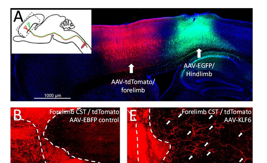
bioRxiv preprint first posted online Feb. 11, 2019; doi: http://dx.doi.org/10.1101/546374. The copyright holder for this preprint
      (which was not peer-reviewed) is the author/funder, who has granted bioRxiv a license to display the preprint in perpetuity.
                                   It is made available under a CC-BY-NC-ND 4.0 International license.

relay circuits to replace lost function, particularly for tasks involving fine motor control. Thus, to complement
progress in creating indirect replacements circuitry after injury, there remains a pressing need to restore the ability
of supraspinal neurons to communicate directly with distal spinal neurons.
    Doing so likely requires regenerative growth by injured axons. Axon regeneration in the central nervous
system (CNS) is blocked by dual barriers: a low neuron-intrinsic capacity for axon growth in adults (11, 12) and
an inhospitable environment around the injury site (13–15). Corticospinal tract (CST) neurons, important
mediators of fine motor control in both rodents and humans, have historically regenerated poorly but more
recently have responded, albeit partially, to several pro-regenerative treatments (16, 17). The neuron-intrinsic
growth ability of CST neurons can be enhanced by deleting growth-inhibitory genes (18, 19) or forcing the
expression of pro-regenerative factors (20, 21). A transcription factor called KLF6 has emerged as a particularly
potent promoter of CST axon growth by activating a set of complementary gene networks involved in axon
extension (22). Yet these neuron-intrinsic interventions only partially restore growth ability, and stimulated axons
generally circumvent partial injuries, but fail to traverse complete injuries, suggesting persistent environmental
inhibition (23). To address extrinsic inhibition, an important advance has been the demonstration that grafts of
neural progenitor cells (NPCs) placed into sites of spinal injury can attract regenerative ingrowth from CST axons
(10). Nevertheless, one major limitation is that a majority of regenerating CST axons extend only about 1mm into
the grafts, and rarely re-enter distal host tissue caudal to the injury site (10). This finding hints that even when
extrinsic inhibition is neutralized with improved tissue, the low growth ability of CST axons limits full growth.
    Here we test a novel combinatorial strategy to enhance CST regeneration by simultaneously addressing
intrinsic and extrinsic barriers to growth. Using a murine model of deep cervical injury, we combine KLF6 gene
delivery and NPC grafting to evoke robust CST regeneration that extends completely through injuries and into
distal host tissue. Both forelimb and hindlimb populations of CST axons participated in the regenerative growth.
Optogenetic stimulation of regenerated axons and single unit recordings of spinal neurons confirmed the ability
of CST axons to evoke spinal activity distal to injury sites, indicating restoration of direct communication between
cortex and spinal tissue distal to the injury. Finally, by harnessing a newly developed retrograde AAV vector to
improve gene delivery, we found that combined KLF6 expression and NPC grafting significantly improved fine
motor control in a horizontal ladder task and pellet retrieval task. These data indicate that combined NPC grafting
and delivery of pro-regenerative transcription factors can restore direct neural communication across sites of
spinal injury.
Results
Combined KLF6 expression and NPC grafts enhance CST regeneration. KLF6 is a pro-regenerative
transcription factor that is expressed by corticospinal tract (CST) neurons during periods of axon growth, and then
downregulated with maturation (22, 24). We showed previously that forced re-expression of KLF6 in adult CST
neurons enhances compensatory CST sprouting and regenerative growth after partial spinal injuries (22).
Importantly, however, this KLF6-stimulated growth occurred through spared grey matter. Here we tested whether
KLF6 over-expression can stimulate CST regeneration through a deeper and more challenging injury. AAV9-
KLF6 or AAV9-EBFP control, mixed with AAV-EYFP tracer, were injected to the left motor cortex of adult
C57Bl/6J mice. Animals then received an injury to cervical spinal cord in which a wife-knife was inserted at the
midline, the blade extended toward the right edge of the spinal cord and lowered/raised in three successive cycles
to a depth of 1.1mm. In contrast to the prior injuries, this produced a more tear-like injury, affecting the majority
of grey matter (Supp. Fig. 1). Eight weeks later, sections of spinal cord were prepared, and CST growth was
quantified as the number of EYFP+ axons that intersected virtual lines at set distances below the injury,
normalized to total EYFP+ axons counted in the medullary pyramids. As expected, AAV-EBFP-treated animals
showed minimal CST growth below the injury site (Supp. Fig. 2A,B). In contrast to prior findings (22), KLF6-
treated animals displayed only a non-significant trend toward enhanced CST growth in this injury paradigm
(Supp. Fig. 2C,D). These data indicate that when confronted with a deeper and more severe injury, KLF6-
stimulated axons are largely unable to regenerate, a finding reminiscent of prior findings with closely related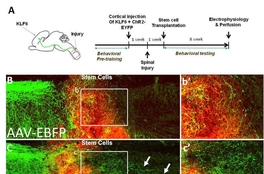
bioRxiv preprint first posted online Feb. 11, 2019; doi: http://dx.doi.org/10.1101/546374. The copyright holder for this preprint
(which was not peer-reviewed) is the author/funder, who has granted bioRxiv a license to display the preprint in perpetuity.
It is made available under a CC-BY-NC-ND 4.0 International license.
KLF7 (21, 23). Thus, although KLF-based
manipulations elevate intrinsic regenerative ability in
CST axons (22), extrinsic barriers continue to
constrain growth.
We therefore employed grafts of embryonic spinal
cells supported by a fibrin matrix and growth factors,
shown previously to create growth-permissive
interfaces with host tissue and to support ingrowth by
injured CST axons (7, 8, 10). To confirm the ability of
these grafts to attract regenerative CST growth, adult
mice received cortical injection of control AAV-EBFP
and AAV-ChR2-EYFP tracer and deep wire knife
injury, followed one week later by transplantation of
cells from E12.5 embryonic spinal cord, labeled by
transgenic tdTomato expression (Fig. 1A). Eight
weeks after transplantation, examination of spinal
sections showed consistent survival and integration of
grafts and ingrowth by in CST axons (EYFP+),
confirming the grafts’ suitability as a growth substrate
(Supp. Table 1). Consistent with prior reports,
however, most of this CST growth penetrated less than
1mm into the grafts, and rarely traversed the entire
graft to re-enter distal host tissue (10). Thus, the
availability of growth-permissive embryonic tissue
restores axon regeneration by CST neurons only
partially, likely reflecting continued neuron-intrinsic
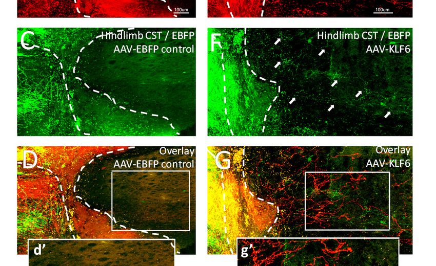
constraints to growth. To simultaneously improve both Fig. 1. Combined KLF6 expression and NPC grafting improves CST
regeneration. (A) Adult mice received cortical injection of AAV-ChR2-
neuron-intrinsic and -extrinsic conditions for growth, EYFP and AAV-KLF6 or AAV-EBFP control, followed by cervical
adult mice received cortical injection of AAV-KLF6 hemisection and transplantation of NPCs labeled with tdTomato. (B-E)
and AAV-ChR2-EYFP tracer, followed by spinal Eight weeks after NPC grafting, CST axons (EYFP+, green) regenerate
into NPC grafts (red). In animals treated with KLF6, CST axons extend
injury and NPC grafting as described above. Eight beyond the distal graft/host border and into spinal tissue caudal to the
weeks later, animals that received combined lesion (arrows, C). (D) Quantification of the number of axonal profiles
KLF6/NPCs showed a significant increase in CST that extend beyond the injury shows a significant elevation of CST
regeneration in KLF6-treated animals. N=12 per group, *p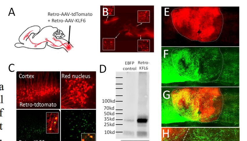
bioRxiv preprint first posted online Feb. 11, 2019; doi: http://dx.doi.org/10.1101/546374. The copyright holder for this preprint
      (which was not peer-reviewed) is the author/funder, who has granted bioRxiv a license to display the preprint in perpetuity.
                                   It is made available under a CC-BY-NC-ND 4.0 International license.

from forelimb and hindlimb populations. These data
indicate that KLF6’s ability to promote axon growth
is robust across different sub-regions of cortex and
consistent across experiments, highlighting its
versatility.
Regenerated CST axons form functional
synapses on distal spinal neurons. To test the
ability of regenerated CST axons to form effective
synapses we paired selective optogenetic
stimulation of CST terminals with single-unit
recording from spinal neurons in intact animals. As
described above, animals received mixed cortical
injection of AAV-ChR2-EYFP and AAV-EBFP or
AAV-KLF6, followed by cervical injury. Half of
the animals then received NPC grafts. Eight weeks
later animals were anesthetized and their spinal
cords exposed between C3 and C6. 473nm light was
focally directed to locations up to 1000µm above
the injury site, to the center of the injury, and up to
1000µm distal to the injury. In this way optical
stimulation specifically triggers synaptic release
from CST terminals, which uniquely express ChR2
in spinal tissue. At each location, a 32-multichannel
electrode was inserted 1000µm into the spinal cord,
with sensitivity to activity from individual cell
bodies but not fibers of passage (26). Spontaneously
active units were identified and the average change
in firing during light exposure was calculated for
each unit. Units that displayed significant increases
in firing during light stimulation (p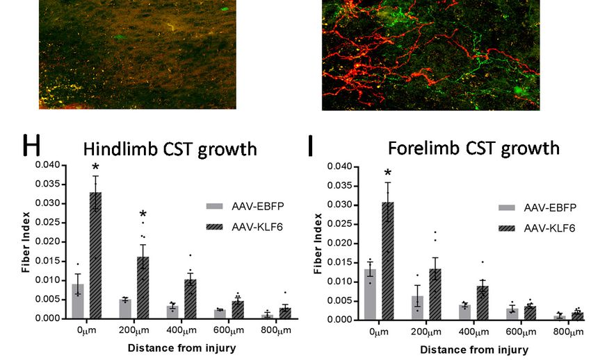
bioRxiv preprint first posted online Feb. 11, 2019; doi: http://dx.doi.org/10.1101/546374. The copyright holder for this preprint
       (which was not peer-reviewed) is the author/funder, who has granted bioRxiv a license to display the preprint in perpetuity.
                                    It is made available under a CC-BY-NC-ND 4.0 International license.

                                                                                                  location, the enhanced firing rate during
                                                                                                  optical stimulation likely reflected
                                                                                                  synaptic integration of CST axons with
                                                                                                  graft-derived neurons, consistent with
                                                                                                  prior findings (10). Distal to the injury,
                                                                                                  10% and 7% of host neurons responded to
                                                                                                  CST stimulation at 500um and 1mm,
                                                                                                  respectively. These data indicate that host
                                                                                                  CST axons, which enter grafts and
                                                                                                  occasionally exit distally, likely form
                                                                                                  functional synapses on both graft-derived
                                                                                                  and host neurons, albeit with modest
                                                                                                  frequency. Finally, in animals that receive
                                                                                                  both grafts and KLF6 treatment, a
                                                                                                  substantially larger fraction of units
                                                                                                  responded to CST stimulation both within
                                                                                                  and beyond grafts. Indeed, 34% and 29%
                                                                                                  of spontaneously active spinal units
                                                                                                  responded to CST activation at positions
                                                                                                  500um and 1000um beyond the spinal
                                                                                                  injury, respectively. In addition, in CST-
                                                                                                  responsive units the average change in
                                                                                                  firing rate below the injury was
                                                                                                  significantly larger in KLF6 than in
 Fig. 3. Selective optogenetic stimulation and single unit recording reveal synaptic              EBFP-treated animals (KLF6: 16.9 (±2.1
 connectivity between regenerated CST axons and spinal neurons. Adult mice received
 cortical injection of AAV-ChR2-EYFP and AAV-KLF6 or AAV-EBFP control,                            SEM) spikes/s, EBFP: 6.0 (±0.6 SEM)
 followed by cervical hemisection and NPC transplants or saline control. Eight weeks              spikes/s p=.0068, 2 tailed t-test). Thus
 later, 473nm light was focally delivered to sites rostral to, within, and caudal to the injury   KLF6 treatment of CST axons produces
 site while single-unit recordings were performed with a 32-channel electrode. ChR2 is
 present only on CST axons, thus enabling CST-selective stimulation. (A-D) Each dot               substantial elevation of direct neural
 represents a single spinal unit, with position indicating the average change in firing rate      communication distal to a site of spinal
 during light stimulation and green indicating significance (p
bioRxiv preprint first posted online Feb. 11, 2019; doi: http://dx.doi.org/10.1101/546374. The copyright holder for this preprint
      (which was not peer-reviewed) is the author/funder, who has granted bioRxiv a license to display the preprint in perpetuity.
                                   It is made available under a CC-BY-NC-ND 4.0 International license.

therefore repeated behavioral assessment of animals
treated with combined KLF6 and NPCs, this time
delivering KLF6 by injection of Retro-AAV to the spinal
cord at the time of NPC grafting. Cortical tracing in a
subset of animals confirmed extensive regeneration by
CST axons in the combined treatment group (Fig. 4E-H).
Combined Retro-KLF6 and NPC grafting produced a
significant reduction in mistargeted steps on the horizontal
ladder task and a significant increase in the number of
pellets that were displaced and retrieved by the right
forelimb (Fig. 4I, J). Thus, in the presence of growth-
permissive tissue grafts, KLF6 delivery with a retrograde
vector produces significant improvements in forelimb
function after spinal injury.
Discussion
        We have shown that following spinal injury,
regeneration of the corticospinal tract is substantially
enhanced by simultaneously expressing a pro-regenerative
transcription factor in injured neurons and supplying growth-
permissive tissue bridges to injured axons. Injured CST
axons traversed the grafted tissue and re-entered distal host                          Fig. 4. Widespread delivery of KLF6 with retrograde
                                                                                       vectors, combined with NPC grafts, improves forelimb
tissue, where they established synaptic connections. This                              function after cervical spinal injury. (A-D) Adult mice
outcome was consistent across multiple experiments and was                             received cervical injection of AAV2-Retro-KLF6 with AAV2-
observed in both forelimb and hindlimb-directed populations                            Retro-tdTomato tracer. Two weeks later, tdTomato
                                                                                       fluorescence confirmed widespread retrograde transduction (B)
of CST neurons. Moreover, when delivered with a retrograde                             and       immunohistochemistry       confirmed        successful
vector, KLF6 combined with NPC grafts produced                                         overexpression of KLF6 in retrogradely transduced cells (C).
significant restoration of forelimb function in cervically                             (D) Western blotting confirmed elevated KLF6 expression in
                                                                                       cortical tissue two weeks after retrograde delivery to cervical
injured mice. These findings establish conditions that                                 spinal cord. (E-H) Adult mice received cervical hemisection
reliably evoke functional regeneration by corticospinal tract                          injury and cervical injection of AAV2-Retro-KLF6, followed
neurons across a site of spinal injury.                                                one week later by NPC transplants. Twelve weeks later, AAV9-
                                                                                       EGFP was injected to cortex to trace CST axons. (E-H) Similar
        The promotion of CST growth after spinal injury has                            to anterograde KLF6, retrogradely expressed KLF6 produced
                                                                                       robust CST growth into and beyond NPC grafts. (I,J) Compared
been a long-standing goal in regeneration research. In work                            to control animals that received EBFP and no NPCs, animals
spanning several decades, a great diversity of cell types have                         treated with AAV2-Retro-KLF6 and NPC grafts displayed
been transplanted into animal models of spinal injury in an                            significant improvements in forelimb placement on the
                                                                                       horizontal ladder task, and in the ability to retrieve pellets on
effort to provide CST axons with a more growth-permissive
tissue environment (30–38). Grafted cells have succeeded in eliciting growth from other supraspinal populations
(e.g. propriospinal, raphespinal, bulbospinal, and sometimes rubrospinal), but CST axons often respond
minimally or not at all (30–36). Thus, compared to other populations, CST neurons appear to possess an
intrinsically low capacity for growth and/or more stringent requirements for extrinsic growth substrates. In this
context, it is highly significant that NPC grafts, supported by growth factors and a fibrin/thrombin matrix, were
recently shown to act as suitable substrates for CST axon growth (10). Here we confirm the ability of NPC grafts
to attract growth from injured CST axons; the reproducibility of this effect across research labs marks important
progress toward the long-sought goal of evoking regenerative growth from this critical population.
        Also consistent with prior observations, however, we found CST growth through the grafts to be
incomplete, mostly reaching only the proximal half of the graft and very rarely extending to distal host tissue
(10). One important implication of this pattern of growth is that to provide functional improvements, NPC grafts
alone are unable restore the original CST circuitry but instead must act by creating novel relay circuits through
grafted neurons. There is now substantial evidence that new relay circuits do form in grafts, based on the growth
and synaptogenesis by host neurons into grafts and exuberant extension of graft-derived axons into distal host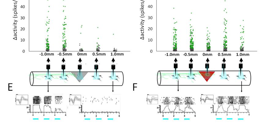
bioRxiv preprint first posted online Feb. 11, 2019; doi: http://dx.doi.org/10.1101/546374. The copyright holder for this preprint
      (which was not peer-reviewed) is the author/funder, who has granted bioRxiv a license to display the preprint in perpetuity.
                                   It is made available under a CC-BY-NC-ND 4.0 International license.

tissue (7, 8). Moreover, recent findings indicate that these relay circuits contribute functionally to improvements
in hindlimb stepping motions(9). There may be limits, however, to the ability of these relay circuits to restore
motor control, particularly for fine forelimb movements normally controlled by the CST. Timing and synchrony
of firing between neural circuits is essential for both the learning and execution of fine motor tasks (39). From a
temporal perspective, relay circuits in NPC grafts create a built-in delay for movement and the return of sensory
information, greatly complicating coincidence detection. Spatially, as host axons invade the graft they appear to
terminate in a highly disorganized pattern that lacks clear topography (8, 10, 40), although there is some evidence
that some populations do select appropriate cell types as synaptic targets (40). Again, this initial disorganization
complicates any engagement of Hebbian processes to sculpt effective movement (41, 42). It is notable that despite
the robust growth in and out of NPC grafts and the existence of new relay connections, improvements even in
stereotyped motions such as locomotion remain only partial(7, 9).
        To address this constraint, we harnessed forced expression of a pro-regenerative transcription factor to
drive substantial regeneration of CST axons completely through NPC grafts and into distal host tissue. KLF
factors comprise a 17-member family that play widespread roles in development, including both positive and
negative regulation of axon growth (24, 43). KLF6 and the closely related KLF7 stand out as the only members
known to be pro-regenerative (21, 22, 24). In contrast to KLF7, which required addition of a VP16 transcriptional
activation domain to evoke CST regeneration (21), KLF6 effectively promotes CST axon growth in its wild type
form(22). Here we showed that when environmental constraints are relieved by cell transplants into the site of
injury, CST neurons with forced expression of KLF6 extended axons through sites of spinal injury and back into
distal host tissue. CST regeneration was consistent across experiment and between cortical regions, highlighting
the potential of combined intrinsic and extrinsic-targeted treatments to reliably evoke CST regeneration.
Importantly, direct optical stimulation of these regenerated axons produced robust post-synaptic responses from
host neurons in distal cord, indicating the re-establishment of direct synaptic connectivity. Thus, this combined
approach reliably yielded a neural substrate for direct communication between the cortex and distal spinal cord,
directly through a site of spinal injury.
        Finally, a key finding was that delivery of KLF6 by retrograde vectors, but not direct cranial injection,
improved forelimb function after spinal injury. We recently characterized retrograde transduction after spinal
injections of AAV2-Retro, and showed widespread transduction of CST neurons throughout the cortex, in
addition to subcortical nuclei that participate in skilled movements, including the red nucleus and reticular
formation (29). The likely explanation for the difference in behavioral outcomes is that compared to focal delivery
to subregions of motor cortex, retrograde vectors deliver therapeutic transgenes to a larger number and greater
diversity of supraspinal cell types. Importantly, retrograde gene delivery also offers substantial advantages over
direct cortical injection from a translational perspective. Retrograde injections could be readily combined with
existing cell transplantation procedures, and in human patients a small number of injections to spinal targets could
potentially reach widespread supraspinal populations in a way that is highly impractical using direct cranial
injections. Thus, these data highlight the potential for retrograde KLF6 vectors to supplement current grafting
approaches and further improve functional outcomes after spinal injury.
Materials and Methods
Injury and AAV delivery. Spinal cord injury was performed between cervical region C4-C5 using a wire knife
to create a lesion of 1.1mm depth. AAV9-KLF6 and AAV-EBFP were created by PCR-based cloning into a
pAAV-MCS vector, followed by production at the Univ. of N. Carolina vector core, as described previously (22).
AAV9-CamKII-ChR2(H134R)-EYFP was obtained from the Univ. of North Carolina Viral Vector Core. pAAV-
CAG-tdTomato (codon diversified) was a gift from Edward Boyden (Addgene viral prep # 59462-AAV5).
Vectors were delivered to five locations spanning the motor cortex of adult female C57/Bl6 mice, (1X10¹³
particles/ml, 0.3μl each) using a Kopf stereotaxic instrument, Hamilton syringe, and Stoelting QSI infusion pump.

NPC preparation and transplantation. Spinal cords from tdTomato-expressing (Jax 007676) E12 embryos
were dissociated by trituration in 1X HBSS buffer and resuspended in Neurobasal medium. 1 million cells were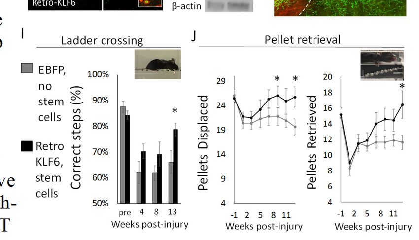
bioRxiv preprint first posted online Feb. 11, 2019; doi: http://dx.doi.org/10.1101/546374. The copyright holder for this preprint
       (which was not peer-reviewed) is the author/funder, who has granted bioRxiv a license to display the preprint in perpetuity.
                                    It is made available under a CC-BY-NC-ND 4.0 International license.

injected by Picospritzer 5 to five locations surrounding the injury site in thrombin / fibrinogen and BDNF, VEFF,
bFGF and MDL28170VEGF (BDNF 50ng/µl, bFGF 10ng/µl, VEGF 10ng/µl, MDL28170 50µM)

Behavior. As described previously (26) mice were pre-trained on a horizontal ladder (30cm long, 1cm rung
spacing) and staircase pellet apparatus (Lafayette Instruments, Lafayette IN) until performance plateaued, then
tested every four weeks (ladder) or bi-weekly (retrieval) after injury.

In vivo optogenetics and electrophysiology. Extracellular recording were obtained using a multichannel silicon
electrode inserted 1000 µm from the surface of the spinal cord. 473 nm laser was focused via collimator on the
point of electrode insertion to stimulate terminal firing of ChR2-expressing CST axons. The recordings were
sampled at 30KS/s and recorded using an integrated data acquisition system (Smartbox; NeuroNexus
Technologies). Paired t-test was conducted to determine baseline change in firing rate before and after the light
stimulation. Units that exhibited mean firing rate change greater than 2 spikes/sec and a significant difference (p<
.0001) were classified as exhibiting laser-evoked activity in this analysis.

Axon growth quantification. Spinal cord sections were cut using a vibratome (Leica VT100S) at 50 µm
thickness. Axon growth was quantified by a blinded observed from sagittal sections imaged on Nikon A1
Confocal Microscope and analyzed with Advanced Research software. Fluorescent axons that intersected virtual
lines at set distances from the injury site were quantified and normalized to total fluorescent axons counted in the
medullary pyramids

References
1.      Bareyre FM, et al. (2004) The injured spinal cord spontaneously forms a new intraspinal circuit in adult
        rats. Nat Neurosci 7(3):269–277.
2.      Filli L, et al. (2014) Bridging the Gap: A Reticulo-Propriospinal Detour Bypassing an Incomplete Spinal
        Cord Injury. J Neurosci 34(40):13399–13410.
3.      Siegel CS, Fink KL, Strittmatter SM, Cafferty WBJ (2015) Plasticity of intact rubral projections mediates
        spontaneous recovery of function after corticospinal tract injury. J Neurosci 35(4):1443–57.
4.      Asboth L, et al. (2018) Cortico-reticulo-spinal circuit reorganization enables functional recovery after
        severe spinal cord contusion. Nat Neurosci 21(4):576–588.
5.      Tohyama T, et al. (2017) Contribution of propriospinal neurons to recovery of hand dexterity after
        corticospinal tract lesions in monkeys. Proc Natl Acad Sci 114(3):604–609.
6.      Chen B, et al. (2018) Reactivation of Dormant Relay Pathways in Injured Spinal Cord by KCC2
        Manipulations. Cell 174(3):521–535.e13.
7.      Lu P, et al. (2012) Long-Distance Growth and Connectivity of Neural Stem Cells after Severe Spinal
        Cord Injury. Cell 150(6):1264–1273.
8.      Adler AF, Lee-Kubli C, Kumamaru H, Kadoya K, Tuszynski MH (2017) Comprehensive Monosynaptic
        Rabies Virus Mapping of Host Connectivity with Neural Progenitor Grafts after Spinal Cord Injury. Stem
        Cell Reports 8(6):1525–1533.
9.      Dell’Anno MT, et al. (2018) Human neuroepithelial stem cell regional specificity enables spinal cord
        repair through a relay circuit. Nat Commun 9(1):3419.
10.     Kadoya K, et al. (2016) Spinal cord reconstitution with homologous neural grafts enables robust
        corticospinal regeneration. Nat Med 22(5):479–487.
11.     Tedeschi A, Bradke F (2017) Spatial and temporal arrangement of neuronal intrinsic and extrinsic
        mechanisms controlling axon regeneration. Curr Opin Neurobiol 42:118–127.
12.     Sun F, He Z (2010) Neuronal intrinsic barriers for axon regeneration in the adult CNS. Curr Opin
        Neurobiol 20(4):510–8.
13.     Kaplan A, Ong Tone S, Fournier AE (2015) Extrinsic and intrinsic regulation of axon regeneration at a
        crossroads. Front Mol Neurosci 8:27.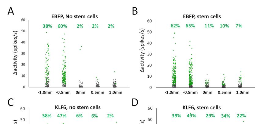
bioRxiv preprint first posted online Feb. 11, 2019; doi: http://dx.doi.org/10.1101/546374. The copyright holder for this preprint
       (which was not peer-reviewed) is the author/funder, who has granted bioRxiv a license to display the preprint in perpetuity.
                                    It is made available under a CC-BY-NC-ND 4.0 International license.

14.     Silver J, Schwab ME, Popovich PG (2015) Central Nervous System Regenerative Failure: Role of
        Oligodendrocytes, Astrocytes, and Microglia. Cold Spring Harb Perspect Biol 7(3):a020602.
15.     Dunham I, et al. (2012) An integrated encyclopedia of DNA elements in the human genome. Nature
        489(7414):57–74.
16.     Tuszynski MH, Steward O (2012) Concepts and Methods for the Study of Axonal Regeneration in the
        CNS. Neuron 74(5):777–791.
17.     Lemon RN (2008) Descending Pathways in Motor Control. Annu Rev Neurosci 31(1):195–218.
18.     Liu K, et al. (2010) PTEN deletion enhances the regenerative ability of adult corticospinal neurons. Nat
        Neurosci 13(9):1075–1081.
19.     Jin D, et al. (2015) Restoration of skilled locomotion by sprouting corticospinal axons induced by co-
        deletion of PTEN and SOCS3. Nat Commun 6:8074.
20.     Wang Z, Reynolds A, Kirry A, Nienhaus C, Blackmore MG (2015) Overexpression of Sox11 Promotes
        Corticospinal Tract Regeneration after Spinal Injury While Interfering with Functional Recovery. J
        Neurosci 35(7):3139–3145.
21.     Blackmore MG, et al. (2012) Kruppel-like Factor 7 engineered for transcriptional activation promotes
        axon regeneration in the adult corticospinal tract. Proc Natl Acad Sci 109:7517–7522.
22.     Wang Z, et al. (2018) KLF6 and STAT3 co-occupy regulatory DNA and functionally synergize to
        promote axon growth in CNS neurons. Sci Rep 8(1):12565.
23.     Wang Z, Winsor K, Nienhaus C, Hess E, Blackmore MG (2017) Combined chondroitinase and KLF7
        expression reduce net retraction of sensory and CST axons from sites of spinal injury. Neurobiol Dis
        99:24–35.
24.     Moore DL, et al. (2009) KLF family members regulate intrinsic axon regeneration ability. Science
        326(5950):298–301.
25.     Kamiyama T, et al. (2015) Corticospinal tract development and spinal cord innervation differ between
        cervical and lumbar targets. J Neurosci 35(3):1181–91.
26.     Jayaprakash N, et al. (2016) Optogenetic Interrogation of Functional Synapse Formation by Corticospinal
        Tract Axons in the Injured Spinal Cord. J Neurosci 36(21):5877–90.
27.     Esposito MS, Capelli P, Arber S (2014) Brainstem nucleus MdV mediates skilled forelimb motor tasks.
        Nature 508(7496):351–356.
28.     Wang X, et al. (2017) Deconstruction of Corticospinal Circuits for Goal-Directed Motor Skills. Cell
        171(2):440–455.e14.
29.     Wang Z, Maunze B, Wang Y, Tsoulfas P, Blackmore MG (2018) Global connectivity and function of
        descending spinal input revealed by 3D microscopy and retrograde transduction. J Neurosci:1196–18.
30.     Richardson PM, Issa VM, Aguayo AJ (1984) Regeneration of long spinal axons in the rat. J Neurocytol
        13(1):165–82.
31.     Fouad K, et al. (2005) Combining Schwann cell bridges and olfactory-ensheathing glia grafts with
        chondroitinase promotes locomotor recovery after complete transection of the spinal cord. J Neurosci
        25(5):1169–78.
32.     Hollis ER, Lu P, Blesch A, Tuszynski MH (2009) IGF-I gene delivery promotes corticospinal neuronal
        survival but not regeneration after adult CNS injury. Exp Neurol 215(1):53–9.
33.     Bregman BS, Kunkel-Bagden E, McAtee M, O’Neill A (1989) Extension of the critical period for
        developmental plasticity of the corticospinal pathway. J Comp Neurol 282(3):355–370.
34.     Grill RJ, Blesch A, Tuszynski MH (1997) Robust growth of chronically injured spinal cord axons
        induced by grafts of genetically modified NGF-secreting cells. Exp Neurol 148(2):444–52.
35.     Lu P, et al. (2006) Olfactory Ensheathing Cells Do Not Exhibit Unique Migratory or Axonal Growth-
        Promoting Properties after Spinal Cord Injury. J Neurosci 26(43):11120–11130.
36.     Lu P, Jones LL, Snyder EY, Tuszynski MH (2003) Neural stem cells constitutively secrete neurotrophic
        factors and promote extensive host axonal growth after spinal cord injury. Exp Neurol 181(2):115–29.
37.     Liu K, Tedeschi A, Park KK, He Z (2011) Neuronal Intrinsic Mechanisms of Axon Regeneration. Annu
        Rev Neurosci 34(1):131–152.
38.     Geoffroy CG, Meves JM, Zheng B (2017) The age factor in axonal repair after spinal cord injury: A
        focus on neuron-intrinsic mechanisms. Neurosci Lett 652:41–49.
bioRxiv preprint first posted online Feb. 11, 2019; doi: http://dx.doi.org/10.1101/546374. The copyright holder for this preprint
(which was not peer-reviewed) is the author/funder, who has granted bioRxiv a license to display the preprint in perpetuity.
It is made available under a CC-BY-NC-ND 4.0 International license.
39. van Wijk BCM, Beek PJ, Daffertshofer A (2012) Neural synchrony within the motor system: what have
we learned so far? Front Hum Neurosci 6:252.
40. Dulin JN, et al. (2018) Injured adult motor and sensory axons regenerate into appropriate organotypic
domains of neural progenitor grafts. Nat Commun 9(1):84.
41. Harel NY, Carmel JB (2016) Paired Stimulation to Promote Lasting Augmentation of Corticospinal
Circuits. Neural Plast 2016:1–11.
42. Hebb DO (1949) The Organization of Behavior; A Neuropsychological Theory (Wiley, New York).
43. Pearson R, Fleetwood J, Eaton S, Crossley M, Bao S (2008) Krüppel-like transcription factors: A
functional family. Int J Biochem Cell Biol 40(10):1996–2001.You can also read