Resuscitation - European Resuscitation Council ...
←
→
Page content transcription
If your browser does not render page correctly, please read the page content below
RESUS 8905 No. of Pages 50

                                                        RESUSCITATION XXX (2021) XXX       XXX

                                                 Resuscitation
                                 journal homepage: www.elsevier.com/locate/resuscitation

European Resuscitation Council and European
Society of Intensive Care Medicine Guidelines 2021:
                                                               $
Post-resuscitation care

Jerry P. Nolan a,b,1, * , Claudio Sandroni c,d,1 , Bernd W. Böttiger e , Alain Cariou f ,
Tobias Cronberg g , Hans Friberg h, Cornelia Genbrugge i,j , Kirstie Haywood k ,
Gisela Lilja l , Véronique R.M. Moulaert m , Nikolaos Nikolaou n,
Theresa Mariero Olasveengen o , Markus B. Skrifvars p , Fabio Taccone q , Jasmeet Soar r
a
  University of Warwick, Warwick Medical School, Coventry CV4 7AL, UK
b
  Royal United Hospital, Bath, BA1 3NG, UK
c
  Department of Intensive Care, Emergency Medicine and Anaesthesiology, Fondazione Policlinico Universitario A. Gemelli-IRCCS, Rome, Italy
d
  Institute of Anaesthesiology and Intensive Care Medicine, Università Cattolica del Sacro Cuore, Rome, Italy
e
  University Hospital of Cologne, Kerpener Straße 62, D-50937 Cologne, Germany
f
  Cochin University Hospital (APHP) and University of Paris (Medical School), Paris, France
g
  Department of Clinical Sciences, Neurology, Lund University, Skane University Hospital, Lund, Sweden
h
  Department of Clinical Sciences, Anaesthesia and Intensive Care Medicine, Lund University, Skane University Hospital, Lund, Sweden
i
  Acute Medicine Research Pole, Institute of Experimental and Clinical Research (IREC) Université Catholique de Louvain, Brussels, Belgium
j
  Emergency Department, University Hospitals Saint-Luc, Brussels, Belgium
k
  Warwick Research in Nursing, Room A108, Division of Health Sciences, Warwick Medical School, University of Warwick, Coventry CV4 7AL, UK
l
  Lund University, Skane University Hospital, Department of Clinical Sciences Lund, Neurology, Lund, Sweden
m
   University of Groningen, University Medical Center Groningen, Department of Rehabilitation Medicine, Groningen, The Netherlands
n
  Cardiology Department, Konstantopouleio General Hospital, Athens, Greece
o
  Department of Anesthesiology, Oslo University Hospital and Institute of Clinical Medicine, University of Oslo, Norway
p
  Department of Emergency Care and Services, University of Helsinki and Helsinki University Hospital, Finland
q
  Department of Intensive Care, Hôpital Erasme, Université Libre de Bruxelles, Route de Lennik, 808, 1070 Brussels, Belgium
r
  Southmead Hospital, North Bristol NHS Trust, Bristol BS10 5NB, UK

    Abstract
    The European Resuscitation Council (ERC) and the European Society of Intensive Care Medicine (ESICM) have collaborated to produce these post-
    resuscitation care guidelines for adults, which are based on the 2020 International Consensus on Cardiopulmonary Resuscitation Science with
    Treatment Recommendations. The topics covered include the post-cardiac arrest syndrome, diagnosis of cause of cardiac arrest, control of
    oxygenation and ventilation, coronary reperfusion, haemodynamic monitoring and management, control of seizures, temperature control, general
    intensive care management, prognostication, long-term outcome, rehabilitation, and organ donation.

    $
    This article is co-published in the journals Intensive Care Medicine and Resuscitation.
 * Corresponding author at: University of Warwick, Warwick Medical School, Coventry, CV4 7AL.
    E-mail address: jerry.nolan@nhs.net (J.P. Nolan).
  1
    Joint first authors.
https://doi.org/10.1016/j.resuscitation.2021.02.012
Available o
0300-9572/© 2021 European Resuscitation Council and European Society of Intensive Care Medicine. Published by Elsevier B.V. All rights reserved

        Please cite this article in press as: J.P. Nolan, et al., European Resuscitation Council and European Society of Intensive Care Medicine
        Guidelines 2021: Post-resuscitation care, Resuscitation (2021), https://doi.org/10.1016/j.resuscitation.2021.02.012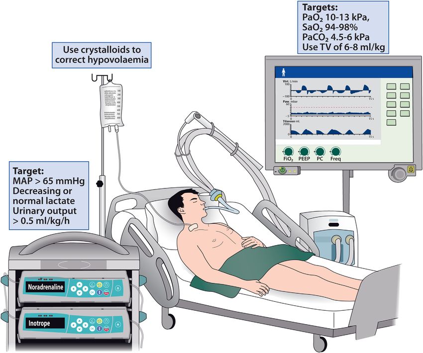
RESUS 8905 No. of Pages 50

2                                                     RESUSCITATION XXX (2021) XXX      XXX

                                                                           representation and diversity (gender, physician and non-physician,
Introduction and scope                                                     and geography (Northern and Southern Europe).
                                                                               These ERC-ESICM guidelines on post-resuscitation care for
In 2015 the European Resuscitation Council (ERC) and the European          adults are based mainly on the Advanced Life Support section of the
Society of Intensive Care Medicine (ESICM) collaborated to produce         2020 CoSTR document and represent consensus among the writing
their first combined post-resuscitation care guidelines, which were co-    group, which included representatives of the ERC and the ESICM.9
published in Resuscitation and Intensive Care Medicine.1,2 These           Where treatment recommendations are provided by ILCOR, these
post-resuscitation care guidelines have been extensively updated for       have been adopted by the ERC and ESICM. In the absence of an
2020 and incorporate the science that has been published since 2015.       ILCOR recommendation, ERC-ESICM guidance was based on
The topics covered include the post-cardiac arrest syndrome, control       review and discussion of the evidence by the working group until
of oxygenation and ventilation, haemodynamic targets, coronary             consensus was achieved. The writing group chairs ensured that
reperfusion, targeted temperature management, control of seizures,         everyone on the working group had the opportunity to present and
prognostication, rehabilitation, and long-term outcome.                    debate their views and ensured that discussions were open and
                                                                           constructive. All discussions took place during eight 2-h Zoom
                                                                           videoconferences that were held between January 2020 and
Methods                                                                    November 2020. Consensus was achieved by all 15 writing group
                                                                           members on all the treatment recommendations using an open
A comprehensive description of the guideline development process is        process.
provided in an electronic supplement.                                          These guidelines were drafted and agreed by the Post-
                                                                           Resuscitation Care Writing Group members before posting on the
The international consensus on cardiopulmonary                             ERC website for public comment between 21 October and 5 Novem-
resuscitation science evidence review process                              ber 2020. The opportunity to comment on the guidelines was
                                                                           advertised through social media (Facebook, Twitter) and the ERC
The International Liaison Committee on Resuscitation (ILCOR, www.          network of 33 national resuscitation councils. Nine individuals from
ilcor.org) includes representatives from the American Heart Associa-       four countries made 25 comments. One of these individuals was a lay
tion (AHA), the European Resuscitation Council (ERC), the Heart and        person. Review of these comments led to eight changes.
Stroke Foundation of Canada (HSFC), the Australian and New
Zealand Committee on Resuscitation (ANZCOR), the Resuscitation
Council of Southern Africa (RCSA), the Inter-American Heart                Summary of the key changes
Foundation (IAHF), and the Resuscitation Council of Asia (RCA).
From 2000 to 2015 researchers from the ILCOR member councils               A summary of the main changes from the 2015 ERC-ESICM Post-
evaluated resuscitation science in 5-yearly cycles. After publication of   resuscitation care guidelines is set out in Table 1.
the 2015 International Consensus on CPR and ECC Science with                  Key messages from the section are presented in Fig. 1.
Treatment Recommendations (2015 CoSTR),3 ILCOR committed to a
continuous evidence-evaluation process, with topics prioritised for
review by the task forces and with CoSTR updates published annually.       Concise guidelines for clinical practice
4 6
     For the 2020 CoSTR, the six ILCOR task forces performed three
types of evidence evaluation: the systematic review, the scoping           This section includes only a summary of the main recommendations.
review, and the evidence update, which covered 184 topics in total.7 It    The evidence underpinning each recommendation is detailed in the
was agreed that only systematic reviews (these used Grading of             section on ‘evidence informing the guidelines’.
Recommendations Assessment, Development, and Evaluation
(GRADE) methodology) could result in new or modified treatment             Immediate post-resuscitation care
recommendations.8 The data analysis from each systematic review
was presented to the task force, and the task force drafted the              Post-resuscitation care is started immediately after sustained
summary consensus on science and the treatment recommendations.               ROSC, regardless of location (Fig. 2).
Each treatment recommendation indicated the strength of the                  For out-of-hospital cardiac arrest consider transport to a cardiac
recommendation (recommends = strong, suggests = weak) and the                 arrest centre.
certainty of the evidence. Draft 2020 CoSTRs were posted on the
ILCOR website (ilcor.org) for a 2-week comment period after which          Diagnosis of cause of cardiac arrest
final wording of science statements and treatment recommendations
were completed by the task forces and published in Resuscitation and         If there is clinical (e.g. haemodynamic instability) or ECG evidence
Circulation as the 2020 Consensus on Science and Treatment                    of myocardial ischaemia, undertake coronary angiography first.
Recommendations (CoSTR).                                                      This is followed by CT brain and/or CT pulmonary angiography if
                                                                              coronary angiography fails to identify causative lesions.
The European Resuscitation Council and European Society                      Early identification of a respiratory or neurological cause can be
for intensive care medicine process for developing post-                      achieved by performing a brain and chest CT-scan at hospital
resuscitation care guidelines                                                 admission, before or after coronary angiography (see coronary
                                                                              reperfusion).
Fifteen individuals were selected for the ERC-ESICM Post-Resusci-            If there are signs or symptoms pre-arrest suggesting a
tation Care Writing Group based on their expertise, ERC and ESICM             neurological or respiratory cause (e.g. headache, seizures or

    Please cite this article in press as: J.P. Nolan, et al., European Resuscitation Council and European Society of Intensive Care Medicine
    Guidelines 2021: Post-resuscitation care, Resuscitation (2021), https://doi.org/10.1016/j.resuscitation.2021.02.012
RESUS 8905 No. of Pages 50

                                                              RESUSCITATION XXX (2021) XXX               XXX                                                               3

  Table 1 – Summary of changes since the 2015 Guidelines on Post-resuscitation care.
            2015 Guidelines                                                       2021 Guidelines                                    Rationale for change
  Coronary angiography
  It is reasonable to discuss and consider emergent cardiac       In patients with ROSC after OHCA without ST-            A randomised controlled trial showed no
  catheterisation laboratory evaluation after ROSC in             elevation on the ECG, emergent cardiac catheter-        difference in 90-day survival following out of
  patients with the highest risk of a coronary cause for their    isation laboratory evaluation should be considered if   hospital VF cardiac arrest among patients
  cardiac arrest                                                  there is an estimated high probability of acute         without ST-elevation on the ECG allocated to
                                                                  coronary occlusion (e.g. patients with haemody-         immediate coronary angiography versus de-
                                                                  namic and/or electrical instability).                   layed angiography.10 Recent ESC guidelines
                                                                                                                          state that ‘Delayed as opposed to immediate
                                                                                                                          angiography should be considered in haemo-
                                                                                                                          dynamically stable patients without ST-seg-
                                                                                                                          ment elevation successfully resuscitated after
                                                                                                                          an out-of-hospital cardiac arrest’.11

  Blood pressure target
  Target the mean arterial blood pressure to achieve an           Avoid hypotension (0.5 mL kg 1 h 1) (37.7  C) for at least 72 h after      33  C versus 37  C.13 This has enabled the
     (36  C) temperatures remains unknown, and further              ROSC in patients who remain in coma.                 recommendation to be extended to all rhythms
     research may help elucidate this.                                                                                    and locations.
    TTM is recommended for adults after OHCA with an                                                                     The definition of fever (>37.7  C) is consistent
     initial shockable rhythm who remain unresponsive                                                                     with that used in the TTM2 trial.14
     after ROSC (strong recommendation, low-quality
     evidence).
    TTM is suggested for adults after OHCA with an initial
     non-shockable rhythm who remain unresponsive after
     ROSC (weak recommendation, very low-quality
     evidence).
    TTM is suggested for adults after IHCA with any initial
     rhythm who remain unresponsive after ROSC (weak
     recommendation, very low-quality evidence).
    If targeted temperature management is used, it is
     suggested that the duration is at least 24 h (weak
     recommendation, very low-quality evidence).

  General intensive care management
  Short-acting drugs (e.g., propofol, alfentanil, remifentanil)     Use short acting sedatives and opioids.              The 2015 guidelines included very few state-
  will enable more reliable and earlier neurological                Avoid using a neuromuscular blocking drug            ments on general intensive care management.
  assessment and prognostication                                     routinely in patients undergoing TTM, but it may     For 2020 we have several best practice state-
  Following ROSC maintain the blood glucose at                       be considered in case of severe shivering            ments based mainly on data extrapolated from
  10 mmol L 1 (180 mg dL 1) and avoid hypoglycaemia.                during TTM.                                          other critically ill patient groups.
                                                                    Provide stress ulcer prophylaxis routinely in
                                                                     cardiac arrest patients.
                                                                    Provide deep venous thrombosis prophylaxis.
                                                                    Target a blood glucose of 7.8 10 mmol L 1
                                                                     (140 180 mg dL 1) using an infusion of insulin if
                                                                     required; avoid hypoglycaemia (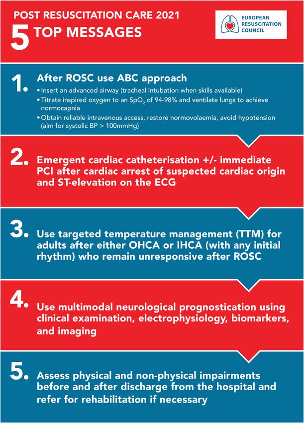
RESUS 8905 No. of Pages 50

4                                                             RESUSCITATION XXX (2021) XXX               XXX

    Table  1 (continued)
    neurological  deficits, shortness of breath or documented hypo-
            in patients
    xaemia 2015         with known respiratory disease), perform a CT2021 Guidelines
                  Guidelines                                                                                                          Rationale for change
    brain and/or a CT pulmonary angiogram.
                                                                     Start enteral feeding at low rates (trophic
                                                                      feeding) during TTM and increase after re-
                                                                      warming if indicated. If TTM of 36  C is used as
                                                                      the target temperature, trophic gastric feeding
                                                                      rates may be increased early during TTM.
                                                                     We do not recommend using prophylactic
                                                                      antibiotics routinely.

    Prognostication
    The prognostication strategy algorithm is applicable to all    In a comatose patient with M  3 at 72 h from          There has a very large amount of data published
    patients who remain comatose with an absent or extensor        ROSC, in the absence of confounders, poor               on prognostication since the 2015 guidelines. A
    motor response to pain at 72 h from ROSC. Results of          outcome is likely when two or more of the following     recent systematic review identified 94 studies
    earlier prognostic tests are also considered at this time      predictors are present:                                 that included over 30,000 patients, all published
    point.                                                            no pupillary and corneal reflexes at 72 h,         since January 2013.15
    One or both of the following indicate that a poor outcome is      bilaterally absent N20 SSEP wave at 24 h,          The two-stage prognostication algorithm in the
    very likely (FPR < 5%, narrow 95% CIs):                           highly malignant EEG (suppressed background         2015 guidelines has been simplified so that a
       No pupillary and corneal reflexes                               or burst-suppression) at >24 h,                    poor outcome is considered likely when two or
       Bilaterally absent N20 SSEP wave                              NSE >60 mg L 1 at 48 h and/or 72 h,                 more of the listed predictors are present. The
    Two or more of the following indicate that a poor outcome         status myoclonus 72 h,                             algorithm is valid for comatose patients with a
    is likely:                                                        or a diffuse and extensive anoxic injury on brain   Glasgow Motor Score 3 (compared with 2 in
       Status myoclonus 48 h after ROSC                               CT/MRI.                                            the 2015 version). A threshold value for NSE is
       High NSE levels                                                                                                    now stated. The EEG patterns suppression and
       Unreactive burst-suppression or status epilepticus on                                                              burst-suppression are the most consistent
          EEG                                                                                                              predictors of poor neurological outcome. Con-
       Diffuse anoxic injury on brain CT/MRI                                                                              versely, absence of EEG reactivity has been
                                                                                                                           only inconsistently associated with poor neu-
                                                                                                                           rological outcome in recent studies.
                                                                                                                           We suggest using the 2021 ACNS terminology
                                                                                                                           when assessing these patterns for prognosti-
                                                                                                                           cation, to ensure an unequivocal identification.

    Rehabilitation
    Follow-up care should be organised systematically and             Perform functional assessments of physical          The authorship of the 2021 guidelines now
    can be provided by a physician or specialised nurse. It            and non-physical impairments before dis-            includes 3 individuals with expertise on long-
    includes at least the following aspects:                           charge from the hospital to identify early          term outcomes and rehabilitation after cardiac
       Screening for cognitive impairments                            rehabilitation needs and refer to rehabilitation    arrest compared with one author in 2015. The
       Screening for emotional problems                               if necessary.                                       2021 guidelines include greater emphasis on
       Provision of information                                      Organise follow-up for all cardiac arrest survi-    functional assessments of physical and non-
                                                                       vors within 3 months after hospital discharge,      physical impairments before discharge and
                                                                       including:                                          long-term follow up and rehabilitation. There is
                                                                    1. Screening for cognitive problems.                   greater recognition of the importance of survi-
                                                                    2. Screening for emotional problems and fatigue.       vorship after cardiac arrest. The recommen-
                                                                    3. Providing information and support for survivors     dations in this section are all best practice
                                                                       and family members.                                 statements

    Cardiac arrest centres
    No specific recommendation                                     Adult patients with non-traumatic OHCA should be An expert consensus paper published by
                                                                   considered for transport to a cardiac arrest centre several European organisations including the
                                                                   according to local protocol.                        Association of Acute Cardiovascular Care
                                                                                                                       (ACVA) of the European Society of Cardiology
                                                                                                                       (ESC), the ERC and the ESICM, states that the
                                                                                                                       minimum requirements for a cardiac arrest
                                                                                                                       centre are 24/7 availability of an on-site
                                                                                                                       coronary angiography laboratory, an emer-
                                                                                                                       gency department, an ICU, imaging facilities,
                                                                                                                       such as echocardiography, CT, and MRI.16
                                                                                                                       Based on evidence from a systematic review,
                                                                                                                       ILCOR suggests that wherever possible, adult
                                                                                                                       patients with non-traumatic OHCA cardiac
                                                                                                                       arrest should be cared for in cardiac arrest
                                                                                                                       centres.17

    ACNS American Clinical Neurophysiology Society; CT computed tomography; ESC European Society of Cariology; EEG electroencephalogram; FPR false
    positive rate; ILCOR International Liaison Committee on Resuscitation; IHCA in-hospital cardiac arrest; MAP mean arterial pressure; MRI magnetic resonance
    imaging; NSE neuron specific enolase; OHCA out-of-hospital cardiac arrest; ROSC return of spontaneous circulation; SSEP somatosensory evoked potential;
    TTM targeted temperature management; VF ventricular fibrillation.

    Please cite this article in press as: J.P. Nolan, et al., European Resuscitation Council and European Society of Intensive Care Medicine
    Guidelines 2021: Post-resuscitation care, Resuscitation (2021), https://doi.org/10.1016/j.resuscitation.2021.02.012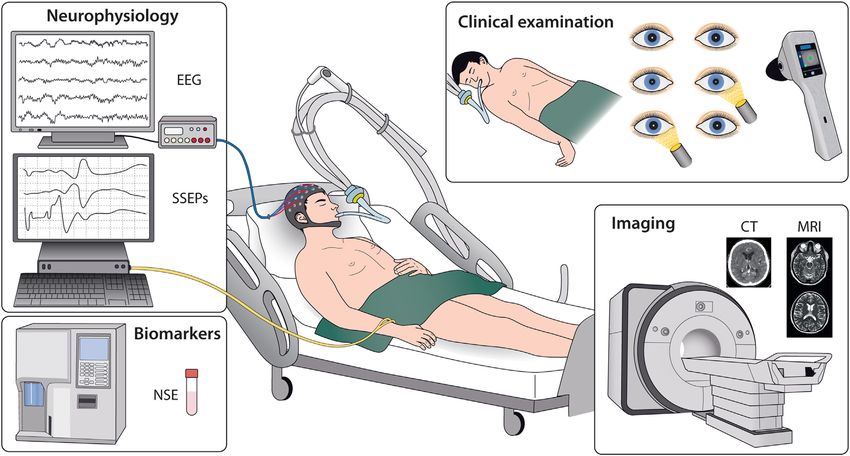
RESUS 8905 No. of Pages 50

                                                   RESUSCITATION XXX (2021) XXX       XXX                                                     5

                                      Fig. 1 – Post-resuscitation care infographic summary.

Airway and breathing                                                       Patients who remain comatose following ROSC, or who have
                                                                            another clinical indication for sedation and mechanical ventilation,
  Airway management after return of spontaneous circulation                should have their trachea intubated if this has not been done
  Airway and ventilation support should continue after return of           already during CPR.
   spontaneous circulation (ROSC) is achieved.                             Tracheal intubation should be performed only by experienced
  Patients who have had a brief period of cardiac arrest and an            operators who have a high success rate.
   immediate return of normal cerebral function and are breathing          Correct placement of the tracheal tube must be confirmed with
   normally may not require tracheal intubation but should be given         waveform capnography.
   oxygen via a facemask if their arterial blood oxygen saturation is      In the absence of personnel experienced in tracheal intubation,
   less than 94%.                                                           it is reasonable to insert a supraglottic airway (SGA) or

   Please cite this article in press as: J.P. Nolan, et al., European Resuscitation Council and European Society of Intensive Care Medicine
   Guidelines 2021: Post-resuscitation care, Resuscitation (2021), https://doi.org/10.1016/j.resuscitation.2021.02.012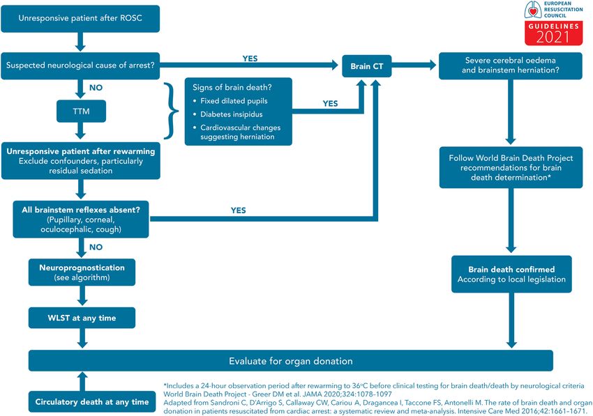
RESUS 8905 No. of Pages 50

6                                                   RESUSCITATION XXX (2021) XXX       XXX

Fig. 2 – Post resuscitation care algorithm.
SBP Systolic blood pressure; PCI Percutaneous coronary intervention; CTPA Computed tomography pulmonary angiogram; ICU Intensive care
unit; EEG electroencephalography; ICD implanted cardioverter defibrillator.

    Please cite this article in press as: J.P. Nolan, et al., European Resuscitation Council and European Society of Intensive Care Medicine
    Guidelines 2021: Post-resuscitation care, Resuscitation (2021), https://doi.org/10.1016/j.resuscitation.2021.02.012
RESUS 8905 No. of Pages 50

                                                        RESUSCITATION XXX (2021) XXX       XXX                                                       7

    maintain the airway with basic techniques until skilled                     Consider mechanical circulatory support (such as intra-aortic
    intubators are available.                                                    balloon pump, left-ventricular assist device or arterio-venous
                                                                                 extra corporal membrane oxygenation) for persisting cardio-
Control of oxygenation                                                           genic shock from left ventricular failure if treatment with fluid
  After ROSC, use 100% (or maximum available) inspired oxygen                   resuscitation, inotropes, and vasoactive drugs is insufficient.
   until the arterial oxygen saturation or the partial pressure of arterial      Left-ventricular assist devices or arterio-venous extra corporal
   oxygen can be measured reliably.                                              membrane oxygenation should also be considered in haemo-
  After ROSC, once SpO2 can be measured reliably or arterial blood              dynamically unstable patients with acute coronary syndromes
   gas values are obtained, titrate the inspired oxygen to achieve an            (ACS) and recurrent ventricular tachycardia (VT) or ventricular
   arterial oxygen saturation of 94 98% or arterial partial pressure of          fibrillation (VF) despite optimal therapy.
   oxygen (PaO2) of 10 13 kPa or 75 100 mmHg (Fig. 3).
  Avoid hypoxaemia (PaO2 < 8 kPa or 60 mmHg) following ROSC.                 Disability (optimising neurological recovery)
  Avoid hyperoxaemia following ROSC.
                                                                              Control of seizures
Control of ventilation                                                          We recommend using electroencephalography (EEG) to diag-
  Obtain an arterial blood gas and use end tidal CO2 in mechanically            nose electrographic seizures in patients with clinical convulsions
   ventilated patients.                                                          and to monitor treatment effects.
  In patients requiring mechanical ventilation after ROSC, adjust              To treat seizures after cardiac arrest, we suggest levetiracetam or
   ventilation to target a normal arterial partial pressure of carbon            sodium valproate as first-line antiepileptic drugs in addition to
   dioxide (PaCO2) i.e. 4.5 6.0 kPa or 35 45 mmHg.                               sedative drugs.
  In patients treated with targeted temperature management (TTM)               We suggest that routine seizure prophylaxis is not used in post-
   monitor PaCO2 frequently as hypocapnia may occur.                             cardiac arrest patients.
  During TTM and lower temperatures use consistently either a
   temperature or non-temperature corrected approach for measur-              Temperature control
   ing blood gas values.                                                        We recommend targeted temperature management (TTM)
  Use a lung protective ventilation strategy aiming for a tidal volume          for adults after either OHCA or in-hospital cardiac arrest
   of 6 8 mL kg 1 ideal body weight.                                             (IHCA) (with any initial rhythm) who remain unresponsive after
                                                                                 ROSC.
Circulation                                                                     Maintain a target temperature at a constant value between 32  C
                                                                                 and 36  C for at least 24 h.
Coronary reperfusion                                                            Avoid fever (>37.7  C) for at least 72 h after ROSC in patients who
  Emergent cardiac catheterisation laboratory evaluation (and                   remain in coma.
   immediate PCI if required) should be performed in adult patients             Do not use pre-hospital intravenous cold fluids to initiate
   with ROSC after cardiac arrest of suspected cardiac origin with               hypothermia.
   ST-elevation on the ECG.
  In patients with ROSC after out-of-hospital cardiac arrest (OHCA)          General intensive care management
   without ST-elevation on the ECG, emergent cardiac catheter-
   isation laboratory evaluation should be considered if there is an            Use short acting sedatives and opioids.
   estimated high probability of acute coronary occlusion (e.g.                 Avoid using a neuromuscular blocking drug routinely in patients
   patients with haemodynamic and/or electrical instability).                    undergoing TTM, but it may be considered in case of severe
                                                                                 shivering during TTM.
Haemodynamic monitoring and management                                          Provide stress ulcer prophylaxis routinely in cardiac arrest
  All patients should be monitored with an arterial line for continuous         patients.
   blood pressure measurements, and it is reasonable to monitor                 Provide deep venous thrombosis prophylaxis.
   cardiac output in haemodynamically unstable patients.                        Target a blood glucose of 7.8 10 mmol L 1 (140 180 mg dL 1)
  Perform early (as soon as possible) echocardiography in all                   using an infusion of insulin if required; avoid hypoglycaemia
   patients to detect any underlying cardiac pathology and quantify              (
RESUS 8905 No. of Pages 50
8 RESUSCITATION XXX (2021) XXX XXX
Fig. 3 – Haemodynamic, oxygenation and ventilation targets.
treatments based on the patient's chances of achieving a Index tests for neurological prognostication are aimed at
neurologically meaningful recovery (Fig. 4). assessing the severity of hypoxic-ischaemic brain injury. The
No single predictor is 100% accurate. Therefore, a multimodal neurological prognosis is one of several aspects to consider in
neuroprognostication strategy is recommended. discussions around an individual's potential for recovery.
When predicting poor neurological outcome, a high
specificity and precision are desirable, to avoid falsely pessimistic Multimodal prognostication
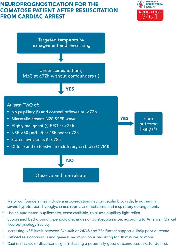
predictions. Start the prognostication assessment with an accurate clinical
The clinical neurological examination is central to prognostication. examination, to be performed only after major confounders
To avoid falsely pessimistic predictions, clinicians should avoid (e.g. residual sedation, hypothermia) have been excluded
potential confounding from sedatives and other drugs that may (Fig. 5).
confound the results of the tests. In a comatose patient with M 3 at 72 h from ROSC, in the
When patients are treated with TTM, daily clinical examination is absence of confounders, poor outcome is likely when two or more
advocated but final prognostic assessment should be undertaken of the following predictors are present: no pupillary and corneal
only after rewarming. reflexes at 72 h, bilaterally absent N20 SSEP wave at 24 h,
Clinicians must be aware of the risk of a self-fulfilling prophecy highly malignant EEG at >24 h, neuron specific enolase (NSE)
bias, occurring when the results of an index test predicting poor >60 mg L 1 at 48 h and/or 72 h, status myoclonus 72 h, or a
outcome is used for treatment decisions, especially regarding life- diffuse and extensive anoxic injury on brain CT/MRI. Most of these
sustaining therapies. signs can be recorded before 72 h from ROSC, however their
Please cite this article in press as: J.P. Nolan, et al., European Resuscitation Council and European Society of Intensive Care Medicine
Guidelines 2021: Post-resuscitation care, Resuscitation (2021), https://doi.org/10.1016/j.resuscitation.2021.02.012RESUS 8905 No. of Pages 50
RESUSCITATION XXX (2021) XXX XXX 9
Fig. 4 – Prognostication modes. EEG electroencephalography; NSE neuron specific enolase; SSEP somatosensory
evoked potential.
results will be evaluated only at the time of clinical prognostic The presence of unequivocal seizures on EEG during the first 72 h
assessment. after ROSC is an indicator of a poor prognosis.
Absence of background reactivity on EEG is an indicator of poor
Clinical examination prognosis after cardiac arrest.
Clinical examination is prone to interference from sedatives, Bilateral absence of somatosensory evoked cortical N20-
opioids or muscle relaxants. A potential confounding from residual potentials is an indicator of poor prognosis after cardiac arrest.
sedation should always be considered and excluded. Always consider the results of EEG and somatosensory evoked
A Glasgow Motor Score of 3 (abnormal flexion or worse in potentials (SSEP) in the context of clinical examination findings
response to pain) at 72 h or later after ROSC may identify patients and other tests. Always consider using a neuromuscular
in whom neurological prognostication may be needed. blocking drug when performing SSEP.
In patients who remain comatose at 72 h or later after ROSC the
following tests may predict a poor neurological outcome: Biomarkers
The bilateral absence of the standard pupillary light reflex. Use serial measurements of NSE in combination with other
Quantitative pupillometry methods to predict outcome after cardiac arrest. Increasing values
The bilateral absence of corneal reflex between 24 and 48 h or 72 h in combination with high values at 48
The presence of myoclonus within 96 h and, in particular, status and 72 h indicate a poor prognosis.
myoclonus within 72 h
We also suggest recording the EEG in the presence of myoclonic Imaging
jerks to enable detection of any associated epileptiform activity or Use brain imaging studies for predicting poor neurological
EEG signs, such as background reactivity or continuity, suggest- outcome after cardiac arrest in combination with other
ing a potential for neurological recovery. predictors, in centres where specific experience in these studies
is available.
Neurophysiology Use presence of generalised brain oedema, manifested by a
Perform an EEG in patients who are unconscious after the arrest. marked reduction of the grey matter/white matter ratio on brain CT,
Highly malignant EEG-patterns include suppressed background or extensive diffusion restriction on brain MRI to predict poor
with or without periodic discharges and burst-suppression. We neurological outcome after cardiac arrest.
suggest using these EEG-patterns after the end of TTM and after Always consider findings from imaging in combination with other
sedation has been cleared as indicators of a poor prognosis. methods for neurological prognostication.
Please cite this article in press as: J.P. Nolan, et al., European Resuscitation Council and European Society of Intensive Care Medicine
Guidelines 2021: Post-resuscitation care, Resuscitation (2021), https://doi.org/10.1016/j.resuscitation.2021.02.012RESUS 8905 No. of Pages 50 10 RESUSCITATION XXX (2021) XXX XXX Fig. 5 – Prognostication strategy algorithm. EEG electroencephalography; NSE neuron specific enolase; SSEP somatosensory evoked potential; ROSC return of spontaneous circulation. Please cite this article in press as: J.P. Nolan, et al., European Resuscitation Council and European Society of Intensive Care Medicine Guidelines 2021: Post-resuscitation care, Resuscitation (2021), https://doi.org/10.1016/j.resuscitation.2021.02.012
RESUS 8905 No. of Pages 50

                                                       RESUSCITATION XXX (2021) XXX       XXX                                                  11

Fig. 6 – Recommendations for in-hospital functional assessments, follow-up and rehabilitation after cardiac arrest.

Withdrawal of life-sustaining therapy                                          Organise follow-up for all cardiac arrest survivors within 3 months
                                                                                after hospital discharge, including:
  Separate discussions around withdrawal of life-sustaining therapy            1. Screening for cognitive problems.
   (WLST) and the assessment of prognosis for neurological                      2. Screening for emotional problems and fatigue.
   recovery; WLST decisions should consider aspects other than                  3. Providing information and support for survivors and family
   brain injury such as age, co-morbidity, general organ function and              members.
   the patients’ preferences.
  Allocate sufficient time for communication around the level-of-           Organ donation
   treatment decision within the team and with the relatives.
                                                                               All decisions concerning organ donation must follow local legal
Long-term outcome after cardiac arrest                                          and ethical requirements.
                                                                               Organ donation should be considered in those who have achieved
  Perform functional assessments of physical and non-physical                  ROSC and who fulfil neurological criteria for death (Fig. 7).
   impairments before discharge from the hospital to identify early            In comatose ventilated patients who do not fulfil neurological
   rehabilitation needs and refer to rehabilitation if necessary (Fig. 6).      criteria for death, if a decision to start end-of-life care and

   Please cite this article in press as: J.P. Nolan, et al., European Resuscitation Council and European Society of Intensive Care Medicine
   Guidelines 2021: Post-resuscitation care, Resuscitation (2021), https://doi.org/10.1016/j.resuscitation.2021.02.012
RESUS 8905 No. of Pages 50
12 RESUSCITATION XXX (2021) XXX XXX
withdrawal of life support is made, organ donation should be
considered for when circulatory arrest occurs.
Fig. 7 – Organ donation after cardiac arrest algorithm.
Cardiac arrest centres deaths.23,26,27 Post-cardiac arrest hypoxic-ischaemic brain injury is
associated with hypotension, hypoxaemia, hyperoxaemia, pyrexia,
Adult patients with non-traumatic OHCA should be considered for hypoglycaemia, hyperglycaemia and seizures. Significant myocardial
transport to a cardiac arrest centre according to local protocol. dysfunction is common after cardiac arrest but typically starts to
recover by 2 3 days, although full recovery may take significantly
longer.28 33 The whole-body ischaemia/reperfusion of cardiac arrest,
Evidence informing the guidelines CPR and ROSC activates immune and coagulation pathways
contributing to multiple organ failure and increasing the risk of
Post-cardiac arrest syndrome infection.34 43 Thus, the post-cardiac arrest syndrome has many
features in common with sepsis, including intravascular volume
The post-cardiac arrest syndrome comprises post-cardiac arrest depletion, vasodilation, endothelial injury and abnormalities of the
hypoxic-ischaemic brain injury, post-cardiac arrest myocardial microcirculation.44 53
dysfunction, the systemic ischaemia/reperfusion response, and the
persistent precipitating pathology.18 21 The severity of this syndrome Diagnosis of cause of cardiac arrest
will vary with the duration and cause of cardiac arrest. It may not occur
at all if the cardiac arrest is brief. Among patients surviving to intensive These guidelines are informed by expert consensus.
care unit (ICU) admission but subsequently dying in-hospital, Cardiac causes of OHCA have been studied extensively in the last
withdrawal of treatment following prognostication of poor neurological few decades; conversely, little is known about non-cardiac causes.
outcome is the cause of death in approximately two-thirds after Early identification of a respiratory or neurological cause would enable
OHCA and approximately 25% after in-hospital cardiac arrest.22 26 transfer of the patient to a specialised ICU for optimal care. Improved
Cardiovascular failure accounts for most deaths in the first three days, knowledge of prognosis also enables discussion about the appropri-
while, in many countries, WLST based on a prognostication of severe ateness of specific therapies, including TTM. Several case series
hypoxic-ischaemic brain injury accounts for most of the later showed that this strategy enables diagnosis of non-cardiac causes of
Please cite this article in press as: J.P. Nolan, et al., European Resuscitation Council and European Society of Intensive Care Medicine
Guidelines 2021: Post-resuscitation care, Resuscitation (2021), https://doi.org/10.1016/j.resuscitation.2021.02.012RESUS 8905 No. of Pages 50
RESUSCITATION XXX (2021) XXX XXX 13
arrest in a substantial proportion of patients.54,55 There is consider- oxygenation targets for varying durations immediately and up to
able regional variation in the incidence of sub-arachnoid haemorrhage 48 h after ROSC.74 79 A sub-group analysis of a large RCT targeting
as a cause of cardiac arrest among those with sustained ROSC at an arterial blood oxygen saturation of 90 97% compared with 90
hospital admission. Published case series report 16.2% in Japan,56 100% showed that in patients at risk of hypoxic-ischaemic brain
11.4% in Korea57 and 7% in France.58 In those with cardiac arrest injury 180-day mortality was lower in the lower oxygen target group74;
associated with trauma or haemorrhage a whole-body CT scan is however, this difference was no longer statistically significant when
likely indicated.9,59,60 adjusted for baseline differences.80 A pilot RCT targeting a PaO2 of
10 15 kPa compared with 20 25 kPa showed no difference in the
Airway and breathing values of biomarkers of neurological injury.75 Overall, the evidence is
mixed but suggests targeting normal oxygenation rather than
Airway management after return of spontaneous circulation hyperoxaemia. Observational data suggests avoiding hypoxaemia
These guidelines are informed by expert consensus. but there are no RCTs on this topic.
Patients can have their trachea intubated before, during or following In most post-cardiac arrest patients, controlled oxygenation will
cardiac arrest depending on the setting or particular circumstances.61 require tracheal intubation and mechanical ventilation for at least
Following most cardiac arrests tracheal intubation will occur during CPR 24 72 h. The exception being the completely conscious patient with a
or if the patient remains comatose after ROSC.62 patent airway who should be treated with an oxygen mask or non-
Tracheal intubation following ROSC in comatose patients will invasive ventilation targeting a peripheral oxygen saturation (SpO2) of
facilitate post-resuscitation care that includes controlled oxygen- 94 98%. During cardiac arrest, patients’ lungs are ventilated with the
ation and ventilation, protection of the lungs from aspiration of maximum feasible inspired oxygen, which is usually 100% during
stomach contents, control of seizures, and TTM see below for advanced resuscitation.9 After ROSC the goal should be to monitor
further details. oxygenation either with a pulse oximeter or preferably with an early
Post ROSC patients are haemodynamically unstable and, arterial blood gas sample. Oxygenation measured early after ROSC is
depending on their level of consciousness, may require drug assisted highly variable, varying from hypoxaemia to extreme hyperoxaemia.81
tracheal intubation. The same level of care should be provided as for Thus, it is appropriate to titrate the inspired oxygen by adjusting either
any other critically ill patient in terms of skills of the provider, the oxygen flow if using bag-mask ventilation or the fraction inspired
monitoring, and choice of drugs.63,64 There are no recommendations oxygen (FiO2) if using a mechanical ventilator.82 Prolonged use of
for a specific drug combination,65 but use of a low dose of a sedative, 100% inspired oxygen, for example during transport, will lead
an analgesic and a rapid onset neuromuscular blocking drug is commonly to extreme hyperoxaemia.83 Another method for monitor-
probably optimal. ing is using cerebral oxygen monitoring with near infrared spectros-
copy, but its role during post resuscitation care is uncertain.84,85
Control of oxygenation
These guidelines are informed by the ILCOR systematic review on Control of ventilation
oxygenation and ventilation targets after cardiac arrest, which These guidelines are informed by the same ILCOR systematic review
identified seven RCTs and 36 observational studies.66 and CoSTR.9 noted in the section on oxygenation.9,66 The ILCOR treatment
The ILCOR treatment recommendations in relation to oxygenation recommendations in relation to ventilation are:
are: There is insufficient evidence to suggest for or against targeting
We suggest the use of 100% inspired oxygen until the arterial mild hypercapnia compared with normocapnia in adults with
oxygen saturation or the partial pressure of arterial oxygen can be ROSC after cardiac arrest.
measured reliably in adults with ROSC after cardiac arrest in any We suggest against routinely targeting hypocapnia in adults with
setting (weak recommendation, very low-certainty evidence). ROSC after cardiac arrest. (weak recommendation, low-certainty
We recommend avoiding hypoxaemia in adults with ROSC after evidence).
cardiac arrest in any setting (strong recommendation, very low-
certainty evidence). After ROSC, blood carbon dioxide values (PaCO2) are commonly
We suggest avoiding hyperoxaemia in adults with ROSC after increased because of intra-arrest hypoventilation and poor tissue
cardiac arrest in any setting (weak recommendation, low-certainty perfusion,86 causing a mixed respiratory acidosis and metabolic
evidence). acidosis.87 Carbon dioxide is a well-known regulator of blood vessel
tone and cerebral blood flow.88 Increased PaCO2 (hypercapnia)
From a pathophysiological perspective, post cardiac arrest increases cerebral blood flow, cerebral blood volume and intracere-
patients are at risk of developing hypoxic-ischaemic brain injury bral pressure. Hypocapnia causes vasoconstriction that may
and accompanying organ dysfunction.9,21,67,68 The role of blood decrease blood flow and cause cerebral ischaemia.89
oxygen values in the disease process is poorly understood.69 Studies The main method for controlling PaCO2 in a mechanically
show that cerebral ischaemia in post cardiac arrest patients is ventilated patient is adjusting the minute volume by changing the
associated with poor outcome.70 Administering more oxygen can ventilation frequency and or tidal volume. In general, limiting the tidal
increase brain oxygenation.71 On the other hand, higher oxygen volume and using a lung protective ventilation strategy is the standard
values would logically cause an increase in harmful oxygen free of care, especially in patients with acute respiratory distress syndrome
radicals.72 It is also likely that the effect of oxygen values varies (ARDS).9,90,91 Acute respiratory distress syndrome is not uncommon
between different organs such as the heart and brain. in cardiac arrest patients and is associated with worse out-
Numerous experimental studies have assessed the impact of comes.9,92,93 Low lung compliance predicts poor functional outcome
hyperoxaemia on neurological injury with mixed findings.73 Six in OHCA patients94 ; however, ventilation with lower tidal volumes is
randomised controlled trials (RCTs) have compared different not standard practice in neurointensive care.95
Please cite this article in press as: J.P. Nolan, et al., European Resuscitation Council and European Society of Intensive Care Medicine
Guidelines 2021: Post-resuscitation care, Resuscitation (2021), https://doi.org/10.1016/j.resuscitation.2021.02.012RESUS 8905 No. of Pages 50
14 RESUSCITATION XXX (2021) XXX XXX
Two pilot studies have compared different carbon dioxide targets recommend emergency cardiac catheterisation laboratory evaluation
during post resuscitation care.75,96 One study found targeting mild in comparison with cardiac catheterisation later in the hospital stay or no
hypercapnia (50 55 mmHg) compared with normocapnia (35 45 catheterization in select adult patients with ROSC after OHCA of
mmHg) resulted in lower neuron specific enolase (NSE) values, a suspected cardiac origin with ST elevation on ECG (strong recommen-
marker of the magnitude of neurological injury.96 Another pilot study dation, low-quality evidence). The 2017 European Society of
compared the lower and higher end of the range for normocapnia (33 Cardiology Guidelines for the management of acute myocardial
45 mmHg) for the first 36 h of post resuscitation care and found no infarction with ST-segment elevation state that ‘a primary PCI strategy
difference in markers of neurological injury.75 Both of these studies is recommended in patients with resuscitated cardiac arrest and an
showed that a higher PaCO2 was associated with higher cerebral ECG consistent with STEMI’.113
oxygenation measured with near infrared spectroscopy (NIRS), but the
clinical implications of this are uncertain.85 Several large observational Percutaneous coronary intervention following ROSC without
studies have aimed to define the optimal CO2 during post-cardiac arrest ST-elevation
care.97 102 The results are mixed, with some studies indicating harm In OHCA patients without ST segment elevation, several large
from both hypo- and hypercapnia and some suggesting better outcome observational series showed that absence of ST segment elevation
with mild hypercapnia. Recent UK observational data suggest a does not completely exclude the presence of a recent coronary
relationship between arterial oxygen and carbon dioxide. Data from the occlusion.114 Therefore, the decision for early CAG should be based
first 24 h of post resuscitation care observed a combination of hypoxia on meticulous patient assessment for the presence of haemodynamic
and hypocapnia was associated with a worse outcome and did not or electrical instability and ongoing myocardial ischaemia taking into
report harm from hyperoxia.103 Previous observational data from account multiple factors including previous medical history, warning
Finnish ICUs reported similar findings.97 symptoms before arrest, initial cardiac rhythm for CA,115 ECG pattern
Observational data suggest that patients undergoing TTM are prone post ROSC, and echocardiography, as well as comorbidities. When
to hypocapnia.104 This may be avoided by frequent measurement of an ischaemic cause is considered likely, a similar approach as for
carbon dioxide with arterial blood gas analysis and use of end tidal CO2 patients with STEMI should be followed. In patients with a low
monitoring. In patients undergoing TTM with lower temperature targets, probability of an ischaemic cause of cardiac arrest, delaying CAG for
PaCO2 management including measurement is particularly challeng- few hours or days may buy time for initial management in ICU,
ing.105 There is limited evidence to support a particular method for enabling early initiation of post-resuscitation care (haemodynamic
measuring PaCO2 during hypothermia, therefore the guidance to use optimisation, protective ventilation, TTM) and prognostication. This
either a temperature or non-temperature corrected approach for ‘wait and see’ management may also avoid performing CAG in
measuring blood gases is based on expert opinion.106 patients with the lowest probability of an acute coronary lesion. These
The recommendation for tidal volume is based on current guidance two strategies (early versus delayed CAG) were evaluated in patients
for lung protective ventilation in the ICU107 and limited observational with VF arrest and without shock in an RCT that showed no difference
data from post cardiac arrest patients.108 One observational study in 90-day survival, the primary outcome (odds ratio 0.89; 95%
suggests that using a tidal volume of 6 8 mL kg 1 to ventilate the confidence interval [CI], 0.62 to 1.27; P = 0.51),10 In this study, the
lungs of post-cardiac arrest patients may be associated with improved median time to target temperature was 5.4 h in the immediate
outcome.108 This study also showed that by using higher ventilation angiography group and 4.7 h in the delayed angiography group (ratio
frequency normocapnia may be achieved.108 of geometric means, 1.19; 95% CI, 1.04 to 1.36). Another recently
published pilot RCT comparing early with delayed CAG also showed
Circulation no difference in the primary outcome, which was a composite of
efficacy and safety measures.116 Further trials testing the same
Coronary reperfusion hypothesis are ongoing (DISCO NCT02309151, COUPe
NCT02641626, TOMAHAWK NCT02750462, EMERGE
Percutaneous coronary intervention following ROSC with NCT02876458). The 2020 European Society of Cardiology Guide-
ST-elevation lines for the management of acute coronary syndromes in patients
Arrhythmia caused by myocardial ischaemia is the commonest cause of without persistent ST-segment elevation state that ‘delayed as
sudden cardiac death (SCD) in adults.109,110 Immediate reperfusion opposed to immediate angiography should be considered in
using percutaneous coronary intervention (PCI) of the culprit coronary haemodynamically stable patients without ST-segment elevation
lesion has been used for more than 20 years. This strategy is supported successfully resuscitated after an out-of-hospital cardiac arrest’.11
by many observational studies that reported a significant association Ideally, coronary interventions would be undertaken only in those
between early PCI with survival and favourable neurological outcome patients without permanent severe neurological injury. Patients with
after OHCA. Whilst the benefit of early PCI in OHCA caused by a recent irreversible hypoxic-ischaemic brain injury are unlikely to benefit from
coronary occlusion is universally acknowledged, the main challenge is PCI, even if a culprit coronary lesion is successfully treated.117
to identify the best candidates for coronary angiography (CAG) among However, the absence of a universally acceptable prognostic tool in
all resuscitated patients. In patients with ST segment elevation (STE) or the first hours after ROSC makes it impossible to identify such patients
left bundle branch block (LBBB) on the post-ROSC electrocardiogram with high sensitivity and specificity at the time of hospital admission.
(ECG) more than 80% will have an acute coronary lesion.111 A
systematic review completed for the 2015 ILCOR CoSTR identified Haemodynamic monitoring and management
15 observational studies enrolling 3800 patients showing a mortality
benefit for emergent versus delayed or no cardiac catheterisation Haemodynamic monitoring
among patients with ROSC after cardiac arrest with evidence of STE on Post-resuscitation myocardial dysfunction and low cardiac index may
their ECG.112 The treatment recommendation from 2015 was to occur in up to 60% of post-cardiac arrest patients30,118 and may be
Please cite this article in press as: J.P. Nolan, et al., European Resuscitation Council and European Society of Intensive Care Medicine
Guidelines 2021: Post-resuscitation care, Resuscitation (2021), https://doi.org/10.1016/j.resuscitation.2021.02.012RESUS 8905 No. of Pages 50
RESUSCITATION XXX (2021) XXX XXX 15
even more common in patients with an acute myocardial infarction outcome, we do not have high certainty evidence to guide an optimal
(AMI) as the cause of the arrest.119 Early echocardiography can MAP target.
identify underlying cardiac pathology, quantify the degree of Mean arterial pressure (MAP) is one of the main determinants of
myocardial dysfunction and help guide haemodynamic management. cerebral blood flow (CBF).143 Although a high MAP is generally
Serial echocardiography or invasive monitoring with a pulmonary required in non-anoxic brain injured patients because of cerebral
artery catheter quantifies myocardial dysfunction and indicates swelling and increased intracranial pressure (ICP),144 few data on ICP
trends.28,29,120 Impaired cardiac function is most common during values are available in cardiac arrest survivors. In many post-cardiac
the first 24 48 h after which it gradually resolves.30,118 Whether low arrest patients, CBF autoregulation is impaired or the lower limit is
cardiac output (or index) is associated with poor outcome is currently right-shifted.133,145 This means that at lower MAP values, in some
unclear. A sub-study of the TTM trial showed that low cardiac index patients CBF may be MAP-dependent with an increased risk of
may not be associated with outcome if lactate clearance is cerebral hypoperfusion (i.e. hypotension) or hyperaemia and
maintained.121 These findings were independent of target tempera- intracranial hypertension (i.e. hypertension).
ture. Both non-invasive and invasive monitoring with echocardiogra- The use of cerebral oxygen saturation or ICP monitoring to
phy, arterial lines and measurement of cardiac output are commonly determine the presence of autoregulation and to determine an optimal
used in intensive care and it is reasonable to use these to guide MAP may enable a more individualised approach.146 In a retrospec-
treatment in cardiac arrest patients (best practice statement). tive study, the estimated optimal MAP (i.e. MAP target at which the
autoregulation is more effective) was 85 mmHg in post-cardiac arrest
Haemodynamic management patients with preserved autoregulation and 100 mmHg when the
autoregulation was impaired.133 Another small observational study
Mean arterial pressure and cerebral perfusion calculated a median optimal MAP of 89 mmHg in the same setting.147
A systematic review completed for the 2015 ILCOR CoSTR searched However, there are no prospective studies evaluating whether an
for studies that compared titration of therapy to achieve a specific autoregulation-driven MAP target may influence neurological injury
haemodynamic goal with no haemodynamic goal.122 At that time, only and/or outcome. A more recent study has shown that after cardiac
observational studies were identified.123 127 That systematic review arrest, in particular in cases of non-cardiac origin, episodes of
also identified observational studies that compared a bundle of elevated ICP and/or brain hypoxia are frequent and a higher MAP is
therapies with a specific blood pressure target with no bundle.128 130 necessary to improve brain oxygenation.147 Preliminary evidence
The 2015 CoSTR treatment recommendations were: based on measurement of brain tissue oxygenation (PbtO2) has
We suggest haemodynamic goals (e.g., MAP, systolic blood shown that in resuscitated comatose patients impairment of oxygen
pressure) be considered during post-resuscitation care and as diffusion to the brain may cause persisting brain hypoxia despite
part of any bundle of post-resuscitation interventions (weak optimisation of oxygen delivery to the brain.148 The implementation
recommendation, low-quality evidence). and the safety of these invasive monitoring tools in cardiac arrest
There is insufficient evidence to recommend specific haemody- patients need to be further evaluated. While these are all
namic goals; such goals should be considered on an individual observational findings, they indicate optimal MAP targets may need
patient basis and are likely to be influenced by post-cardiac arrest to be individualised and support further research into identification of
status and pre-existing comorbidities (weak recommendation, optimal MAP targets for individual cardiac arrest survivors receiving
low-quality evidence). intensive care. In the post cardiac arrest patient, transcranial Doppler
(TCD) can give information about cerebral haemodynamics and, in the
An evidence update for this topic was included in the 2020 ILCOR future, may have a role in optimising haemodynamics in these
CoSTR and included two RCTs9,131,132 and 11 observational patients.149 Changes in cerebral blood flow can be seen using TCD
studies121,133 142 published since the 2015 systematic review.122 and this may be a target to for treatment.150 152 However, the
Two RCTs (including 232 patients) compared a blood pressure target technique and interpretations of the images is operator dependent and
of 65 75 mmHg to 80 100 mmHg with131 and without132 goal- requires an acoustic window in the patient. Moreover, cerebral
directed optimisation of cardiac function. These studies were not haemodynamics are continuously changing and serial measurements
powered for clinical outcomes but used surrogate markers of are possible only intermittently and the monitoring is labour-intensive.
neurological injury such as MRI131 and NSE.132 Whilst these studies Based on the evidence summarised by ILCOR9 we suggest avoiding
showed that higher MAP targets with vasopressors are safe, and do hypotension (MAP < 65 mmHg) and targeting MAP to achieve
not, for example, lead to cardiac arrhythmias, they failed to show any adequate urine output (>0.5 mL 1 kg h 1) and normal or decreasing
clear improvement in surrogate markers of brain injury with a higher lactate values (best practice statement).
MAP target.
Nine observational studies found hypotension was associated with Heart rate
poor outcome.134 139,141,142 One study found time spent below Tachycardia was associated with poor outcome in one retrospective
optimal MAP (assessed by correlation between near-infrared study.153 During mild induced hypothermia the normal physiological
spectroscopy and blood pressure) was associated with poor response is bradycardia. In animal models this has been shown to
outcome;133 one study did not find low cardiac output to be associated reduce the diastolic dysfunction that is usually present early after
with poor outcome,121 while the last study documented better cardiac arrest.154 Bradycardia was previously considered to be a side
outcomes among patients given fluids compared with vasopressors effect, especially below a rate of 40 min 1; however, bradycardia has
to increase MAP.140 These observations are similar to the five been shown to be associated with a good outcome.155,156 Similar
observational studies included in the 2015 ILCOR Guidelines.122 association between bradycardia and improved long-term outcome
While hypotension (RESUS 8905 No. of Pages 50
16 RESUSCITATION XXX (2021) XXX XXX
Sedation, controlled ventilation and a temperature between 32 arrest, and hydrocortisone in those with post-ROSC shock compared
36 C lowers oxygen consumption in cardiac arrest patients. with only adrenaline and placebo (18/130 [13.9%] versus 7/138
Although bradycardia generally reduces cardiac output, this is well [5.1%]; RR, 2.94; 95% CI, 1.16 6.50) 163 Only the third RCT confined
tolerated in this post-arrest setting. We suggest bradycardia (heart the use of steroids to the post-resuscitation phase; it did not show any
rate < 30 40 min 1) be left untreated as long as there are no signs of benefit for steroid post-ROSC but included only 50 patients.166
hypoperfusion (i.e. increasing lactate, reduced urinary output etc.) One trial has recently been completed but is not yet published
(best practice statement). (NCT02790788). ILCOR recommended a systematic review be
undertaken once the recently completed trial is published, and
Fluid resuscitation, vasoactive and inotropic drugs therefore left the treatment recommendation unchanged from
There is limited evidence to guide optimal fluid therapy for post- 2010:167
cardiac arrest patients. One study during which invasive monitoring There is insufficient evidence to support or refute the use of
and filling pressures were used observed that up to 5 7 L of fluid were corticosteroids for patients with ROSC following cardiac arrest.
given during the first 24 h.30 One retrospective study indicated that
with a treatment algorithm involving the pulse contour continuous Until there is higher-certainty evidence supportive of their use, we
cardiac output (PiCCO) system larger fluid volumes (range 4 5 L suggest that steroids are not given routinely to post-cardiac arrest
during the first 24 h) were associated with a lower incidence of acute patients (weak recommendation, low-certainty evidence).
kidney injury.158
There is little direct evidence comparing various vasoactive drugs Potassium
for post-cardiac arrest patients, therefore this recommendation is Hyperkalaemia is common immediately after cardiac arrest. Subse-
based on indirect evidence from critically ill patients in general. The quent endogenous catecholamine release and correction of metabolic
most recent Cochrane review on vasopressors for hypotensive shock and respiratory acidosis promotes intracellular transportation of
included 28 RCTs (n = 3497 patients) and did not find any mortality potassium, causing hypokalaemia. Hyperkalaemia in the post-cardiac
benefit from any of the six vasopressors assessed. Acknowledging arrest period is associated with worse outcome.168 Hypokalaemia, on
noradrenaline as the most commonly used vasopressor, their the other hand may predispose to ventricular arrhythmias. Based on
suggestion was that major changes in clinical practice were not these observational studies we suggest that potassium be given to
needed.159 As noradrenaline is the most widely used vasoactive agent maintain the serum potassium concentration between 4.0 and
for post-cardiac arrest patients, we suggest using noradrenaline as 4.5 mmol L 1 (best practice statement).
the first-line vasoactive agent in hypotensive post-cardiac arrest
patients. A recent RCT comparing noradrenaline with adrenaline in Mechanical circulatory support
57 patients with acute myocardial infarction and cardiogenic shock If treatment with fluid resuscitation, inotropes and vasoactive drugs is
was terminated early because of significantly more refractory shock in insufficient to support the circulation, consider insertion of a
patients treated with adrenaline.160 The COMACARE and NEURO- mechanical circulatory assistance device (e.g. IMPELLA, Abiomed,
PROTECT pilot trials also used noradrenaline as the drug of choice to USA).126,169,170 One study indicated that 10 15% of patients with
achieve higher MAP targets.131,132 None of the studies showed any OHCA and ongoing cardiogenic shock eventually require mechanical
evidence of relevant tachycardia, arrhythmias or recurrent shock in circulatory support.171 In patients with cardiogenic shock without
the higher MAP group, despite the use of significantly higher doses of cardiac arrest some centres still advocate use of an intra-aortic
noradrenaline compared with the lower MAP group. This suggests balloon pump (IABP), although the IABP-SHOCK II Trial failed to show
that noradrenaline is well tolerated in post-cardiac arrest patients.131 that use of the IABP improved 30-day mortality in patients with
Post-resuscitation myocardial dysfunction often requires inotropic myocardial infarction and cardiogenic shock.172,173 One recent small
support. Based on experimental data, dobutamine is the most RCT found no difference in outcome in patients with acute myocardial
established treatment in this setting,161,162 but the systemic infarction and cardiogenic shock treated with an IMPELLA device
inflammatory response that occurs frequently in post-cardiac arrest compared with an IABP.174 Another retrospective study including only
patients also causes vasoplegia and severe vasodilation.30 The post-cardiac arrest patients found no difference in clinical outcome but
NEUROPROTECT trial used dobutamine to increase cardiac index in higher incidence of bleeding with the use of IMPELLA compared with
the higher MAP group. Although this did not decrease neurological IABP.169 Thus far, the evidence about which type of mechanical
injury it also did not increase myocardial injury.131 device is superior appears inconclusive and thus their use should be
decided on a case-by-case basis.
Steroids The 2015 ESC Guidelines for the management of patients with
ILCOR performed an evidence update on use of steroids for post- ventricular arrhythmias and the prevention of sudden cardiac death
cardiac arrest patients for the 2020 guidelines.9 Three small RCTs and include the following recommendation for the use of mechanical
a large observational study have addressed the use of steroids in post- circulatory support: left-ventricular assist devices or arterio-venous
cardiac arrest patients.163 166 Two of the RCTs used steroids both extra corporal membrane oxygenation should also be considered in
during CPR for IHCA and after ROSC.163,164 The first of these RCTs haemodynamically unstable patients with acute coronary syndromes
showed improved survival to discharge with a combination of (ACS) and recurrent ventricular tachycardia (VT) or ventricular
methylprednisolone, vasopressin, and adrenaline during cardiac fibrillation (VF) despite optimal therapy.175
arrest and hydrocortisone after ROSC for those with shock, compared
with the use of only adrenaline and placebo (9/48 [19%] versus 2/52 Implantable cardioverter defibrillators
[4%];RR, 4.87; 95% CI, 1.17 13.79).164 The second RCT showed An implantable cardioverter defibrillator (ICD) is a device used for
improved survival to discharge with favourable neurological outcome the treatment of certain life-threatening arrhythmias. The Europe-
with methylprednisolone, vasopressin, and adrenaline during cardiac an Society of Cardiology has published guidelines on the
Please cite this article in press as: J.P. Nolan, et al., European Resuscitation Council and European Society of Intensive Care Medicine
Guidelines 2021: Post-resuscitation care, Resuscitation (2021), https://doi.org/10.1016/j.resuscitation.2021.02.012You can also read