Reversal of senescence associated beta galactosidase expression during in vitro three dimensional tissue engineering of human chondrocytes in a ...
←
→
Page content transcription
If your browser does not render page correctly, please read the page content below
www.nature.com/scientificreports
OPEN Reversal of senescence‑associated
beta‑galactosidase expression
during in vitro three‑dimensional
tissue‑engineering of human
chondrocytes in a polymer scaffold
Shojiro Katoh1,2, Atsuki Fujimaru2, Masaru Iwasaki3, Hiroshi Yoshioka4,
Rajappa Senthilkumar5, Senthilkumar Preethy5 & Samuel J. K. Abraham3,6,7,8*
Regenerative medicine applications require cells that are not inflicted with senescence after in vitro
culture for an optimal in vivo outcome. Methods to overcome replicative senescence include genomic
modifications which have their own disadvantages. We have evaluated a three-dimensional (3D)
thermo-reversible gelation polymer (TGP) matrix environment for its capabilities to reverse cellular
senescence. The expression of senescence-associated beta-galactosidase (SA-βgal) by human
chondrocytes from osteoarthritis-affected cartilage tissue, grown in a conventional two-dimensional
(2D) monolayer culture versus in 3D-TGP were compared. In 2D, the cells de-differentiated into
fibroblasts, expressed higher SA-βgal and started degenerating at 25 days. SA-βgal levels decreased
when the chondrocytes were transferred from the 2D to the 3D-TGP culture, with cells exhibiting a
tissue-like growth until 42–45 days. Other senescence associated markers such as p16INK4a and p21
were also expressed only in 2D cultured cells but not in 3D-TGP tissue engineered cartilage. This is
a first-of-its-kind report of a chemically synthesized and reproducible in vitro environment yielding
an advantageous reversal of aging of human chondrocytes without any genomic modifications. The
method is worth consideration as an optimal method for growing cells for regenerative medicine
applications.
Cell and tissue engineering-based regenerative therapies warrant good-quality cells and tissues for optimal
clinical outcomes. Cellular senescence is a multifaceted process that arrests cell p roliferation1. The term was
first mentioned in the landmark paper by Leonard Hayflick, who reported that in vitro cultured primary human
fibroblasts have a restricted lifespan, which is approximately 50 cell divisions, known as “Hayflick’s limit”2. A
tissue’s ability to regenerate decreases when a significant proportion of proliferating cells in it undergoes cellular
senescence. The number of senescent cells increases with age in multiple types of t issues3. Cellular senescence
is triggered in response to a variety of stressors, including telomere shortening, oxidative stress, DNA damage
and oncogene a ctivation4. Telomere shortening is the major cause underlying replicative s enescence5. While
most human somatic cell types express little or no telomerase activity, leading to telomere loss, and proliferating
normal stem cells though express regulated telomerase, the expression level is insufficient to maintain telomeres,
and gradual telomere erosion occurs. Progressive telomere shortening leads to in vitro replicative s enescence6.
Regarding age-related diseases like osteoarthritis (OA), chondrocytes primarily are thought to play a major role in
1
Edogawa Evolutionary Lab of Science, Edogawa Hospital Campus, 2‑24‑18, Higashi Koiwa, Edogawa‑Ku,
Tokyo 133‑0052, Japan. 2Department of Orthopaedic Surgery, Edogawa Hospital, 2‑24‑18, Higashi Koiwa,
Edogawa‑Ku, Tokyo 133‑0052, Japan. 3Centre for Advancing Clinical Research (CACR), University of
Yamanashi-Faculty of Medicine, 1110, Shimokato, Chuo, Yamanashi 409‑3898, Japan. 4Mebiol Inc., 1‑25‑8,
Nakahara, Hiratsuka, Kanagawa 254‑0075, Japan. 5The Fujio-Eiji Academic Terrain (FEAT), Nichi-In Centre for
Regenerative Medicine (NCRM), PB 1262, Chennai, Tamil Nadu 600034, India. 6The Mary-Yoshio Translational
Hexagon (MYTH), Nichi-In Centre for Regenerative Medicine (NCRM), PB 1262, Chennai, Tamil Nadu 600034,
India. 7JBM Inc., 3‑1‑14, Higashi Koiwa, Edogawa‑Ku, Tokyo 133‑0052, Japan. 8 Antony‑ Xavier Interdisciplinary
Scholastics (AXIS), GN Corporation Co. Ltd., 3‑8, Wakamatsu, Kofu, Yamanashi 400‑0866, Japan. *email: drsam@
nichimail.jp
Scientific Reports | (2021) 11:14059 | https://doi.org/10.1038/s41598-021-93607-9 1
Vol.:(0123456789)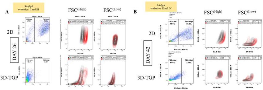
www.nature.com/scientificreports/
OA induction as they become senescent due to progressive telomere shortening with age. Senescent chondrocytes
are absent from normal cartilage and are present near osteoarthritic lesions7. When such senescent cells were
transplanted into the knee joint of wild type mice, an OA-like state was induced, thus showing that senescence
of chondrocytes is a major factor driving O A8. When chondrocytes are cultured in vitro, especially in monolayer,
they easily lose their native phenotype, de-differentiate and express various senescence- and dedifferentiation-
related genes. Replicative senescence in vitro has been observed after 30–40 passages during in vitro culture
of chondrocytes, which then exhibit features of the senescent phenotype, including enlarged flattened cells in
culture and the expression of SA-βgal9. The cellular senescence of in vitro cultured cells is usually overcome by
inducing telomerase activity or initiating recombination-mediated alternative lengthening of telomeres (ALT)
pathway(s) or genomic modifications such as reprogramming using specific transcription factors, all of which
carry a risk of oncogenesis10. An in vitro culture method which does not involve any such genomic modifications
would be ideal for use in regenerative therapies. The capabilities of a three-dimensional (3D) thermo-reversible
gelation polymer (TGP) to maintain the native phenotype for a longer time in vitro have been reported for
several cell types such as corneal endothelial precursor c ells11, corneal limbal stem c ells12, mesenchymal stem
cells13, buccal epithelial c ells14 and c hondrocytes15–18. This 3D-TGP can maintain the native hyaline phenotype
of knee-cartilage-derived chondrocytes from bovine15, rabbit16 and human17,18 sources for 16–18 weeks, both
in vitro and in vivo. In vitro 3D-TGP-tissue-engineered cartilage tissue expressed pluripotency-related markers
in a lectin micro-array18, higher miRNA21 and 140 expression indicative of healthy cartilage p henotype19 and
20
mesenchymal-chondroprogenitor markers . We sought to examine the expression of senescence-associated
beta-galactosidase (SA-βgal) in human chondrocytes derived from elderly donors affected by OA, cultured in
2D- followed by 3D-TGP.
Methods
The institutional ethics committee of Edogawa Hospital, Tokyo, Japan, approved the study. Discarded cartilage
biopsy tissues were obtained from elderly patients with severe OA (aged between 60 and 85 years) who underwent
arthroscopy and were employed in the study. The study was conducted in accordance with relevant guidelines/
regulations, and informed consent was obtained from all of the participants and/or their legal guardians.
The cartilage tissue samples were subjected to chondrocyte isolation and culture, following our methodology
reported earlier16–18. Nine samples were used in the study. The cartilage tissues were subjected to digestion with
0.25% trypsin for 30 min in an orbital shaker at 150 rpm, at 37 °C, followed by digestion in 2 mg/ml collagenase
digestion for 12–18 h at 37 °C in an orbital shaker. After the digestion, the cells were isolated by filtering with a
100-μm cell strainer. The cells were centrifuged at 1000 rpm for 10 min and cultured in two-dimensional (2D)
monolayer using media containing low-glucose DMEM, 10% autologous plasma, 1% penicillin streptomycin,
50 μg/ml gentamicin and 0.25 μg/ml amphotericin B and L-ascorbic acid (50 mg/ml) for 17–25 days at 37 °C
with 5% CO2. After 17–25 days when the 2D-cultured cells started degenerating, a portion of the cells was seeded
into the 3D-TGP scaffold in a cylindrical silastic tube for culture using high-glucose DMEM, 10% autologous
plasma, 1% penicillin streptomycin, 50 μg/ml gentamicin and 0.25 μg/ml amphotericin B and L-ascorbic acid
(50 mg/ml), placed on an orbital shaker at 80 RPM in 5% CO2 incubator, while the other portion was continued
as 2D culture. The 3D cultures were maintained for 42–45 days. For the tissue harvest from 3D-TGP, the culture
supernatant was removed and cold phosphate buffered saline (PBS) kept at 4° C was added to liquify the TGP
and the tissue like construct was retrieved.
For histological staining, the 2D and the 3D-TGP cultures were fixed using formalin, and then embedded in
paraffin blocks. Serial sections were deparaffinized and stained with haematoxylin and eosin (H&E), safranin
O/fast Green and toluidine blue employing standard histological techniques. The 2D-cultured cells were sub-
jected to SA-βgal measurement before transfer to 3D-TGP and on days 22–45, in whichever sample they did not
degenerate while the 3D-TGP- cultured cells were subjected to SA-βgal measurement on days 26, 36 and 42–45 of
culture. The 2D and 3D-TGP cultures were also subjected to evaluation for p16INK4a and p21 mRNA expression.
The time-points of measurement in 2D and 3D-TGP are illustrated in Fig. 1.
For SA-βgal measurement, cells from the 2D and 3D-TGP cultures were stained using a SPiDER-βgal cellular
senescence plate assay kit (Dojindo Laboratories, Japan) to measure their SA-βgal. For the 3D-TGP cultured cells,
single cells were obtained from spheroids harvested from the 3D culture by incubation with Collagenase Type II
(1 mg/ml) for 2 min. The cell culture supernatant from the cells in the cell culture plate/dish was removed, and
the cells were washed with phosphate buffered saline (PBS) once. Lysis buffer was added, and the plate/dish was
incubated at room temperature for 10 min. The lysate solution was added to each well of the cell culture plate.
Then, the SPiDER-βgal working solution was added to each well and incubated at 37 °C for 30 min. The stained
cells were measured with FACSVia and analysed with the FlowJo software (BD).
The cells from 2 and 3D-TGP were also subjected to qRT-PCR for the expression of other senescence associ-
ated markers p16 INK4a and p2121,22.
Total RNA was isolated and RT-PCR was performed using the One Step TB Green PrimeScript PLUS RT-
PCR Kit (Perfect Real Time; Takara, Japan) and Thermal Cycler Dice Real Time System Lite (TP700, TaKaRa).
The primers employed are provided below.
p16 INK4a
5′ to 3′: GGCACCAGAGGCAGTAACCA
3′ to 5′: CCTACGCATGCCTGCT TCTACA
p21
5′ to 3′: GCGATGGAACTTCGACTTTGT
3′ to 5′: GGGCTTCCTCTTGGAGAAGAT
Scientific Reports | (2021) 11:14059 | https://doi.org/10.1038/s41598-021-93607-9 2
Vol:.(1234567890)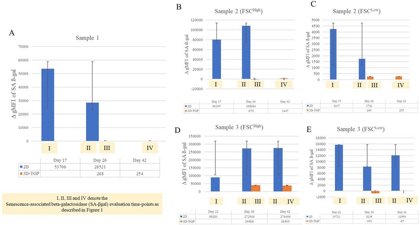
www.nature.com/scientificreports/
Figure 1. Illustration of the study groups and time-points of evaluation of SA-βgal, p16INK4a and p21 in two-
dimensional (2D) and three-dimensional (3D) thermo-reversible gelation polymer (3D-TGP) cultures.
For mRNA quantification, the relative expression of the genes of interest was normalized against the GAPDH
housekeeping gene by employing the comparative cycle threshold (Ct) method.
∆Ct = Ct (gene of interest) – Ct (housekeeping gene (GAPDH))
∆∆Ct = ∆Ct (treated sample or experimental sample) – ∆Ct (control sample)
All of the data were analysed using the Microsoft Office Excel software package. Student’s paired t-tests were
also calculated using this package. P-values < 0.05 were considered significant.
Ethical approval. The institutional ethics committee of Edogawa Hospital, Tokyo, Japan approved the study.
Results
The cells grew individually in a monolayer 2D culture with de-differentiating into fibroblast like cells and could
be maintained in the in vitro culture in a healthy manner for only up to 25 days after which they started showing
signs of degeneration. After transfer to 3D-TGP, the cells in the 3D-TGP grew as a tissue-like morphology with
native hyaline phenotype maintenance observed in H&E staining, Safranin O/Fast Green and Toluidine blue
throughout the culture period of 42–45 days (Fig. 2).
Regarding the mRNA expression of p16 INK4a and p21, they were expressed only in 2D cultured samples and
not in 3D-TGP indicating presence of senescent cells in 2D cultures but not in 3D-TGP (Fig. 3). The average
delta G of mean fluorescence intensity (ΔG MFI) of the expression of SA-βgal in the cells from 2D culture was
42,016.6 while after transfer to 3D-TGP the value greatly decreased to an average of − 144.66 (Fig. 4) and it only
slightly increased after 42–45 days of culture in 3D-TGP. These values are of the larger cells observed during gat-
ing in the flow-cytometry (the forward scatter [FSC]high group) (Fig. 5). For another population (FSClow group),
the average of ΔG MFI was 190,314 on day 17 in 2D, which decreased to 19,706.5 after transfer to 3D-TGP and
again only slightly increased till day 42–45 in 3D-TGP (Figs. 4, 5). The difference between the higher expression
of total ΔG MFI of SA-βgal in cells from 2D compared to decreased expression in 3D-TGP was however not
statistically significant (p-value = 0.089723).
Discussion
The most widely used biomarker for identifying senescent and aging cells in vitro is SA‐β‐gal, which is the activ-
ity of an enzyme called β‐galactosidase, which is detectable at pH 6.0 in cells undergoing replicative or induced
senescence in vitro. It is absent in proliferating cells23. SA-βgal originates from lysosomal β‐galactosidase activ-
ity, which increases in senescent cells due to increased lysosome content, whose levels become detectable at
pH 6.0 when it surpasses a threshold limit23. Relevant to chondrocytes and aging, a study which compared the
SA-βgal expression of normal (controls) versus OA cartilage reported that no SA-βgal staining was observed in
the normal articular cartilage samples, while the percentage of SA-βgal-positive chondrocytes was 13.00 ± 5.77%
in mild lesions, 31.65 ± 6.91% in moderate lesions and 51.95 ± 6.21% in severe lesions24, thus implying that
SA-β-gal expression is associated with progressive knee joint damage from OA and is a potential indicator of
disease severity24. Another study also reported that cultured chondrocytes isolated from near the lesion sites of
OA contained a greater percentage of SA-β-gal-positive cells than cultures isolated from distal sites or normal
cartilage did25. In the present study, the 2D monolayer culture led to rapid replicative senescence in 17 days,
evident from the higher levels of SA‐β‐gal expression (Fig. 4), expression of p16 INK4a and p21 (Fig. 3) compared
Scientific Reports | (2021) 11:14059 | https://doi.org/10.1038/s41598-021-93607-9 3
Vol.:(0123456789)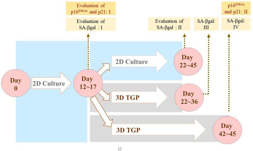
www.nature.com/scientificreports/
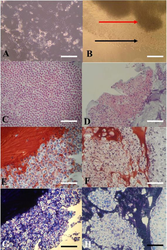
Figure 2. (A,B) In vitro culture images: (A) Chondrocytes in two-dimensional (2D) culture de-differentiating
into fibroblast-like cells; (B) In vitro cultured chondrocytes growing in a tissue-like manner in three-
dimensional (3D) thermo-reversible gelation polymer (3D-TGP) culture (the red arrow indicates the tissue; the
black arrow indicates the cells migrating out into the 3D environment into the tissue); (C,D) H-and-E staining
images: (C) Chondrocytes in 2D observed as individual cells. (D) 3D-TGP tissue-engineered chondrocytes
exhibiting continuous tissue morphology with hyaline phenotype; (E,F) Safranin O/Fast Green staining images:
(E) Chondrocytes in 2D observed as individual cells. (F) 3D-TGP tissue-engineered chondrocytes exhibiting
continuous tissue morphology; (G,H) Toluidine blue images: (E) Chondrocytes in 2D observed as individual
cells. (F) 3D-TGP tissue-engineered chondrocytes exhibiting continuous tissue morphology (All scale
bars = 100 μm).
Scientific Reports | (2021) 11:14059 | https://doi.org/10.1038/s41598-021-93607-9 4
Vol:.(1234567890)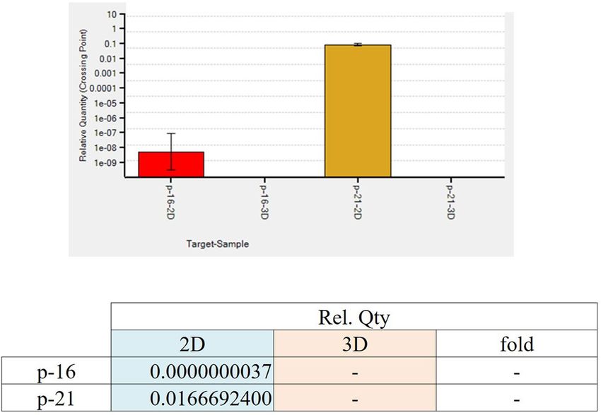
www.nature.com/scientificreports/
Figure 3. Relative expression of p16 INK4a and p21 only in 2D cultured chondrocytes and not in 3D-TGP
indicating presence of senescent cells in 2D cultures but not in 3D-TGP.
Figure 4. Mean fluorescence intensity (ΔG MFI) of the expression of SA-βgal evaluated by flow cytometry in
2D compared to 3D-TGP at different durations of culture with 2D-cultured chondrocytes (evaluation I and II)
showing higher levels of SA-βgal as culture, while the 3D-cultured cells (evaluation III and IV) showed very low
levels of SA-βgal throughout the culture period. (A) Sample 1; (B) Sample 2 ( FSC(High)); (C) Sample 2 ( FSC(Low));
(D) Sample 3 ( FSC(High)); (E) Sample 3 ( FSC(Low)). I, II, III and IV denote the Senescence-associated beta-
galactosidase (SA-βgal) evaluation time-points as described in Fig. 1.
Scientific Reports | (2021) 11:14059 | https://doi.org/10.1038/s41598-021-93607-9 5
Vol.:(0123456789)www.nature.com/scientificreports/
Figure 5. Gating in the flow cytometric analysis of osteoarthritic chondrocytes grown in 2D and 3D-TGP with
the SA-βgal expression in two heterogeneous populations sorted by flow cytometry ( FSChigh versus F SClow) with
the 2D-grown cells showing higher SA-βgal expression than 3D-TGP cultured cells on both day 26 and day 42
of culture (A) Day 26; (B) Day 42. II, III and IV denote the Senescence-associated beta-galactosidase (SA-βgal)
evaluation time-points as described in Fig. 1.
to 3D-TGP, whose environment re-differentiates the de-differentiated fibroblasts to yield younger cells that
grow as a tissue maintaining the native hyaline phenotype (Fig. 2) for a longer period. Having shown earlier that
chondrocytes grown in TGP express pluripotency-associated markers in a lectin microarray, the current study
further substantiates the presence of a heterogeneous cell population (Fig. 5) containing FSChigh and FSClow, with
FSClow comprising smaller cells, presumably progenitor cells, which needs validation, apart from the study of
other markers of senescent cells such as γ-H2AX, the formation of senescence-associated heterochromatin foci
(SAHF) and the acquisition of a senescence-associated secretory phenotype (SASP)26. Both in vitro and in vivo
studies have reported that senolytics which eliminate senescent cells improve physiological function in tissues
and decrease a ging27. These drugs comprise natural products, synthetic small molecules and peptides that target
proteins involved in senescent-cell anti-apoptotic pathways (SCAPs). However, they are not free of side effects23,
and any interference with the natural mechanisms lead to risks of genomic mutations and tumorigenicity. Here,
we have employed a synthetic polymer which could help to grow younger cells in vitro for more efficient tissue-
engineering and regenerative medicine applications. TGP maintains the native phenotype of cells without induc-
ing gene abnormalities13, and its safety has been established in pilot clinical studies on h
umans13,14,28. A recent
study28 has reported that there is age-associated cell-intrinsic defects in hematopoietic stem cells (HSCs) which
cannot be restored by rejuvenating the niche alone. In the present study, the cells’ SA-βgal expression could be
reversed by using a 3D-Poylmer scaffold which probably restores both the niche and the age-related intrinsic
changes in the cells; however this needs further validation. The present study is only a preliminary study on the
possibility of employing 3D-TGP culture technology for the in vitro reversal of aging by using a 3D polymer
without using genomic modifications/proteins which may have other unwanted adverse effects. The technology’s
efficacy can be confirmed further by studying the telomere length of the cells cultured in 2D- and 3D-TGP, in
studies that are underway by our team.
Conclusion
This is the first study to provide proof-of-concept evidence that in vitro cellular senescence analysed by SA-βgal
expression can be reversed through culturing in a 3D-TGP scaffold-based culture platform that provides an
environment which nurtures cells in the native phenotype, apart from making them younger by decreasing the
senescence-associated characteristics, importantly without use of genomic modification techniques. This study
opens a new avenue for evaluating techniques such as 3D-TGP to reverse in vitro aging, in order to produce
younger good-quality cells for regenerative medicine applications.
Received: 12 January 2021; Accepted: 28 June 2021
References
1. Campisi, J. & Adda di Fagagna, F. Cellular senescence: When bad things happen to good cells. Nat. Rev. Mol. Cell Biol. 8, 729–740
(2007).
2. Hayflick, L. The limited in vitro lifetime of human diploid cell strains. Exp. Cell Res. 37, 614–636 (1965).
3. Velarde, M. C. & Menon, R. Positive and negative effects of cellular senescence during female reproductive aging and pregnancy.
J. Endocrinol. 230, R59–R76 (2016).
4. González-Gualda, E., Baker, A. G., Fruk, L. & Muñoz-Espín, D. A guide to assessing cellular senescence in vitro and in vivo. FEBS
J. 2, 2 (2020).
5. Bernadotte, A., Mikhelson, V. M. & Spivak, I. M. Markers of cellular senescence. Telomere shortening as a marker of cellular
senescence. Aging 8, 3–11 (2016).
Scientific Reports | (2021) 11:14059 | https://doi.org/10.1038/s41598-021-93607-9 6
Vol:.(1234567890)www.nature.com/scientificreports/
6. Jafri, M. A., Ansari, S. A., Alqahtani, M. H. & Shay, J. W. Roles of telomeres and telomerase in cancer, and advances in telomerase-
targeted therapies. Genome Med. 8, 69 (2016).
7. Rim, Y. A., Nam, Y. & Ju, J. H. The role of chondrocyte hypertrophy and senescence in osteoarthritis initiation and progression.
Int. J. Mol. Sci. 21, 2358 (2020).
8. Xu, M. et al. Transplanted senescent cells induce an osteoarthritis-like condition in mice. J. Gerontol. 72, 780–785 (2017).
9. Murillo-Ortiz, B. et al. Increased telomere length and proliferative potential in peripheral blood mononuclear cells of adults of
different ages stimulated with concanavalin A. BMC Geriatr. 13, 99 (2013).
10. Kalmbach, K., Robinson, L. G. Jr., Wang, F., Liu, L. & Keefe, D. Telomere length reprogramming in embryos and stem cells. BioMed
Res. Int. 2014, 925121 (2014).
11. Rao, S. K. et al. Successful transportation of human corneal endothelial tissues without cool preservation in varying Indian tropical
climatic conditions and in vitro cell expansion using a novel polymer. Indian J. Ophthalmol. 62, 130–135 (2014).
12. Sitalakshmi, G. et al. Ex vivo cultivation of corneal limbal epithelial cells in a thermoreversible polymer (Mebiol Gel) and their
transplantation in rabbits: An animal model. Tissue Eng. Part A 15, 407–415 (2009).
13. Hishikawa, K. et al. Gene expression profile of human mesenchymal stem cells during osteogenesis in three-dimensional ther-
moreversible gelation polymer. Biochem. Biophys. Res. Commun. 317, 1103–1107 (2004).
14. Vaddi, S. P., Reddy, V. B. & Abraham, S. J. Buccal epithelium expanded and encapsulated in scaffold-hybrid approach to urethral
stricture (BEES-HAUS) procedure: A novel cell therapy-based pilot study. Int. J. Urol. 26, 253–257 (2019).
15. Yasuda, A. et al. In vitro culture of chondrocytes in a novel thermoreversible gelation polymer scaffold containing growth factors.
Tissue Eng. 12, 1237–1245 (2006).
16. Arumugam, S. et al. Transplantation of autologous chondrocytes ex-vivo expanded using thermoreversible gelation polymer in a
rabbit model of articular cartilage defect. J. Orthop. 14, 223–225 (2017).
17. Arumugam, S. et al. In vitro expansion and characterization of human chondrocytes using a novel Thermoreversible Gelation
Polymer (TGP). J. Orthopaed. 8, e5 (2011).
18. Katoh, S., Fujimaru, A., Senthilkumar, R., Preethy, S. & Abraham, S. J. Articular chondrocytes from osteoarthritic knee joints of
elderly, in vitro expanded in thermo-reversible gelation polymer (TGP), exhibiting higher UEA-1 expression in lectin microarray.
Regen. Therapy 14, 234–237 (2020).
19. Katoh, S., Yoshioka. H., Senthilkumar, R., Preethy, S., & Abraham, S. J. Enhanced miRNA-140 expression of osteoarthritis-affected
human chondrocytes cultured in a polymer based three-dimensional (3D) matrix. Life Sci. 278, 119553 (2021).
20. Katoh, S. et al. A three-dimensional in vitro culture environment of a novel polymer scaffold, yielding chondroprogenitors and
mesenchymal stem cells in human chondrocytes derived from osteoarthritis-affected cartilage tissue. J. Orthop. 23, 138–141 (2021).
21. Coryell, P. R., Diekman, B. O. & Loeser, R. F. Mechanisms and therapeutic implications of cellular senescence in osteoarthritis.
Nat. Rev. Rheumatol. 17, 47–57 (2021).
22. Vinatier, C., Domínguez, E., Guicheux, J. & Caramés, B. Role of the inflammation-autophagy-senescence integrative network in
osteoarthritis. Front. Physiol. 9, 706 (2018).
23. Lee, B. Y. et al. Senescence-associated beta-galactosidase is lysosomal beta-galactosidase. Aging Cell 5, 187–195 (2006).
24. Gao, S. G. et al. Correlation between senescence-associated beta-galactosidase expression in articular cartilage and disease severity
of patients with knee osteoarthritis. Int. J. Rheum. Dis. 19, 226–232 (2016).
25. Price, J. S. et al. The role of chondrocyte senescence in osteoarthritis. Aging Cell 1, 57–65 (2002).
26. Noren Hooten, N. & Evans, M. K. Techniques to induce and quantify cellular senescence. J. Vis. Exp. 5, 55533 (2017).
27. Kirkland, J. L., Tchkonia, T., Zhu, Y., Niedernhofer, L. J. & Robbins, P. D. The clinical potential of senolytic drugs. J. Am. Geriatr.
Soc. 65, 2297–2301 (2017).
28. Kuribayashi, W. et al. Limited rejuvenation of aged hematopoietic stem cells in young bone marrow niche. J. Exp. Med. 218,
e20192283 (2021).
Acknowledgements
The authors wish to acknowledge Ms. Takako Fujisaki, Ms. Emi Nagahama & Ms. Junko Tomioka of Edogawa
Hospital, Tokyo, Japan for their assistance in sample collection and documentation; Ms Eiko Amemiya and Ms.
Sayaka Shimizu of II Dept of Surgery, Yamanashi University, Japan for their assistance with literature collection;
Dr. Fumihiro Ijima and Dr. Hiroshi Hirano of Hasumi International Research Foundation, Asagaya, Tokyo,
Japan for their assistance with the cell culture work described in the manuscript. Mr. Mathaiyan Rajmohan
and Mr. Ramalingam Karthick from the Fujio-Eiji Academic Terrain (FEAT), Nichi-In Centre for Regenerative
Medicine (NCRM), Chennai, Tamil Nadu, India, for their assistance with data collection of the study described
in the manuscript; Dr. Madasamy Balamurugan, Department of Pathology, Jawaharlal Institute of Postgraduate
Medical Education and Research (JIPMER), Karaikal, India for his guidance with the immunohistochemis-
try staining; Loyola ICAM College of Engineering Technology (LICET) Chennai, India for their support to our
research work. The authors dedicate this article to the memory of Mr. Venkatesan Sampath Kumar (IT systems
Admin manager, Nichi-In Centre for Regenerative Medicine (NCRM) and NCRM NICHE) who assisted with
the management of the data included in this manuscript. While the manuscript was under review, he passed
away due to COVID-19′s complications. May his soul rest in peace.
Author contributions
S.K. and S.A. contributed to conception and design of the study. A.F. and R.S. helped in data collection and
analysis. S.A. and S.P. drafted the manuscript. M.I., H.Y. and S.K. performed critical revision of the manuscript.
All the authors read, and approved the submitted version.
Competing interests
Potential Conflicts of Interest: 1. Dr. Katoh is an employee of Edogawa Hospital, Japan and is an applicant /inven-
tor to several patents on biomaterials and cell culture methodologies, some of them described in this manuscript.
2. Dr. Yoshioka is an employee of Mebiol Inc and an applicant to several patents on TGP and its applications 3.
Dr. Abraham is a shareholder in GN Corporation Co. Ltd., Japan and is an applicant /inventor to several patents
on biomaterials and cell culture methodologies, some of them described in this manuscript. 4. Other authors
don’t have any potential conflict of interests.
Additional information
Correspondence and requests for materials should be addressed to S.J.K.A.
Scientific Reports | (2021) 11:14059 | https://doi.org/10.1038/s41598-021-93607-9 7
Vol.:(0123456789)www.nature.com/scientificreports/
Reprints and permissions information is available at www.nature.com/reprints.
Publisher’s note Springer Nature remains neutral with regard to jurisdictional claims in published maps and
institutional affiliations.
Open Access This article is licensed under a Creative Commons Attribution 4.0 International
License, which permits use, sharing, adaptation, distribution and reproduction in any medium or
format, as long as you give appropriate credit to the original author(s) and the source, provide a link to the
Creative Commons licence, and indicate if changes were made. The images or other third party material in this
article are included in the article’s Creative Commons licence, unless indicated otherwise in a credit line to the
material. If material is not included in the article’s Creative Commons licence and your intended use is not
permitted by statutory regulation or exceeds the permitted use, you will need to obtain permission directly from
the copyright holder. To view a copy of this licence, visit http://creativecommons.org/licenses/by/4.0/.
© The Author(s) 2021
Scientific Reports | (2021) 11:14059 | https://doi.org/10.1038/s41598-021-93607-9 8
Vol:.(1234567890)You can also read