Review Article Ultrasonography of the Kidneys in Healthy and Diseased Camels (Camelus dromedarius)
←
→
Page content transcription
If your browser does not render page correctly, please read the page content below
Hindawi
Veterinary Medicine International
Volume 2020, Article ID 7814927, 12 pages
https://doi.org/10.1155/2020/7814927

Review Article
Ultrasonography of the Kidneys in Healthy and Diseased Camels
(Camelus dromedarius)

                                            1,2
          Mohamed Tharwat
          1
            Department of Veterinary Medicine, College of Agriculture and Veterinary Medicine, Qassim University, P.O. Box 6622,
            Buraidah, 51452, Saudi Arabia
          2
            Department of Animal Medicine, Faculty of Veterinary Medicine, Zagazig University, 44519, Zagazig, Egypt

          Correspondence should be addressed to Mohamed Tharwat; mohamedtharwat129@gmail.com

          Received 18 March 2020; Revised 15 September 2020; Accepted 17 September 2020; Published 21 October 2020

          Academic Editor: Antonio Ortega-Pacheco

          Copyright © 2020 Mohamed Tharwat. This is an open access article distributed under the Creative Commons Attribution License,
          which permits unrestricted use, distribution, and reproduction in any medium, provided the original work is properly cited.
          This review article is written to describe the results of ultrasonography of the kidneys in healthy camels as well as camels with some
          renal disorders. In the dromedary camel, the physiology of the kidney is of interest in view of the specialization of the camel to hot
          dry deserts and to prolonged periods without water. It plays an important role in water conservation through the production of
          highly concentrated urine that may predispose animal to varieties of renal disorders. Examples of kidney affections in dromedary
          camels are renal capsular pigmentation, medullary hyperemia, subcapsular calcification, cortical and medullar discoloration,
          hemorrhage in renal pelvis, nephrolithiasis, and hydatidosis. Congestion, hemorrhage, hydronephrosis, acute glomerulonephritis,
          subacute glomerulonephritis, chronic glomerulonephritis, diffuse interstitial nephritis, focal interstitial nephritis, renal cyst,
          hyaline degeneration, renal amyloidosis, tubular nephrosis, pyelonephritis, hemosiderosis, and renal toxicity. When the kidney is
          examined by ultrasonography, the clinician can get sufficient information about the size, position, and echo patterns of the renal
          cortex and medulla and renal pelvis and outlines of the renal blood vessels. In recent years, ultrasonography has been used in
          camels for scanning of the healthy status as well as evaluation and determining the diagnosis and prognosis of diseased cases.
          Examples of diseases evaluated by ultrasonography are paratuberculosis, trypanosomiasis, pneumonia, pleurisy, gastrointestinal
          neoplasms, chronic peritonitis, splenic abscessation, and hepatic disorders. Of the renal disorders assessed by ultrasonography are
          nephrolithiasis, hydronephrosis, pyelonephritis, renal abscessation, and renal neoplasms. Ultrasound guidance in biopsy of renal
          specimens has also been reported in dromedary camels.

1. Introduction                                                           hyperemia, and hydatid cyst were confirmed by histo-
                                                                          pathological examination. In another study conducted in
The kidney of the dromedary camel (Camelus dromedarius)                   Rajasthan, India, 121 renal samples were examined for
plays a vital role in water conservation through the pro-                 various pathological abnormalities [2]. Lesions included
duction of highly concentrated urine that may predispose                  congestion, hemorrhage, hydronephrosis, acute glomeru-
camel to varieties of renal disorders. In an abattoir based               lonephritis, subacute glomerulonephritis, chronic glomer-
study conducted on 100 camels in Najaf-Abad, Iran, the                    ulonephritis, diffuse interstitial nephritis, focal interstitial
frequency and types of renal affections were determined [1].               nephritis, renal cyst, hyaline degeneration, renal amyloid-
These disorders included renal capsular pigmentation,                     osis, tubular nephrosis, pyelonephritis, hemosiderosis, and
medullary hyperemia, subcapsular calcification, cortical and               renal toxicity [2]. In a third study conducted in Al-Ahsa
medullar discoloration, hemorrhage in the renal pelvis,                   abattoir, Saudi Arabia, gross and microscopic lesions of the
nephrolithiasis, and hydatidosis [1]. Of the later report,                kidney were investigated in 50 adult camels [3]. Of the
capsular melanosis, acute tubular necrosis, chronic inter-                camels, 33 (66%) had gross kidney lesions, whereas 17 (34%)
stitial nephritis, caseous necrosis, calcification, medullary              were found apparently healthy. Renal lesions included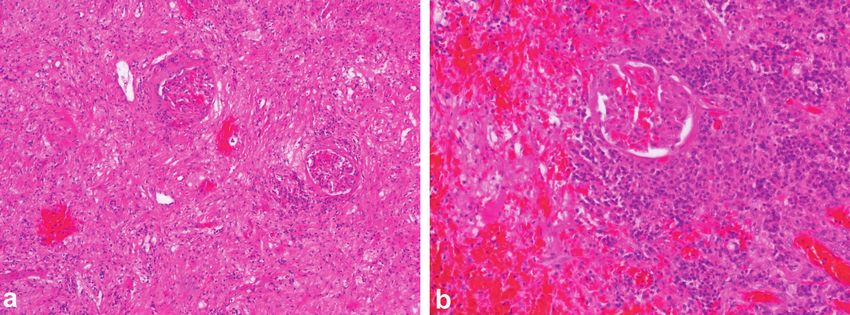
2 Veterinary Medicine International
hydronephrosis, renal hemorrhages, and renal necrosis. In process of the 2nd to the 4th lumbar vertebrae. Its cranial
the camels with renal lesions, microscopic changes included pole is round and it fits into the renal impression of the
glomerular shrinkage and hyalinization, proteinaceous casts, caudate lobe of the liver. Its caudal pole is not as rounded
cortical and medullary congestion, tubular cell swelling, and is slightly flattened dorsoventrally. The left kidney is
interstitial hemorrhage, and thickening of the glomerular regular in shape and lies below the left transverse processes
tufts [3]. of the last three lumbar vertebrae [36]. The position of the
In bovine, thoracic and abdominal ultrasonography is right and left kidneys in a camel carcass preserved in 10%
commonly performed to evaluate the type and severity of formalin solution is presented in Figure 3.
lesions in animals suspected to have cardiopulmonary,
gastrointestinal, hepatic, renal, splenic, and pancreatic dis- 3. Ultrasonographic Examination of the
ease [4–10]. Therefore, ultrasonography of thoracic and Kidneys in Camels and Normal Findings
abdominal organs in bovines is a minimally invasive and
cost-effective method for early detection of thoracic and The right kidney can be visualized in camels at the level of the
abdominal disorders [8, 11, 12]. 10th and 11th intercostal spaces (ICS) and the upper right flank.
In camel medicine, ultrasonography was rarely used with The left kidney can be imaged from the caudal left flank
the exception in the reproduction field. Recently, in healthy (Figure 4). The differentiation between the renal cortex and
camels, our research group has used ultrasound for scanning medulla in both kidneys is visible in most cases. The renal cortex
of the lungs and pleura [13, 14], echocardiography [14, 15], is relatively hyperechoic compared to the renal medulla and the
ultrasonography of the gastrointestinal tract (GIT) [16, 17], renal sinus is hyperechogenic and more differentiated than the
hepatic and renal imaging [17, 18], and abdominal ultra- cortex and medulla. The right and left renal parenchyma are less
sonography [19]. Ultrasound-guided hepatic and renal bi- echogenic than the neighbouring hepatic and splenic paren-
opsy and portocentesis have also been carried out in camels chyma, respectively. The medullary pyramids have a conic
[17, 20, 21]. In diseased camels, diagnostic ultrasonography triangular appearance and are less echogenic than the remaining
has been done for the evaluation and prognosis of abdominal parenchyma. The renal hilus can be imaged when the trans-
distension [22], Johne’s disease [23], trypanosomiasis [24], ducer is placed in the paralumbar fossa and rotated about its
abdominal disorders [19, 25, 26], urinary disorders [27], longitudinal axis (Figure 5). However, the renal artery and vein
thoracic affections [14, 28], renal neoplasms [29], pyelo- and the ureter are difficult to be accurately identified. Ultra-
nephritis and renal abscessation [30, 31], GIT masses sonography via the so-called hepatic and splenic windows also
[32, 33], chronic peritonitis [34], splenic abscessation [35], results in good images of the right and left kidneys, respectively
and hepatic disorders [17]. (Figure 6). When examined transrectally, the left kidney is
The physiology of the kidney of the dromedary camel is accessible. In most camels, the entire left kidney is accessible and
of interest in view of the specialization of the camel to hot the cranial pole can be reached. The left kidney can also be easily
dry deserts and to prolonged periods without water. In this imaged transrectally in a cross-sectional view (Figure 7) [18].
species, ultrasonographic diagnosis of renal disorders has Table 1 shows the measurements of the right and left
received lesser attention compared to other animals; thus kidneys, including the distance to the body surface, the
there is a shortage of information in this area. The procedure thickness of the cortex, medulla and renal sinus, and the vertical
has been adopted widely as a diagnostic procedure and and horizontal diameters of both kidneys. The distance between
research tool in animals. This review article is written to the body surface and the left kidney is greater than that for the
describe the results of ultrasonography of the kidneys in right kidney. The vertical diameter of both kidneys is signifi-
healthy camels as well as camels with some renal disorders. cantly smaller than the horizontal diameter [18].
2. Anatomy of the Kidneys in Camels 4. Ultrasound-Guided Renal Biopsy of the Right
and Left Kidneys in Camels
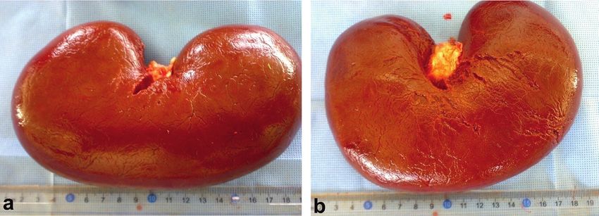
The kidneys in camels are bean-shaped and smooth exter-
nally. The right kidney is more elongated than the left one, In the camel, a biopsy of the right and/or left kidney is
with an average weight of 1.08 kg and 1.13 kg for the right carried out in a sternal recumbency position. The fore-and-
and left kidneys, respectively (Figure 1). On a longitudinal hind legs are tied by a rope near the carpal and hock joints,
section made through the kidney, the cortex and the medulla respectively. The distance between the 10th ICS to the flanks
are easily distinguished (Figure 2). There is a well-developed on both sides of the body is clipped and skin shaved. The
Crista renalis. The renal cortex occupies about 50% of the shaved abdominal area was sterilized using standard surgical
volume of the kidney and the ratio of the thickness of the disinfection techniques. To obtain adequate restraint, camels
medulla to that of the cortex is approximately 4 : 1. Both are slightly sedated with xylazine (0.1 mg/kg·BW), and the
kidneys are situated against the dorsal body wall and are region chosen for collecting hepatic or renal biopsy is
retroperitoneal. The right kidney lies below the transverse infiltrated with 10 ml of 2% lidocaine hydrochloride. The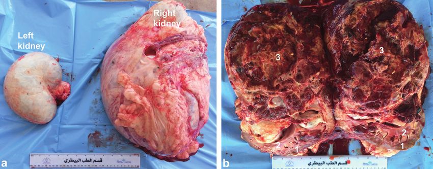
Veterinary Medicine International                                                                                                    3

                   Figure 1: Right (a) and left (b) kidneys in an adult camel (both with removed renal capsules).

     Figure 2: Longitudinal section through the kidney in an adult camel. RC, renal cortex; RM, renal medulla; RS, renal sinus.

Figure 3: Anatomical position of the right (white arrow) (a) and left (black arrow) (b) kidneys in a camel carcass preserved with 10%
formalin solution. C1 � first gastric compartment.

    Figure 4: Imaging of the right kidney in the upper right flank (a) and the left (b) kidney in the caudal part of the left flank.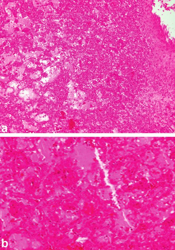
4                                                                                                   Veterinary Medicine International

Figure 5: Ultrasonogram of a longitudinal section of the right kidney in a healthy camel. The image was taken from the upper right flank.
RH � renal hilus; DS � dorsal; VT � ventral.

Figure 6: Ultrasonograms of a longitudinal section of the right (a) and left (b) kidneys in a healthy camel. Image (a) was taken from right
10th intercostal space through the so-called hepatic window. Image (b) was taken from the middle left flank through the so-called splenic
window. 1, cortex; 2, medulla; 3, renal sinus’ DS, dorsal; VT, ventral.

Figure 7: Transrectal ultrasonography of the left kidney in a healthy camel. The cranial pole of the left kidney is apparent (a). A cross-
sectional view of the left kidney is also apparent (b). C � cortex; M � medulla; RS � renal sinus; RC � renal capsule; RW � rumen wall.
Veterinary Medicine International                                                                                                    5

Table 1: Dimensions (means ± SD) of the right and left kidneys
imaged by ultrasound, from the upper right and caudal left
paralumbar fossa in healthy camels [18].
Variable                             Right kidney     Left kidney
Distance from body surface (cm)        1.8 ± 0.5        2.5 ± 0.6
Cortex (cm)                            1.7 ± 0.6        1.7 ± 0.3
Medulla (cm)                           2.7 ± 0.6        3.0 ± 1.0
Renal sinus (cm)                       3.0 ± 0.7        3.6 ± 0.9
Vertical diameter (cm)                 8.4 ± 1.4        9.8 ± 1.9
Horizontal diameter (cm)               18.1 ± 2.6      14.5 ± 3.0

kidneys are firstly scanned to determine the optimal biopsy
                                                                     Figure 8: Renal biopsy in a camel. The needle is clearly visible
sites. After the application of transmission gel to the              within the renal cortex as a sharp bright line. Ds � dorsal;
transducer, the right and left kidneys are examined at the           Vt � ventral.
upper right and caudal left paralumbar fossa [21].
     Prior to biopsy, and under aseptic conditions, a small in-
cision is made in the skin over the suggested biopsy site with the
point of a scalpel blade. Using a free-hand technique, a
14 G × 150 mm spinal biopsy needle was used. The biopsy
needle is then advanced through the skin incision and then
under real-time ultrasound guidance toward the hepatic or
renal parenchyma. During the biopsy of the kidneys, the ad-
vancement of the needle is halted when the tip of the needle is
seen to penetrate the renal capsule. The needle is directed
obliquely in an attempt to sample cortical tissue only and avoid
the renal medulla, renal pelvis, and hilar and renal vessels. When
the needle was considered to be in the correct position, the plain
stylet is withdrawn and a notched part is inserted and advanced
1 cm into the renal cortex beyond the renal capsule. The needle
can be identified on the ultrasound within the renal cortex as a
sharp bright line (Figure 8), while the specimen was being
obtained, thus confirming the location of the biopsy. Both the        Figure 9: Under ultrasound guidance, renal biopsy needle path is
needle and forked stylet were then removed with a sample of          usually easily identified within the renal cortex at post-mortem
renal tissue. Repeat passes were performed, if required, to obtain   examination.
sufficient biopsy specimens. It is advisable to immediately scan
the kidneys after biopsy to assess the presence of hematoma or
active bleeding [21].
     Low complications of renal biopsy are always achieved if
the needle biopsy is advanced under ultrasound guidance,
which provided more accurate localization of the needle in
relation to the kidney and subsequent biopsy site in the renal
cortex (Figure 9). Direct real-time ultrasound control allows
the correction of the needle position at any moment during the
biopsy procedure. Knowledge of the exact location of the
needle in the cortex prevents deep penetration into the medulla
[21].

5. Renal Disorders                                                   Figure 10: Nephrolithiasis (1 and 2) in a female camel with red
5.1. Nephrolithiasis. Urolithiasis is common as a subclinical        urine for a 6-month period. 3, renal cortex; 4, renal medulla; 5,
                                                                     renal sinus. Stars point to acoustic shadowing under the calculi.
disorder among ruminants raised in management systems
where the ration is composed primarily of grain or where
animals graze certain types of pasture. In these situations,         obstruction of urine flow [38]. Urinary calculi are formed in
40–60% of the animals may form calculi in their urinary tract        males and females equally, but the bore (diameter) of the female
[37]. Urinary calculi (uroliths, nephrolith, bladder stone, and      urethra generally allows free passage of a calculus that may enter
cystolith) are formed in either the calyces of the kidney or more    the urethra. Thus, obstructive urolithiasis is rare in the female
commonly in the urinary bladder. In camels, small uroliths may       and exclusive to the male animal. The majority of the calculi
enter the ureter or urethra and cause partial or complete            lodge in the sigmoid flexure of the penis, particularly in the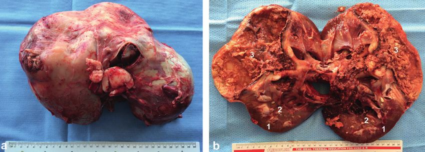
6                                                                                                  Veterinary Medicine International

                                            (a)                                             (b)

                                            (c)                                             (d)

Figure 11: Bilateral hydronephrosis in two female camels with urinary calculi. Both the right (a) and left (b) kidneys were scanned
transcutaneously. In the other case, the right kidney (c) was imaged transcutaneously and the left kidney (d) was imaged transcutaneously.

distal segment. The composition of the calculi differs with             parenchyma is present [44]. Although diseased animals usually
geographic location. Silicate, phosphate, and calcium carbonate        presented with some signs or symptoms, hydronephrosis can be
crystals have been observed [39]. High concentrate diets, im-          an incidental finding encountered during the evaluation of an
proper calcium phosphorus balance, high silica or oxalate              unrelated process. If unrecognized or left untreated, hydro-
pasture, hypovitaminosis A, hypervitaminosis D, reduced water          nephrosis can lead to loss of renal function and sepsis. Con-
intake, and increased salts in drinking water are all possible         sequently, all animals found to have hydronephrosis should
factors for urolithiasis [40]. Calculus may be identified ultra-        undergo a thorough evaluation [11]. In cases of bilateral
sonographically in the kidneys of the camel as acoustic en-            hydronephrosis, transrectal ultrasonography showed a dis-
hancement with distal acoustic shadowing (Figure 10).                  tended urinary bladder, anechoic fluid in the uterus, and
                                                                       hydronephrosis of the left kidney. By transcutaneous ultraso-
                                                                       nography, the clinician can visibly scan and evaluate hydro-
5.2. Hydronephrosis. Chronic, unilateral, and ureteral ob-             nephrosis of the right and/or left kidneys (Figure 11). The
structions are more likely to lead to hydronephrosis that is           condition is usually identified postmortem as unilateral or
defined as a dilation of the renal pelvis with progressive              bilateral affection (Figure 12).
atrophy of the renal parenchyma. Any urinary tract ob-
struction can lead to hydronephrosis, but the extent and
duration of the obstruction are important in determining the           5.3. Pyelonephritis and Renal Abscessations. In camels, renal
severity of the renal lesion. Bilateral hydronephrosis may be          pathologies such as abscess and pyelonephritis are rarely
caused by pelvic masses compressing the urethra or ureters.            reported compared to other animals [39, 45]. Out of 121
     The disease in ruminants can be a life-threatening, such as       slaughterhouse-obtained renal lesions in dromedary camels,
infected condition leading to pyelonephritis [41, 43]. To dis-         only 7.43% were pyelonephritis [45]. Renal ultrasonography
tinguish acute and chronic hydronephrosis, one may consider            provides a precise, noninvasive, and fast technique for the
acute as hydronephrosis that, when corrected, allows full re-          evaluation and subsequent clinical decision-making of renal
covery of renal function. Conversely, chronic hydronephrosis is        abscessation and chronic active pyelonephritis in dromedary
a situation in which the loss of function is irreversible even with    camels. Recently, bilateral, and unilateral chronic active
the correction of the obstruction. A chronically dilated               pyelonephritis together with renal abscessation caused by
hydronephrotic system may be associated with compression of            E. coli and Staphylococcus lugdunensis infection was reported
the papillae and thinning of the parenchyma; eventually, cortical      in a 12-year-old male and 6-year-old female camel, re-
atrophy progresses to the point at which only a thin rim of            spectively [30, 31]. In the male camel with bilateral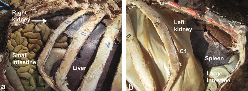
Veterinary Medicine International                                                                                                         7

Figure 12: Unilateral hydronephrosis of the left kidney (b) in a female camel with pelvic abscessation detected postmortem compared to a
healthy right kidney (a). Images (c) and (d) show bilateral hydronephrosis in a male camel with penile calculi.

                                           (a)                                               (b)

Figure 13: Transabdominal ultrasonographic examination of pyelonephritis in camels. Image (a) shows transcutaneous scanning of the
right kidney with pyelonephritis in a male camel caused by E. coli. The renal capsule was imaged hyperechogenic with fibrin tags and the
renal cortex appeared echogenic. A hypoechoic fluid was imaged surrounding the kidney. Image (b) shows transrectal view of unilateral
pyelonephritis in a 6-year-old female camel caused by Staphylococcus lugdunensis. Heterogeneous contents were identified with multi-
chambers containing echogenic fluid (stars). The cortex could not be differentiated from the medulla. 1, cortex; 2, medulla; 3, renal capsule;
4, perirenal abscessation.

pyelonephritis caused by E. coli, the transabdominal ultra-             renal cortex. A hypoechoic fluid was imaged surrounding
sonographic examination of the right kidney revealed a                  the kidney from all sides and the kidney is floating within it.
hyperechogenic renal capsule with fibrin tags and echogenic              In the caudoventral left flank, the left kidney was imaged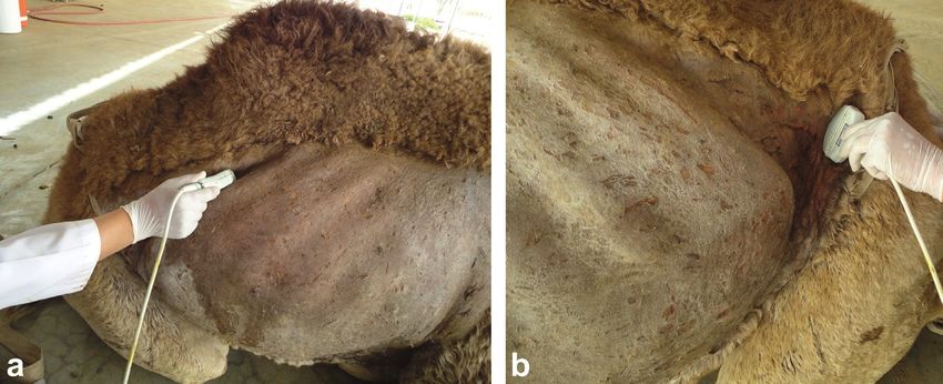
8                                                                                                    Veterinary Medicine International

Figure 14: Gross pathological findings in a male camel with bilateral renal abscessation and chronic active pyelonephritis caused by
Escherichia coli. Image (a) shows gross appearance of the left kidney and thick creamy pus evacuated from the kidney, weighting about 18 kg.
Image (b) shows a longitudinal section through the affected kidney after complete evacuation of the large abscess revealed thickening and
dilatation of the renal pelvis in relation to the renal abscess [30]. Stars point to the renal tissue.

Figure 15: Gross pathological findings in a female dromedary camel with chronic suppurative pyelonephritis caused by Staphylococcus
lugdunensis. Image (a) shows an enlarged abscessed kidney, while image (b) shows a longitudinal section of the affected kidney with
corticomedullary abscess [31]. 1, cortex; 2, medulla; 3, abscess cavity.

Figure 16: Histopathological findings of renal specimen of the right (a) and left (b) kidney in a camel with bilateral chronic active
pyelonephritis showing renal cortical and medullary tissue with congested glomeruli (white arrows) and tubules with colloid casts and a
fibrous and granulation tissue part at the periphery (black arrows). The interstitial tissue shows active and chronic cells infiltrate (H & E,
×100) [31].

with a large volume of hypoechoic contents (Figure 13(a)).              identified with multichambers containing echogenic fluid.
In the female camel with unilateral pyelonephritis caused by            The cortex could not be differentiated from the medulla
Staphylococcus lugdunensis, heterogeneous contents were                 (Figure 13(b)). In both cases of pyelonephritis and
Veterinary Medicine International 9
Figure 17: Histopathological section of the affected kidney showing congestion and periglomerular fibrosis, with atrophic tubules, hyaline,
and RBCs casts. The interstitial tissue shows fibrosis, thick-walled blood vessels, and dense mixed inflammatory cell infiltrates. Area of
necrosis with suppurative exudation is also seen (H & E, image a × 200; image b × 400) [30].
(a) (b)
Figure 18: Transabdominal ultrasonographic examination of renal masses ( ∗ ∗ ) in 2 camels. Image (a) with echogenic mass was captured
transrectally from the left kidney. Image (b) with hypoechoic mass in the right kidney was taken transcutaneously from the upper right
paralumbar fossa. Both masses were confirmed to be abscesses through ultrasound-guided aspiration of pus. 1, cortex; 2, medulla.
(a) (b)
Figure 19: Transrectal ultrasonographic findings in a female camel with renal cell carcinoma of the right kidney. Image (a) shows a
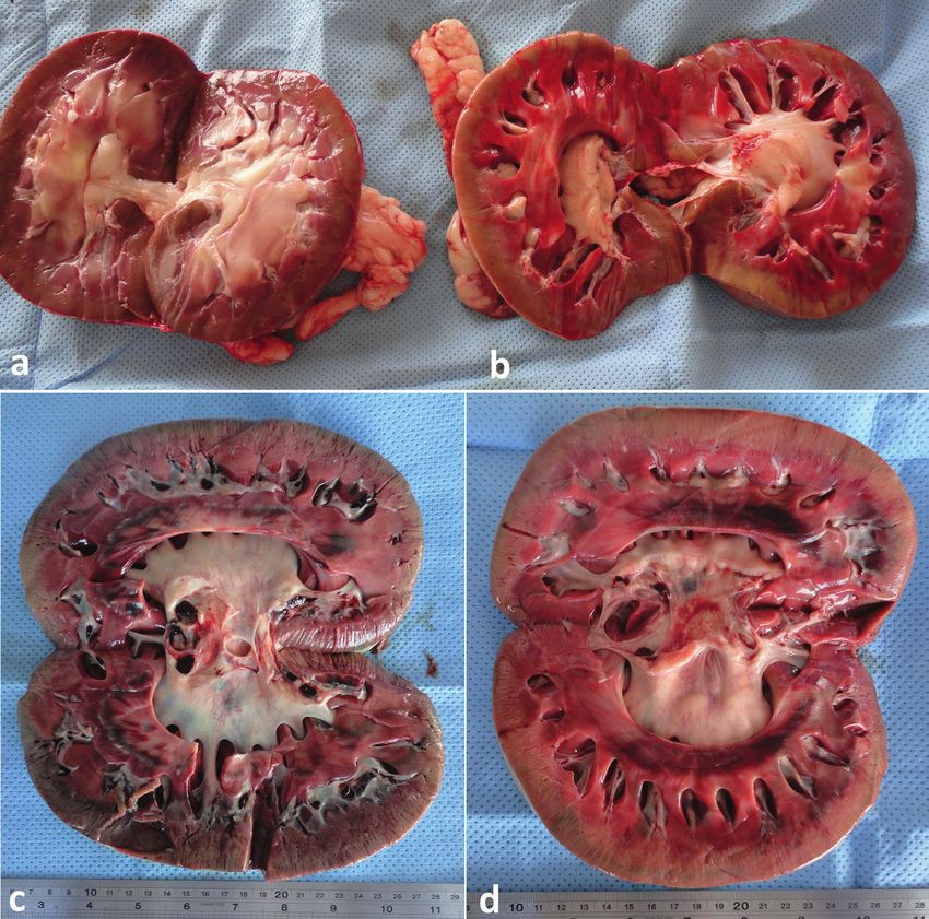
hypoechoic mass involving the right renal parenchyma, while image (b) shows the normal left kidney.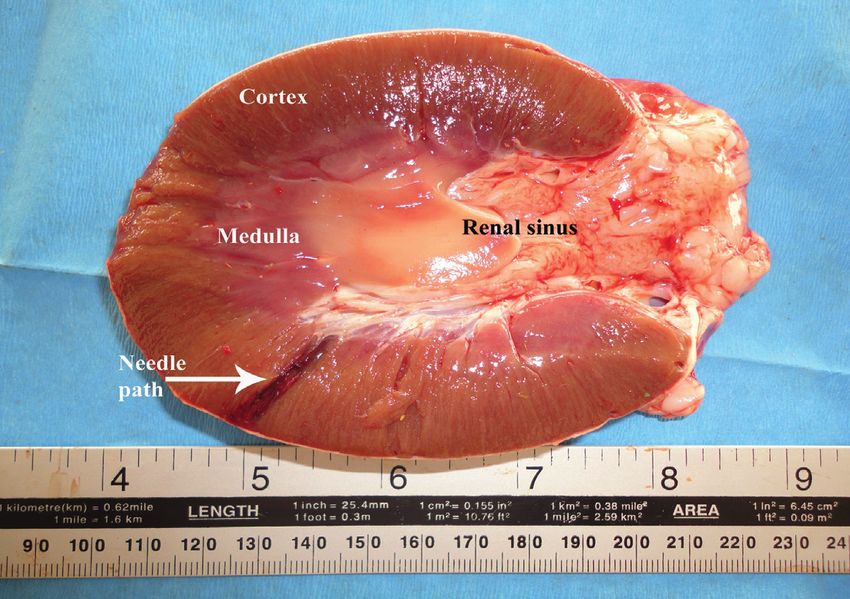
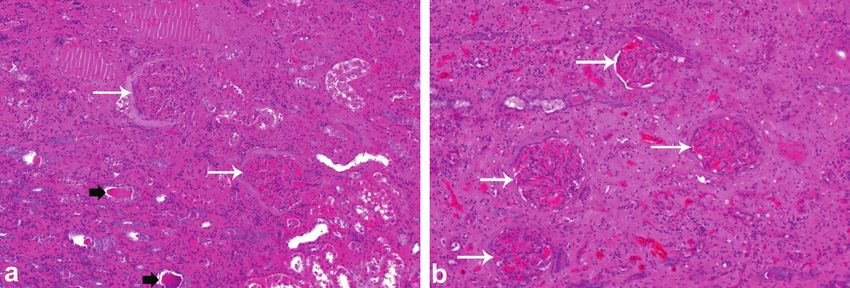
10                                                                                                   Veterinary Medicine International

Figure 20: Postmortem findings in a female camel with renal cell carcinoma. Image (a) shows 18 Kg right kidney compared to 1.5 Kg left
kidney. Image (b) shows cross section through the right kidney large, hemorrhagic, irregular shaped, and cavitated tumor [29]. 1, cortex; 2,
medulla; 3, tumor mass.

Figure 21: Renal cell carcinoma showing tubular differentiation with malignant epithelial lining and nuclear anaplasia (H&E, image a × 100;
image b × 400) [29].

abscessations, the diagnosis was verified postmortem (Fig-               colloid casts and a fibrous and granulation tissue part at the
ures 14 and 15).                                                        periphery. The interstitial tissue showed active and chronic
   In the male camel with bilateral chronic active pyelo-               cells infiltrate (Figure 16) [31]. In the other female camel
nephritis caused by E. coli, histopathological findings of both          with unilateral pyelonephritis caused by Staphylococcus
kidneys showed congested renal glomeruli and tubules with               lugdunensis, histopathological findings of the affected kidney
Veterinary Medicine International                                                                                                  11

showed congestion and periglomerular fibrosis, with atro-          [3] S. E. M. Barakat, F. A. A. Hizab, and M. S. Moqbel, “Path-
phic tubules, hyaline, and RBCs casts. The interstitial tissue        ological and serobiochemical studies on naturally occurring
showed fibrosis, thick-walled blood vessels, and dense mixed           kidney affections in camels (Camelus dromedarius),” Journal
inflammatory cell infiltrates. Area of necrosis with suppu-             of Camel Practice and Research, vol. 24, no. 1, pp. 55–59, 2017.
rative exudation was also seen (Figure 17) [30]. Renal ab-        [4] M. Tharwat, “Ultrasonographic findings in cattle and buffa-
                                                                      loes with chronic hepatic fascioliosis,” Tropical Animal Health
scesses may be imaged localized as hyperechoic or
                                                                      and Production, vol. 44, no. 7, pp. 1555–1560, 2012.
hypoechoic masses (Figure 18).                                    [5] M. Tharwat, “Ultrasonography as a diagnostic and prognostic
                                                                      approach in cattle and buffaloes with fatty infiltration of the
5.4. Renal Neoplasia. In dromedary camels, the most                   liver,” Polish Journal of Veterinary Sciences, vol. 15, no. 1,
common tumors are squamous cell carcinoma, fibroma,                    pp. 83–93, 2012.
                                                                  [6] M. Tharwat and S. Buczinski, “Clinicopathological findings
adenocarcinoma, fibromyxosarcoma, leiomyoma, angio-
                                                                      andechocardiographic prediction of the localisation of bovine
sarcoma, schwannoma, lipoma, microcystic adnexal carci-
                                                                      endocarditis,” Veterinary Record, vol. 169, no. 7, p. 180, 2011.
noma, renal cell carcinoma, Sertoli-Leydig cell tumour, and       [7] M. Tharwat and S. Oikawa, “Ultrasonographic evaluation of
granulosa cell tumor [29, 46, 47]. In a case of confirmed right        cattle and buffaloes with respiratory disorders,” Tropical
kidney renal cell carcinoma, transrectal ultrasonography              Animal Health and Production, vol. 43, no. 4, pp. 803–810,
revealed a caudally protruded, large, irregular shaped,               2011.
hypoechoic, and cavitated mass involving the right renal          [8] M. Tharwat and S. Oikawa, “Ultrasonographic characteristics
parenchyma. However, the left kidney appeared subjectively            of abdominal and thoracic abscesses in cattle and buffaloes,”
normal (Figure 19). The ultrasound imaging is still to be             Journal of Veterinary Medicine A, vol. 54, no. 9, pp. 512–517,
verified at postmortem examination (Figure 20). Histolog-              2007.
ical examination of the renal specimen revealed renal cell        [9] M. Tharwat, H. Sato, T. Kurosawa, S. Oikawa, and A. Nitanai,
carcinoma showing tubular differentiation with malignant               “Ultrasonographic imaging of experimentally induced pan-
                                                                      creatitis in cattle,” Veterinary Journal, vol. 165, no. 3,
epithelial lining and nuclear anaplasia (Figure 21). No
                                                                      pp. 314–324, 2003.
metastasis was found in other organs or even in the left         [10] M. Tharwat, “Clinicopathological and ultrasonographic
kidney [29].                                                          findings in 40 water buffaloes (Bubalus bubalis) with trau-
    In conclusion, ultrasonography of the kidneys in                  matic pericarditis,” Veterinary Record, vol. 167, no. 21,
dromedary camels is an important tool for imaging of the              pp. 819–824, 2010.
normal renal parenchyma as well as diagnosis, prognosti-         [11] M. Tharwat and S. Oikawa, “Efficacy and safety of ultrasound-
cation, and follow-up of treatment of kidney affections. This          guided percutaneous biopsy of the right kidney in cattle,”
imaging modality can scan focal as well as diffuse renal               Journal of Veterinary Medical Science, vol. 70, no. 2,
lesions. Ultrasound-guided collection of renal specimens              pp. 175–179, 2008.
remains the final modality for confirmation of kidney              [12] M. Tharwat, H. Sato, T. Kurosawa, and S. Oikawa, “Trans-
lesions.                                                              cutaneous ultrasound-guided pancreatic biopsy in cattle and
                                                                      its safety: a preliminary report,” Veterinary Journal, vol. 166,
                                                                      no. 2, pp. 188–193, 2003.
Data Availability                                                [13] M. Tharwat, “Ultrasonography of the lungs and pleura in
                                                                      healthy camels (Camelus dromedarius),” Acta Veterinaria
The data used to support the findings of this study are in-
                                                                      Hungarica, vol. 61, no. 3, pp. 309–318, 2013.
cluded within the article.                                       [14] M. Tharwat, “Ultrasonography of the cardiopulmonary sys-
                                                                      tem in camels (Camelus dromedarius),” Journal of Camel
Conflicts of Interest                                                 Health, vol. 1, pp. 37–46, 2019.
                                                                 [15] M. Tharwat, F. Al-Sobayil, A. Ali, and S. Buczinski, “Echo-
The author declares that he has no conflicts of interests.             cardiography of the normal camel (Camelus dromedarius)
                                                                      heart: technique and cardiac dimensions,” BMC Veterinary
Acknowledgments                                                       Research, vol. 8, no. 1, p. 130, 2012.
                                                                 [16] M. Tharwat, F. Al-Sobayil, A. Ali, and S. Buczinski, “Trans-
The author would like to express his gratitude to Dr.                 abdominal ultrasonographic appearance of the gastrointes-
N. Peacy, former English teacher, Deanship for Educational            tinal viscera of healthy camels (Camelus dromedarius),”
Services, Qassim University, Saudi Arabia, for language               Research in Veterinary Science, vol. 93, no. 2, pp. 1015–1020,
revising.                                                             2012.
                                                                 [17] M. Tharwat, “Ultrasonography of the liver in healthy and
References                                                            diseased camels (Camelus dromedarius),” Journal of Veteri-
                                                                      nary Medical Science, vol. 82, no. 4, pp. 399–407, 2020.
 [1] G. A. Kojouri1, H. Nourani, S. Sadeghian, H. Imani, and     [18] M. Tharwat, F. Al-Sobayil, A. Ali, and S. Buczinski, “Ultra-
     A. Raisi, “Pathological findings of slaughtered camels’           sonography of the liver and kidneys of healthy camels
     (Camelus dromedarius) kidneys in Najaf-Abad, Iran,” Vet-         (Camelus dromedarius),” The Canadian Veterinary Journal-
     erinary Research Forum, vol. 5, no. 3, pp. 231–235, 2014.         � La Revue Veterinaire Canadienne, vol. 53, no. 12,
 [2] K. Saini, H. Dadhich, M. Mathur, and A. Tripathi, “Histo-        pp. 1273–1278, 2012.
     pathological studies on renal lesions in dromedary camel    [19] M. Tharwat, “Ultrasonography of the abdomen in healthy and
     (Camelus dromedarius),” Journal of Camel Practice and Re-        diseased camels (Camelus dromedarius),” Journal of Applied
     search, vol. 22, no. 1, pp. 113–119, 2015.                       Animal Research, vol. 48, no. 1, pp. 300–312, 2020.
12 Veterinary Medicine International
[20] M. Tharwat, F. Al-Sobayil, A. Ali et al., “Percutaneous ul- [36] M. M. S. Smuts and A. J. Bezuidenhout, Anatomy of the
trasound-guided portocentesis in camels (Camelus drome- Dromedary, Clarendon Press, Oxford, England, 1987.
darius),” Journal of Camel Practice and Research, vol. 19, no. 2, [37] O. M. Radostits, C. C. Gay, D. C. Blood, and P. D. Constable,
pp. 193–196, 2012. “Veterinary medicine,” in A Textbook of the Diseases of Cattle,
[21] M. Tharwat, F. Al-Sobayil, and S. Buczinski, “Ultrasound- Sheep, Pigs, Goats and Horses, O. M. Radostits, C. C. Gay,
guided hepatic and renal biopsy in camels (Camelus drom- D. C. Blood, and K. W. Hinchcliff, Eds., W. B. Saunders,
edarius): technique development and assessment of the London, UK, 10th edition, 2007.
safety,” Small Ruminant Research, vol. 103, no. 2-3, [38] I. Köhler-Rollefson, P. Mundy, and E. Mathias, “Managing
pp. 211–219, 2012. and treating camels,” in A Field Manual of Camel Diseases:
[22] M. Tharwat, F. Al-Sobayil, A. Ali, and S. Buczinski, “Ultra- Traditional and Modern Healthcare for the Dromedary,
sonographic evaluation of abdominal distension in 52 camels pp. 1–67, ITDG Publishing, London, UK, 2001.
(Camelus dromedarius),” Research in Veterinary Science, [39] F. K. Al-Ani, Camel Management and Diseases, Al-Sharq
vol. 93, no. 1, pp. 448–456, 2012. Printing Press, Amman, Jordan, 2004.
[23] M. Tharwat, F. Al-Sobayil, A. Ali et al., “Clinical, ultraso- [40] M. E. Fowler, “Urinary system,” in Medicine and Surgery of
nographic, and pathologic findings in 70 camels (Camelus Camelids, pp. 479–486, Blackwell Publishing, Ames, IA, USA,
dromedarius) with Johne’s disease,” Canadian Veterinary 3rd edition, 2010.
[41] A. Rosenbaum, C. L. Guard, and B. L. Njaa, “Slaughterhouse
Journal, vol. 53, no. 5, pp. 543–548, 2012.
survey of pyelonephritis in dairy cows,” Veterinary Record,
[24] M. Tharwat, “Ultrasonographic findings in camels (Camelus
vol. 157, no. 21, pp. 652–655, 2005.
dromedarius) with trypanosomiasis,” Journal of Camel
[42] I. Yeruham, D. Elad, Y. Avidar, and T. Goshen, “A herd level
Practice and Research, vol. 20, no. 2, pp. 283–287, 2013.
analysis of urinary tract infection in dairy cattle,” The Vet-
[25] M. Tharwat and F. Al-Sobayil, “Ultrasonographic and post-
erinary Journal, vol. 171, no. 1, pp. 172–176, 2006.
mortem findings in camels (Camelus dromedarius) with ab-
[43] M. Floeck, “Sonographic application in the diagnosis of py-
dominal disorders,” Journal of Camel Practice and Research,
elonephritis in cattle,” Veterinary Radiology & Ultrasound,
vol. 23, no. 2, pp. 291–299, 2016. vol. 48, no. 1, pp. 74–77, 2004.
[26] M. Tharwat, “Ultrasonography of the gastrointestinal tract in [44] K. J. Chandler, B. O. Brien, and J. N. Huxley, “Hydronephrosis
healthy and diseased camels (Camelus dromedarius),” Journal and renal failure in two Friesian cows,” Veterinary Record,
of Camel Health, vol. 2, pp. 10–15, 2020. vol. 146, no. 22, pp. 646–648, 2000.
[27] M. Tharwat and F. Al-Sobayil, “Ultrasonographic findings in [45] S. Karmveer, H. Dadhich, M. Mathur, and T. Ashutosh,
camels (Camelus dromedarius) with different urinary affec- “Histopathological studies on renal lesions in dromedary
tions,” Journal of Camel Practice and Research, vol. 23, no. 2, camel (Camelus dromedarius),” Journal of Camel Practice and
pp. 301–308, 2016. Research, vol. 22, no. 1, pp. 113–119, 2015.
[28] M. Tharwat and F. Al-Sobayil, “Ultrasonographic findings in [46] A. Ali, R. Derar, F. A. Sobayil, M. Tharwat, A. Fathy, and
camel calves (Camelus dromedarius) withthoracic affections,” M. Khodeir, “Adenocarcinoma in the genital tract of infertile
Journal of Camel Practice and Research, vol. 23, no. 2, female dromedary camels,” Journal of Camel Practice and
pp. 287–290, 2016. Research, vol. 25, no. 2, pp. 181–187, 2018.
[29] M. Tharwat, F. Al-Sobayil, A. Ali, D. Derar, and M. Khodeir, [47] F. A. Alsobayil, A. Ali, D. R. Derar, M. Tharwat, A. F. Ahmed,
“Renal cell carcinoma in A female Arabian camel,” Journal of and M. Khodeir, “Tumours in dromedary camels: prevalence,
Camel Practice and Research, vol. 24, no. 1, pp. 61–66, 2017. types and locations,” Journal of Camel Practice and Research,
[30] M. Tharwat, M. Sadan, E. El-Shafaey, A. Al-Hawas, and vol. 25, no. 2, pp. 189–197, 2018.
E. M. A. Saeed, “Unilateral nephrectomy in a female drom-
edary camel with pyelonephritis caused by Staphylococcus
lugdunensis,” Pakistan Veterinary Journal, vol. 38, pp. 116–
118, 2018.
[31] M. Tharwat, M. Sadan, E.-S. El-shafaey, E.-H. Saeed, and
A. Al-hawas, “Bilateral renal abscessation and chronic active
pyelonephritis in a male camel (Camelus dromedarius) caused
by Escherichia coli,” Journal of Veterinary Medical Science,
vol. 80, no. 5, pp. 778–783, 2018.
[32] E. M. Elmanakhly, M. Tharwat, E. El-Shafaey, M. Sadan, and
A. S. M. Aljohani, “First report of benign intraluminal
esophageal inflammatory fibroid polyp infected with Candida
albicans in camel: a case report,” Journal of Camel Health,
vol. 2, pp. 29–33, 2020.
[33] M. Tharwat, E.-S. El-Shafaey, M. Sadan, A. Ali, F. Al-Sobayil,
and A. Al-Hawas, “Omaso-abomasal adenocarcinoma in a
female Arabian camel (Camelus dromedarius),” Journal of
Applied Animal Research, vol. 46, no. 1, pp. 1268–1271, 2018.
[34] M. Tharwat, “Chronic peritonitis in dromedary camels:
clinical, ultrasonographic and pathologic findings,” Journal of
Camel Practice and Research, vol. 26, no. 2, pp. 169–172, 2019.
[35] M. Tharwat, “Multiple splenic abscessation in a camel: case
report,” Journal of Camel Practice and Research, vol. 26, no. 3,
pp. 273–276, 2019.You can also read