Ribosomal Proteins S5 and L6: High-resolution Crystal Structures and Roles in Protein Synthesis and Antibiotic Resistance
←
→
Page content transcription
If your browser does not render page correctly, please read the page content below
Article No. mb981780                                                          J. Mol. Biol. (1998) 279, 873±888

Ribosomal Proteins S5 and L6: High-resolution
Crystal Structures and Roles in Protein Synthesis and
Antibiotic Resistance
Christopher Davies1{, Dirksen E. Bussiere2{, Barbara L. Golden3
Stephanie J. Porter1, Venki Ramakrishnan4* and Stephen W. White1,5*
1
 Department of Structural         Antibiotic resistance is rapidly becoming a major medical problem. Many
Biology, St. Jude Children's      antibiotics are directed against bacterial ribosomes, and mutations within
Research Hospital, 332 North      both the RNA and protein components can render them ineffective. It is
Lauderdale St., Memphis           well known that the majority of these antibiotics act by binding to the
TN 38105, USA                     ribosomal RNA, and it is of interest to understand how mutations in the
2                                 ribosomal proteins can produce resistance. Translational accuracy is one
  Abbott Laboratories
                                  important target of antibiotics, and a number of ribosomal protein
Department of Scienti®c
                                  mutations in Escherichia coli are known to modulate the proofreading
Information, Analysis and
                                  mechanism of the ribosome. Here we describe the high-resolution struc-
Management, 100 Abbott Park       tures of two such ribosomal proteins and characterize these mutations.
Rd., Abbott Park, IL 60064
                                     The S5 protein, from the small ribosomal unit, is associated with two
USA
                                  types of mutations: those that reduce translational ®delity and others that
3
  Department of Chemistry &       produce resistance to the antibiotic spectinomycin. The L6 protein, from
Biochemistry, Box 215             the large subunit, has mutations that cause resistance to several amino-
University of Colorado            glycoside antibiotics, notably gentamicin. In both proteins, the mutations
Boulder, CO 80309, USA            occur within their putative RNA-binding sites. The L6 mutations are par-
4                                 ticularly drastic because they result in large deletions of an RNA-binding
Department of Biochemistry        region. These results support the hypothesis that the mutations create
University of Utah School of      local distortions of the catalytic RNA component.
Medicine, Salt Lake City             When combined with a variety of structural and biochemical data, these
UT 84132, USA                     mutations also become important probes of the architecture and function
5
 Department of Biochemistry       of the translational machinery. We propose that the C-terminal half of S5,
University of Tennessee           which contains the accuracy mutations, organizes RNA structures associ-
Memphis, 858 Madison              ated with the decoding region, and the N-terminal half, which contains
Suite G01, Memphis                the spectinomycin-resistance mutations, directly interacts with an RNA
TN 38163, USA                     helix that binds this antibiotic. As regards L6, we suggest that the
                                  mutations indirectly affect proofreading by locally distorting the
                                  EF-Tu GTP  aminoacyl tRNA binding site on the large subunit.
                                                                               # 1998 Academic Press Limited
                                  Keywords: Ribosome architecture; X-ray crystallography; protein-RNA
*Corresponding authors            interactions; protein evolution; translocation

Introduction                                             ribosomal components that confer resistance to
                                                         these compounds. With the increased use of anti-
   Ef®cient protein synthesis is essential to cell       biotics to treat bacterial infections, pathogenic
growth, and many antibiotics have evolved to inca-       strains are now acquiring antibiotic resistance, and
pacitate the ribosome, the cell's ubiquitous transla-    many important drugs that target bacterial ribo-
tional machinery. In response to this selective          somes are becoming ineffective. This represents an
pressure, mutations have evolved within bacterial        extremely serious medical problem that has
                                                         prompted extensive searches for new antibacterial
  {These authors contributed equally to this work.       agents.
  Abbreviations used: dsRBD, double-stranded RNA-           An important ribosome function targeted by a
binding domain; EF, elongation factor; RRM, RNA          number of antibiotics is proofreading, which
recognition motif.                                       ensures accurate reading and decoding of the

0022±2836/98/240873±16 $25.00/0                                                   # 1998 Academic Press Limited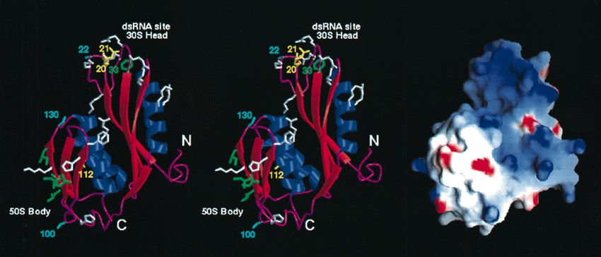
874                                                                             Ribosomal Proteins S5 and L6

mRNA. Early insights into ribosomal ®delity were       S5 from the 30 S subunit, and L6 from the 50 S
provided by the antibiotic streptomycin, which         subunit. As noted above, mutations within both
had been shown to reduce the accuracy of protein       proteins can in¯uence ribosome accuracy, and they
synthesis. Further studies revealed that ®delity can   are clearly adjacent to important functional centers
also be altered by mutations in three 30 S subunit     of their respective subunits.
proteins, S4, S5 and S12 (Gorini & Kataja, 1964).         S5 is particularly interesting, since three distinct
Mutations in S4 and S5 affect the ribosome in a        types of mutations have been identi®ed, the ram
way similar to streptomycin and reduce the level       mutations described above (Piepersberg et al.,
of accuracy (the so-called ram or ribosome ambigu-     1975a,b), cold sensitivity (Guthrie & Nomura,
ity mutations), whereas mutations in S12 have the      1969) and ribosomal resistance to the antibiotic
opposite effect and increase translational accuracy    spectinomycin (Bollen et al., 1969; Funatsu et al.,
(reviewed in Kurland et al., 1990). These factors      1972; Piepersberg et al., 1975b). Spectinomycin is
appear to be additive, since S12 mutations confer      usually associated with the translocation mechan-
resistance to streptomycin, causing the ribosome to    ism of the ribosome (Burns & Cundliffe, 1973;
become dependent on the antibiotic for optimal         Wallace et al., 1974), and the S5 locus therefore
accuracy. This dependency can be relieved by sub-      appears to be close to not one, but two important
sequent mutations in either S4 or S5. More             functions in the 30 S subunit. This central location
recently, de®ned regions of 16 S rRNA have also        is supported by RNA footprinting (Stern et al.,
come to be associated with the ribosome's accuracy     1989; Powers & Noller, 1995) and crosslinking
function (Montandon et al., 1986; MelancËon et al.,    experiments (Osswald et al., 1987) that show S5 to
1988; Powers & Noller, 1991).                          be in the vicinity of the three-way junction that
   Although largely a feature of the 30 S subunit,     connects the major subdomains of the 16 S rRNA
translational accuracy can also be affected by two     molecule.
components of the 50 S subunit, ribosomal protein         L6 has been localized by immunoelectron
L6 and the 2660 loop region of 23 S rRNA.              microscopy (StoÈf¯er-Meilicke et al., 1983; Hackl &
Mutations have been identi®ed in these com-            StoÈf¯er-Meilicke 1988) and protein-protein cross-
                                                       linking (Traut et al., 1986; Walleczek et al., 1989a,b)
ponents that result in a decreased rate of trans-
                                                       to a region between the base of the L7/L12 stalk
lation, greater accuracy in protein synthesis, and
                                                       and the central protuberance (Walleczek et al.,
increased resistance to many of the misreading-
                                                       1988). L6 appears to be at the 50 S ± 30 S interface
inducing aminoglycoside antibiotics, in particular
                                                       because it can be cross-linked to S13 in the 30 S
gentamicin (Buckel et al., 1977; Kuhberger et al.,
                                                       subunit (Lambert & Traut, 1981). As regards its
1979; Hummel et al., 1980; Tapprich & Dahlberg,
                                                       23 S rRNA environment, L6 has been cross-linked
1990; MelancËon et al., 1992).
                                                       to the end of a stem-loop structure (nucleotides
   For several years, we have been studying the        2455 to 2496 in Escherichia coli) that projects from
structures of individual proteins from the prokar-     domain V (Wower et al., 1981). Domain V is a
yotic ribosome as part of a concerted effort in        highly conserved region of the 23 S molecule that
many laboratories to understand the structure and      has been implicated in peptide bond formation
mechanism of the translational machinery. The role     (Vester & Garrett, 1988) and the binding of tRNA
of ribosomal proteins now appears to be largely        at the A and P sites (Steiner et al., 1988; Mitchell
architectural, helping to direct and maintain the      et al., 1990; Brimacombe et al., 1993). L6 also binds
fold of the catalytic RNA component, and it is of      to a fragment of 23 S rRNA corresponding to
interest to understand how mutations in the pro-       domain VI (Leffers et al., 1988). Domain VI con-
teins exert their effects on ribosome function. Our    tains the a-sarcin loop and is the binding site for
studies have shown that antibiotic resistance          the EF-Tu  GTP aminoacyl tRNA ternary complex
mutations, and other mutations that affect ribo-       (Leffers et al., 1988; Moazed et al., 1988).
some function and assembly, tend to cluster in            Several years ago we reported the medium-res-
their putative RNA-binding sites (Ramakrishnan &       olution crystal structures of S5 and L6 from Bacil-
White, 1992; Golden et al., 1993a; Hoffman et al.,     lus stearothermophilus, and showed that each is a
1996). This suggests that they act indirectly by       two-domain molecule with clear putative RNA-
locally altering the ribosomal RNA conformation.       binding regions (Ramakrishnan & White, 1992;
If correct, these proteins represent powerful probes   Golden et al., 1993b). Here, we present the
of the structure and function of the ribosome, since   re®ned structures of both proteins, and use them
their mutant forms and associated phenotypes can       to re-evaluate their roles in the organization of
be directly correlated with their RNA environ-         the large and small ribosomal subunits. In the
ments. These results also provide important            case of L6, we have also characterized the
insights into the mechanism of antibiotic action       mutations that produce gentamicin resistance.
and the acquisition of antibiotic resistance. We       Consistent with contemporary ideas on the role
have been investigating the crystal structures of      of ribosomal proteins, S5 and L6 appear to
two proteins that exemplify this approach to           organize important local regions of the 16 S and
studying the mechanism of the ribosome, namely         23 S rRNA molecules, respectively.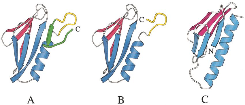
Ribosomal Proteins S5 and L6 875
Results Table 1. Data collection and re®nement parameters for
ribosomal protein S5
Ribosomal protein S5 A. Data Collection
Resolution (AÊ) 50.0±2.2
Refinement Total number of observations 66,420
Number of unique reflections 11,138
The starting point of the re®nement was the R-merge (%)a 8.15
Ê structure previously published, which
2.7 A Completeness of data (%) 93
included residues 4 to 148 of the 166 amino acid Ê ±2.3 A
Completeness of data (%) (2.2 A Ê) 66
residue protein (Ramakrishnan & White, 1992). B. Refinement
This structure had an R-factor of 22% with devi- Resolution (A Ê) 10 ±2.2
ations from ideality in bond lengths and angles of Sigma cut-off 0.0
0.017 AÊ and 3.74 , respectively. To obtain an initial Residues in model 5± 27, 30± 148
Ê
2.2 A structure, a series of Powell minimizations Number of protein atoms 1,037
Number of water molecules 187
was performed in which the high-resolution data Number of reflections 10,470
were added in 0.2 A Ê increments every 100 cycles. R-value (%) 21.66
This structure was compared to Fo ÿ Fc and R-value (%) (2.2 AÊ ± 2.3 A
Ê) 26.76
2Fo ÿ Fc electron density maps, manual adjust- Rfreeb value (%) 26.63
Overall G factorc ÿ0.02
ments were made, and water molecules were r.m.s. deviation on bonds (A Ê) 0.009
added. The criteria for the latter were clear peaks r.m.s. deviation on angles ( ) 1.485
in both maps, and adjacency to suitable hydrogen r.m.s. deviation on improper angles ( ) 2.440
bond donors and acceptors. Following the rebuild- r.m.s. deviation on dihedral angles ( ) 27.5
Mean B factor (main-chain) (A Ê 2) 36.2
ing, the model was subjected to 200 cycles of Ê 2)
r.m.s. deviation in B factor (main-chain) (A 2.683
Powell minimization, simulated annealing at Mean B factor (side-chain) (A Ê 2) 44.3
2000 K and individual B-factor re®nement. Weak Ê 2)
r.m.s. deviation in B factor (side-chain) (A 5.556
electron density now became visible for the disor- Residues in most favored regions (%) 87.7
dered N and C termini, and for residues 28 to 30 Residues in additional allowed regions (%) 10.5
within a ¯exible loop. Following three more macro- a
R-merge jIi ÿ Imj/Im.
b
cycles of model adjustment, Powell minimization, c
BruÈnger (1992).
simulated annealing and individual B-factor re®ne- Laskowski et al. (1993).
ment, convergence was reached. Summaries of the
re®nement statistics and the model geometry are
shown in Table 1. The density for the N and C dues. Valine 16, 24, 38 and 46, alanine 17, leucine
termini showed little further improvement, and the 36, phenylalanine 48 and isoleucine 72 from the
termini appear to be essentially disordered and N-terminal domain are on the outer surface of
loosely associated with nearby ordered regions the b-sheet, and proline 110 and 133, alanine 113
within the unit cell. Within the body of the mol- and 118, valine 114 and 137, leucine 117, isoleu-
ecule, only residues 28 to 30 are not clearly de®ned cine 134 and phenylalanine 141 from the C-term-
in the electron density. The ®nal model comprised inal domain are on the outer surface of the
residues 5 to 27 and 31 to 148. The S5 structure has a-helices (Figure 2). The two phenylalanine rings,
been brie¯y described (Ramakrishnan & White, one from each domain, are adjacent and interact
1992), and a detailed description is given below. in the usual edge-to-face orientation (Burley &
Petsko, 1985). A short loop between residues 41
S5 comprises two domains and 44 in the N-terminal domain is adjacent to
another loop in the C-terminal domain between
S5 is an a/b class of protein that is organized residues 118 and 123, and residues 118 and 119
into two a/b domains, each containing a single are constrained to be alanine and glycine, respect-
b-sheet with a-helical elements on one side ively, by the close approach. There is a speci®c
(Figure 1). The two domains are directly connected hydrogen-bonded interaction between the two
at valine 74. The side-chain of this residue forms loops involving the amide protons of aspartate 41
part of the C-terminal domain hydrophobic core, and lysine 42 and the carbonyl oxygen of atom
and its amide proton is hydrogen-bonded to the leucine 117. Finally, there is a conserved interdo-
carbonyl oxygen atom of glycine 44 in the N-term- main salt-bridge between arginine 14 and gluta-
inal domain. The two halves of the protein pack mate 116.
closely together such that the N-terminal domain
b-sheet associates with the C-terminal domain The N-terminal domain
a-helices. The packing of the domains creates alter-
nate layers of a-helices and b-sheets, which results The N-terminal half of S5 is somewhat elongated
in an unusual a-b-a-b sandwich architecture. The and contains ®ve main secondary structural
two domains appear to be structurally very similar, elements, four b-strands and an a-helix, linearly
but closer inspection reveals that their topologies arranged as bbbab. The locations of these elements
are in fact quite different (see later). in the sequence are as follows: strand 1, residues
The domains interact extensively through a 11 to 19; strand 2, residues 33 to 41; strand 3, resi-
highly conserved network of hydrophobic resi- dues 44 to 53; helix 1, residues 55 to 70; strand 4,876 Ribosomal Proteins S5 and L6
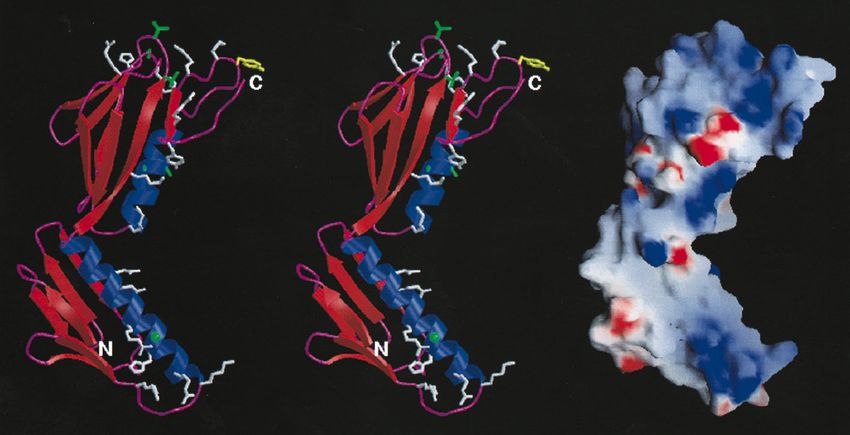
Figure 1. The overall structure of ribosomal protein S5. Left, stereo views of the Ca trace of the S5 backbone with
every tenth residue labeled and marked with a ®lled circle. Right, a ribbon representation of S5 showing the elements
of secondary structure. The unstructured C-terminal 19 amino acid residues have not been included for clarity. The
Figure was produced using MOLSCRIPT (Kraulis, 1991).
residues 71 to 74. These elements are arranged as a on one surface of the b-sheet on the outer surface
four-stranded anti-parallel b-sheet with the single of the S5 molecule. The a-helix appears to be stabil-
a-helix inserted between strands 3 and 4 (Figure 1). ized by a number of salt bridges on its outer sur-
As a result, strand 4 is quite short. The a-helix sits face (arginine 61 and lysine 62 with glutamate 58;
Figure 2. The structurally import-
ant features within the two
domains of ribosomal protein S5.
Top, stereo views of the N-terminal
domain. Regions that are discussed
in the text are numbered as fol-
lows. Region 1 is the interdomain
hydrophobic patch. Region 2 is the
small hydrophobic core within the
putative RNA-binding loop cen-
tered on phenylalanine 33. Region
3 is the exposed hydrophobic core.
Region 4 is the conserved salt-
bridge between glutamate 13 and
lysine 68. Region 5 is the close
approach between strand 3 and
helix 1 that is mediated by con-
served glycine and alanine resi-
dues. For clarity, individual
residues are not labeled. Bottom,
stereo views of the C-terminal
domain. The residues in the inter-
domain hydrophobic patch are in
the region labeled 1. The conserved
glycine and alanine residues in the
tight turns at the top of the domain
are labeled. The bottom half of the
domain is covered by three loops,
and the ends of the loops are
labeled. Aspartate 122 and threo-
nine 80 form important inter-loop
interactions. See the text for details.
The unstructured C-terminal 19
amino acid residues have not been
included for clarity. The Figure was
produced using MOLSCRIPT
(Kraulis, 1991).Ribosomal Proteins S5 and L6 877
lysine 68 with glutamate 65; lysine 69 with aspar- ¯anked by proline residues and includes a short
tate 66). b-ribbon with a b-turn between residues 77 and 80.
An unusual feature of this domain is that the Loop 8, between strands 6 and 7 (residues 97 to
a-helix is shifted towards one edge of the b-sheet, 104), is a highly conserved region of S5 and con-
and makes a very close approach to strand 3. They tains a b-turn between residues 100 and 103. Pro-
are so close in fact that the contacting surfaces of line 98, which initiates the loop, is present in all
strand 3 and helix 1 are constrained to contain prokaryotic-like S5 sequences, and threonine 103,
exclusively glycine (44, 47, 49 and 51) and alanine whose side-chain oxygen atom makes a hydrogen
residues (59, 63 and 67), respectively (Figure 2). bond across the b-turn to the amide proton of ser-
Conversely, the gap between helix 1 and strand 1 ine 100, is also highly conserved. Finally, loop 10,
on the opposite edge of the b-sheet exposes the between helix 2 and strand 8 (residues 119 to 123),
hydrophobic core residues in this region (Figure 2). traverses the domain and connects the a-helical
This gap is minimized and held roughly constant side to the b-sheet side. This loop is also conserved
by a kink in strand 1 that results from a b-bulge at and provides the main-chain and side-chain hydro-
residues 16 and 17. Speci®cally, the amide protons gen bonding elements that knit this region
of these residues form a bifurcated hydrogen bond together. One of the side-chain oxygen atoms
to the carbonyl oxygen atom of residue 36 on the of aspartate 122 interacts with the amide protons
adjacent strand 2. At the lower end of the domain, of residues 103 and 104, and the amide protons of
the gap is spanned by a highly conserved salt- residues 121 and 122 bond to the carbonyl oxygen
bridge between glutamate 13 and lysine 68 atom of residue 79. Finally, the side-chain oxygen
(Figure 2). atom of threonine 80 bonds to the amide proton of
Loop 2 (residues 19 to 33) that connects strands residue 99 to complete the network.
1 and 2 is convoluted, ¯exible, and highly posi-
tively charged. Apart from the N and C termini, The N-terminal domain is a double-stranded
this region is the most poorly de®ned in the elec-
RNA-binding element
tron density map, and density for residues 28 to 30
is barely visible. The loop is folded around a mini A number of RNA-binding motifs have now
hydrophobic core that contains a central and been described that can be identi®ed on the basis
highly conserved phenylalanine (residue 33; of their characteristic amino acid sequences (Burd
Figure 2). We have previously identi®ed this loop & Dreyfuss, 1994). Structural information is now
as a probable RNA-binding region, and subsequent available for three of these motifs: the RRM (Nagai
results suggest that it may be speci®c for double- et al., 1990; Hoffman et al., 1991), the double-
stranded RNA. This will be discussed later. stranded RNA-binding domain (dsRBD: Bycroft
et al., 1995; Kharrat et al., 1995) and the KH domain
The C-terminal domain (Castiglione et al., 1995). The RRM domain has
now been found in several ribosomal proteins
The C-terminal half of S5 contains six secondary (Ramakrishnan & White, 1998), and it was recently
structural elements, four b-strands and two shown that the N-terminal half of S5 represents a
a-helices, linearly arranged as bbbaba. The classic example of a dsRBD (Bycroft et al., 1995).
locations of these elements in the sequence is as The only major difference is that the dsRBD nor-
follows: strand 5, residues 85 to 89; strand 6, resi- mally contains an N-terminal a-helix that is posi-
dues 92 to 97; strand 7, residues 104 to 107; helix 2, tioned next to the C-terminal a-helix on one
residues 108 to 119; strand 8, residues 123 to 129; surface of the b-sheet, but this a-helix is not present
helix 3, residues 132 to 146. Like the N-terminal in S5.
domain, these elements are also arranged as a An analysis of the dsRBD consensus sequence
b-sheet with a-helical elements packed onto one combined with site-directed mutagenesis and
surface, but the topology differs signi®cantly. In RNA-binding experiments have shown that the
this case, the outer short b-strand is parallel with RNA-binding surface comprises the extended
its neighbor and sequentially the third element. loop between strands 1 and 2, and the N-term-
One surface of the b-sheet is exposed, and the inal part of the a-helix (Bycroft et al., 1995).
other is packed against the two a-helices in this These regions contain a number of highly con-
domain. Curiously, the domain also resembles the served basic residues that cannot be mutated
RNA recognition motif (RRM) structure that is without seriously impairing the interaction with
found in a number of other ribosomal proteins dsRNA. A model has been proposed in which
(Ramakrishnan & White, 1998), but again the top- these two regions of the protein straddle the
ology is quite different. dsRNA groove and make non-speci®c ionic inter-
The connections between the secondary structur- actions with the two sugar-phosphate backbones
al elements at the top of the domain are short, and (Bycroft et al., 1995). Recently, the crystal struc-
are populated with glycine and alanine residues to ture of a dsRBD-dsRNA complex has been deter-
accommodate the tight turns (Figure 2). However, mined that con®rms the major features of this
those at the bottom are convoluted and serve to model (J. Ryter & S. Schultz, personal communi-
cover this end of the protein (Figure 2). Loop 6 cation). The N-terminal domain of S5 also con-
between strands 4 and 5 (residues 74 to 85) is tains a number of highly conserved lysine and878 Ribosomal Proteins S5 and L6 arginine residues in these two locations, and is and that it interacts with the minor groove of the likely to interact with a double-stranded region RNA helix (J. Ryter & S. Schultz, personal com- of 16 S rRNA in the same way. Such an inter- munication). action has recently been supported by site- directed hydroxyl radical probing of the S5 RNA environment. A probe placed at residue 22 (21 in Functional regions of the C-terminal domain E. coli) within the extended loop of S5 clearly labeled helix 34 of the 16 S rRNA molecule S5 is not a primary rRNA-binding protein, but (Heilek & Noller, 1996). Figure 3 shows two rep- the C-terminal domain does have a surface patch resentations of the S5 N-terminal domain that that contains the conserved aromatic and basic highlight the conserved functional residues and residues that are diagnostic of speci®c interactions the associated surface electropotential, respect- with RNA (Oubridge et al., 1994; ValegaÊrd et al., ively. 1997). This region, which contains histidine 83 and In both of the NMR structures of the dsRBD, the 88, phenylalanine 89 and arginine 138 is shown in extended loop between strands 1 and 2 is poorly Figure 3. We previously suggested that the C-term- de®ned and apparently ¯exible. In the S5 crystal inal domain might also interact with 16 S rRNA in structure, this loop is wrapped around a phenyl- the vicinity of glycine 104 and arginine 112 alanine side-chain in a loosely ordered confor- (Ramakrishnan & White, 1992). These amino acid mation, and it probably adopts a de®ned structure residues are adjacent, highly conserved and the when bound to RNA. We have recently deter- sites of the ram mutations. RNA footprinting exper- mined the crystal structure of ribosomal protein iments have shown that the RNA-binding sites of S7, and it contains an extended b-ribbon structure S5 cover several de®ned regions of the 16 S rRNA that we propose interacts with a region of double- molecule (Stern et al., 1989; Powers & Noller, 1995). stranded RNA (Wimberley et al., 1997). The S5 Recently, more precise site-directed hydroxyl rad- loop could also adopt this type of conformation, ical cleavage data have been obtained for speci®c and a pair of adjacent conserved glycines residues residues within the C-terminal domain (Heilek & (27 and 28) could mediate a tight turn at the end of Noller, 1996). Position 130 (129 in E. coli) shows lit- such a b-ribbon. It has now been con®rmed from tle interaction with rRNA, whereas position 100 the dsRBD-dsRNA crystal structure that the loop (99 in E. coli) shows a more extensive interaction. does become extended into a b-ribbon structure, Neither position is directly within the putative Figure 3. The putative functional sites of ribosomal protein S5. Left, stereo views of the protein with key residues labeled. Individual residues are color-coded as follows: white, basic and aromatic residues; green, exposed hydro- phobic residues; yellow, sites of mutations for ram (112) and spectinomycin resistance (20, 21 and 22); cyan, residues used for site-speci®c hydroxyl radical footprinting (22, 100 and 130; Heilek & Noller, 1996). Also shown in green is the conserved phenyalanine 33, which is buried within the putative RNA-binding loop. It is proposed that S5 con- tains two regions of interaction with ribosomal RNA, one on each domain. The N-terminal domain contains a double-stranded RNA-binding site that interacts with the head of the 30 S subunit. The C-terminal domain interacts with RNA elements in the body of the 30 S subunit. This domain also has an exposed hydrophobic patch on the left that is proposed to interact with another ribosomal protein, possibly S8. The unstructured C-terminal 19 amino acid residues have not been included for clarity. The Figure was produced using MOLSCRIPT (Kraulis, 1991). Right, The surface electrostatic potential of ribosomal protein S5 calculated using GRASP (Nicholls et al., 1991). The orientation of the molecule is identical with that shown on the left. The extreme ranges of red (negative) and blue (positive) rep- resent electrostatic potentials of 10 kbT, where kb is the Boltzmann constant and T is the temperature.
Ribosomal Proteins S5 and L6 879
RNA-binding sites, but they are immediately adja- simulated annealing omit maps in which residues
cent (Figure 3). were deleted from the model in blocks of ten
A rather striking surface feature of the C-term- along the polypeptide chain. A summary of the
inal domain is an extended hydrophobic patch that re®nement statistics and model geometries is
covers the b-sheet towards the lower end of the shown in Table 2.
protein as viewed in Figure 1. This patch com-
prises isoleucine 86, 95, 106 and 128, and leucine Structure description
124, and is shown in Figure 3. We have reported
similar hydrophobic regions on the surfaces of L14 The structure of the L6 molecule has been
and S8, and suggested that they represent sites of described in some detail (Golden et al., 1993b), and
interaction with other ribosomal proteins (Davies only a brief description will be given here, empha-
et al., 1996a,b). A number of proteins are known to sizing features that have been revealed by the
be close to S5 in the ribosome, including S2, S3, S4, re®nement process. L6 has an elongated L-shaped
S8, S12, S15 and S16 (Lambert et al., 1983; Capel structure, and is divided into two structurally
et al., 1987), but the only protein for which there is homologous domains that appear to have evolved
evidence of a direct interaction is S8. This protein by gene duplication (Figure 4), possibly from an
has been shown to form a complex with S5 in sol- RNA-binding RRM-like motif. The domains are
ution (Tindall & Aune, 1981), and a protein-protein approximately equal in size; the N-terminal
cross-link between the two molecules has been domain consists of amino acids 1 through 79, and
reported (Allen et al., 1979). We recently deter- the C-terminal domain 80 through 177. Each
mined the structure of S8 and identi®ed a hydro- domain comprises a pair of three-stranded antipar-
phobic concave surface on the molecule adjacent to allel b-pleated sheets and an a-helix, and these are
the cross-link site that could mediate its interaction arranged like a triangular prism in which two
with S5 (Davies et al., 1996b). b-sheets form two of the faces and the a-helix the
Murzin (1995) has noted that the C-terminal third. The two b-sheets contain strands b1-b5-b6
domain contains a rare left-handed bab-unit (sheet 1) and b2-b3-b4 (sheet 2), and the a-helix is
between the parallel strands 7 and 8 of the mixed at the C-terminus. To re¯ect the homology, corre-
b-sheet. He has further noted that the overall top- sponding elements within each domain are labeled
ology of the C-terminal domain including this Nb1, Cb1 etc.
unusual turn is found also in the second domain of There are two major differences between the two
the gyrase B molecule and domain IV of elongation domains. The ®rst is at the N terminus, where Nb1
factor G. From a detailed comparison of these is very short and contains only four residues,
three protein modules, he has concluded that they
probably diverged from a common ancestor, most
likely a primitive ribosomal protein. Table 2. Data collection and re®nement parameters for
ribosomal protein L6
A. Data Collection
Ribosomal protein L6 Resolution (AÊ) 20.0±2.0
Total number of observations 39,577
Refinement Number of unique reflections 13,166
R-merge (%)a 8.00
The starting point of the re®nement was the Completeness of data (%) 97.5
2.6 AÊ structure previously published, which had Ê ±2.09 A
Completeness of data (%) (2.00 A Ê) 97.4
an R-factor of 18.3%, and deviations from ideality B. Refinement
in bond lengths and angles of 0.016 A Ê and 3.37 Resolution (A Ê) 8.0±2.0
respectively (Golden et al., 1993b). In this model, Sigma cut-off 0.0
which contained 161 residues, seven and nine Residues in model 7±170
Number of protein atoms 1251
residues were omitted from the N and C termini, Number of water molecules 140
respectively, due to weak or absent electron den- Number of reflections 12,972
sity. During initial rounds, the model was re®ned R-value (%) 23.0
against 2.2 AÊ resolution data, and the resolution R-value (%) (2.00 AÊ ±2.09 AÊ) 32.8
was extended to the 2.0 A Ê limit at later stages. Rfreeb value (%) 30.1
Overall G factorc 0.24
Between each round of re®nement, the model r.m.s. deviation on bonds (A Ê) 0.014
was inspected and manually adjusted as necess- r.m.s. deviation on angles ( ) 1.89
ary. Water molecules were introduced into the r.m.s. deviation on improper angles ( ) 1.45
model based on peaks in both the 2Fo ÿ Fc and r.m.s. deviation on dihedral angles ( ) 24.9
Mean B factor (main-chain) (A Ê 2) 29.3
Fo ÿ Fc electron density maps, and suitably adja- r.m.s deviation in B factor (main-chain) (A Ê 2) 1.590
cent hydrogen-bonding partners. Particular effort Mean B factor (side-chain) (A Ê 2) 40.0
was made to extend the N and C termini of the Ê 2)
r.m.s. deviation in B factor (side-chain) (A 3.182
molecule using the gradually improving electron Residues in most favored regions (%) 88.8
density in these regions. It was eventually Residues in additional allowed regions (%) 11.2
possible to include one additional residue at the a
R-merge jIi ÿ Imj/Im.
b
N terminus and two residues at the C terminus. BruÈnger (1992).
c
Laskowski et al. (1993).
The ®nal model was inspected using a series of880 Ribosomal Proteins S5 and L6 Figure 4. The overall structure of ribosomal protein L6. Left, stereo views of the Ca trace of the L6 backbone with every tenth residue labeled and marked with a ®lled circle. Right, a ribbon representation of L6 showing the elements of secondary structure. The Figure was produced using MOLSCRIPT (Kraulis, 1991). whereas Cb1 is longer with eight residues. How- an appropriately located hydrophobic depression ever, the re®nement has revealed weak but clear that could accommodate this short element electron density at the N terminus, which indicates (Figure 5). As described below, these N-terminal that some of the disordered residues at the extreme residues probably become more ordered upon N terminus extend the strand Nb1. It appears that binding RNA. The second difference is at the C ter- the end of this outer strand is frayed out from the minus. Na1 contains six helical turns and leads b-sheet, and the N-terminal domain does contain directly into Cb1, whereas Ca1 is shorter with four Figure 5. The putative functional sites of ribosomal protein L6. Left, stereo views of the protein with key residues labeled. Basic and aromatic residues are white, exposed hydrophobic residues are colored green, and the yellow side- chain is tyrosine 156 that has been cross-linked to ribosomal RNA (Urlaub et al., 1995). L6 has two putative sites of interaction with ribosomal RNA at opposite ends of the molecules (top and bottom). The Figure was produced using MOLSCRIPT (Kraulis, 1991). Right, the surface electrostatic potential of ribosomal protein L6 calculated using GRASP (Nicholls et al., 1991). The orientation of the molecule is identical with that shown on the left. The extreme ranges of red (negative) and blue (positive) represent electrostatic potentials of 10 kbT, where kb is the Boltzmann constant and T is the temperature.
Ribosomal Proteins S5 and L6                                                                                  881

helical turns and is followed by a structured loop-        salt-bridge between arginine 148 and glutamate
strand-loop. The additional b-strand attaches to the       166.
outside of sheet 1, which signi®cantly increases the         With the aid of the large number of L6
homology of the C-terminal domain to the RRM               sequences that have been characterized since we
motif.                                                     ®rst published the structure, it is now apparent
                                                           that a second region of L6 probably interacts with
RNA-binding regions                                        rRNA. This new region is located at the tip of the
                                                           N-terminal domain, at the opposite end of the mol-
   We previously identi®ed a putative RNA-bind-            ecule from the ®rst region described above, and
ing site on the C-terminal domain of L6 (Golden            includes the disordered N terminus (Figure 5). The
et al., 1993b). It is centered on the loop-strand-loop     residues involved are arginine 2, 53, 61 and 68,
at the C terminus, and comprises arginine 34, 94,          lysine 5, 6 and 58, and histidine 64. Glycine 65,
162 and 169, lysine 84, 137, 157 and 159, tyrosine         which is 100% conserved in all bacterial L6
108, 156 and 163, histidine 110, asparagine 73 and         sequences, is also in this region and appears to cre-
serine 109. The disordered C terminus, which con-          ate a surface gap, perhaps to allow the intimate
tains three conserved lysine residues, 171, 174 and        contact with rRNA. Residues 1 to 6 are disordered,
177, probably contributes to this region. Recently, a      but as noted above, at least three of these appear
cross-link has been obtained between 23 S rRNA             to constitute a frayed out b-strand that may
and tyrosine 156, which con®rms that this region           become more ordered upon binding rRNA.
of L6 is immediately adjacent to RNA in the 50 S
particle (Urlaub et al., 1995). It should also be          Mutations that encode gentamicin resistance
noted that this region is located within the RRM-
like part of the C-terminal domain, close to the              The L6 gene is encoded by nucleotides 2251 to
known RRM RNA-binding site on the surface of               2784 in the E. coli spc operon (Cerretti et al., 1983),
the b-sheet (Oubridge et al., 1994). This site prob-       and positions in the gene described below are with
ably interacts with a highly ordered region of the         respect to these numbers. Initial attempts to clone
23 S rRNA molecule, because the central loop-              the intact L6 gene from the strains GE 20-8 and GS
strand-loop has four conserved features that com-          50-15 failed because the E. coli cells that incorpor-
bine to produce a rigid conformation (Figure 6).           ated the recombinant DNA were not viable.
First, as described above, the b-strand is secured to      Assuming that this was due to a severe toxicity of
the outside of sheet 1. Second, the loop preceding         the mutant L6 protein, the gene was cloned in two
the b-strand contains three proline residues (152,         segments that corresponded to the N and C-term-
153 and 155) to provide minimal conformational             inal domains of the protein (nucleotides 2251 to
¯exibility. Third, the side-chain of glutamate 154,        2550 and 2517 to 2850, respectively). Clones for the
which is between the proline residues, reaches             N-terminal domain were obtained, but cells con-
across and makes hydrogen bonds to the main-               taining the C-terminal domain construct still failed
chain amide groups further down the loop, just             to grow. Since the C-terminal domain can probably
prior to the b-strand. Finally, the second loop is         fold in the absence of the N-terminal domain, we
®rmly anchored to the end of Ca1 by a conserved            surmised that the former was the toxic half of the

  Figure 6. A stereo view of the putative RNA-binding loop-strand-loop region of ribosomal protein L6. Each of the
residues shown is highly conserved and provides conformational rigidity to the C terminus of the protein. The
majority of this region is deleted in the gentamicin-resistant mutants of L6. See the text for details.
882 Ribosomal Proteins S5 and L6 Figure 7. Details of the L6 gentamicin resistance mutants from E. coli. Two mutants have been characterized, GE 20-8 and GE 50-15, and both have deletions at the C terminus resulting from the introduction of STOP codons at the 30 end of the gene. The nucleotide and amino acid sequences prior to the mutation are shown for the wild-type and mutant forms, and the corresponding peptide sequence from B. stearothermophilus is shown for comparison. The linear arrangement of the L6 secondary structure elements is shown at the top. protein and further subdivided its gene into two a stutter of the previous 7 nt, and this introduced a overlapping portions (nucleotides 2516 to 2698 and frame shift and premature stop codon into the 2607 to 2854) that would code for two non-folding gene. When this gene is translated, the wild-type polypeptides. This strategy was successful, and the sequence of the L6 protein would end prematurely entire sequence was obtained. at residue 158. In strain GS 50-15, an 11 residue The two gentamicin-resistant strains contain deletion occurred after nucleotide 2703, which different mutations, but both result in similar and would result in a frame shift, alterations in resi- drastic changes at the extreme C terminus of the dues 151 and 152, and a premature end at residue molecule. In strain GE 20-8, there was a 7 nt inser- 152. The results are summarized in Figures 7 and tion at nucleotide 2722. These 7 nt were a repeat or 8. Both of these deletions completely remove Figure 8. Structural mutagenesis of the two domains of ribosomal protein L6. Shown are the wild-type C-terminal domain (A), the mutated gentamicin-resistant C-terminal domain (B) and the N-terminal domain (C). Secondary structure elements are color-coded to emphasize the homology to the RRM motif (blue/yellow/green), and the sheet 2 insertion (red). The yellow and green regions are deleted in the two gentamicin-resistant mutants of L6. Note that the resulting domain is homologous to the N-terminal domain, suggesting that the mutants would have structurally viable C-terminal domains. The image of the deletion mutant does not take into account any structural rearrange- ments caused by the deletion and is merely a useful model.
Ribosomal Proteins S5 and L6                                                                              883

strand Cb7, part of the loop connecting Cb7 to Ca1      machinery by creating locally distorted regions of
and the disordered C terminus. These elements           RNA. In support of this general idea, it has been
contain the majority of the residues within C-term-     shown that mutations in S4 and S12, and transla-
inal domain RNA-binding site, and they also con-        tional miscoding agents all elicit structural changes
stitute the major structural differences between the    within regions of the 16 S rRNA associated with
two homologous halves of the L6 molecule.               translational accuracy (Allen & Noller, 1989;
                                                        Powers & Noller, 1994).
                                                           When a mutation results in the deletion of a pro-
                                                        tein region, the possibility always exists that the
Discussion                                              phenotype is simply the result of a misfolded or
                                                        unfolded protein. We were unable to verify this
The RNA-binding sites of S5 and L6
                                                        directly for L6, because it was impossible to clone
   It is now generally accepted that the role of        either the full-length mutant proteins or their
ribosomal proteins is to assist in the folding of the   C-terminal domains to perform structural analysis.
ribosomal RNA and to help maintain its functional       We believe that the proteins are folded for three
structure. Consistent with this role, many of the       reasons. First, attempts to clone the mutant C-term-
proteins, in particular the primary rRNA-binding        inal domain failed, probably due to their toxicity to
proteins, appear to have multiple RNA-binding           E. coli. This suggests that low-level expression of
sites that presumably bring together two or more        the domain is restrictive to cell growth, implying a
regions of the RNA at the protein locus                 folded, functional structure. Secondly, as is evident
(Ramakrishnan & White, 1998). In the ``larger'' pro-    from Figures 6 and 8, the deleted region is packed
teins, these binding sites are often on two separate    on the outside of the C-terminal domain and con-
domains that probably originated from the fusion        tributes very little to the domain's integrity.
of two primitive RNA-binding proteins. The S5           Indeed, the deletion simply generates a structure
and L6 molecules both contain these features, and       similar to that of the N-terminal domain, and the
the general arrangement of their RNA-binding            overall stability of L6 is unlikely to be severely
sites is in fact quite similar. Each has a well-        compromised. Finally, the mutated L6 proteins
ordered site on its C-terminal domain, and a more       have actually been shown to be present within the
disordered site on the N-terminal domain, largely       ribosomes of the gentamicin-resistant E. coli strains
populated with basic residues. Recent hydroxyl          (Buckel et al., 1977). This raises the question of how
radical footprinting experiments on S5 demonstrate      an L6 molecule with a substantially reduced major
how this type of architecture can mediate the fold-     RNA-binding site can assemble into the ribosome.
ing of ribosomal RNA (Heilek & Noller, 1996). The       L6 is added late in the 50 S assembly process
protein initially binds rRNA using the site in the      (Herold & Nierhaus, 1987), and protein-protein
C-terminal domain. Although S5 is not a primary         interactions are likely to be important for its incor-
RNA-binding protein, this is apparently a highly        poration into the ribonucleoprotein complex. We
speci®c interaction involving a 16 S rRNA locus         have previously suggested which surfaces of L6
created early in the assembly process. The second       are involved in protein-protein interactions, and
RNA-binding event occurs late in assembly and           these are in regions of the molecule unaffected by
appears to be a relatively non-speci®c tethering of     the deletion (Golden et al., 1993b).
an adjacent RNA moeity, in this case a region of
dsRNA. L6 is also incorporated into the ribosome        The role of S5 in the 30 S subunit
at a later stage of assembly (Herold & Nierhaus,
1987), and it probably has a very similar role in          Based on extensive biochemical and functional
folding the 23 S rRNA molecule.                         data, we have previously suggested that S5 inter-
                                                        acts with two regions of 16 S rRNA
The L6 gentamicin resistance mutations                  (Ramakrishnan & White, 1992). The C-terminal
                                                        half, which contains the sites of mutation relating
   Two mutations in L6 that give rise to gentamicin     to ribosomal accuracy, is located at the so-called
resistance result in severe truncations at the C ter-   proofreading domain that contains all the 30 S
minus of the C-terminal domain; in GE 20-8 the          components associated with ribosomal accuracy
C-terminal 18 residues have been deleted, and in        (Lambert et al., 1983; StoÈf¯er & StoÈf¯er-Meilicke,
GS 50-15 the C-terminal 25 residues are missing.        1984; Capel et al., 1987; Moazed & Noller, 1987;
These deletions have a dramatic effect on the puta-     Stern et al., 1989; Powers & Noller, 1995). The N-
tive speci®c RNA-binding site of L6 (Figures 5 and      terminal half, which contains the sites of mutation
6), and also result in the loss of b-strand Cb7,        that confer resistance to the antibiotic spectinomy-
which is at the heart of the RRM homology.              cin, binds non-speci®cally to helix 34 (Mueller et al.,
Although most of the previously characterized pro-      1995). This helix encompasses nucleotides 1046 to
tein mutations that produce antibiotic resistance       1067 and 1211 to 1189, and contains the speci®c
are the result of single amino acid changes             binding site for spectinomycin (Sigmund et al.,
(Ramakrishnan & White, 1992; Golden et al.,             1984; Moazed & Noller, 1987). This proposal was
1993a), the results with L6 support the general         controversial because it disagreed with contempor-
notion that the mutations affect the translational      ary models of the 30 S subunit (Brimacombe et al.,
884 Ribosomal Proteins S5 and L6
1988; Stern et al., 1988), but it has since been sup- cing antibiotics directly, by reducing their binding
ported by a number of ®ndings. af®nities (Powers & Noller, 1991; Leclerc et al.,
Brimacombe and co-workers have shown that 1991). However, the 50 S mutations do not affect
mRNA at the A-site and the P-site can be cross- these af®nities (MelancËon et al., 1992) but rather
linked to the 530 loop, helix 34, the decoding site slow the translational machinery and increase the
of 16 S rRNA, and proteins S5 and S7 (Dontsova opportunity for proofreading at the decoding cen-
et al., 1991, 1992). These data place S5 at a central ter. The underlying mechanism appears to involve
location in the 30 S subunit, not only adjacent to the process by which the EF-Tu GTP aminoacyl
helix 34 and the 530 loop, but also to the decoding tRNA ternary complex binds to the ribosome and
site. The data also suggest that the proofreading is subsequently turned over (Thompson, 1988).
and decoding regions are not separated, as they Aminoacyl tRNA is brought to the ribosome as a
were in previous models, but adjacent. This complex with elongation factor Tu (EF-Tu) and
important conclusion was supported by additional GTP. However, the interaction of the 30 acceptor
cross-linking data, which revealed that the 530 stem with the peptidyl transferase site on the 50 S
loop is close to S7 and the 1400 region of 16 S subunit and subsequent peptide bond formation
rRNA, which are both accepted components of the occurs only after GTP turnover and the dis-
decoding site (Muralikrishna & Cooperman, 1994; sociation of the EF-Tu GDP complex (Moazed &
Alexander et al., 1994). Recently, Noller and co- Noller, 1989). Recently, this proofreading mechan-
workers showed directly that the putative RNA- ism was directly observed in an electron micro-
binding loop in the N-terminal domain of S5 is scope image of the ribosome with the bound
indeed adjacent to helix 34, and further showed ternary complex (Stark et al., 1997). Any mutation
that the interaction occurs only in the assembled of a 50 S component that alters the rate of this
30 S particle (Heilek & Noller, 1996). Finally, the dissociation either increases or decreases the speed
®nding that the N-terminal domain of S5 is a of translation.
dsRBD provides a simple model for its interaction L6 is well placed to be such a component. First,
with helix 34. This homology is particularly satisfy- it is directly at the subunit interface (Lambert &
ing, since the structure of the N-terminal domain
Traut, 1981) and is positioned close to the tRNA-
was originally thought to be very unique, but it
binding site below the central protuberance
now has a clear function that is consistent with the
(StoÈf¯er-Meilicke et al., 1983; Hackl & StoÈf¯er-
S5 environment in the 30 S subunit.
Meilicke, 1988). Second, L6 is close to the GTPase
The decoding and proofreading regions are
center in the region of the L7/L12 stalk (Walleczek
located in the body of the 30 S subunit, and the
spectinomycin binding site within helix 34 is et al., 1988; Spirin & Vasiliev, 1989), and can be
located in the head region. Thus, a picture emerges cross-linked to EF-G (SkoÈld, 1982), which binds in
that S5 domains straddles these major subdomains. the same location as EF-Tu. Third, L6 has been
This location of S5 may have implications for the associated with domains V and VI of 23S rRNA
translocation function of the ribosome, since specti- (Wower et al., 1981; Leffers et al., 1988), both of
nomycin is thought to inhibit this process (Burns & which have been implicated in the binding of the
Cundliffe, 1973; Wallace et al., 1974). For example, EF-Tu GTP aminoacyl tRNA ternary complex
could translocation involve a relative movement of (Leffers et al., 1988; Moazed et al., 1988; Moazed &
the head and body subdomains that is mediated Noller, 1989; Mitchell et al., 1990).
by the binding of S5 to helix 34? It has long been The only other 50 S component in which
suspected that the translational process is associ- mutations affect proofreading is the a-sarcin loop.
ated with dynamic changes in the ribosome struc- Mutations at nucleotide 2661 within the loop result
ture, and that these may be mediated by in an increase in the binding af®nity of the ternary
conformational switches in the RNA. Recently, complex, a reduction in the rate of translation, and
clear evidence for such a conformational switch in negation of the misreading affects of streptomycin,
16 S rRNA has been reported, which appears to be gentamicin and neomycin without reducing their
mediated by S5 (Lodmell & Dahlberg, 1997). Inter- binding af®nites (Tapprich & Dahlberg, 1990;
estingly, alternative base-pairing schemes are also MelancËon et al., 1992). These results are consistent
possible within helix 34 (Glotz & Brimacombe, with independent data that show that the a-sarcin
1980), and these have been suggested to be the loop forms part of the ternary complex binding site
basis of a spectinomycin-sensitive conformational (Leffers et al., 1988; Moazed et al., 1988). The
switch (Sigmund et al., 1984). mutations at nucleotide 2661 cause a phenotype
that is very similar to that of the L6 mutants, and it
has been proposed that L6 stabilizes or modulates
The role of L6 in the 50 S subunit
the RNA structure that forms the ternary complex
It has been shown that the mechanism of binding site (MelancËon et al., 1992). Our analysis of
L6-mediated antibiotic resistance is distinct from the L6 mutants suggests that this interaction
that by which S12 mutations negate the effects of would be mediated by the highly ordered RNA-
streptomycin and ram mutations in the 30 S sub- binding site in the C-terminal domain. If correct,
unit (Hummel et al., 1980). In general, the 30 S L6 should prove to be a useful vehicle for probing
mutations produce resistance to misreading-indu- this important local RNA structure in the sameRibosomal Proteins S5 and L6 885
way that S5 has been used to study the 16 S RNA Cloning and sequencing of the mutant L6 genes
molecule (Heilek & Noller, 1996).
The two mutant genes encoding ribosomal protein L6
were PCR-ampli®ed and cloned in three parts because it
was impossible to clone the intact genes. The oligonu-
Materials and Methods cleotides used in the PCR ampli®cation were 29 to 40
bases in length and designed to amplify the nucleotides
High-resolution data collection encoding the N-terminal domain, and overlapping
New native data were collected for both S5 and L6 to halves of the C-terminal domain. The oligonucleotides
extend the resolution of the two models to the diffraction also introduced unique restriction sites for XbaI and
limits of the crystals. Diffraction data were collected BamHI at the 50 and 30 ends for cloning. The oligonucleo-
using an RAXIS II image plate system equipped with tide sequences are listed below, and the restriction sites
Yale mirror optics (Molecular Structure Corporation), that were incorporated are underlined.
mounted on an RU300 X-ray generator operating at oligo 1a 50 -ATGACTGATCTAGAAGCGCGCCAA-
40kV and 80mA. Data were collected at room tempera-
ture using the standard oscillation method (Wonacott, GCTGGTCTTGGTGGCG-30
1977) and a crystal-to-detector distance of 100 mm. S5 oligo 1b 50 -CCTTTAACGGATCCACGGTAACCT-
crystallizes in the trigonal space group P3221 with cell ACACCAACCAGCTGC-30
dimensions a b 59.3 A Ê and c 109.8 AÊ . Due to crys-
tal morphology, which results in a preferred alignment olgio 2a 50 -GGTTGGTCTAGATTACCGTGCAGC-
of the c axis normal to the X-ray beam, it was necessary GGTTAAAGGGAATG-30
to collect two data sets from two separate crystals oligo 2b 50 -CGCGCGGATCCGCTGCAACCTGGC-
mounted orthogonally to ®ll in the blind region. The
data comprised 45 2 oscillation frames collected CGATC-30
around the a* axis, and 30 2 oscillation frames around oligo 3a 50 -GGTATCACTCTAGAATGTCCGACT-
the c* axis, using an exposure time of 15 minutes per CAGACTG-30
degree. These data were processed and merged using
the RAXIS suite of programs to give a ®nal data set that oligo 3b 50 -GCTCCTGGATCCTGCGGCGTGCGC-
was 93% complete to 2.2 A Ê (Table 1). L6 crystallizes in GGGTCGCACG-30
the hexagonal space group P6122 with cell dimensions
a b 71.8 AÊ and c 124.5 AÊ . A single data set was PCR products were quanti®ed on a 1% (w/v) agarose
collected comprising 80 0.35 frames, using an gel, and digested with XbaI and BamHI. The digested
exposure time of 45 minutes per degree. The data were DNA fragments were gel-puri®ed, and ligated into the
integrated, scaled and merged using HKL (Otwinowski, XbaI/BamHI sites of pET13a. These were transformed
1993) to give a ®nal data set that was 97.5% complete to into competent E. coli DH5a cells and were then plated
2.0 AÊ (Table 2). onto LB agar with 25 mg/ml kanamycin and grown
overnight at 37 C. Plasmid DNA from resulting colonies
was extracted and sequenced using oligonucleotides
Refinement complementary to the plasmid sequences ¯anking the
gene
The re®nement was performed using the program
X-PLOR (BruÈnger et al., 1987), and model building was
done using the O program (Jones et al., 1991). The stereo-
chemical parameters of the ®nal model were analyzed
by the program PROCHECK (Laskowski et al., 1993). Acknowledgments
This work was supported by grant GM44973 from the
Preparation of genomic DNA National Institutes of Health (to S.W.W. and V.R.), and
by the American Lebanese Syrian Associated Charities
The E. coli strains GE 20-8 and GS 50-15 containing (ALSAC).
the mutant L6 genes were grown in LB containing
20 mg/l gentamicin. Total genomic DNA was isolated
from cells grown in 50 ml overnight cultures. The cells References
were harvested by centrifugation at 12,000 rpm for 15
minutes, washed with TE buffer (10 mM Tris-HCl Alexander, R. W., Muralikrishna, P. & Cooperman, B. S.
(pH 8.0), 1 mM EDTA), and frozen at ÿ80 C to promote (1994). Ribosomal components neighboring the con-
cell lysis. The cell pellet was thawed, and resuspended in served 518± 533 loop of 16S rRNA in 30S subunits.
4 ml of TE buffer and 0.1 M EDTA, and SDS was added Biochemistry, 33, 12109± 12118.
to 0.5% (w/v). To complete the cell lysis, 1 mg of lyso- Allen, G., Capasso, R. & Gualerzi, C. (1979). Identi®-
zyme was added to the solution, which was then placed cation of the amino acid residues of proteins S5 and
on ice for ®ve minutes. To remove protein, 2 mg of pro- S8 adjacent to each other in the 30S ribosomal sub-
teinase K was added, and the solution was further incu- unit of Escherichia coli. J. Biol. Chem. 254, 9800±9806.
bated at 65 C overnight. The mixture was then brought Allen, P. N. & Noller, H. F. (1989). Mutations in riboso-
to room temperature, and extracted twice with phenol/ mal proteins S4 and S12 in¯uence the higher order
chloroform/isoamyl alcohol (25:24:1, by vol.). The DNA structure of 16 S ribosomal RNA. J. Mol. Biol. 208,
was precipitated by overlaying the solution with ice-cold 457± 468.
100% ethanol, and it was removed from the interface by Bollen, A., Davies, J., Ozaki, M. & Mizushima, S. (1969).
spooling onto a Pasteur pipette. The DNA was ®nally Ribosomal protein conferring sensitivity to the anti-
transferred to an Eppendorf tube, and re-dissolved in TE biotic spectinomycin in Escherichia coli. Science 165,
buffer at room temperature. 85 ±86.You can also read