ROLE AND APPLICATION OF STEM CELLS IN DENTAL REGENERATION: A COMPREHENSIVE OVERVIEW
←
→
Page content transcription
If your browser does not render page correctly, please read the page content below
EXCLI Journal 2021;20:454-489 – ISSN 1611-2156
 Received: December 27, 2020, accepted: February 09, 2021, published: February 22, 2021

 Review article:

 ROLE AND APPLICATION OF STEM CELLS IN DENTAL
 REGENERATION: A COMPREHENSIVE OVERVIEW

Armin Soudi1, Mohsen Yazdanian1,* , Reza Ranjbar1, Hamid Tebyanian1,* ,
Alireza Yazdanian2, Elahe Tahmasebi1, Ali Keshvad1, Alexander Seifalian3
1
 Research Center for Prevention of Oral and Dental Diseases, Baqiyatallah University of
 Medical Sciences, Tehran, Iran
2
 Department of Veterinary, Science and Research Branch, Islamic Azad University,
 Tehran, Iran
3
 Nanotechnology and Regenerative Medicine Commercialization Centre (Ltd),
 The London Bioscience Innovation Centre, London, UK

* Corresponding authors: Mohsen Yazdanian, Research Center for Prevention of Oral
 and Dental Diseases, Baqiyatallah University of Medical Sciences, Tehran, Iran,
 Phone: +989122937539, Fax: +982182482549; E-mail: myazdaniandr@gmail.com
 Hamid Tebyanian, Research Center for Prevention of Oral and Dental Diseases,
 Baqiyatallah University of Medical Sciences, Tehran, Iran, Phone: +989198045743,
 Fax: +982182482549, E-mail: tebyan.hamid@yahoo.com

http://dx.doi.org/10.17179/excli2021-3335

This is an Open Access article distributed under the terms of the Creative Commons Attribution License
(http://creativecommons.org/licenses/by/4.0/).

 ABSTRACT
Recently, a growing attention has been observed toward potential advantages of stem cell (SC)-based therapies in
regenerative treatments. Mesenchymal stem/stromal cells (MSCs) are now considered excellent candidates for
tissue replacement therapies and tissue engineering. Autologous MSCs importantly contribute to the state-of-the-
art clinical strategies for SC-based alveolar bone regeneration. The donor cells and immune cells play a prominent
role in determining the clinical success of MSCs therapy. In line with the promising future that stem cell therapy
has shown for tissue engineering applications, dental stem cells have also attracted the attention of the relevant
researchers in recent years. The current literature review aims to survey the variety and extension of SC-applica-
tion in tissue-regenerative dentistry. In this regard, the relevant English written literature was searched using key-
words: “tissue engineering”, “stem cells”, “dental stem cells”, and “dentistry strategies”. According to the avail-
able database, SCs application has become increasingly widespread because of its accessibility, plasticity, and
high proliferative ability. Among the growing recognized niches and tissues containing higher SCs, dental tissues
are evidenced to be rich sources of MSCs. According to the literature, dental SCs are mostly present in the dental
pulp, periodontal ligament, and dental follicle tissues. In this regard, the present review has described the recent
findings on the potential of dental stem cells to be used in tissue regeneration.

Keywords: Dental, stem cells, regeneration, stem cell therapy

 454
EXCLI Journal 2021;20:454-489 – ISSN 1611-2156
 Received: December 27, 2020, accepted: February 09, 2021, published: February 22, 2021

Abbreviations: JBMMSCs: Jaw Bone Marrow Mesenchymal Stem
2D: Two-Dimensional Cells
3D: Three-Dimensional LLLI: Low-Level Laser Irradiation
Ab: Antibody LOXL2: Lysyl Oxidase-Like 2
AM: Adrenomedullin MAPK: Mitogen-Activated Protein Kinase
APA: Alkaline Phosphatase Activity MSCs: Mesenchymal Stem Cells
ASC: Adult Stem Cell MTA: Mineral Trioxide Aggregate
BBB: Basso Beattie and Bresnahan scores NCPs: Non-collagenous Proteins
BMMSCs: Bone Marrow-derived Mesenchymal Stem NETs: Neutrophil Extracellular Traps
Cells NGS: Next-Generation Sequencing
BMPs: Bone Morphogenetic Proteins PBMC: Peripheral Blood Mononuclear Cell
BMP7: Bone Morphogenetic Protein-7 PDGF: Platelet-Derived Growth Factor
BMS-345541: 4(2'-aminoethyl) amino-18-dime- PDLSCs: Periodontal Ligament Stem Cells
thylimidazo(12-a)quinoxaline PGE2: Prostaglandin E2
BMSCs: Bone Marrow-derived MSCs pLN: Pancreatic Lymph Nodes
Ca(OH)2: Calcium Hydroxide-Based Materials PRF: Platelet-Rich Fibrin
CDMSCs: Craniofacial-Derived Mesenchymal Stem PRP: Platelet Rich Plasma
Cells PTH: Parathyroid Hormone
CIA: Collagen-Induced Arthritis QCT: Quantitative Computed Tomography
CK: Cytokeratin 3 rh-PDGF: Recombinant Human Platelet-Derived
CRE: cAMP Response Elements Growth Factor
CREB: cAMP Response Element-Binding ROCK: Rho-Associated Coiled-Coil Containing Pro-
DBCs: Dental Bud Cells tein Kinase
DC: Dendritic Cell ROS: Reactive Oxygen Species
DESCs: Dental Epithelial Stem Cells RTKs: Receptor Tyrosine Kinase Cascades
DEXA: Dual-Energy X-Ray Absorptiometry RT-PCR: Reverse Transcription-Polymerase Chain
DFSCs: Dental Follicle Stem Cells Reaction
DPPSC: Dental Pulp Pluripotent-like Stem Cells SAOS-2: Sarcoma Osteogenic Cell Line
DPSC: Dental Pulp Stem Cells SCAP: Stem Cell from Apical Papilla
hDPSCs: Human Dental Pulp Stem Cells SCI: Spinal Cord Injury
DSC: Dental Stem Cell SDF1: Stromal-Derived Factor-1
DSPP: Dentin Sialophosphoprotein SGSCs: Salivary Gland Stem Cells
EAE: Experimental Autoimmune Encephalomyelitis SHED: Stem Cells from Human Exfoliated Deciduous
ECM: Extracellular Matrix Teeth
EGF: Epidermal Growth Factor STZ: Streptozotocin
EMD: Enamel Matrix Derivative T1DM: Type 1 Diabetes
ESCs: Embryonic Stem Cells TBI: Traumatic Brain Injury
FDA: US Food and Drug Administration TCP: Tricalcium Phosphate
GDF: Growth/Differentiation Factor TGFs: Transforming Growth Factors
GDNF: Glial Cell Line-Derived Neurotrophic Factor TGF‐β: Tumor Growth Factor β
GMSCs: Gingiva-derived Mesenchymal Stem Cells TLR-4: Toll-Like Receptor-4
HA: Hydroxyapatite TMJ: Temporomandibular Joint
HBGF: Heparin-Binding Growth Factor TNF- : Tumor Necrosis Factor-
hDPC: Human Dental Pulp Cells VEGF: Vascular Endothelial Growth Factor.
hDPSC: Human Dental Pulp Stem Cells
HGF: Hepatocyte Growth Factor
HI: Hypoxia-Ischemia INTRODUCTION
HSCs: Hematopoietic Stem Cells
HUVECs: Human Umbilical Vein Endothelial Cells Any trauma, disease, or congenital abnor-
ICCs: Islet-Like Cell Clusters malities that lead to tissue loss in the cranio-
IDCs: Immature Dendritic Cells facial region that affect the aesthetic and/or
IDO: Indoleamine 23-Dioxygenase
IFN: Interferon
 function of the craniofacial area, can raise se-
IGF: Insulin-like Growth Factors vere physiological and psychological sequela
IKK: IκB Kinase for the patients (Zaky and Cancedda, 2009).
IPAPCs: Inflamed Periapical Progenitor Cells Losing alveolar bone for any reason is also a
iPSCs: Induced Pluripotent Stem Cells challenge for clinicians since conventional
ISCT: International Society for Cellular Therapy

 455
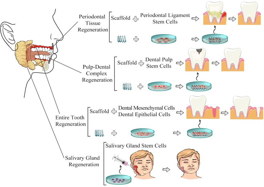
EXCLI Journal 2021;20:454-489 – ISSN 1611-2156
 Received: December 27, 2020, accepted: February 09, 2021, published: February 22, 2021

treatments such as dentures or implants usu- cells resulted in their substitution with more
ally do not achieve enough satisfactory out- biocompatible polymers and synthetic materi-
comes that content patients (Mitsiadis et al., als (‘70s and ‘80s) (Yan et al., 2010). Concur-
2017), mainly because the implanted or den- rently with the extending surgical-free mate-
ture teeth are weaker than the natural tooth rial-based reconstruction approaches in clini-
(Feng et al., 2016). Significant bone tissue cal prosthodontics, stem cell (SC)-based re-
loss in the craniofacial area frequently occurs generation and associated therapies in perio-
due to periodontal disease, congenital abnor- dontal diseases started developing too (Wang
malities, tumors, traumatic injury, or resorp- et al., 2010; Yan et al., 2010). The modern
tion secondary to tooth loss (Mani et al., SC-based regenerative medicine has facili-
2014). In general, the reconstruction strate- tated various reconstruction therapies in clin-
gies are extended from using medical devices ical implant dentistry such as regeneration
to tissue grafts and/or tissue engineering ap- (Yan et al., 2010). However, most studies ma-
proaches. This review mainly addresses the nipulate SCs in ex-vivo conditions using dif-
researches regarding the modern methods for ferent physical matrices (Wang et al., 2010).
orofacial reconstruction approaches that use One of the most state-of-the-art dental mate-
stem cells for tissue engineering. These inno- rial studies has focused on designing and us-
vative methods utilize specific bioactive/bio- ing natural and degradable biologic-based
degradable synthetic or natural scaffolds of- materials as scaffolds for regenerating perio-
ten together with advanced molecular tech- dontal tissues in-vivo (Wang et al., 2010;
niques to return the function and appearance Abou Neel et al., 2014). For this purpose, the
to the damaged tissue as much as possible. required stem cells have been obtained from
Here, the scaffold types and methodologies different sources, including bone marrow
which are shown in the literature that enable (BM), periodontal ligament (PDL), etc., and
cells to produce the efficient extracellular ma- have been applied with different types of bone
trix (ECM) are briefly implied. An efficient grafts such as autografts, xenografts, allo-
ECM is desired to ultimately convert to a grafts, and alloplastic materials (Wang et al.,
functional tissue with eligible geometry, size, 2010). It cannot be concluded yet from the
and composition. By looking at the proceed- current literature that which donor sources
ing of regenerative medicine, it can be notably provide the most appropriate cell isolation
observed that the medical devices and whole- (Wang et al., 2010). SC-based approaches
tissue grafts are increasingly replaced by the have developed to the point that makes it pos-
tissue regenerative engineering approaches. sible to replace the missing teeth with bioen-
These neoteric strategies include using spe- gineered ones that have already brought the
cific materials as scaffolds (sometimes com- dental stem cell (DSC)-banking for future re-
bined with certain molecules) for growing generative uses to the market (Egusa et al.,
compatible cells to substitute the diseased or 2012b). In this regard, understanding the fun-
damaged tissue with a functional one in-situ damentals of SCs and their associated tech-
(Zaky and Cancedda, 2009). Dentistry has nologies seems to be necessary for dentistry
passed several eras each of which is distin- clinicians and relevant-fields’ researchers
guished by trending different materials and (Yan et al., 2010). Accordingly, the current
methodologies. The most recent progress in study has critically reviewed the applications
dentistry is distinguished by the extensive at- of stem cells in reconstructive dentistry.
tempts to use biomaterials for replacing in-
jured craniofacial tissue with a functional nat- STEM CELL TYPES AND SOURCES
ural-like tissue (Abou Neel et al., 2014). The The SC types ever investigated for appli-
first predominantly used materials were metal cation in regenerative medicine can be di-
implants and associated devices (the 1950s) vided into two categories: embryonic stem
whose effects on the surrounding tissues and cells (ESCs) and adult stem cells (ASCs).

 456
EXCLI Journal 2021;20:454-489 – ISSN 1611-2156
 Received: December 27, 2020, accepted: February 09, 2021, published: February 22, 2021

ESCs are pluripotent stem cells originating several mesenchymal stem cells are intro-
from the inner cell mass of the blastocyst‐ duced in the literature from non-marrow oro-
stage embryos (Mahla, 2016; Hu et al., 2018). facial or extra-oral sources such as; stem cells
They can differentiate into almost all specific of the primary tooth (SHEDs), stem cells of
lines. Whereas, ASCs are typically catego- apical papilles (SCAPs), stem cells of perio-
rized as non-pluripotent cells, instead, as mul- dontal ligament (PDLSCs), and precursor
tipotent stem cells that exist in few numbers cells of the dental follicle (DFPCs). In the re-
within adult tissues and are responsible for generative medicine, the key products in suc-
maintaining tissues healthy and repairing cessful outcomes are not only stem cells, but
damages by self-regeneration and differentia- also the 3D scaffold, growth factors for differ-
tion into specific cell types (Paz et al., 2018). entiation and proliferation, as well as bioreac-
ASCs are also known as somatic stem cells or tors. The highlights of recent researches on
postnatal stem cells and can be isolated from the oral stem cells are summarized in Table 1.
various adult organs, including bone, muscle,
skin, nerve, pancreas, heart, and dental tissues EMBRYONIC STEM CELLS
(Mahla, 2016). Furthermore, multiple adult
SC lines can now be induced to be repro- The embryonic stem cell (ESC) is a more
grammed and produce induced pluripotent general terminology for pluripotent human
stem cells (iPSCs) (Paz et al., 2018) recalled embryonic stem (hES) cells with stem cell-
as plasticity potential (Towns and Jones, like developmental quality in-vitro (Zaky and
2004). The first stem cells used in regenera- Cancedda, 2009). The hES cells show three
tive medicine applications were isolated from properties which profound them as a qualified
bone marrow; however, today, it is demon- platform for developing an extensive range of
strated that the unspecialized cells called cell types; 1) hES cells surprisingly remain in
“stem cells” present not only in the bone mar- the second week of development within in-
row but also in many other tissues and organs, vivo niches, 2) they are much higher scalable
including dental pulp cells (Potdar and in the undifferentiated state compared to other
Deshpande, 2013). The postnatal dental stem SC types (Zaky and Cancedda, 2009), 3) they
cells are primarily originated from either epi- can be clonally isolated probably due to the
thelial cells or mesenchymal cells (Lymperi et presence of specific transcription factors
al., 2013). Likely, the only niche for the epi- (homeobox genes); however, the exact mech-
thelial dental SCs is recognized to be in the anism and the uniformity of these genes have
apical end of rodents’ incisors (Paz et al., remained to be studied in more details
2018). The mesenchymal dental SCs can be (Gebhard et al., 2007). Findings from animal
derived from different sources, including model studies and cellular/molecular studies
bone marrow and non-marrow tissues from suggest that the clonal derivation of hES cells
either extra-oral or intra-oral niches. The bone in-vitro might be similar to what happens for
marrow-derived stem cells (BMSCs) used for neural crest cells when their fate is specified
regenerating dental tissues are generally iso- before migration to construct the mesen-
lated from extra-oral origins (femur and iliac chyme of embryonic branchial arches struc-
crest) or orofacial bones (maxilla and mandi- tures (Zaky and Cancedda, 2009; Sternberg et
ble bone marrow) obtained through dental al., 2012). The clonal isolation potential of
treatments. Despite the early positive out- hES cells is essential for producing a wide
come of autologous craniofacial bone graft- range of stem cells in vitro for research and
ing, there are some drawbacks and challenges therapy purposes (Sternberg et al., 2012).
such as the invasive isolation method of extra- While other SC types differentiate into heter-
oral BMSCs and lower alternative sources of ogeneous cell lines that should be purified and
dental stem cells (Abdel Meguid et al., 2018; may become problematic by producing un-
Hu et al., 2018; Paz et al., 2018). Therefore, wanted cell lines that grow to ectopic tissues

 457
EXCLI Journal 2021;20:454-489 – ISSN 1611-2156
 Received: December 27, 2020, accepted: February 09, 2021, published: February 22, 2021

Table 1: Applications of oral stem cells in regeneration
 Stem Tissue sources Experiment Outcomes Reference
 Cells
 type
 SCAPs Inflamed periapical Comparing SCAPs from inflamed immature Human SCAPs under inflammatory conditions retained Chrepa et al.,
 pulp tissue (necro- mandibular premolar tissue with normal SCAPs their stemness partially and attained more osteogenic and 2017
 sis and periodonti- and IPAPCs in flow cytometry and quantitative angiogenesis potentials.
 tis) osteogenesis experiments.
 SCAPs Human third molar Mycoplasma contamination evaluation. Evaluation of hSCAPs mycoplasma contamination and Kim et al.,
 and premolars Cell proliferation capacity was tested. elimination process is required before application in tissue 2015
 engineering and regenerative medicine.

 SCAPs Root canal system Cell viability evaluation using Porphyromonas Pretreatment with different LPS concentrations had no ef- Lertchirakarn
 gingivalis LPS. Cell proliferation investigated by fect on cell viability, cell proliferation, and mineralization. and Aguilar,
 resazurin-based assay. Mineralization capacity No significant difference between DSPP and OPN gene 2017
 determination. Marker detection for odonto- expression levels at all concentrations
 blast, general bone, and cementum, using
 DSPP, OPN, and BSP gene expression and
 PCR.

 SCAPs Human third molars 3D engineering of micro nerve tissue using Generation of three-dimensional nerve-like tissue with ax- Kim et al.,
 postnatal hSCAPs in-vitro by organotypic cul- ons and myelin structures was possible. 2017
 ture and using an integrated bioprocess.

 SCAPs Immature impacted Effect evaluation of different periods of expo- In comparison with EDTA, MTAD, QMix, and NaOCl, Farhad Mol-
 mandibular third sure to some irrigating solutions on hSCAPs. chlorhexidine solution was least cytotoxic, and its cytotox- lashahi et al.,
 molars icity stayed invariable over time. 2016
 SCAPs Freshly extracted SCAPs and HUVECs coculture under artificial Hypoxia promoted angiogenesis (formation of endothelial Yuan et al.,
 human third molars hypoxia. tubules and blood vessel networks). 2015
 with immature roots

 458
EXCLI Journal 2021;20:454-489 – ISSN 1611-2156
 Received: December 27, 2020, accepted: February 09, 2021, published: February 22, 2021

 Stem Tissue sources Experiment Outcomes Reference
 Cells
 type
SCAPs Immature impacted Evaluation of SCAPs potential of motor recov- Early postsurgery persistent functional impairment was De Berdt et
 mandibular third ery after SCI in a rat model by transplanting observed in all sham models, while a significant reversed al., 2015
 molars of rats them in the injured spinal cord wound via Cat- impairment was observed in transplanted subjects.
 Walk analyses and BBB locomotor scores.

GMSCs Human gingiva Producing GMSCs/NO-releasing microspheres They could produce hybrid aggregates of GMSCs/NO-re- Regmi et al.,
 samples by hanging drop technique. Their osteogenic leasing microspheres by for in situ delivery of exogenous 2017
 differentiation ability was evaluated by APA NO under in-vitro culture conditions, which promoted os-
 assay and alizarin red staining. teogenic differentiation.
GMSCs Human gingiva Evaluating the therapeutic potential of GMSCs GMSCs inhibited the proliferation of PBMC and T cells Huang et al.,
 samples collected in preventing the xeno-GVHD condition by de- in-vitro. 2017
 following routine veloping a mouse xeno-GVHD model. Co-transfer of GMSC with human PBMC significantly
 dental procedures suppressed human cell engraftment and markedly pro-
 longed the mouse survival.
 GMSC inhibited the xeno-GVHD via a mechanism involv-
 ing IDO and CD39/CD73/adenosine signals.
GMSCs Gingiva of the oral Cell-based therapy by GMSCs and evaluating GMSCs-based therapy suppressed the experimental co- Zhang et al.,
 cavity their capability of ameliorating inflammation- litis in animal models by immunomodulatory functions. 2009
 related tissue destruction via systemic infusion The parameters of clinical symptoms, histopathological
 in experimental colitis. severity, the injury of gastrointestinal mucosal tissues, di-
 arrhea, and weight loss improved. Suppression of inflam-
 matory infiltrates, and inflammatory cytokines/mediators
 were felt to be involved in the therapeutic effects of
 GMSCs.
GMSCs STZ-induced T1DM GMSCs transfer to STZ-induced T1DM. GMSCs transfer could significantly control blood glucose Zhang et al.,
 Blood glucose levels, disease severities, and levels, prolong diabetes onset, mitigate pathology scores 2017b
 GMSC distribution were analyzed. T cell sub- of the pancreas, and reduce IL-17 and IFN-γ expression
 sets (CD4+ and CD8+) in blood, spleen, and in T cells of spleens and lymph nodes (e.g., pLN).
 lymph nodes were detected dynamically by
 flow cytometry.

 459
EXCLI Journal 2021;20:454-489 – ISSN 1611-2156
 Received: December 27, 2020, accepted: February 09, 2021, published: February 22, 2021

 Stem Tissue sources Experiment Outcomes Reference
 Cells
 type
GMSCs Oral cavity of Testing the potential of GMSCs transplanta- GMSCs transfer significantly reduced the severity of ex- Gu and Shi,
 C57BL/6 J mouse tion to mitigate CIA symptoms and studying perimental arthritis and the immunosuppressant effects 2016
 the probable involvement of the FasL/Fas and balanced Th cell subsets. The inability of FasL-/-
 pathway. GMSCs to bring about apoptosis in T cells compared to
 FasL TF GMSCs showed the involvement of the
 FasL/Fas pathway in the underlying mechanism of the
 CIA treatment.
GMSCs Subjects to clinical Evaluating the dexamethasone effects on The short-term application of dexamethasone increased Kim et al.,
 crown-lengthening hGMSCs when it is applied in the short term (2 the expression of 7 mRNAs and reduced the expression 2017
 procedures hours) using NGS. of 25 mRNAs, including RUNX2 and β-catenin in
 hGMSCs.
SHED Human exfoliated Evaluating the SHED potential for managing SHED-originated exosomes showed therapeutic effects Li et al., 2017
 deciduous teeth TBI and motor recovery by local injection of on TBI in rats and reduced neuroinflammation via shifting
 SHED and SHED-originated exosomes into microglia polarization.
 TBI rat models using BBB scores assessment.
SHED Human exfoliated Evaluating the effects of three compounds Magnesium borate or zinc borate could induce more os- Liu et al.,
 deciduous teeth (magnesium borate, zinc borate, boric acid) on teogenic differentiation with increased alkaline phospha- 2018
 the SHED osteoblastic differentiation inter- tase activity and collagen type I gene expression in the
 mixed with the chitosan scaffold. differentiated cells.
SHED Exfoliated decidu- Evaluation of modulatory effects of SHED ad- SHED could adjust responses of peripheral CD4+ T cells. Rossato et
 ous teeth from ministration on EAE models, e.g., clinical signs Its immunomodulatory potential was strongly proved, al., 2017
 healthy volunteers and cellular patterns. The animal model was which confirmed SHED as a proper candidate for cellular
 Foxp3 GFP+ transgenic mice (C57Bl/6- therapy in CNS-associated autoimmune diseases.
 Foxp3GFP).
SHED Dental pulp sam- Measuring the potential of SHED differentia- In-vitro co-culture of SHED with immortal corneal epithe- Tsai et al.,
 ples from exfoliated tion into corneal epithelium-like cells and the lium cells showed the potential of SHED to be used for 2015
 deciduous teeth of expression levels of mature corneal epithe- clinical applications in ocular surface regeneration.
 5-7 years old chil- lium-specific marker CK3 (immunofluores- SHED could trans-differentiate into corneal epithelium-
 dren cence) and corneal epithelial progenitor like cells. The study represented exfoliated teeth as an
 marker CK19 ( RT-PCR). alternative SC resource.
SHED Dental pulp sam- Evaluating the LLLI effect on the proliferation The LLLI parameters (660 nm, 30 mW, 1.0 J/cm2) used Ginani et al.,
 ples from exfoliated and viability of SHED from healthy human vol- in the study could stimulate the SHED proliferation and 2018
 deciduous teeth unteers. SCs were irradiated with two different maintain the viability of cells.
 energy densities of InGaAlP laser diode.

 460
EXCLI Journal 2021;20:454-489 – ISSN 1611-2156
 Received: December 27, 2020, accepted: February 09, 2021, published: February 22, 2021

 Stem Tissue sources Experiment Outcomes Reference
 Cells
 type
SHED Perivascular niche Evaluating the SHED effects on neuronal SHED transplantation one hour after SCI was shown to Nicola et al.,
 of the dental pulp death in an experimental model of SCI. Spinal reduce early neuronal apoptosis, preserves motor neu- 2017
 injury and SHED transplantation effects on the rons, and recover hind limb function, partially by adjust-
 behavior, tissue protection and motor neuron ing pro- and anti-apoptotic factors.
 survival were confirmed by functional evalua-
 tions and morphological analysis.
hDPSCs Deciduous teeth in Evaluating the effects of intracardiac trans- hDPSC could recover HI-induced cognitive deficits. The Sanches et
 resorption plantation of hDPSCs on HI damage using the exact mechanism underlying its neuroprotective effects al., 2018
 motor and cognitive tests. remained unclear.
hDPSCs Human dental pulp SC identification and localization in the human SCs contributed to reparative dentinogenesis during pul- Ustiashvili et
 samples with pulpi- dental pulp and assessing their function in nor- pitis through perivascular mobilization, proliferation, and al., 2014
 tis mal and inflammation processes. migration to the Hohle layer, especially in acute disease
 cases in response to irritation/stimulation.
hDPSCs Deciduous teeth in Assessing the contribution of histone acetyla- The histone acetylation of the dentin sialophosphoprotein Gu et al.,
 resorption tion in regulating odontoblast-like differentia- gene represented to positively influence on mineral for- 2013
 tion of DPSCs using Western blot analysis mation, stimulates odontoblast-like differentiation, and
 regulates DPSCs maturation via controlling DSPP ex-
 pression.
hDPSCs Third molars Searching for intercellular purinergic signaling The survival and proliferation of hDPSCs reduced by Zhang et al.,
 pathways in hDPSCs and measuring its effect blocking both P2Y and P2X receptors and enhanced by 2019b
 on the cells’ survival and proliferation using inhibition of ecto-ATPase activity. Solo-blocking of P2X
 whole-cell patch-clamp recordings of ATP-in- receptors just reduced the hDPSC proliferation. Auto-
 duced currents, immunofluorescence, and en- crine/paracrine purinergic signaling was shown to be es-
 zymatic histochemistry staining. sential for hDPSC survival and proliferation.
SHED Human exfoliated Evaluating the potential of postnatal SCs for The SHED yielded more ICCs than DPSCs. SHED-de- Kanafi et al.,
and deciduous teeth proliferating insulin-producing cells with quali- rived ICCs could restore normoglycemia in STZ-induced 2013
DPSCs Dental pulp from fied physiological parameters and using them diabetic mice within almost one month and persisting for
 permanent teeth for transplantation. more than two months, while the control groups showed
 hyperglycemia.
hDPSCs Third molars Protein profiling of hDPSCs during differentia- LOXL2 protein expression and secretion were down-reg- Kim et al.,
 tion into odontoblast-like cells in mineralization ulated during hDPSCs odontogenic differentiation. Treat- 2013
 media using LC-MS/MS proteomics ap- ing hDPSCs with recombinant LOXL2 decreased hDP-
 proaches. SCs proliferation into odontoblast-like cells, and blocking
 it assisted odontogenic differentiation of hDPSC.

 461
EXCLI Journal 2021;20:454-489 – ISSN 1611-2156
 Received: December 27, 2020, accepted: February 09, 2021, published: February 22, 2021

 Stem Tissue sources Experiment Outcomes Reference
 Cells
 type
DPPSCs Third molars ex- Analyzing growth factors produced by DPP- Treatment with DPPSC induced re-epithelialization, im- Martinez-
 tracted from young SCs using Ab-arrays. DPPSCs' potential of proved collagen storage and structure in healing Sarra et al.,
 patients producing endothelium and both smooth and wounds, extended the cross-sectional area of type II 2017
 skeletal muscles was evaluated by culturing in fast-glycolytic fibers, and reduced fibrosis and collagen
 differentiation media. content.

DPPSCs Third molars from Evaluating the osteogenic potential of DPPSC DPPSC expressed bone-related markers confirming its Nunez-Toldra
 healthy patients compared to hSAOS-2. osteogenesis capacity as well as SAOS-2 and higher ad- et al., 2017
 hesion markers higher initial adhesion potential to bio-
 materials.
DPPSCs Third molars from Comparing the effects of three pretreatments The newly manufactured pure MZ-endodontic cement Maher et al.,
 healthy humans (ProRoot-MTA, Biodentine, or Portland ce- showed increased cell proliferation compared to other 2018
 ment Med-PZ) of the conditional media on the pretreatments and more osteogenic capacity represent-
 osteogenic differentiation potential of DPP- ing a promising cement for endodontic therapies.
 SCs.

DPPSCs Third molars of Evaluation of the effect of some inorganic ions Inorganic ions dissolved from Bioactive Glass extracts Nunez-Toldra
 healthy patients in a cell co-culture endothelial medium on stimulated both vascular-like structures and mineraliza- et al., 2019
 forming vascularized bone in-vitro. tion, resulting in enhanced endothelial and osteogenic
 processes simultaneously.
SGSCs Human nonmalig- Assessing the self-renewal and differentiation The cultured SGSCs from human salivary glands could Pringle et al.,
 nant submandibular properties of hSGSCs and their potential for form small cell aggregates, self-renew, differentiate, and 2016
 salivary gland tis- in-vivo engraftment and functionality. produce saliva that suggested salisphere-cell therapy as
 sue a promising treatment for xerostomia.
SGSCs Human submandib- Evaluating the potential of hSGSCs transplan- Human SGSCs could restore the acinar and duct cell Jeong et al.,
 ular salivary gland tation for treating hyposalivation in the radia- structure and decrease the number of apoptotic cells, re- 2013
 tion-damaged salivary glands of the rat model. sulting in controlled hyposalivation and body weight loss.
SGSCs Salisphere of Gdnf- Studying the involvement of GNDF in the ef- GDNF does not protect mSGSCs against irradiation but Peng et al.,
 hypermorphic and fects of irradiation on mice SGSCs and its po- seems to promote mSGSCs proliferation through the 2017
 wild type mice tential of salivary gland regeneration using ani- GDNF-RET signaling pathway.
 mal models (Gdnfwt/hyper and Gdnfwt/wt).

 462
EXCLI Journal 2021;20:454-489 – ISSN 1611-2156
 Received: December 27, 2020, accepted: February 09, 2021, published: February 22, 2021

 Stem Tissue sources Experiment Outcomes Reference
 Cells
 type
 SGSCs Salivary glands and Characterizing the regenerative potentials of Aged SGSCs could retain their regenerative potential in- Maimets et
 sphere from young an aged SGSC population using murine mod- vivo, similar to young SGSCs following exposure to ade- al., 2015
 and old mice els. quate growth conditions. Results suggest aged SGSCs
 as potential candidates for regenerative therapy of
 age/medication associated dysfunctions of salivary
 glands.
 ASCs Raw human ab- Inducing single MSC populations to generate ASCs could transdifferentiate into a specific 3D structure Ferro et al.,
 dominal lipoaspi- dental bud-like structures in-vitro. The hASCs in-vitro with a dental bud-like phenotype even when no 2011
 rates were primarily cultured in a dental-inducing structural matrix or scaffold existed to assist the develop-
 medium, and the obtained aggregates were mental progress.
 cultured in three-dimensional scaffolds.
 PDLSCs Premolars from Investigating the regenerative potential of PDLSC-amnion could improve periodontal tissue regen- Iwasaki et al.,
 healthy patients PDLSC-amnion through transplanting it in rats eration. 2014
 with surgically created periodontal defects in
 maxillary molars and assessing the result by
 micro-CT and histology.
 PDLSCs Normal healthy pre- Evaluating the bone regenerative capacity of a PRP intervention enhanced the extracellular matrix gen- Xu et al.,
 molars mixture of PDLSC sheets and PRP using eration and osteogenic differentiation of PDLSCs at an 2017
 scanning electron microscopy and the osteo- optimized ion concentration and improved the total
 genic gene expression measurement. PDLSC sheets behavior in periodontal tissue regenera-
 tion.
 PDLSCs Third impacted mo- Studying the biological effects of a Chinese Transplantation of osthole extract-treated cell sheets of Gao et al.,
 lars with healthy herbal extract treatment (osthole) of hPDLSCs PDLSCs and JBMMSCs with the optimized concentra- 2013
 PDL tissue and JBMMSCs on their osteogenic character- tion and stimulation mode induced more proliferation and
 istics and cell sheet formation behavior by bone generation in-vivo than the control subjects receiv-
 measuring cell proliferation and ALP activity. ing no osthole intervention.
 Determining the best transplantation modality
 for each cell type by measuring ECM amount
 and osteogenic-related gene expression.
MTAD: Mixture of Tetracycline isomer, Acid, Detergent, NaOCl: Sodium Hypochlorite, EDTA: Ethylenediaminetetraacetic acid, QMix™ is a 2-in-1 solution con-
taining a bisbiguanide antimicrobial agent

 463
EXCLI Journal 2021;20:454-489 – ISSN 1611-2156
 Received: December 27, 2020, accepted: February 09, 2021, published: February 22, 2021

in the graft (Sternberg et al., 2012). Previ- compared to other MSCs such as BM- or adi-
ously, the common method for restricting un- pose-derived SCs relates to their ability to ex-
defined cell types has been using clonal em- press higher trophic factors including brain-
bryonic progenitor cell lines from available derived neurotrophic factor (BDNF), glial cell
cell banks. This method contaminates the de- line-derived neurotrophic factor (GDNF),
signed grafts for clinical applications (Stern- nerve growth factor (NGF), neurotrophin 3
berg et al., 2012). Using clonal embryonic SC (NT-3), vascular endothelial growth factor
lines leads to more scalability and less lot-to- (VEGF), and platelet-derived growth factor
lot variability that allows repeating funda- (PDGF) (Parsa et al., 2010). Also, the higher
mental experiments on the same lines and fa- cytokine expression in DPSCs promotes neu-
cilitate producing more site-specific tissues to ronal differentiation in them (Ullah et al.,
be applied in regenerative clinical therapies 2017). The secreted cyto-protective factors
(Sternberg et al., 2012). help DPSCs to present both direct and indirect
 neuroprotective properties in nervous dis-
 DENTAL PULP PLURIPOTENT-LIKE eases and injuries leading to a decreased neu-
 STEM CELLS rodegeneration in the early stages of patholo-
 gies (Parsa et al., 2010; Mead et al., 2013;
 Dental pulp pluripotent stem cells (DPP-
 Ullah et al., 2017). DPSCs have also shown
SCs or DPSCs) originate from the cranial
 axon regeneration ability even in the presence
neural crest in the embryonic stage (Zhang et
 of axon growth inhibitors in a spinal cord in-
al., 2017a). The isolation of dental stem cells
 jury model (Parsa et al., 2010) and protection
can be done by size-sieved isolation, stem cell
 against cell death in an ischemic astrocyte in-
colony cultivation, magnetically activated
 jury model (Ullah et al., 2017).
cell sorting (MACS), and fluorescence-acti-
vated cell sorting (FACS). In addition, the
preservation of dental stem cells can be cate- PLURIPOTENT STEM CELLS FROM
gorized as cryopreservation and magnetic HUMAN EXFOLIATED DECIDUOUS
freezing (Bansal and Jain, 2015). Markers that TEETH
can detect DPSCs are STRO-1 and CD146 Stem cells derived from human exfoliated
(Yang et al., 2018; Zhai et al., 2018). DPSCs deciduous teeth (SHED) or the primary teeth
are invasively obtained from the third molar are among the most studied SC types and the
(wisdom teeth) with less ethical concerns and most valuable source of stem cells in tissue
show favorable MSC-like characteristics such engineering studies and cell-based regenera-
as multipotency and self-renewal procedures tive medicine therapies (Martinez Saez et al.,
(Caseiro et al., 2016). DPSCs are unique in 2016). The reason is that these immature SC
lineage differentiation as they express neu- population advantage from 1) non-invasive
ron-related markers before developing into isolation procedures, 2) low immune reac-
functional neuron-like cells with the ability to tions or rejection after transplantation, 3) abil-
produce neurotrophic factors such as neuro- ity to remain undifferentiated and stable after
trophin (NT), which makes them promising long-term cryopreservation, 4) being highly
candidates for SC-based nerve regeneration proliferative, 5) being easily accessible, 6) po-
therapies (Ullah et al., 2017). Differentiating tential of multi-lineage differentiation, 7)
DPSCs into neurons have been experimen- non-invasiveness, and finally, 8) few ethical
tally induced through various protocols, concerns (Martinez Saez et al., 2016). SHED
which are usually relying on: 1) growth fac- can be detected by STRO-1, CD146/MUC18,
tors, 2) culture supplements, and 3) some CD90, CD29, CD44, CD166, CD105, and
small molecules as neurotrophic factors CD13 (Zhai et al., 2018). Either cultured in-
(Zhang et al., 2017a). The higher efficacy of vitro or in-vivo, SHED populations can suc-
DPSCs in nervous regenerative therapies cessfully differentiate into various specialized

 464
EXCLI Journal 2021;20:454-489 – ISSN 1611-2156
 Received: December 27, 2020, accepted: February 09, 2021, published: February 22, 2021

cell populations such as odontoblasts, osteo- ADULT STEM CELLS
blasts, chondrocytes, adipocytes, and neural
 Induced pluripotent stem cells
cells (Martinez Saez et al., 2016). In 2003, a Induced pluripotent stem cells (iPSCs)
mixture of SHED and hydroxyapatite/trical- have already shown great promises in animal
cium phosphate (HA/TCP) was suggested to model trials for regenerative treatment of Par-
be used for dental pulp tissue regeneration for kinson’s disease and sickle cell anemia (Jung
the first time (Casagrande et al., 2011). This et al., 2012). Human iPSC-derived MSCs can
research was a landmark since dental pulp re- produce osteoblasts, adipocytes, and chon-
generation is a required step for pulp tissue drocytes in-vitro (Wu et al., 2010), and some
engineering practices in clinics (Martinez
 disease‐specific iPSC lines are used for differ-
Saez et al., 2016). The SHED mixture has
 ent purposes such as “diseases in a dish” stud-
been first implanted in animal models and
 ies (modeling genetic disorders using in-vitro
then was used to proliferate within the scaf-
 induced pluripotent cells), drug develop-
fold and form dentin-like tissue (Casagrande
 ments, and inventing novel therapies (Jung et
et al., 2011). Fortunately, SHED has shown
 al., 2012). The iPSCs can also be induced to-
potency in adhering to the dentin walls and
 ward vascular and muscle regeneration (Wu
proliferating within the full-length root canals
 et al., 2010). The higher telomerase activity
in-vitro (Martinez Saez et al., 2016). In 2008,
 and less senescence of iPSCs-derived MSCs
further advances were achieved regarding the
 compared to BM‐MSCs have introduced
in-vivo development of dental pulp from
 them as up-and-coming regenerative alterna-
SHED on biodegradable poly-L-lactic acid-
 tives that provide higher survival and engraft-
based scaffolds, and a pulp-like tissue with a
 ment after transplantation (Wu et al., 2010).
functional vascular network plus odontoblast-
 Despite the extensive suggested applications
like cells substituted the scaffold (Casagrande
 for iPSCs, their clinical application is not rec-
et al., 2011). The odontoblast-like cells lie on
 ommended due to the tumorigenesis possibil-
the dentin surface and have an eccentric nu-
 ity, which is attributed to the mutagens (e.g.,
cleus and express dentin sialoprotein (DSP)
 c‐Myc) that cause cancers via integration or
(Casagrande et al., 2011). More recently,
 disrupting tumor suppressor genes (Jung et
SHED was used to proliferate odontoblasts
 al., 2012). Another reason for critics of iPSCs
with the expected markers (DSPP, DMP-1,
 applications is the perturbations in epigenetic
and MEPE) within the full-length root canals
 memories and aberrations in the genomic
on the injected scaffolds in-vitro and generate
 properties of reprogrammed cells (Jung et al.,
functional dental pulp in the subcutaneous
 2012). Therefore, many precautions are gen-
space of mice (in-vivo) (Martinez Saez et al.,
 erally recommended while using iPSCs for
2016). In this trial, cells could well occupy the
 clinical applications.
root canal space and the tissue regeneration
occurred with a promising growth rate
 ORIGINATED STEM CELLS IN DEN-
(10 μm/day) providing adequate timing for
 TAL REGENERATIVE TREATMENTS
the clinical uses (Martinez Saez et al., 2016;
Rosa et al., 2016). The scaffold composition Stem cells from apical papilla
seems to be ineffective on SHED proliferation The stem cells from the apical papilla
(Martinez Saez et al., 2016). Tetracycline la- (SCAPs) belong to a unique SC line locating
beling is a common method for revealing the at the apical tissues of the growing tooth roots
newly formed dentin. No in-natura study has when at least two-thirds of the root have
been conducted to evaluate the SHED poten- formed (Lin et al., 2018; Nada and El Backly,
tial to proliferate and differentiate in the oral 2018). While SCAP derives from the dental
environment (Rosa et al., 2016). papilla, they express a mesenchymal surface
 marker (STRO-1) and contribute to the epi-
 thelial-mesenchymal interactive process of

 465
EXCLI Journal 2021;20:454-489 – ISSN 1611-2156
 Received: December 27, 2020, accepted: February 09, 2021, published: February 22, 2021

tooth development (Nada and El Backly, multipotency of differentiating into osteo-
2018). SCAPs can be detected by STRO-1, genic, adipogenic, and chondrogenic lineages
CD146, and CD24 (Zhai et al., 2018). SCAP was distinguished long time ago (Pittenger et
has an infection-resistant nature that is ex- al., 1999). Furthermore, they are praised for
plained by its histo-morphologic position their “stem cell plasticity” feature which ena-
concerning the dental pulp (Lin et al., 2018). bles them to induce similar developing cell
The apical papilla is separated from the epi- lines from typically different origins (Paz et
thelial diaphragm with a cell-rich zone and al., 2018). A challenge for the recognition of
has access to a collateral circulation that ena- MSCs has been defining their distinguishing
bles the apical papilla to survive a necrotic markers among the non-homogeneous popu-
pulp in just adjacent tissues (Lin et al., 2018). lations of adherent cells obtained from the
Today, SCAP is readily isolated from the tips bone marrow (Paz et al., 2018). MSCs are de-
of the developing roots of an extracted tooth fined by ISCT as tissue-culture-treated plastic
and treated with a well-established protocol adherent stem cells regardless of the tissue
(dissection, digestion using collagenase and from which they are isolated; however, Hor-
protease, and culturing the obtained cell sus- witz et al. have suggested them to be termed
pensions) to be used for the consequent re- as fibroblast-like plastic-adherent cells from
search or clinical processes (Nada and El any source (Horwitz et al., 2005).
Backly, 2018). Compared to DPSCs and Emersion and recovery of dental diseases
PDLSCs, SCAP seems to be a better source to such as deterioration, is substantially under
be used in cell-based tooth regeneration be- the effect of the teeth microenvironment, so
cause of its higher proliferative and minerali- that any pathologic alteration that can affect
zation properties. Primary odontoblasts are the endogenous MSCs’ functions and their re-
mainly differentiated from SCAP during root generation capacity may lead to substantial
dentin formation, while replacement odonto- bone loss. Similarly, transplanted exogenous
blasts are likely derived from DPSCs leading MSCs are highly influenced by the microen-
to reparative dentin formation (Zhai et al., vironment of both donor and recipient niches
2018). that creates a major challenge to using MSCs
 for therapeutic regeneration purposes in dis-
 eased microenvironments (Zheng et al.,
 MESENCHYMAL STEM CELLS
 2019).
 Mesenchymal stem cells (MSCs) were
first obtained from the bone marrow of the EXTRA-ORAL DERIVED MSCS
iliac crest (Pittenger et al., 1999). However,
the term “mesenchymal stromal cells” was at- Adipose tissue-derived stem cells
 Adipose tissue-derived stem cells (ASCs)
tributed to this derivative of adherent cells by
the International Society for Cellular Therapy are considered an abundant MSC source that
 can be obtained through lipectomy or lipoas-
(ISCT) (Horwitz et al., 2005). After their first
 piration from different adipose tissues such as
introduction, their other subsets have also
 the chin, hips, upper arms, and abdomen
been found in several differentiated tissues
 (Mizuno et al., 2012). The ASCs eliciting pro-
such as skin, adipose tissue, and various den-
 cedure is considered a low invasive process,
tal tissues (Egusa et al., 2012a). MSCs have a
 and they show a robust osteogenesis potency;
specifically better coating on surface-treated
 hence, they can be considered as a promising
plastic with plasma gas (so-called tissue-cul-
 alternative source of MSCs for periodontal re-
ture-treated plates), which makes them distin-
 generation therapies while their efficiency in
guishable among all SC types (Horwitz et al.,
 guided bone regeneration (GBR) and implant
2005). This type of adult stem cell has shown
great promise for clinical applications. Their surgery has been demonstrated (Mizuno et al.,
 2012).

 466
EXCLI Journal 2021;20:454-489 – ISSN 1611-2156
 Received: December 27, 2020, accepted: February 09, 2021, published: February 22, 2021

 INTRA-ORAL DERIVED MSCS pulp and produce supportive connective tis-
 sue and odontoblasts, yet the origin of these
Dental tissue-derived MSCs
 Epithelial stem cells and MSC-like cells cells has not been characterized, and the pos-
are reported to locate within particular niches sible existence of a postnatal DPSC was never
in dental pulp, dental follicle, and periodontal heretofore proposed (Apatzidou et al., 2018).
tissues (ligament stem cells) (Egusa et al., Odontoblasts, as one of these precursor cell
 populations, have a polarized columnar mor-
2012b; Paz et al., 2018). Up to now, several
 phology, with eccentric nuclei located at the
oral tissues have been introduced as sources
 outer edges of dentin and form reparative den-
for stem cells, including exfoliated deciduous
 tin in response to general mechanical erosion
teeth, orofacial bone marrow, apical papilla,
 or disruption (Honda et al., 2010). In 2007,
dental follicle, dental pulp tissue, periodontal
 d'Aquino et al. isolated DPSCs acting as
ligaments, oral epithelium, periosteum, sali-
 odontogenic progenitors from adult dental
vary glands, and gingival lamina propria tis-
 pulp tissue and early developing dental root
sues (Ercal et al., 2018). These are multipo-
 tissue, and showed that similar to bone mar-
tential cells producing different dental organs
 row cells, DPSCs could differentiate into
such as reparative dentin (Paz et al., 2018).
 odontoblast-like cells and develop mineral-
Other tissues differentiated from dental pulp-
 ized nodules in-vitro (d'Aquino et al., 2007).
derived SCs include osteogenic, dentino-
 Further studies have identified DPSCs as
genic, adipogenic, chondrogenic, myogenic,
 clonogenic and highly proliferative stem cells
and neurogenic cells (Egusa et al., 2012b).
 especially in animal testing (Apatzidou et al.,
Periosteum-derived stem/progenitor cells
 2018). The “transplanted” DPSCs produce
with osteogenesis potential differentiate into
 dentin and pulp-like tissue in much more vol-
osteoblasts and chondrocytes. Their MSC-
 ume than it would be formed in situ during the
markers make them suitable candidates for
 lifetime of an organism (Apatzidou et al.,
tissue engineering and bone regeneration (Paz
 2018).
et al., 2018).
 DENTAL FOLLICLE STEM CELLS
POSTNATAL HUMAN DENTAL PULP
 STEM CELLS The dental follicle stem cells (DFSCs) are
 located within the dental follicle or bilayered
 Similar to muscles’ and nerves’ tissues, Hertwig’s epithelial root sheath (HERS); they
the dental pulp is a specialized tissue during originate from the ectomesenchymal progeni-
postnatal life that also contains SCs confer- tor cell population and differentiate into ce-
ring it the tissue regeneration ability in re- mentoblasts or osteoblasts (cementogenesis)
sponse to injury (Apatzidou et al., 2018). during tooth root formation (Honda et al.,
Dental pulp stem cells (DPSCs) are a subpop- 2010). DFSCs can be detected by STRO-1,
ulation of mesenchymal cells residing within CD90, CD105, nestin, notch-1 (Zhai et al.,
the pulp tissue that differentiate into odonto- 2018). DFSCs are discussed in predominant
blasts during tooth formation under the influ- studies to differentiate consecutively into ce-
ence of epithelial and dental papilla cells’ in- mentum, PDL, and alveolar bone (collec-
teractions (Honda et al., 2010). Generally, the tively termed as periodontium), which con-
origin and nature of postnatal cells as the pre- firms DFSCs as the origin of cementoblasts
cursor of various specialized tooth-associated (Fawzy El-Sayed and Dörfer, 2016). There-
cell types are little known (d'Aquino et al., fore, the dental follicle and its containing stem
2007). Further characterization of DPSCs has cells exert several critical functions during
been facilitated by transplanting human tooth development, including periodontium
DPSCs into immunocompromised mice development, bone resorption, tooth eruption,
(Apatzidou et al., 2018). These progenitors osteoclastogenesis and osteogenesis regula-
are hypothesized to be originated from dental

 467
EXCLI Journal 2021;20:454-489 – ISSN 1611-2156
 Received: December 27, 2020, accepted: February 09, 2021, published: February 22, 2021

tion, and proliferation into stratified squa- tissue, mostly referred to as lamina propria
mous epithelium under pathological condi- (Ercal et al., 2018). The gingiva (both at-
tions to generate dental cysts (Honda et al., tached and free parts) shows similar immuno-
2010). phenotypic characteristics to MSC-like cells
 from healthy periodontal tissues and is an ac-
 PERIODONTAL LIGAMENT STEM cessible source for collecting SCs via a mini-
 CELLS mally invasive route even in cases of inflamed
 gingiva, gingival hyperplasia, or periodontal
 Human periodontal ligament stem cells
 lesions (Apatzidou et al., 2018). The immuno-
(PDLSCs) are a few mesenchymal progenitor
 modulatory properties and multipotency for
cells within the PDL that remain proliferative,
 differentiation into various mesenchymal lin-
and their differentiation potential provides
 eages of GMSCs have made them a useful
great promises for SC-based regenerative
 candidate for application in antitumor thera-
therapies in dentistry (Wang et al., 2010;
 pies, skin wound repair, periodontal tendon
Bright et al., 2015). PDLSCs can be detected
 regeneration, peri-implantitis treatment, bone
by STRO-1, CD146, CD73, CD90, and
 defect regeneration, as well as treating oral
CD105 (Zhai et al., 2018). The envisages for
 mucositis, experimental colitis, collagen-in-
clinical use of PDLSCs come from many pos-
 duced arthritis, and contact hypersensitivity
itive preclinical results in a wide range of in-
 (Fawzy El-Sayed and Dörfer, 2016).
vitro and in-vivo studies (Bright et al., 2015).
Yet, they are not economically competitive
enough to be moved through the commercial SALIVARY GLAND-DERIVED STEM
usage phase compared to the current root ca- CELLS
nal therapies and dental implants (Wang et al., Salivary gland-derived stem cells
2010). PDLSCs are multipotent cells that can (SGSCs) were firstly isolated from a rat sub-
produce structures similar to cementum and mandibular gland (Emmerson and Knox,
periodontal ligament in-vivo, which can be 2018). Similar to DPSCs, SGSCs can be used
used for regeneration of the periodontium in as an autologous graft in different procedures,
periodontal diseases (Chen et al., 2016). But, including SC-based dental regeneration ther-
the non-lethal nature of periodontitis has apies as well as head and neck cancer (HNC)
caused the periodontal tissues not to be a res- (Emmerson and Knox, 2018). SGSCs are
olute target for SC-therapy researches (Wang multipotent and highly proliferative progeni-
et al., 2010). PDLSCs are reported to be capa- tor cells that can produce acinar, myo-epithe-
ble of forming a complete periodontal attach- lial, and ductal cell lineage markers and can
ment apparatus by generating several perio- be induced to differentiate into chondrogenic,
dontal tissues containing multiple develop- osteogenic, and adipogenic cells (Emmerson
mental lineages such as osteoblasts, fibro- and Knox, 2018). The SGSCs isolated from
blasts, and cementoblasts (Wang et al., 2010; the human salivary gland can express both
Bright et al., 2015). In periodontal regenera- embryonic and adult stem cell markers
tion trials, the affected teeth have been used (Emmerson and Knox, 2018).
as ideal models for evaluating new treatments
and facilitating surgical-free therapies (Chen GROWTH FACTORS
et al., 2016). Growth factors (GFs) are natural biologi-
 cal molecules with growth-promoting activi-
 NON-DENTAL TISSUE-DERIVED ties that usually have been initially identified
 MSCS for their functions as mediators and regulators
Gingiva-derived mesenchymal stem cells in cellular events (Mercola and Stiles, 1988;
 Gingival mesenchymal stem cells Mani et al., 2014). GFs are generally clustered
(GMSCs) originate from gingival connective into the superfamilies of epidermal growth

 468
EXCLI Journal 2021;20:454-489 – ISSN 1611-2156
 Received: December 27, 2020, accepted: February 09, 2021, published: February 22, 2021

factors (EGF), insulin-like growth factors et al., 2011). Considering the extensive cur-
(IGF), transforming growth factors (TGF), rent applying of EMD and PRP in periodontal
heparin-binding growth factors (HBGF), and tissue regeneration, identifying their active in-
platelet-derived growth factors (PDGF) gredients and defining the role of their numer-
(Mercola and Stiles, 1988). Different sub- ous proteins in osteogenesis and cementogen-
classes in each superfamily have similar esis can improve the future clinical outcomes
structures but multiple functions (Zhu et al., (Ribatti et al., 2011). Several studies have tar-
2017a). These scaffolds (such as methylcellu- geted identifying the mechanism of these in-
lose and β-TCP) play a role as GF delivery gredients, including recombinant growth fac-
vehicle and mechanical support for cell mi- tors (rGFs). The most extensively used rGFs
gration and also contribute to the formation of in bone regenerative therapies contain bone
new bone, cementum, and/or periodontal lig- morphogenetic protein (BMP)-2, platelet-de-
ament (Kaigler et al., 2011). Although, the di- rived growth factor (PDGF)-BB, and fibro-
rect application of GFs has also been shown blast growth factor (FGF)-2 (Ribatti et al.,
that significantly helped to improve retention, 2011). BMP-2, PDGF-BB, and FGF-2 have
bone height increase, and alveolar bone for- shown commendable results in bone and per-
mation to fill osseous defects (Howell et al., iodontal regeneration (Sculean et al., 2007).
1997). In this regard, several in-vitro and in- Therefore, several studies are assessing the
vivo researches imply the mediation of cell capability of different GF combinations for
chemo-attraction, differentiation, and prolif- future applications in tissue regeneration ther-
eration in the GFs capability of increasing tis- apies.
sue regeneration capacity (Kaigler et al.,
2011). Based on their promising records on BMPS
the GFs’ positive effect on tissue regenera- Bone morphogenetic proteins (BMPs) are
tion, GF delivery has been introduced as an multi-functional growth factors from the
assistive strategy in scaffold-based bone tis-
 transforming growth factor-beta (TGFβ) su-
sue regeneration procedures (Özdemir and perfamily (Chen et al., 2004). The growth-
Ökte, 2012). Despite some promising out- promoting function of BMPs includes regu-
comes of PRP clinical trial treatments, its ap- lating chondrogenesis and osteogenesis dur-
plication is controversial and has not yet ing embryo development (Zhu et al., 2017a).
gained the acceptable results of β‐tricalcium Recently, the role of BMPs in the formation
phosphate (β-TCP) in tissue replacement for of many organs and tissues, especially in teeth
repairing intrabony defects (Sculean et al., and dentin regeneration, is increasingly iden-
2007; Özdemir and Ökte, 2012). Next to PRP, tified (Zhu et al., 2017a). BMPs have proved
GFs are obtained from platelet-rich fibrin beneficial for use in tissue engineering, crani-
(PRF), which is prepared by centrifugation of ofacial complex regenerative therapy, and
the natural blood with no additives and con- even regenerating a complete tooth to be used
tains a fibrin matrix embedding higher con- in endodontics and periodontal surgeries for
tent of platelet cytokines, growth factors, and tooth replacement (Kaigler et al., 2011). The
leucocytes (Naik et al., 2013). Another fre- applicability of BMPs is for their potential to
quent GF-based treatment for bone regenera- induce cell proliferation, collagen synthesis,
tion following tissue defects is applying ALP activity, and osteocalcin synthesis in os-
enamel matrix derivative (EMD) for stimulat- teoblast cells. Hence, they involve in regulat-
ing regeneration of the soft and hard tissues ing the expression of the molecular markers
surrounding the teeth (Sculean et al., 2007). (e.g., ALP, DSPP, dentin matrix protein 1,
EMD is an extract of porcine fetal tooth ma- and Nestin) in odontoblasts in human dental
terial involved in cementogenesis via stimu- pulp, stimulating the odontoblast differentia-
lating proliferation and growth of fibroblasts tion, and inducing the formation of dentin-
while inhibits that of epithelial cells (Ribatti

 469
You can also read


