Role of cesarean section in the development of neonatal gut microbiota: A systematic review
←
→
Page content transcription
If your browser does not render page correctly, please read the page content below
Open Medicine 2021; 16: 624–639
Review Article
Negin Shaterian, Fatemeh Abdi*, Nooshin Ghavidel, Farzane Alidost
Role of cesarean section in the development of
neonatal gut microbiota: A systematic review
https://doi.org/10.1515/med-2021-0270 the second 3 months of life, and Lactobacillus and Staphylo-
received December 21, 2020; accepted March 22, 2021 coccus after the first year of life.
Abstract Discussion ‒ Delivery mode can affect the type of the
Background ‒ The delivery mode is one of the factors human intestinal microbiota. The CS-born babies had
affecting the type of colonization of the human gut. Gut lower colonization rates of Bifidobacterium and Bacteroides,
colonization affects all stages of the human life cycle, and but they had higher colonization rates of Clostridium, Lacto-
the type of gut microbiome can contribute to immune bacillus, Enterobacter, Enterococcus, and Staphylococcus.
system function, the development of some diseases, and Given the effect of microbiota colonization on neonatal
brain development; and it has a significant impact on a health, it is therefore recommended to conduct further stu-
newborn’s growth and development. dies in order to investigate the effect of the colonization on
Methods ‒ Terms defined as MeSH keywords were searched the delivery mode and on baby’s growth and development.
by the databases, and web search engines such as PubMed, Application to practice ‒ The aim of this study was to
ClinicalTrials.gov, Embase, Scopus, ProQuest, Web of investigate the role of CS in the development of the neo-
Science, and Google Scholar were searched between 2010 natal gut microbiota.
and 2020. The quality of each study was assessed according Keywords: gut microbiome, neonate, cesarean section,
to the Newcastle–Ottawa scale, and seven eligible and high- vaginal delivery
quality studies were analyzed.
Finding ‒ The abundances of Bacteroides and Bifid-
obacterium during the first 3 months of life; Lactobacillus
and Bacteroides during the second 3 months of life; Bacteroides 1 Background
and Bifidobacterium during the second 6 months of life; and
Bacteroides, Enterobacter, and Streptococcus after the first Abundant microbes associated with humans can form
year of life were higher in vaginal delivery-born infants. microbial communities called the human microbiota [1].
While infants born by cesarean section (CS) had higher The neonatal gut microbiota colonization seems to be
abundances of Clostridium and Lactobacillus during the important for his or her health and development because
first 3 months of life, Enterococcus and Clostridium during the developing infant gut microbiome can influence meta-
bolism, immune system function, and brain development
[2]. When the initial colonization occurs in the beginning of
infancy and adulthood, the microbiome can be influenced
by several factors including genetics, maternal prenatal
* Corresponding author: Fatemeh Abdi, Non-communicable stress [3], culture [4], delivery mode, use of antibiotics,
Diseases Research Center, Alborz University of Medical Sciences, nutrition, environment, health, and disease status [5]. The
Karaj, Iran, e-mail: abdi@sbmu.ac.ir, tel: +9802634197000 newborns’ gastrointestinal tract is sterile, but it becomes
Negin Shaterian: Student Research Committee, Nursing and
colonized immediately after birth with the bacteria from
Midwifery faculty, Shahid Beheshti University of Medical Sciences,
Tehran, Iran, e-mail: negin_shaterian@yahoo.com the environment, mainly from the mother [6], and the
Nooshin Ghavidel: Social Determinants of Health Research Center, microbiota of an infant can develop rapidly after birth [7].
Alborz University of Medical Sciences, Karaj, Iran, However, recent studies have shown that microbial accu-
e-mail: ngh_med50@yahoo.com mulation occurs in the uterus [8] and continues to start
Farzane Alidost: Student Research Committee, School of Nursing
accumulating intestinal microbiota until a relatively stable
and Midwifery, Tehran University of Medical Sciences, Tehran, Iran,
e-mail: f.alidost.90@gmail.com
state is reached [9]. The process of early intestinal coloniza-
ORCID: Negin Shaterian 0000-0003-1827-941X; Fatemeh Abdi tion can vary greatly from person to person and is influ-
0000-0001-8338-166X; Nooshin Ghavidel 0000-0003-1511-3772 enced by several factors such as the mode of delivery that
Open Access. © 2021 Negin Shaterian et al., published by De Gruyter. This work is licensed under the Creative Commons Attribution 4.0
International License.Cesarean section and neonatal gut microbiota development 625
can play an important role in the early establishment of gut investigate the role of CS in the development of the neo-
microbiota and the newborn’s immune system [10]. Birth by natal gut microbiota.
cesarean section (CS) and insufficient breastfeeding have
been reported to induce an abnormal gut microbiome com-
position in infants’ gut and may also lead to increased risk
of several serious health conditions in children, including 2 Methods
asthma and allergies, celiac disease, diabetes, and obesity
[11], which may be due to reduced exposure to maternal The guidelines of Preferred Reporting Items for Systematic
microbes during birth [12]. Early colonization patterns can Review and Meta-Analysis (PRISMA) were followed while
be influenced by delivery mode and even types of CS reporting the study protocol [25,26]. Also, in accordance
because the patterns can differ based on the delivery by with the PRISMA guidelines, the following steps were
means of elective or emergency CS [13]. Infants born by taken: a systematic literature search, organization of
vaginal delivery (VD), unlike those born by CS, are mostly documents for the review, abstracting and quality assess-
colonized with the maternal vaginal and intestinal flora ment of each empirical study, synthesizing data, and
[14], and these differences seem to be present during writing the report [27].
infancy [15]. Early gut microbiota may affect subsequent
microbiota [16]. Studies conducted on the neonatal gut
microbiota have been restricted to culture-based enu-
meration, 16S-based profiling, and/or small sample sizes 2.1 Search strategy
[17]. Over the past few decades, there has been a steady
rise in the rate of CS delivery worldwide in spite of the In this systematic review, the databases and web search
absence of any medical indications [18]. In some situa- engines such as Google Scholar, PubMed, ClinicalTrials.gov,
tions if there is no evidence in favor of CS such as mothers Embase, Scopus, ProQuest, and Web of Science were
infected with COVID-19, the type of delivery should be searched between 2010 and 2020. In addition, we searched
based on the usual obstetric indications and maternal according to MeSH as well (Table 1).
requests [19]. In some countries, more than 50% of births
occurs by CS, and more than 15% of all women give birth
by CS for the protection of the health of both themselves
and their babies [20]. The gut microbiota is a highly com- 2.2 Inclusion criteria
plex ecosystem containing 1014 bacteria, and there are
approximately 160 such species in the fecal samples of 2.2.1 Types of studies
each individual [21]; and its genome, which is guessed to
be 100 times greater than that of human genome, can be Cohorts and cross-sections conducted between 2010
defined as a microbiome [22] and the number of its bac- and 2020 were included in this review. Letters, com-
teria is 10 times more than the total number of human ments, controlled trials, randomized-controlled clinical
cells, especially after the bacterial colonization of the trials, and quasi-experimental and observational stu-
infant. The gut microbial composition is unique for each dies, as well as case reports, were excluded. There are
individual although more than 95% can be assigned to no language restrictions to use and enter articles in this
one of four major phyla: Firmicutes, Bacteroidetes, Actino- study. If the language used in an article is other than
bacteria, and Proteobacteria [23]. Knowing the patterns of Persian or English, we asked a translator to translate the
microbial intestinal colonization of healthy infants based article.
on determining the effects of specific health and changeable
risk factors are crucial in the early years of life [24].
2.2.2 Types of participants
The studies were selected if:
1.1 Evidence-based practice purpose – their participants were healthy full-term infants;
– bacteria found in the gut microbiota had no restriction;
Given that the colonization of the neonatal gut micro- – the studies investigated Clostridium, Bacteroides, Bifido-
biome is influenced by several factors including the bacterium, Lactobacillus, Enterobacter, Streptococcus,
mode of delivery, therefore, the aim of this study was to and Enterococcus.626 Negin Shaterian et al.
Table 1: Search strategies for systematic review
1. “Cesarean Sections” [MeSH] OR “Delivery, Abdominal” [MeSH] OR “Abdominal Deliveries” [MeSH] OR “Deliveries, Abdominal”
[MeSH] OR “Caesarean Section” [MeSH] OR “Caesarean Sections” [MeSH] OR “Abdominal Delivery” [MeSH] OR “C-section (OB)”
[MeSH] OR “C Section (OB)” [MeSH] OR “C-sections (OB)” [MeSH]
2. “Gastrointestinal Microbiomes” [MeSH] OR “Microbiome, Gastrointestinal” [MeSH] OR “Gut Microbiome” [MeSH] OR “Gut
Microbiomes” [MeSH] OR “Microbiome, Gut” [MeSH] OR “Gut Microflora” [MeSH] “Gut Microbiota” [MeSH] OR “Gut Microbiotas”
[MeSH] OR “Microbiota, Gut” [MeSH] OR “Gastrointestinal Flora” [MeSH] OR “Flora, Gastrointestinal” [MeSH] OR “Gut Flora”
[MeSH] OR “Flora, Gut” [MeSH] OR “Gastrointestinal Microbiota” [MeSH] OR “Gastrointestinal Microbiotas” [MeSH] OR
“Microbiota, Gastrointestinal” [MeSH] OR “Gastrointestinal Microbial Community” [MeSH] OR “Gastrointestinal Microbial
Communities” [MeSH] OR “Microbial Community, Gastrointestinal” [MeSH] OR “Gastrointestinal Microflora” [MeSH] OR
“Microflora, Gastrointestinal” [MeSH] OR “Gastric Microbiome” [MeSH] OR “Gastric Microbiomes” [MeSH] OR “Microbiome,
Gastric” [MeSH] OR “Intestinal Microbiome” [MeSH] OR “Intestinal Microbiomes” [MeSH] OR “Microbiome, Intestinal” [MeSH] OR
“Intestinal Microbiota” [MeSH] OR “Intestinal Microbiotas” [MeSH] OR “Microbiota, Intestinal” [MeSH] OR “Intestinal Flora”
[MeSH] OR “Flora, Intestinal” [MeSH] OR “Enteric Bacteria” [MeSH] OR “Bacteria, Enteric” [MeSH]
3. “Infant” [MeSH] OR “Infants” [MeSH] OR “Newborn” [MeSH]
4. #1 AND #2 AND #3
2.2.3 Types of interventions the most important factor). A maximum of three stars can
be given to the outcome (such as assessment of the out-
The studies were reviewed if studies mentioned the per- come and statistical test). Studies of high-quality score
centages/means of each gut microbiota colonization in nine or ten stars, and studies with a score of seven or
each stage of life (after birth to after 1 year). eight stars are considered to be of medium quality, and
also studies scoring less than six stars are considered to
be of low quality [29]. The quality score for each article is
2.2.4 Types of outcome measure summarized in Table 2.
The method to identify and detect the bacteria is sum-
marized in Table 3. 2.4 Data extraction
Two investigators independently searched for relevant scien-
2.2.5 Study selection tific publications, carried out validity assessments [30], and
any disagreements were resolved [31]. The demographic data
The titles and abstracts of articles and the eligible studies of each selected article such as reference, study type, location,
were first reviewed. Then two authors independently delivery mode (number), gestational age (week), feeding type,
reviewed the full text of articles and they discussed dis- use of antibiotics, sample collection, and quality score are
crepancies until agreement was reached. Afterward, a summarized in Table 2. In addition, the method to identify
table was prepared by reviewing several articles that and detect the bacteria, sampling time (days), and microbiota
best reflect the data of each article in order to make a (mean/number) % are summarized in Table 3.
decision by collecting data from articles on CS and neo-
natal gut microbiota.
2.5 Eligible criteria
2.3 Quality assessment
The study inclusion criteria were as follows: all studies
The quality of each study was assessed according to the published in English between 2010 and 2020 in which
Newcastle–Ottawa scale (NOS) [28]. A maximum of ten healthy full-term infants were examined, and bacteria
stars can be given to each study based on the NOS. A found in the gut microbiota had no restriction. The studies
maximum of five stars can be given to the selection investigating Clostridium, Bacteroides, Bifidobacterium, Lacto-
(such as sample size, nonrespondents, and ascertain- bacillus, Enterobacter, Streptococcus, and Enterococcus and
ment of the exposure). A maximum of two stars can be studies that mentioned the percentages/means of each gut
given to the comparability (such as the study control for microbiota colonization in each stage of life (after birth toTable 2: Overview of all included studies in systematic review
Reference Study type Location Delivery mode Gestational Feeding type Use of antibiotics Sample Quality
(number) age (week) collection time score
CS VD
Lee [32] Cross- Korea 3 3 Not reported A combination of breastfeeding No use of antibiotics 1–3 days 8
sectional formula feeding 1 month after birth
6 months after birth
Nagpal [33] Cohort Japan 17 134 38 Exclusive breastfeeding Antibiotic is given to 3 VD-born infants 24–48 h after birth 9
3–7 days after birth
1, 3, and 6 months
after birth
3 years after birth
Martin Cross- Belgium 28 80Table 2: Continued
628
Reference Study type Location Delivery mode Gestational Feeding type Use of antibiotics Sample Quality
(number) age (week) collection time score
CS VD
Azad [36] Cohort Canada 6 18 37–41 9 babies not breastfed, 5 babies 19 infants not used antibiotics, not to 3–4 months after 8
breastfed partially, and 10 mention the use of antibiotics for 2 birth
babies breastfed exclusively babies who were given ampicillin and
Negin Shaterian et al.
gentamicin to 1 VD-born infant (2 days),
amoxicillin is given to 2 CS-born infants
(6–12 weeks), 12 women not use the
antibiotics, 1 woman did not use
antibiotic seriously, ampicillin is given
to 1 woman after delivery, during 20
weeks of gestation azithromycin
penicillin G are given to 1 woman,
Cephalexin is given to one woman
during the 31st week of pregnancy,
Cefazolin is given to 2 women prior to
cesarean section,
Clindamycin is given to 1 woman prior
to cesarean section,
Penicillin G is given to three women,
Cephalexin is given to 1 woman after
cesarean section,
Cefazolin and metronidazole are given
to 1 woman after cesarean sectionTable 3: The diversity and colonization rates of neonatal gut microbiota
Author The method to Sampling Microbiota (mean/number) %
identify and detect time (days)
Clostridium Bacteroides Bifidobacterium Lactobacillus Staphylococcus Enterobacter Streptococcus Enterococcus
the bacteria
NVD CS NVD CS NVD CS NVD CS NVD CS NVD CS NVD CS NVD CS
Lee [32] 16SrRNA gene From birth to 0.2 0.0 11.9 0.0 0.6 0.3 0.3 33.3 1.0 18.7 NR* NR NR NR NR NR
analysis the 7th day
performed by 454 8–30 0.0 33.1 0.4 1.2 24.3 1.1 4.4 4.1 4.09 0.007
pyrosequencing of 91–180 0.1 31.2 0.0 1.8 2.6 0.4 0.0 0.0 0.007 0.002
the V1–V3 regions
Nagpal [33] RNA Extraction and After the 1st 4.0 ± 0.9 3.9 ± 0.6 5.3 ± 2.0 5.0 ± 2.3 5.7 ± 1.7 5.7 ± 1.6 3.7 ± 0.9 5.0 5.4 ± 1.5 5.9 ± 1.7 7.0 ± 1.8 5.9 ± 1.6 4.4 ± 1.2 3.9 ± 0.6 5.0 ± 1.8 4.9 ± 1.8
RT-qPCR year of life
Martin DNA was extracted From birth to 12.8 23.2 23.6 0.2 41.8 11.7 30.9 11.7 NR NR NR NR NR NR 52.2 49.3
et al. [2] from the PBS- the 7th day
suspension 8–30 24.1 53.6 33.9 3 61.5 35.7 34.1 33.9 72.2 92.9
mentioned above 31–90 33.3 35.7 40.0 15.4 69.8 65.1 31.4 35.7 89.7 96.5
and subjected 91–180 36.4 34.6 46.2 26.2 77.3 79.8 28.6 23.0 97.4 100
to qPCR
Azad [34] DNA extraction 31–90 0.5 1.1 29.4 0.2 5.0 5.8 NR NR NR NR 16.6 27.9 NR NR NR NR
and amplification 181–360 0.1 0.2 50.2 42.2 2.0 1.3 1.0 1.2
Illumina 16S rRNA
sequencing, and
taxonomic
classification
Madan next-generation 31–90 5.1 8.8 34.6 20.7 23.3 17.4 2.5 4.2 1.6 3.4 NR NR 12.1 14.0 4.3 8.7
et al. [24] sequencing of the
16S rRNA gene
Liu [35] polymerase chain From birth to 0.0 5.7 8.2 2.8 1.9 0.0 1.5 1.5 0.0 4.8 3.9 3.0 0.6 5.1 0.7 2.3
reaction the 7th day
(PCR)–denaturing
gradient gel
electrophoresis in
combination with
16S ribosomal RNA
(rRNA) gene
sequencing of the
Cesarean section and neonatal gut microbiota development
clones
corresponding to
the degenerating
gradient gel
electrophoresis
629
(DGGE) bandsTable 3: Continued
630
Author The method to Sampling Microbiota (mean/number) %
identify and detect time (days)
Clostridium Bacteroides Bifidobacterium Lactobacillus Staphylococcus Enterobacter Streptococcus Enterococcus
the bacteria
NVD CS NVD CS NVD CS NVD CS NVD CS NVD CS NVD CS NVD CS
Azad [36] signature gene From birth to 2.8 ± 2.0 2.1 ± 1.0 1.0 ± 0.4 0.0 ± 0.0 36.6 ± 7.8 48.6 NR NR NR NR 13.7 ± 2.7 6.2 ± 3.1 4.7 ± 2.4 8.7 ± 6.8 1.6 ± 0.6 0.9 ± 0.8
used was 16S rRNA the 7th day ± 14.8
*
Negin Shaterian et al.
NR: not reported.Cesarean section and neonatal gut microbiota development 631
after 1 year) were included in the study. The publications, 2.7 Factors examined in the studies
such as reviews, letters, comments, and case reports, studies
in which the difference between VD-born infants and CS-born The factors studied in the studies included the type of
infants were expressed as the number of clones (but not as study, country, number of participants in the VD and
percentages/means), and studies examining the effects of CS groups, gestational age, feeding type, antibiotic use
delivery mode on bacterial colonization leading to a specific in mother and infant, sample collection time, and the
disease in the newborn were excluded from the study. quality score of each article. There was a difference in
the included studies with respect to the infant feeding
type. Three studies showed antibiotic use in infants,
and another three studies indicated antibiotic use in a
2.6 Findings number of mothers. Sample collection time varied from
birth to 3 years after birth in the included studies.
This study was reported based on the PRISMA guidelines
[37]. The systematic search in the databases identified 155
articles. After reviewing their titles and abstracts, 116 irre-
levant articles and 32 full-text articles due to duplication 2.8 Neonatal gut microbiota
were removed. Finally, seven articles were included in
the systematic review. Flowchart of studies included The neonatal gut microbiota examining in the included
in the review is shown in Figure 1. The characteristics studies were as follows: Bifidobacterium, Bacteroides,
of included studies are presented in Table 1 and their Clostridium, Lactobacillus, Enterobacter, Enterococcus, and
main findings are summarized in Table 2. In the order Staphylococcus. Table 3 shows the rates of neonatal gut
of frequency, the countries where the articles were pub- microbiota and their mean according to the following sche-
lished were Canada, the United States, Korea, Japan, dule: the first week of life, during 8 days to 1 month of life,
Belgium, and China. during 31 days to 3 months of life, during 91 days to 6
EM base ProQuest PubMed Scopus Web of Science Google Scholar
N=17 N=41 N=21 N=44 N=17 N=15
All arcles reviewed
N=155
Eliminang irrelevant arcles based on
their tles and abstract
N=116
Irrelevant arcles based on their
tles and abstract
N=39
Removing duplicates aera full-text review
of the arcles
N=32
All arcles included in the
study
N=7
Figure 1: Search flow diagram.632 Negin Shaterian et al.
months of life, during 181 days to 1 year of life, and after 1 CS group = 11.7), Enterobacter (VD group = 8.8 and CS
year of life. group = 4.6), and Enterococcus (VD group = 2.6 and CS
group = 6.9). The colonization rates of Bifidobacterium,
Bacteroides, Enterobacter, and Enterococcus were higher
in the VD group, while the colonization rates of Clostridium,
2.9 The diversity and colonization rates of Lactobacillus, Staphylococcus, and Streptococcus were
neonatal gut microbiota during the first higher in the CD group (Figure 2).
week of life
Colonization rate of gut microbiota in CS-born infants
was lower than that in VD-born infants in their first 2.10 The diversity and colonization rates of
week of life and their means were 10.0 and 10.5, respec- neonatal gut microbiota during 8 days
tively. The colonization rate of neonatal gut microbiota to 1 month of life
during the first week of life was explored in four studies.
The highest rate of colonization of VD-born infants was According to the assessed articles and the mean of gut
related to Bifidobacterium (mean = 20.1), and the highest microbiota rate in both studies that mentioned the rate of
rate of the colonization of CD-born infants was related to gut microbiota during 8 days to 1 month, no significant
Enterococcus (mean = 17.5). Also, the lowest rates of colo- difference was found between CS and VD groups with
nization of CD-born infants and VD-born infants were respect to colonization rates of gut microbiota during
related to Staphylococcus and Bacteroides and their colo- their second week of life to 1 month of life and its mean
nization means were 0.5 and 0.7, respectively. Significant was 25.8. The colonization rate of neonatal gut microbiota
differences were observed between the two groups with during 8 days to 1 month of life was investigated in two
respect to the mean colonization rates of Clostridium (VD studies. The highest rate of colonization of the VD group
group = 3.9 and CS group = 7.7), Bacteroides (VD group = was related to Bifidobacterium (mean = 42.9), and also the
11.1 and CS group: 0.75), Bifidobacterium (VD group = 20.1 highest rate of the colonization of the CS group was
and CS group = 15.1), Lactobacillus (VD group = 10.9 and related to Enterococcus (mean = 92.9). Moreover, the
CS group = 15.5), Staphylococcus (VD group = 0.5 and lowest rates of colonization of VD and CS groups were
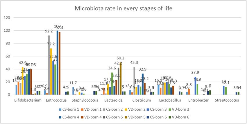
Figure 2: The total colonization rates and gut microbiota colonization rates in each time. CS-born 1 and VD-born 1: 1st week, CS-born 2 and
VD-born 2: 8 to 30 days, CS-born 3 and VD-born 3: 31 to 90 days, CS-born 4 and VD-born 4: 91 to 180 days, CS-born 5 and VD-born 5: 181
days to 1st year, CS-born 6 and VD-born 6: after 1st year.Cesarean section and neonatal gut microbiota development 633
related to Staphylococcus and their colonization means 2.12 The diversity and colonization rates of
were 4.0 and 0.0, respectively. There have been no neonatal gut microbiota during 91 days
reports of the colonization rates of Enterobacter and to 6 months of life
Streptococcus in the included studies. No statistically sig-
nificant difference was observed between the two groups Colonization rate of gut microbiota in VD-born infants
in terms of the mean colonization rate of Lactobacillus was lower than that in CS-born infants during their third
(VD group = 19.2 and CS group = 19). However, there to 6 months of life and their means were 28.8 and 29.7,
were significant differences between the two groups with respectively. The diversity and colonization rates of neo-
respect to the mean colonization rates of Clostridium natal gut microbiota during 91 days to 6 months of life
(VD group = 12.0 and CS group = 43.3), Bacteroides (VD were explored in two studies. Furthermore, the highest
group = 17.1 and CS group = 2.1), Bifidobacterium (VD rates of colonization of the VD and CS groups were
group = 42.9 and CS group = 18.4), Staphylococcus related to Enterococcus and their colonization means
(VD group = 4.0 and CS group = 0.0), and Enterococcus were 100 and 97.4, respectively. Also, the lowest rates
(VD group = 72.2 and CS group = 92.9). The colonization of the colonization of the VD and CS groups were related
rates of Bifidobacterium, Bacteroides, and Staphylococcus to Staphylococcus, and their colonization means were
were higher in the VD group, while the colonization rates 0.007 and 0.002, respectively. There have been no reports
of Clostridium and Enterococcus were higher in the CS group. of the colonization rates of Enterobacter and Streptococcus
in the included studies. No significant difference was
observed between the two groups in terms of the total
mean colonization rates of Bifidobacterium (VD group =
2.11 The diversity and colonization rates of 39.95 and CS group = 40.1). As mentioned earlier, no sig-
neonatal gut microbiota during 31 days nificant difference was found between the two groups with
to 3 months of life respect to the total mean colonization rate of Staphylococcus.
Significant differences were observed between the two
Colonization rate of gut microbiota in CS-born infants groups with respect to the mean colonization rates of
was lower than that in VD-born infants during their Clostridium (VD group = 18.2 and CS group = 32.9),
second month of life to 3 months of life and their means Bacteroides (VD group = 23.1 and CS group = 14),
were 22.5 and 24.9, respectively. The diversity and colo- Lactobacillus (VD group = 14.3 and CS group = 11.5), and
nization rates of neonatal gut microbiota during 31 days Enterococcus (VD group = 97.4 and CS group = 100). The
to 3 months of life were examined in the three studies. colonization rates of Bacteroides and Lactobacillus were
Furthermore, the highest rates of colonization of VD and higher in the VD group, while the colonization rates of
CS groups were related to Enterococcus and their coloni- Clostridium and Enterococcus were higher in the CS group.
zation means were 52.6 and 47.0, respectively. Also, the
lowest rates of colonization of the VD and CS groups were
related to Staphylococcus and their colonization means
were 3.4 and 1.6, respectively. There were significant dif- 2.13 The diversity and colonization rates of
ferences between the two groups with respect to the neonatal gut microbiota during 181
mean colonization rates of Clostridium (VD group = 12.9 days to 1 year of life
and CS group = 15.3), Bacteroides (VD group = 34.6 and
CS group = 12.1), Bifidobacterium (VD group = 32.7 and Colonization rate of gut microbiota in CS-born infants
CS group = 29.4), Lactobacillus (VD group = 16.9 and CS was lower than that of VD-born infants during their 7th
group = 19.9), Staphylococcus (VD group = 1.6 and CS month to 1 year of life and their means were 11.2 and 13.3,
group = 3.4), Enterobacter (VD group = 16.6 and CS group = respectively. The diversity and colonization rates of neo-
27.9), Streptococcus (VD group = 12.1 and CS group = 14.0), natal gut microbiota from 181 days to 1 year of age were
and Enterococcus (VD group = 47 and CS group = 52.6). The investigated in one study. In addition, the highest rates of
colonization rates of Bifidobacterium, Bacteroides, and colonization of the VD and CS groups were related to
Staphylococcus were higher in the VD group, while the colo- Bacteroides and their colonization means were 50.2 and
nization rates of Clostridium, Lactobacillus, Enterobacter, 42.2, respectively. Also, the lowest rate of colonization of
Streptococcus, Enterococcus, and Staphylococcus were higher the VD-born infants was related to Clostridium (mean =
in the CS group. 0.1), and the lowest rate of the colonization of CS-born634 Negin Shaterian et al.
infants was related to Clostridium (mean = 0.2). There colonized the gut of VD-born infants in their first year of life
have been no reports of the colonization rates of Lacto- and Clostridium colonized the gut of the CS-born infants.
bacillus, Staphylococcus, Streptococcus, and Enterococcus
in the included study. No statistically significant differences
were observed between two groups with respect to the
mean colonization rates of Clostridium (VD group = 0.1 3 Discussion
and CS group = 0.2) and Enterobacter (VD group = 0.1
and CS group = 1.2), but statistically significant differ- The results of the present study showed that the diversity
ences were observed between two groups in terms of the and colonization rates of neonatal gut microbiota were
mean colonization rates of Bacteroides (VD group = 50.2 associated with the mode of delivery. Moreover, the colo-
and CS group = 42.2) and Bifidobacterium (VD group = 2 nization rates of Bacteroides and Bifidobacterium in the
and CS group = 1.3). Additionally, the colonization rates of VD group were higher than those in the CS group. Also,
Bacteroides and Bifidobacterium in the VD group were the colonization rates of Clostridium, Lactobacillus,
higher than those in the CS group. Enterobacter, Streptococcus, and Enterococcus in the CS
group were higher than those in the VD group. The mode
of delivery did not significantly affect the colonization
rate of Lactobacillus during the 2nd week to the 1st month
2.14 The diversity and colonization rates of of life, the colonization rate of Bifidobacterium during
neonatal gut microbiota after 1 year the 4th to 6th month of life, the colonization rates of
of life Clostridium and Enterobacter from the 7th month to the
1st year of life, and the colonization rates of Clostridium,
Colonization rate of gut microbiota in VD-born infants Bifidobacterium, and Enterococcus after the 1st year of
was marginally higher than that in the CS-born infants life. However, in the 1st week after delivery and the 2nd
after their first year of life and their means were 5.02 and 3rd months of life, the mode of delivery could affect
and 5.06, respectively. The colonization rate of neonatal the colonization rates of all the bacteria. In a systematic
gut microbiota after 1 year of life was examined in one review, Rutayisire et al. found that during the first 3
study. The highest rate of colonization of VD-born infants months of life, the colonization rates of Bifidobacterium
was related to the Enterobacter (mean = 42.9), and also and Bacteroides were higher in the VD group, while the
the highest rates of the colonization of CS-born infants colonization rates of Clostridium and Lactobacillus were
were related to the Staphylococcus and Enterobacter and higher in the CS group [23], which were consistent with
their means were 5.9 and 5.9, respectively. The lowest our results. In their study, during 6–12 months of life, the
rate of colonization of the VD-born infants was related mode of delivery had less effect on the diversity and the
to Lactobacillus (mean = 3.7), and the lowest rates of colonization rates of Bifidobacterium, Bacteroides, Clos-
the colonization of the CS-born infants were related to tridium, and Lactobacillus; while in our study, during
Clostridium and Streptococcus (mean = 3.9). No statisti- 6–12 months of life, the colonization rates of Bifidobac-
cally significant differences were observed between two terium and Bacteroides in the VD group were greater than
groups with respect to the mean colonization rates of those in the CS group. This difference may be due to the
Bifidobacterium (in both the groups = 5.7) and Clostridium experimental methods presented in the included studies.
(VD group = 4.0 and CS group = 3.9) but statistically sig- The findings of this study are consistent with the previous
nificant differences were observed between two groups in studies showing that the term CS-born infants lack the
terms of the mean colonization rates of Bacteroides (VD colonization of neonatal gut microbiota up to a year, with
group = 5.3 and CS group = 5.0), Lactobacillus (VD group = lower overall microbial diversity [6,38]. Another study
3.7 and CS group = 5.0), Staphylococcus (VD group = 5.4 and demonstrated that a significant difference was observed
CS group = 5.9), Enterobacter (VD group = 7.0 and CS group = between the CS and VD groups in terms of gut microbial
5.9), Enterococcus (VD group = 5.0 and CS group = 4.9), colonization infants up to their 7th year of life [39]. Our
and Streptococcus (VD group = 4.4 and CS group = 3.9). study is in line with the study of Shao et al. (2019), sug-
Furthermore, the colonization rates of Bacteroides, Entero- gesting that the mode of delivery can play an important
bacter, and Streptococcus were higher in those born by VD, factor in the diversity of neonatal gut microbiota, espe-
while the colonization rates of Lactobacillus and Staphyl- cially 4 days after birth. Bifidobacterium, Bacteroides, and
ococcus were higher in CS-born infants. All these bacteria Parabacteroides species in the samples from the VDCesarean section and neonatal gut microbiota development 635 group were more abundant than those in the CS group; Staphylococcus, Streptococcus, Enterococcus, and Clostri- while Enterococcus, Staphylococcus epidermidis, Strepto- dium) and reduced levels of Bacteroides among CS-born coccus, Klebsiella, Enterobacter cloacae, and Clostridium infants [23]. Previous studies have suggested that the Bac- perfringens species were observed in the premature teroides may be transmitted from mother to child during babies delivered via CS in the hospital settings, and birth [48,49]. Increased levels of Clostridium in CS-born they also reported that other clinical factors such as pre- infants may be attributed to nosocomial infections [45]. natal antibiotic use, hospital stay, and breastfeeding had Breast milk contains the beneficial gut bacteria similar to less effects [40]. In consistent with our results, the study probiotics, which can stimulate the growth of Bifidobac- conducted by Chee et al. in Singapore and Indonesia terium and Lactobacillus [51,52]. Infants born by CS are demonstrated that the colonization rate of Lactobacillus deprived of breast milk in the early stages, and there may was higher in the CS group. Other studies have shown be a reason for the decrease in these species in CS-born that the colonization rate of Lactobacillus in the VD group babies. Studies have demonstrated that formula feeding is was significantly higher than that in the CS group [20,41]. associated with increased levels of Clostridium difficile and In anal samples from the exposed infants and the VD- decreased Bifidobacterium [53,54]. A Danish cohort study born infants, there is an early enrichment of Lactobacillus showed that significant changes in the gut microbiota followed by a bloom of Bacteroides from week 2 that is occurred, particularly from age 9 to 18 months, when cessa- not observed in infants not exposed to vaginal fluids [41]. tion of breastfeeding and introduction of a complementary In their study, Shao et al. demonstrated that no signifi- feeding induces replacement of a microbiota [55]. Although cant difference was observed between the two groups there are two theories about infant gut colonization: “sterile with respect to the colonization rate of Lactobacillus gut before birth” and “in uterus colonization hypothesis,” [40]. These differences may be attributed to techniques but the influence of the maternal microbiota on the organi- used for the analysis of the gut microbiota. Studies have zation of microbial population in uterus is yet to be deter- also shown that demographic factors, including breast- mined [2]. Studies have shown that mother-to-baby trans- feeding, age to stop breastfeeding, and antibiotic use, mission of bacteria occurs before birth and continues after can affect the infant’s gut microbiota, but the mode of birth [21,56]. During the third trimester of pregnancy, with delivery would have the greatest effect on it [38]. Studies the development of the nervous system, the fetus swallows a have shown that the incidence of diarrhea is inversely large amount of amniotic fluid, which causes the uterine related to Lactobacillus and Bifidobacterium levels in chil- microbiota to enter the baby’s digestive tract [57]. Recent dren less than 5 years of age. Due to their possible ben- studies have also shown that there are common bacteria eficial effects on human health, Lactobacillus and Bifido- between amniotic fluid and the meconium [58]. During the bacterium as probiotic bacteria have also been used to 1st week of life, the term infant gut is colonized by the prevent or reduce the risk of infant gastroenteritis [42,43]. Actinobacterial family (Bifidobacterium, Propionibacterium, An increase in the level of Bifidobacterium appears to play an Corynebacterium, and Streptomyces), Proteobacteria (Rumi- important role in the development and maturation of the nococcus, Enterobacter, Escherichia coli, Klebsiella, and Aci- immune system, and increased levels of Clostridium difficile netobacter), and Firmicutes (Lactobacillus, Staphylococcus, known as a nosocomial infection can cause gastroenteritis in Streptococcus, Enterococcus, and Clostridium) [59,60]. Colo- infants [23]. Reduced levels of Bifidobacterium and increased nization can be altered by factors such as gestational age, Clostridium in CS-born infants may be due to antibiotic use delivery mode (VD or CS), formula feeding (breastfeeding or [44]. Women undergoing CSs receive antibiotics before, formula feeding), hygiene, and use of antibiotics. During the during, and after delivery, especially during CS complica- first 3 years of life, the environment and feeding have impor- tions such as uterine rupture [45], bladder injury [46], tant roles in achieving an adult gut microbiome that affects etc., which may affect the gut microbiota diversity. Studies the development of the immune and nervous systems. The have shown that postnatal antibiotic use is associated with human gut microbiota reaches the characteristics of an adult increased levels of Clostridium and decreased levels of Bifi- microbiota between the ages of 2 and 5 years [21]. In this dobacterium and Bacteroides, and the use of antibiotics as study, all infants were born at term while studies have a potential factor can affect the composition of gut micro- shown that the duration of pregnancy can also affect neo- biota [38,47]. Also, the reduced levels of bactericides were natal gut microbiota diversity. In preterm infants, there observed in infants born by VD or CS whose mother used was a decrease in gut microbiota diversity and an increase antibiotic prophylaxis during delivery [6,40,44]. The lack of in colonization rate of pathogenic organisms [61,62]. Com- exposure to vaginal microbiota may be another possible pared to the term infants, premature infants have increases in reason for increased levels of Firmicutes species (Lactobacillus, anaerobes (such as Enterococcus, Enterobacter, Lactobacillus,
636 Negin Shaterian et al.
and Staphylococcus) as well as decreases in the anaerobes gut microbiota types, and the colonization of each type of
(such as Bifidobacterium, Bacteroides, and Atopobium) microbiota has an effect on the baby’s growth, develop-
[63,64]. In their study, Gregory et al. found that preterm ment, and health; therefore, it is recommended to con-
infants born by VD had a higher rate of Bacteroides than duct further studies in order to investigate the effect of
CS-born infants, which is consistent with our results, indi- the colonization type on delivery mode and on baby’s
cating that the mode of delivery can affect both term and growth and development.
preterm infants. Some limitations of this study are as follows: failure
Although there are higher complications with advanced to explore the microbiota types in most studies, a small
maternal age [65], it has been shown that variables of number of studies that examined neonatal gut micro-
maternal prenatal factors including geographic location, biota, and a small number of studies that examined the
gestational hypertensive status, and maternal age did not neonatal gut microbiota during 6 months of life, not men-
affect the diversity of gut microbial taxa composition [66]. tioning the number of bacteria found in the infants gut
The mode of delivery affects the colonization of and not distinguishing the type of bacteria found in the
the neonatal gut microbiota. The colonization rates of infant’s gut in terms of the mode of delivery and a small
Bacteroides and Bifidobacterium were lower in CS-born sample size.
infants. Also, the colonization rates of Clostridium,
Lactobacillus, Enterobacter, Streptococcus, Enterococcus, Ethics of approval: This study has a code of ethics number
and Staphylococcus were higher in CS-born infants. The IR.ABZUMS.REC.1399.168 from Alborz University of Medical
mode of delivery did not significantly affect the colonization Sciences.
rate of Lactobacillus during the 2nd week to the 1st month of
life; the colonization rate of Bifidobacterium during the 4th Acknowledgment: The authors acknowledge Alborz
to 6th month of life; the colonization rates of Clostridium and University of Medical Science for their help in this
Enterobacter from the 7th month to the 1st year of life; project.
and the colonization rates of Clostridium, Bifidobacterium,
and Enterococcus after the 1st year of life. Funding information: This study did not receive any
The neonatal gut microbiota colonization seems to be funding support.
important for health and development because the devel-
oping infant gut microbiome can influence metabolism, Conflict of interest: The authors state no conflicts of
immune system function, and brain development [2]. The interest.
gut microbiota has three essential roles, namely, protec-
tive, metabolic, and trophic. Protective role includes pre- Data availability statement: All the data generated or
vention of the proliferation of pathogenic organisms; and analyzed during this study are included in this published
the metabolic role includes the digestion and metabolism article.
of milk and food in infants, the breakdown of toxins and
drugs, vitamin synthesis, and ion absorption. Trophic
role includes the growth and differentiation of the epithe-
lial cells of the intestinal lumen, and the homeostatic References
maintenance of the immune system includes tolerance
to food antigens [67,68]. The neonatal immune system [1] Moya-Pérez A, Luczynski P, Renes IB, Wang S, Borre Y,
will rapidly mature due to the influence of microbiota, Anthony Ryan C, et al. Intervention strategies for cesarean
diet, exposure to new microbes, and other environmental section-induced alterations in the microbiota-gut-brain axis.
Nutr Rev. 2017;75(4):225–40. doi: 10.1093/nutrit/nuw069.
exposures [56,69].
[2] Martin R, Makino H, Cetinyurek Yavuz A, Ben-Amor K,
The main message of this study for practitioners is
Roelofs M, Ishikawa E, et al. Early-life events, including
that CS can cause many problems for infants and babies; mode of delivery and type of feeding, siblings and gender,
moreover, it can change the pattern of infant’s gut micro- shape the developing gut microbiota. PLoS One.
biota, and hence, VD is the best method of delivery and 2016;11(6):e0158498.
we must avoid unnecessary and without medical indica- [3] Zijlmans MA, Korpela K, Riksen-Walraven JM, de Vos WM, de
Weerth C. Maternal prenatal stress is associated with the
tion CS. However, in the 1st week after delivery and the
infant intestinal microbiota. Psychoneuroendocrinology.
2nd and 3rd months of life, the mode of delivery could 2015;53:233–45. doi: 10.1016/j.psyneuen.2015.01.006.
affect the colonization rates of all the bacteria. Given that [4] Matamoros S, Gras-Leguen C, Le Vacon F, Potel G, de La
the mode of delivery affects the colonization of infant’s Cochetiere M-F. Development of intestinal microbiota inCesarean section and neonatal gut microbiota development 637
infants and its impact on health. Trends Microbiol. [19] Shaterian N, Abdi F. Is cesarean section a safe delivery method
2013;21(4):167–73. doi: 10.1016/j.tim.2012.12.001. to prevent mother to child transmission of SARS-CoV-2?
[5] Gregory KE, LaPlante RD, Shan G, Kumar DV, Gregas M. Mode Tehran Univ Med J TUMS Publ. 2020;78(5):337–8.
of birth influences preterm infant intestinal colonization with [20] Dominguez-Bello MG, De Jesus-Laboy KM, Shen N, Cox LM,
bacteroides over the early neonatal period. Adv Neonatal Care. Amir A, Gonzalez A, et al. Partial restoration of the microbiota
2015;15(6):386. doi: 10.1097/ANC.0000000000000237. of cesarean-born infants via vaginal microbial transfer. Nat
[6] Jakobsson HE, Abrahamsson TR, Jenmalm MC, Harris K, Med. 2016;22(3):250.
Quince C, Jernberg C, et al. Decreased gut microbiota diversity, [21] Rodríguez JM, Murphy K, Stanton C, Ross RP, Kober OI, Juge N,
delayed Bacteroidetes colonisation and reduced Th1 et al. The composition of the gut microbiota throughout life,
responses in infants delivered by caesarean section. Gut. with an emphasis on early life. Microb Ecol Health Dis.
2014;63(4):559–66. doi: 10.1136/gutjnl-2012-303249. 2015;26(1):26050. doi: 10.3402/mehd.v26.26050.
[7] Grzeskowiak L, Collado MC, Mangani C, Maleta K, Laitinen K, [22] Putignani L, Del Chierico F, Petrucca A, Vernocchi P,
Ashorn P, et al. Distinct gut microbiota in southeastern African Dallapiccola B. The human gut microbiota: a dynamic interplay
and northern European infants. J Pediatr Gastroenterol Nutr. with the host from birth to senescence settled during child-
2012;54(6):812–6. doi: 10.1097/MPG.0b013e318249039c. hood. Pediatr Res. 2014;76(1):2–10. doi: 10.1038/pr.2014.49.
[8] Hill CJ, Lynch DB, Murphy K, Ulaszewska M, Jeffery IB, [23] Rutayisire E, Huang K, Liu Y, Tao F. The mode of delivery affects
O’Shea CA, et al. Evolution of gut microbiota composition from the diversity and colonization pattern of the gut microbiota
birth to 24 weeks in the INFANTMET cohort. Microbiome. during the first year of infants’ life: a systematic review. BMC
2017;5(1):4. doi: 10.1186/s40168-016-0213-y. Gastroenterol. 2016;16(1):86. doi: 10.1186/s12876-016-0498-0.
[9] Dogra S, Sakwinska O, Soh S-E, Ngom-Bru C, Brück WM, [24] Madan JC, Hoen AG, Lundgren SN, Farzan SF, Cottingham KL,
Berger B, et al. Dynamics of infant gut microbiota are influ- Morrison HG, et al. Association of cesarean delivery and for-
enced by delivery mode and gestational duration and are mula supplementation with the intestinal microbiome of
associated with subsequent adiposity. MBio. 6-week-old infants. JAMA Pediatr. 2016;170(3):212–9.
2015;6(1):e02419–14. doi: 10.1128/mBio.02419-14. doi: 10.1001/jamapediatrics.2015.3732.
[10] Francino MP. Birth mode-related differences in gut microbiota [25] Abdi F, Roozbeh N, Mortazavian AM. Effects of date palm
colonization and immune system development. Ann Nutr pollen on fertility: research proposal for a systematic review.
Metab. 2018;73(3):12–6. doi: 10.1159/000490842. BMC Res Notes. 2017;10(1):1–4. doi: 10.1186/s13104-017-
[11] Nagpal R, Yamashiro Y. Gut microbiota composition in healthy 2697-3.
Japanese infants and young adults born by C-section. Ann Nutr [26] Abdi F, Mobedi H, Bayat F, Mosaffa N, Dolatian M, Tehrani FR.
Metab. 2018;73(3):4–11. doi: 10.1159/000490841. The effects of transdermal estrogen delivery on bone mineral
[12] Bokulich NA, Chung J, Battaglia T, Henderson N, Jay M, Li H, density in postmenopausal women: a meta-analysis. Iran J
et al. Antibiotics, birth mode, and diet shape microbiome Pharm Res IJPR. 2017;16(1):380.
maturation during early life. Sci Transl Med. [27] Abdi F, Mobedi H, Mosaffa N, Dolatian M, Tehrani FR. Hormone
2016;8(343):343ra82. doi: 10.1126/scitranslmed.aad7121. therapy for relieving postmenopausal vasomotor symptoms:
[13] Stokholm J, Thorsen J, Chawes BL, Schjørring S, Krogfelt KA, a systematic review. Arch Iran Med. 2016;19(2):2–12.
Bønnelykke K, et al. Cesarean section changes neonatal gut [28] Modesti PA, Reboldi G, Cappuccio FP, Agyemang C, Remuzzi G,
colonization. J Allergy Clin Immunol. 2016;138(3):8819.e2. Rapi S, et al. Panethnic differences in blood pressure in
doi: 10.1016/j.jaci.2016.01.028. Europe: a systematic review and meta-analysis. PLoS One.
[14] Biasucci G, Rubini M, Riboni S, Morelli L, Bessi E, 2016;11(1):e0147601.
Retetangos C. Mode of delivery affects the bacterial commu- [29] Abdi F, Rahnemaei FA, Shojaei P, Afsahi F, Mahmoodi Z. Social
nity in the newborn gut. Early Hum Dev. 2010;86(1):13–5. determinants of mental health of women living in slum:
doi: 10.1016/j.earlhumdev.2010.01.004. a systematic review. Korean J Obstet Gynecol.
[15] Ajslev T, Andersen C, Gamborg M, Sørensen TI, Jess T. 2021;64(2):143–55. doi: 10.5468/ogs.20264.
Childhood overweight after establishment of the gut micro- [30] Abdi F, Roozbeh N. The effects of Humulus lupulus L. hops on
biota: the role of delivery mode, pre-pregnancy weight and menopausal vasomotor symptoms: a systematic review and
early administration of antibiotics. Int J Obes. meta-analysis. Iran J Obstet Gynecol Infertil.
2011;35(4):522–9. doi: 10.1038/ijo.2011.27. 2016;19(26):9–17.
[16] Eggesbø M, Moen B, Peddada S, Baird D, Rugtveit J, [31] Abdi F, Mahmoodi Z, Afsahi F, Shaterian N, Rahnemaei FA.
Midtvedt T, et al. Development of gut microbiota in infants not Social determinants of domestic violence against suburban
exposed to medical interventions. Apmis. 2011;119(1):17–35. women in developing countries: a systematic review.
doi: 10.1111/j.1600-0463.2010.02688.x. Korean J Obstet Gynecol. 2021;64(2):131–42. doi: 10.5468/
[17] Bäckhed F, Roswall J, Peng Y, Feng Q, Jia H, Kovatcheva- ogs.20211.
Datchary P, et al. Dynamics and stabilization of the human gut [32] Lee E, Kim B-J, Kang M-J, Choi KY, Cho H-J, Kim Y, et al.
microbiome during the first year of life. Cell Host Microbe. Dynamics of gut microbiota according to the delivery mode in
2015;17(5):690–703. doi: 10.1016/j.chom.2015.04.004. healthy Korean infants. Allergy Asthma Immunol Res.
[18] Wampach L, Heintz-Buschart A, Fritz JV, Ramiro-Garcia J, 2016;8(5):471–7.
Habier J, Herold M, et al. Birth mode is associated with earliest [33] Nagpal R, Tsuji H, Takahashi T, Kawashima K, Nagata S,
strain-conferred gut microbiome functions and immunosti- Nomoto K, et al. Sensitive quantitative analysis of the meco-
mulatory potential. Nat Commun. 2018;9(1):1–14. nium bacterial microbiota in healthy term infants born vagin-
doi: 10.1038/s41467-018-07631-x. ally or by cesarean section. Front Microbiol. 2016;7:1997.638 Negin Shaterian et al.
[34] Azad MB, Konya T, Persaud RR, Guttman DS, Chari RS, Field CJ, revealed by deep 16S rRNA sequencing. PLoS Biol.
et al. Impact of maternal intrapartum antibiotics, method of 2008;6(11):e280.
birth and breastfeeding on gut microbiota during the first year [48] Adlerberth I, Strachan DP, Matricardi PM, Ahrné S, Orfei L,
of life: a prospective cohort study. BJOG Int J Obstet Gynaecol. Åberg N, et al. Gut microbiota and development of atopic
2016;123(6):983–93. eczema in 3 European birth cohorts. J Allergy Clin Immunol.
[35] Liu D, Yu J, Li L, Ai Q, Feng J, Song C, et al. Bacterial community 2007;120(2):343–50. doi: 10.1016/j.jaci.2007.05.018.
structure associated with elective cesarean section versus [49] Vaishampayan PA, Kuehl JV, Froula JL, Morgan JL, Ochman H,
vaginal delivery in Chinese newborns. J Pediatr Gastroenterol Francino MP. Comparative metagenomics and population
Nutr. 2015;60(2):240–6. dynamics of the gut microbiota in mother and infant. Gen Biol
[36] Azad MB, Konya T, Maughan H, Guttman DS, Field CJ, Chari RS, Evol. 2010;2:53–66. doi: 10.1093/gbe/evp057.
et al. Gut microbiota of healthy Canadian infants: profiles by [50] Penders J, Thijs C, Vink C, Stelma FF, Snijders B, Kummeling I,
mode of delivery and infant diet at 4 months. Cmaj. et al. Factors influencing the composition of the intestinal
2013;185(5):385–94. microbiota in early infancy. Pediatrics. 2006;118(2):511–21.
[37] Rahnemaie FS, Zare E, Zaheri F, Abdi F. Effects of comple- doi: 10.1542/peds.2005-2824.
mentary medicine on successful breastfeeding and its asso- [51] Underwood MA, German JB, Lebrilla CB, Mills DA.
ciated issues in the postpartum period. Iran J Pediatr. Bifidobacterium longum subspecies infantis: champion colo-
2019;29(1):1–10. doi: 10.5812/ijp.80180. nizer of the infant gut. Pediatr Res. 2015;77(1–2):229–35.
[38] Yap GC, Chee KK, Hong P-Y, Lay C, Satria CD, Soenarto Y, et al. doi: 10.1038/pr.2014.156.
Evaluation of stool microbiota signatures in two cohorts of [52] Martín R, Langa S, Reviriego C, Jimínez E, Marín ML, Xaus J,
Asian (Singapore and Indonesia) newborns at risk of atopy. et al. Human milk is a source of lactic acid bacteria for the
BMC Microbiol. 2011;11(1):193. infant gut. J Pediatr. 2003;143(6):754–8. doi: 10.1016/
[39] Salminen S, Gibson GR, McCartney AL, Isolauri E. Influence of j.jpeds.2003.09.028.
mode of delivery on gut microbiota composition in seven year old [53] Bezirtzoglou E, Tsiotsias A, Welling GW. Microbiota profile in
children. Gut. 2004;53(9):1388–9. doi: 10.1136/gut.2004.041640. feces of breast-and formula-fed newborns by using fluores-
[40] Shao Y, Forster SC, Tsaliki E, Vervier K, Strang A, Simpson N, cence in situ hybridization (FISH). Anaerobe.
et al. Stunted microbiota and opportunistic pathogen coloni- 2011;17(6):478–82. doi: 10.1016/j.anaerobe.2011.03.009.
zation in caesarean-section birth. Nature. [54] Penders J, Vink C, Driessen C, London N, Thijs C,
2019;574(7776):117–21. Stobberingh EE. Quantification of Bifidobacterium spp.,
[41] Dominguez-Bello MG, Costello EK, Contreras M, Magris M, Escherichia coli and Clostridium difficile in faecal samples of
Hidalgo G, Fierer N, et al. Delivery mode shapes the acquisition breast-fed and formula-fed infants by real-time PCR. FEMS
and structure of the initial microbiota across multiple body Microbiol Lett. 2005;243(1):141–7. doi: 10.1016/
habitats in newborns. Proc Natl Acad Sci. j.femsle.2004.11.052.
2010;107(26):11971–5. doi: 10.1073/pnas.1002601107. [55] Bergström A, Skov TH, Bahl MI, Roager HM, Christensen LB,
[42] Solano-Aguilar G, Fernandez KP, Ets H, Molokin A, Vinyard B, Ejlerskov KT, et al. Establishment of intestinal microbiota
Urban JF, et al. Characterization of fecal microbiota of children during early life: a longitudinal, explorative study of a large
with diarrhea in 2 locations in Colombia. J Pediatr cohort of Danish infants. Appl Environ Microbiol.
Gastroenterol Nutr. 2013;56(5):503–11. doi: 10.1097/ 2014;80(9):2889–900. doi: 10.1128/AEM.00342-14.
MPG.0b013e318282aa12. [56] Gritz EC, Bhandari V. The human neonatal gut microbiome:
[43] Preidis GA, Hill C, Guerrant RL, Ramakrishna B, Tannock GW, a brief review. Front Pediatr. 2015;3:17. doi: 10.3389/
Versalovic J. Probiotics, enteric and diarrheal diseases, and fped.2015.00017.
global health. Gastroenterology. 2011;140(1):8. doi: 10.1053/ [57] Madan JC, Salari RC, Saxena D, Davidson L, O’Toole GA,
j.gastro.2010.11.010. Moore JH, et al. Gut microbial colonisation in premature neo-
[44] Yassour M, Vatanen T, Siljander H, Hämäläinen A-M, nates predicts neonatal sepsis. Arch Dis Childhood-Fetal
Härkönen T, Ryhänen SJ, et al. Natural history of the infant gut Neonatal Ed. 2012;97(6):F456–62. doi: 10.1136/fetalneonatal-
microbiome and impact of antibiotic treatment on bacterial 2011-301373.
strain diversity and stability. Sci Transl Med. [58] Ardissone AN, Diomel M, Davis-Richardson AG, Rechcigl KT,
2016;8(343):343ra81. doi: 10.1126/scitranslmed.aad0917. Li N, Drew JC, et al. Meconium microbiome analysis identifies
[45] Jansa V, Laganà AS, Ferrari F, Ghezzi F, Burnik Papler T, bacteria correlated with premature birth. PLoS One.
Vrtacnik Bokal E, et al. Uterine rupture in pregnancy after 2014;9(3):e90784.
hysteroscopic septum resection: a 20-year retrospective ana- [59] Aagaard K, Ma J, Antony KM, Ganu R, Petrosino J, Versalovic J.
lysis. Minim Invasive Ther Allied Technol. 2020;1–8. The placenta harbors a unique microbiome. Sci Transl Med.
doi: 10.1080/13645706.2020.1837884. 2014;6(237):237ra65. doi: 10.1126/scitranslmed.3008599.
[46] Franchi M, Raffaelli R, Baggio S, Scollo M, Garzon S, [60] Adlerberth I, Wold A. Establishment of the gut microbiota in
Laganà AS, et al. Unintentional transvesical caesarean sec- Western infants. Acta Paediatr. 2009;98(2):229–38.
tion: incidence, risk factors, surgical technique and post- doi: 10.1111/j.1651-2227.2008.01060.x.
operative management. Eur J Obstet Gynecol Reprod Biol. [61] Scholtens PA, Oozeer R, Martin R, Amor KB, Knol J. The early
2019;236:26–31. doi: 10.1016/j.ejogrb.2019.02.023. settlers: intestinal microbiology in early life. Annu Rev Food
[47] Dethlefsen L, Huse S, Sogin ML, Relman DA. The pervasive Sci Technol. 2012;3:425–47. doi: 10.1146/annurev-food-
effects of an antibiotic on the human gut microbiota, as 022811-101120.You can also read