Role of ex vivo Expanded Mesenchymal Stromal Cells in Determining Hematopoietic Stem Cell Transplantation Outcome - Frontiers
←
→
Page content transcription
If your browser does not render page correctly, please read the page content below
REVIEW
                                                                                                                                                    published: 04 May 2021
                                                                                                                                            doi: 10.3389/fcell.2021.663316

                                            Role of ex vivo Expanded
                                            Mesenchymal Stromal Cells in
                                            Determining Hematopoietic Stem
                                            Cell Transplantation Outcome
                                            Stefania Crippa 1 , Ludovica Santi 1 , Margherita Berti 1 , Giada De Ponti 1,2 and
                                            Maria Ester Bernardo 1,3,4*
                                            1
                                             San Raffaele Telethon Institute for Gene Therapy, IRCCS San Raffaele Scientific Institute, Milan, Italy, 2 Centro Ricerca M.
                                            Tettamanti, Department of Pediatrics, University of Milano-Bicocca, Monza, Italy, 3 Pediatric Immunohematology and Bone
                                            Marrow Transplantation Unit, San Raffaele Scientific Institute, Milan, Italy, 4 University Vita-Salute San Raffaele, Faculty of
                          Edited by:        Medicine, Milan, Italy
                          Guido Moll,
  Charité – Universitätsmedizin Berlin,
                            Germany         Overall, the human organism requires the production of ∼1 trillion new blood cells per
                       Reviewed by:         day. Such goal is achieved via hematopoiesis occurring within the bone marrow (BM)
               Simon Mendez-Ferrer,
                                            under the tight regulation of hematopoietic stem and progenitor cell (HSPC) homeostasis
             University of Cambridge,
                      United Kingdom        made by the BM microenvironment. The BM niche is defined by the close interactions of
                        Manja Wobus,        HSPCs and non-hematopoietic cells of different origin, which control the maintenance
      Technische Universität Dresden,
                             Germany
                                            of HSPCs and orchestrate hematopoiesis in response to the body’s requirements. The
                 *Correspondence:
                                            activity of the BM niche is regulated by specific signaling pathways in physiological
                Maria Ester Bernardo        conditions and in case of stress, including the one induced by the HSPC transplantation
           bernardo.mariaester@hsr.it
                                            (HSCT) procedures. HSCT is the curative option for several hematological and non-
                    Specialty section:
                                            hematological diseases, despite being associated with early and late complications,
         This article was submitted to      mainly due to a low level of HSPC engraftment, impaired hematopoietic recovery,
                  Stem Cell Research,
                                            immune-mediated graft rejection, and graft-versus-host disease (GvHD) in case of
                a section of the journal
  Frontiers in Cell and Developmental       allogenic transplant. Mesenchymal stromal cells (MSCs) are key elements of the BM
                                Biology     niche, regulating HSPC homeostasis by direct contact and secreting several paracrine
        Received: 09 February 2021          factors. In this review, we will explore the several mechanisms through which MSCs
         Accepted: 17 March 2021
           Published: 04 May 2021
                                            impact on the supportive activity of the BM niche and regulate HSPC homeostasis.
                              Citation:
                                            We will further discuss how the growing understanding of such mechanisms have
            Crippa S, Santi L, Berti M,     impacted, under a clinical point of view, on the transplantation field. In more recent years,
 De Ponti G and Bernardo ME (2021)
                                            these results have instructed the design of clinical trials to ameliorate the outcome of
             Role of ex vivo Expanded
          Mesenchymal Stromal Cells         HSCT, especially in the allogenic setting, and when low doses of HSPCs were available
  in Determining Hematopoietic Stem         for transplantation.
       Cell Transplantation Outcome.
      Front. Cell Dev. Biol. 9:663316.      Keywords: mesenchymal stromal cells, hematopoietic (stem) cell transplantation, hematopoietic stem and
      doi: 10.3389/fcell.2021.663316        progenitor cell, bone marrow niche, immunoregulation

Frontiers in Cell and Developmental Biology | www.frontiersin.org                    1                                                May 2021 | Volume 9 | Article 663316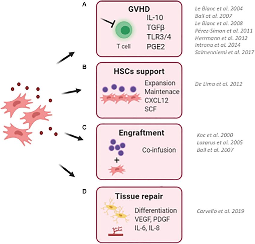
Crippa et al.                                                                                               Hematopoietic Supportive Role of MSCs

INTRODUCTION                                                               of trabecular bone (Morrison and Scadden, 2014). The role of
                                                                           osteoblasts in the control of HSPC hematopoiesis was revaluated
The bone marrow (BM) is the organ responsible for                          in light of few HSPCs found in contact with osteoblasts,
hematopoiesis, the formation of blood cellular components.                 and further experiments demonstrated that osteoblasts do not
At the prenatal stage in humans, hematopoiesis is developed                impact significantly on HSPC proliferation and differentiation
via interactions of hematopoietic stem and progenitor cells                (Greenbaum et al., 2012). MSCs emerge as fundamental cells to
(HSPCs) with a stroma of mesenchymal lineage (Bianco et al.,               physically support HSPCs and sustain HSPC self-renewal and
2010). Postnatally, these interactions are carried out within              differentiation to satisfy the body’s needs at physiological state
the skeleton. Here, HSPCs are maintained, and hematopoiesis                and in the case of stress.
takes place, unless hematopoietic stress causes the process to
transfer to extramedullary locations (Morrison and Scadden,
2014). Over the years, delineating a comprehensive picture of              MESENCHYMAL STROMAL CELLS
both the BM’s components and regulatory mechanisms have
fascinated researchers, due to the potential of such discoveries           Mesenchymal stromal cells are key elements of the BM niche
to benefit the clinical practice, as for example in hematopoietic          regulating the composition of the niche environment and the
stem cell transplantation (HSCT). Furthermore, although                    hematopoietic process. In support of this concept, several studies
the BM is constituted by multiple elements, summarized                     showed the co-localization of MSCs in the sites of hematopoiesis,
in Table 1, this review will revolve specifically around the               starting from embryonic developmental stages E11 in the
role of the BM’s mesenchymal compartment, intended                         aorta–gonad–mesonephros and, following the development of
as mesenchymal stromal cells (MSCs) and MSC-derived                        the hematopoietic systems, in the liver, spleen, and BM
osteoprogenitors/osteoblasts, given its fundamental role in the            (Mendes et al., 2005).
regulation of HSPC homeostasis.                                               In order to better understand the complex interactions within
    The first hypothesis of the existence of a hematopoietic niche         the BM, mice of various strains have been extensively employed
dates back to 1978, when Roy Schofield theorized that a specific           as models to study the human BM niche, revealing fundamental
subgroup of cells within the BM was responsible for conserving             aspects of BM’s dynamic cellular activity. Such results, however,
HSPCs’ self-renewing capacity and preventing their uncontrolled            are based on a few marker genes, such as leptin receptor (LepR),
activation (Schofield, 1978). In the same years, other groups were         nestin (Nes), C-X-C motif ligand 12 (Cxcl12), and Neural/Glia
working to define the BM’s components exerting a nurturing                 antigen 2 (NG2), which are not specific to the BM cells but
effect on HSPCs. These works highlighted the primary role of               are significantly expressed in other tissues as well (Chen et al.,
BM osteoprogenitors in the regulation of HSPC homeostasis, by              2017). Nonetheless, given the important activities of these genes,
underlining HSPCs’ primitive tendency to concentrate toward                their potential employment for therapeutic purposes have led
the endosteal margins of long bones, in direct contact with                scientists to investigate their characteristics. As an example,
osteoblasts supported by N-cadherin (Lord et al., 1975) (Gong,             some Nes+ cells have been proven to originate from the neural
1978#181). Later studies employing labeled immature cells                  crest, co-localize with HSPCs, and support HSPC functions in
confirmed the specific localization of long-term (LT)-HSPCs in             transgenic mice (Mendez-Ferrer et al., 2010; Isern and Mendez-
endosteal sites (Nilsson et al., 1997) (Nilsson et al., 2001 #246)         Ferrer, 2011). More recently, Nes+ MSCs were characterized
(Lo Celso et al., 2009) (Xie et al., 2009 #290). Further significant       in the murine BM. These cells show a greater proliferative
discoveries were made in the early 1990s, when osteoblasts were            capability when compared with Nes- cells, together with an
proven capable of producing hematopoietic cytokines and of                 increased capacity to differentiate into mesodermal cells, release
exerting a supportive role toward primitive hematopoietic cells            chemokines, specifically, colony-stimulating factor-1 (CSF-1)
in vitro (Taichman and Emerson, 1994). In light of these findings,         and tissue inhibitor of metalloproteinase (TIMP)-1 and -2 (Lu
in the following years, several groups observed an impairment              et al., 2019). Such characteristics are important to favor tissue
in the HSPC composition associated with disruption of the                  regeneration following Nes+ cell transplantation (Lu et al., 2019).
normal osteoblastic physiology. Accordingly, the stimulation of               Similarly, Morrison and Scadden (2014) identified Leptin
osteoblast proliferation increases the number of HSPCs in mice             Receptor (LepR), a receptor for a fat cell-specific circulating
and ex vivo in co-culture setting (Raaijmakers et al., 2010). By           hormone, as a marker to prospectively isolate mouse MSCs.
providing evidence that osteoblastic cells played a critical role          LepR+ MSCs show clonogenic capacity and localize around the
in HSPCs’ functioning (Calvi et al., 2003; Zhang et al., 2003;             sinusoidal vessels of the BM (Zhou et al., 2014). Importantly,
Sacchetti et al., 2007), studies in the early decades of the 2000s         LepR+ MSCs produce Cxcl12 regulating HSPC homeostasis.
paved the way for further investigations delineating a highly              In transgenic mice, the conditional reduction of LepR+ MSCs
multi-faceted picture. From that moment, different types of cells,         leads to a sharp decrease in Cxcl12, followed by a reduction
either hematopoietic and have not been observed to be involved             of quiescent HSPCs and increased HSPC mobilization (Ding
in BM’s appropriate functioning, were characterized more as                et al., 2012 #161; Ding and Morrison, 2013 #162; Oguro et al.,
complex systems than as simple groups of cells (Mendez-Ferrer              2013 #250). Finally, in the murine BM cavities, HSPCs show
et al., 2015). The concept arising from these studies highlights the       preferential proximity to CXCL12-abundant reticular (CAR)
perivascular localization of the BM niche, created partly by MSCs          cells, surrounding the sinusoidal endothelial cells close to
and endothelial cells, often associated with the sinusoidal vessels        the endosteum (Sugiyama et al., 2006) as pericyte-like cells.

Frontiers in Cell and Developmental Biology | www.frontiersin.org      2                                       May 2021 | Volume 9 | Article 663316
Crippa et al.                                                                                                                       Hematopoietic Supportive Role of MSCs

TABLE 1 | The BM niche components.

Type of cell                                     Role in the BM

Osteoblastic cells                               The region lining the inner bone surface is enriched with osteoblastic cells, including osteoprogenitors, pre- and mature
                                                 osteoblasts, and osteoclasts. The region is densely vascularized Le et al. (2018). Osteoprogenitors, the most immature
                                                 precursors of the osteoblastic lineage, maintain HSCs and support their proliferation by forming supportive niches within
                                                 the BM environment (Sacchetti et al., 2007). Osteoblastic cells are the primary contributors to the endosteal niche and
                                                 were among the first cells to be identified to regulate hematopoiesis (Lord et al., 1975; Gong, 1978; Taichman and
                                                 Emerson, 1994). The cells, activated by parathyroid hormone (PTH) or locally by PTH-related protein, have been
                                                 observed to produce hematopoietic growth factor (Taichman and Emerson, 1994; Taichman et al., 1996). The number
                                                 of osteoblasts, directly correlated with amount of HSC, is strongly influenced by bone morphogenic protein (BMP)
                                                 signaling (Zhang et al., 2013), whose receptor inhibition leads to an increase of both osteoblasts and HSCs (Galán-Díez
                                                 et al., 2018). Lowering in the amount of osteoblasts has previously shown a suppressing effect towards lymphoid and
                                                 erythroid cells, whilst favoring myeloid cell expansion (Krevvata et al., 2014). Recent results have also suggested the
                                                 degree of their involvement in hematopoiesis to depend on osteoblasts’ state of differentiation (Sacchetti et al., 2007;
                                                 Wu et al., 2008; Mendez-Ferrer et al., 2010; Calvi et al., 2012; Kunisaki et al., 2013; He et al., 2017). Further evidence
                                                 of the osteoblasts involvement in the regulation of HSCs self-renewal ability and correct homeostasis have been
                                                 obtained in Calvi et al. (2003); Zhang et al. (2003).
Osteoclastic cells                               Osteoclasts (OCLs), notoriously bone-resorbing cells and obligate partner of the osteolineage cells (Smith and Calvi,
                                                 2013), play a complex role in HSCs regulation, highly variable depending on the model. Studies have shown that
                                                 administration of granulocyte-stimulating factor (G-CSF) promotes OCL activity in both humans and mice (Takamatsu
                                                 et al., 1998; Watanabe et al., 2003). OCLs are fundamental for BM cavity formation, which, in turn, is necessary for
                                                 HSPC mobilization (Katayama et al., 2006) and establishment of the HSC niche (Mansour et al., 2012). In detail, HSPC
                                                 mobilization is triggered by OCL production of matrix metalloproteinase 9 and cathepsin K, which through cleavage of
                                                 CXCL12, inactivates stromal cell-derived factor 1 (SDF-1) and causes HSPC to be released by the BM (McQuibban
                                                 et al., 2001; Petit et al., 2002; Lévesque et al., 2003). However, results still appear conflicting on OCLs’ activities, with
                                                 some studies showing OCL to actually inhibit mobilization in mice. This was theorized, when mice administered with
                                                 bisphosphonate, which notoriously reduces the number of OCLs, were observed to have greater mobilization
                                                 (Takamatsu et al., 1998; Winkler et al., 2010). Similarly, Miyamoto et al. (2011) observed greater mobilization when
                                                 OCLs were depleted, and reduced following G-CSF serial administration and OCLs increase. Histological analyses
                                                 further suggested that both OCL and the BM cavities may be negative regulators of hematopoiesis, making them
                                                 unnecessary for HSPC maintenance or mobilization (Miyamoto et al., 2011).
Endothelial & Perivascular niche                 Further components of the BM niche include perivascular HSCs, comprising endothelial and Leptin receptor positive
                                                 (LepR+ ) stromal cells (Zhou et al., 2014). In Kiel et al. (2005) localized the perivascular niche in the sinusoidal
                                                 endothelium, whilst endothelial cells (EC) line the lumen of blood vessels aimed at transporting oxygen and nutrients to
                                                 cells. The endothelial niche can be further distinguished between an arteriolar and a sinusoidal niche, identified by
                                                 distinctive markers (Lazzari and Butler, 2018). ECs contribute to HSCs regulation and self-renewal both directly or via
                                                 the release of angiocrine factors, such as C-X-C motif chemokine 12 (CXCL12), VEGF-A, FGF2, ANG1, the Notch
                                                 signaling pathway and TSP1 (Asada et al., 2017; Tikhonova et al., 2019). Moreover, a study by Chen et al. has
                                                 highlighted the important regulatory role of a subpopulation of ECs, named Apln+ ECs, on HSCs correct functioning
                                                 and BM regeneration following transplant (Chen et al., 2019). The BM endothelial components, comprise the arteriolar
                                                 and the sinusoidal niches (Hooper et al., 2009; Poulos et al., 2015). In addition to these, another subset of cells, named
                                                 ‘Type-H’ cells, and characterized by elevated CD31 and Endomucin, has been highlighted. Type-H cells are possibly
                                                 widespread in both the sinusoidal and arteriolar niches (Ramalingam et al., 2017) and have been suggested to be
                                                 significant contributors to regulation of the bone angiogenesis and BM’s microenvironment (Kusumbe et al., 2014;
                                                 Ramasamy et al., 2014; Itkin et al., 2016). However, it has recently been suggested that quiescent HSCs are likely to
                                                 reside in proximity of perivascular niche, closely associated to the sinusoid (Acar et al., 2015; Chen et al., 2016;
                                                 Koechlein et al., 2016).
Neuronal & glial cells                           In the last decade, the sympathetic nervous system (SNS) has been an adjunct to the many regulators for the BM’s
                                                 HSCs, acting via direct contact, via the microenvironment or by releasing catecholamines (Kalinkovich et al., 2009). In
                                                 particular SNS has been observed to regulate HSPCs’ mobilization from the BM to the bloodstream (Katayama et al.,
                                                 2006; Méndez-Ferrer et al., 2008; Mendez-Ferrer et al., 2010) as well as their proliferation and differentiation. Recent
                                                 findings have demonstrated the mechanisms of homing and mobilization of HSPCs to be regulated by coordinated
                                                 activity of the SNS and parasympathetic cholinergic signals in García-García et al. (2019). SNS’s modulating influence of
                                                 the BM have been evidenced in several processes, including bone remodeling, cellular anchorage and egress from the
                                                 BM and HSPC inflammatory outcome (Hanoun et al., 2015; Maryanovich et al., 2018). Additionally, MSCs’ activity has
                                                 been seen to undergo regulation by the sensorial nervous system and, in turn, secrete neurotrophic factors, which
                                                 benefit the nervous system by exerting a neuroprotective effect and promoting regeneration, showing a reciprocal
                                                 relation between the two systems (Teixeira et al., 2013). Moreover CXCL12 downregulation is achieved via
                                                 noradrenaline binding to β 3-ADR located on stromal cells, which causes a decrease in intranuclear Sp1 transcription
                                                 factor (Méndez-Ferrer et al., 2008; García-García et al., 2019). This regulatory mechanism has long been identified as
                                                 essential to HSC/leukocyte preservation within the BM (Nagasawa et al., 1996). As part of the CNS involvement of the
                                                 BM niche, glial cells have demonstrated a supportive role towards the BM nerve fibers in the regulation of HSCs’
                                                 proliferation, by maintaining HSCs’ quiescent state (Yamazaki et al., 2011). By releasing activator molecules, the tumor
                                                 growth factor-β (TGF)/SMAD signaling pathways is activated and Smad2 and Smad3 phosphorylation is increased,
                                                 guaranteeing HSCs’ quiescence (Yamazaki et al., 2011; Blank and Karlsson, 2015).

                                                                                                                                                                  (Continued)

Frontiers in Cell and Developmental Biology | www.frontiersin.org                    3                                                 May 2021 | Volume 9 | Article 663316
Crippa et al. Hematopoietic Supportive Role of MSCs
TABLE 1 | Continued
Type of cell Role in the BM
Adipocytes Bone marrow adipocytes (BMAs) were first hypothesized in the 19th century and officially identified as BM components
in the 1960s (Zakaria and Shafrir, 1967). BMAs are a heterogeneous group of cells both in terms of physical
characteristics and varying among species and genders (Scheller et al., 2015; Lecka-Czernik et al., 2017). They are
present on both the yellow and red BM and represent 50-70% of the total BM volume (Hardouin et al., 2016). Among
the several different cell types making up the BM niche, BMAs play a significant role which, although previously only
considered limited to a negative regulation (Naveiras et al., 2009), it has recently emerged as significant for HSCs
appropriate regulation (Zhou et al., 2017). Imbalanced ratio of BMAs and a lower hematopoietic functional capacity has
been associated with aging (Kirkland et al., 2002; Berkahn and Keating, 2004; Rosen et al., 2009; Van Zant and Liang,
2012). Although the basic function of BMAs is still to store excess energy, yellow and red BMAs have shown a
difference in responsiveness (Craft et al., 2018). Studies have shown that BMAs from the yellow marrow do not respond
to environmental stressors, whilst the red marrow mainly includes react to both endogenous and exogenous cues
(Scheller et al., 2015). One of the mechanism through which BMAs affect hematopoietic stem cells’ function is by
imposing a physical barrier when expanding in the BM. This causes a limitation in mobilization and a necessary
remodeling of the marrow niche via lineage development, to adjust to the limited space available (Boroumand and Klip,
2020). Such limitations may further affect bone appropriate formation (Su et al., 2018) and alter lymphocytes’
maturation (Tokoyoda et al., 2004).
Macrophages Macrophages are a heterogeneous group of mononuclear cells belonging to the innate immune cell group and are
present across all tissue types, including the BM. They primarily act as a first line defense in immune response (Zajd
et al., 2020) but exert several functions including tissue remodeling, clearing of dead cells and releasing of angiogenic
factors (Davies et al., 2013). Macrophages form early in embryogenesis and are believed to migrate towards the sites of
HSC formation to support their development (McGrath et al., 2015). Within the BM, macrophages interact with Nes+
cells, inducing CXCL12 transcription. Once macrophages deplete, CXCL12 expression is lost and HSC are released
from the BM (Mariani et al., 2019). This process involves not only CXCL12, but also several other factors regulating HSC
maintenance, such as ANGPT1 and vascular adhesion protein 1 (VCAM). Experimental depletion of monocytes and
macrophages led to a decrease in BM CXCL12 and a selective inhibition of HSC maintenance genes, which caused
HSC egression into the peripheral circulation (Chow et al., 2011). In the context of HSCT, macrophages are believed to
perform a significant supportive role to guarantee reconstitution, although the mechanisms involved are still unclear
(Qiao et al., 2018). Activation of BM macrophages following allogenic HSCT has been observed to impair correct
recovery, increase mortality and lower overall survival, suggesting macrophages’ critical impact (Takagi et al., 2009). In
mice, reduction in BM macrophages altered the proportion of BM-HSC and megakaryocytes, as well as white blood
cells in the peripheral blood circulation. Such reduction could be associated with a number of damaging consequences,
among which a delay in BM recovery from damage, an increase in apoptotic HSCs and a limited survival rate of
sub-lethal dose irradiation mice confirming the essential role of BM macrophages in BM recovery (Ju et al., 2019).
Table summarizing the non-hematopoietic components of the human BM niche.
As a matter of fact, in vivo analyses of HSPCs following the complexity of the human hematopoietic niche. Recently,
transplantation in mice have provided significant evidence of humanized models of the BM niche were employed as a tool
their preferential localization in vessels displaying a higher to study the human stroma in physiological and pathological
rate of CXCL12, at first, and a subsequent migration toward context, partially overcoming the functional differences between
periosteal locations (Sipkins et al., 2005; Lo Celso et al., 2009). mouse and human BM niches (Reinisch et al., 2016; Abarrategi
CAR cells express adipogenic and osteogenic genes, and control et al., 2017). Within the mature human BM niche, MSCs are
HSPC homeostasis through the secretion of CXCL12 and SCF. localized around the blood vessels, similar to pericytes, in the
Indeed, CAR cell-depleted mice showed an increased expression endosteal and vascular sites (Mangialardi et al., 2016). The
of myeloid-differentiation genes, highlighting the role of CAR endosteal niche is localized in the internal bone shell surface,
cells in the control of HSPC quiescence (Omatsu et al., 2010). close to the endocortical and trabecular surfaces. The endosteum
NG2 perivascular cells were also demonstrated to play a trophic is a thin vascular membrane covering the inner surface of the
effect on HSPCs. with arterioles. Depletion of NG2+ cells was bone mostly formed by osteoblasts and osteoclasts. Endosteal
associated with a sharp reduction in HSPC repopulating ability MSCs line the bone surface where they are physically associated
(Asada et al., 2017). Although differences between humans with both osteoblasts and HSPCs (Mendez-Ferrer et al., 2010).
and mice should be taken into consideration, comparative Several studies have highlighted that long-term (LT)-HSPCs
biology has allowed the advancement in understanding the reside in the endosteal niche supported by endosteal MSCs. MSCs
BM composition and mechanisms in this model, undoubtedly of the vascular niche consist of pericyte-like cells that support
contributing to the current understanding of the interactions cycling HSPCs and regulate HSPC mobilization and homing.
among human BM components and the supportive role that In particular, within the human BM niche, CD146+ MSCs are
MSCs play in this context. Indeed, despite human MSCs showing pericyte-like cells localized in the sinusoidal wall, characterized
different functional characteristics, the molecular mechanisms by high clonogenic and self-renewal capacities, in addition to
underlying the supportive activity of MSCs, first identified in expressing hematopoietic supportive factors, including SDF-1α
the murine models of BM niche, were fundamental to dissect and ANG1 (Sorrentino et al., 2008). CD146+ MSCs are capable
Frontiers in Cell and Developmental Biology | www.frontiersin.org 4 May 2021 | Volume 9 | Article 663316Crippa et al. Hematopoietic Supportive Role of MSCs
of giving rise to hematopoiesis-associated stromal cells in vivo, and favor their engraftment, accelerating the hematological
with a defined developmental sequence in which bone formation reconstitution after transplantation.
precedes the appearance of a sinusoidal system, and ultimately of
hematopoiesis (Sacchetti et al., 2007).
Contrarily, CD271 and CD271+ /CD146−/low MSCs are bone- MESENCHYMAL STROMAL CELLS’
lining MSCs associated with LT-HSPCs in low-oxygen areas. PARACRINE IMMUNOREGULATORY
These specific subsets of MSCs are characterized by higher levels ACTIVITY
of extracellular matrix and chondrogenesis genes (Kuci et al.,
2019). Also, CD271+ MSCs have shown high colony-forming Over time, studies on cultured MSCs have attempted at clarifying
unit—fibroblastic (CFU-F) capacity, the ability to transfer a BM MSCs’ regulatory role, and nowadays, their ability to produce
microenvironment upon transplantation, and to differentiate and release a wide variety of cytokines, chemokines, and growth
in vitro into mesodermal mature cell lineages (Tormin et al., 2011; factors that may affect the immune system, has been extensively
Crippa and Bernardo, 2018). confirmed (Fibbe and Bernardo, 2014). MSC immunosuppressive
Although several studies have clarified the identity of MSC ability was first demonstrated in support to skin graft, which was
subset/s involved in the regulation of HSPC homeostasis in vivo, prolonged after MSC administration (Bartholomew et al., 2002).
most of the available data describing their HSPC supportive Thanks to this immunoregulatory capacity, MSCs may influence
activity have been obtained using ex vivo expanded MSCs. This the activity of both the innate and adaptive immune cells, through
is due to their low frequency in the BM of 0.01–0.001% of the cell-to-cell contact or by paracrine mechanisms. This renders
total mononucleated cells in humans. Hence, MSCs are usually MSCs a promising and attractive candidate for therapeutic
isolated and expanded in vitro to reach an appropriate quantity application against different immune-mediated diseases and to
for biological and functional characterization, and an adequate control T-cell reactivity causing GvHD, in the case of allogenic
number for any clinical application (Alvarez-Viejo et al., 2013). transplants or inducing graft rejection.
MSCs can be easily isolated from BM mononuclear cells by The interaction between MSCs and the T-cell compartment
plastic adherence, and expanded as fibroblast-like cells for several has been demonstrated by several research groups. Francois
passages (p8/9), with an intrinsic capacity to differentiate into et al. (2012b) reported that MSCs inhibit T-cell proliferation
mesodermal cell types when exposed to proper differentiation by secreting indoleamine 2,3-dioxygenase (IDO), especially
factors. Importantly, from a clinical point of view, it has been when MSCs are pre-stimulated with IFNγ and TNF and
shown that MSCs can be also derived from the CD34- fraction when treated with prostaglandin E2 (PGE2). Moreover,
of BM aspirates, highlighting the possibility to isolate autologous it has been shown that the addition of monocytes to a
MSCs from the same sample used to isolate CD34+ HSPCs for PBMCs/MSCs co-culture increases the inhibitory effect of
transplantation (Ingo et al., 2016; Crippa and Bernardo, 2018). MSCs on T-cell proliferation in a dose-dependent manner
Furthermore, MSCs have been isolated from several other (Cutler et al., 2010).
tissues, including the umbilical cord (Erices et al., 2000), The mechanisms at the basis of T-cell immunosuppression
adipose tissue, liver (although with some differing characteristics) rely also upon cytokines, such as interleukin-10 (IL-10),
(Kholodenko et al., 2019), spleen, dental pulp, and lung tissue transforming growth factor-β (TGFβ) (Ryan et al., 2007), and also
(Foronjy and Majka, 2012). To ease the identification of MSCs, through toll-like receptors (TLRs), in particular, TLR3 and TLR4
in 2006, the International Society for Cellular Therapy has listed (Liotta et al., 2008; Figure 1).
some defining criteria (Dominici et al., 2006): The immunoregulatory activity of MSCs is environment
dependent, and MSCs acquire a specific function by sensing
(1) Adherence of MSCs to plastic when cultured. the inflammatory/anti-inflammatory signals in a process
(2) Expression of CD105, CD73, and CD90 surface markers of activation named “licensing” (#97). The activation of
and, at the same time, absence of expression of CD45, immunosuppressive rather than immune-stimulating phenotype
CD34, CD14/CD11b, CD79a, CD19, and HLA-DR of MSCs depends on the balance between different stimuli. Based
surface antigens. on these signals, MSCs can polarize into two acting phenotypes:
(3) Ability to differentiate in vitro in the three lineages: MSC-1 with a proinflammatory profile and MSC-2 with an
osteoblasts, adipocytes, and chondroblasts. anti-inflammatory profile (Waterman et al., 2010). Among all
factors, IFNγ is the key to activate MSC with immunosuppressive
The clinical use of ex vivo expanded MSCs in the properties. IFNγ is the first cytokine to be produced after T-cell
context of HSCT is based on MSC’s capacity to sense the activation, and it would normally act as a boost signal for T
environmental stress and activate a paracrine response to restore cells. However, in the presence of MSCs, IFNγ suppresses T-cell
a proper balanced physiology. This includes the release of anti- proliferation, by binding IFNγ receptors expressed on the cell
inflammatory factors to preserve the BM niche function upon surface of MSCs and activating an immunoregulatory phenotype
the pre-transplant treatment with conditioning regimen and of MSCs. It has also been shown that MSCs treated with IFNγ
the capability to modulate the innate and adaptive immunity, in vitro, behave as an efficient antigen-presenting cell (APC),
reducing the risk of graft-versus-host disease (GvHD) and graft inducing specific immune responses. It was suggested that MSCs
rejection. In addition, the secretion of hematopoietic supportive act in different ways depending on the concentration of IFNγ:
factors by MSCs promote the survival of transplanted HSPCs they may act as APCs at low IFNγ concentration, while exerting
Frontiers in Cell and Developmental Biology | www.frontiersin.org 5 May 2021 | Volume 9 | Article 663316Crippa et al. Hematopoietic Supportive Role of MSCs
FIGURE 1 | Schematic representation describing the clinical use of MSCs in the context of hematopoietic stem cell transplantation (HSCT). (A) MSCs has been
successfully employed to reduce the risk and to treat graft-versus-host disease (GvHD) in transplanted pediatric and adult patients. Thanks to their ability to sense
inflammatory stimuli, MSCs are capable to modulate T-cell proliferation and activation through the release of specific immunomodulatory cytokines (IL10, TGFb, and
PGE2). (B) MSCs have been used as a feeder to expand and maintain UCB-CD34+ HSCs before transplantation due to their ability to secrete HSC supportive
factors. (C) The co-infusion of MSCs has been demonstrated to promote HSC engraftment and accelerate hematological reconstitution in transplanted patients.
Despite only a small percentage of infused MSCs reaches the BM niche, the production and release of supportive factors by co-infused MSCs ameliorates the
outcome of HSCT. (D) The ability of MSC to repair and differentiate in bone cells makes them an attractive cell candidate to restore a proper BM niche, with MSCs
and MSC-derived osteoblasts capable of supporting HSCs. (BM, bone marrow; UCB, umbilical cord blood). For each clinical application, works describing clinical
trials for the use of MSCs are reported.
an inhibitory effect at higher concentration of IFNγ (Chan et al., Furthermore, MSCs have shown the ability to variably respond
2006). The release of other inflammatory cytokines, such as to IFN-γ depending on the circumstances. For example, IFN-
TNF-α and IL-1a, are important for MSC activation, but the γ may enhance the immunosuppressive activity of MSCs on T
effect of these stimuli is only effective in combination with IFNγ. lymphocytes (Krampera et al., 2003), but it may also elicit a
An important role for the activation of immunosuppressive completely different response, by inducing MSCs to behave as
MSCs is played by TLRs. MSCs express different types of a non-conventional antigen-presenting cell (APC) (Stagg et al.,
TLRs, which trigger specific molecular signaling and biological 2006), demonstrating a high immunological plasticity. While
properties. For example, TLR2 induces the secretion of effects on T cells is clearer, regulation of B-cell function remains
IL-6 and inhibits differentiation of MSCs into adipocytes controversial. Several studies show inhibition of B cells, through
and osteoblasts (Flynn et al., 2019); TLR3 induces MSC cell cycle arrest elicited by MSCs, mainly due to the production
migration and secretion of immunosuppressive cytokines and and release of soluble factors (Augello et al., 2005; Corcione
chemokines like IL-10 and TNFα (Rashedi et al., 2017). et al., 2006). This aspect supports the prospective employment
On the contrary, TLR4 priming induces MSCs to release of MSCs in immune-mediated disease. Conversely, Healy et al.
proinflammatory cytokines. In conclusion, the licensing process have shown a completely opposite effect, as MSCs determined
depends on three factors: presence of inflammatory cytokines, activation and proliferation of B lymphocytes (Healy et al., 2015),
their concentration, and timing of exposure (Romieu-Mourez highlighting the necessity to continue the research to improve our
et al., 2010; Sangiorgi and Panepucci, 2016). understanding of the underlying regulatory mechanisms.
Frontiers in Cell and Developmental Biology | www.frontiersin.org 6 May 2021 | Volume 9 | Article 663316Crippa et al. Hematopoietic Supportive Role of MSCs
Importantly, although the mechanisms still need to be excessive HSPC activation (Taniguchi Ishikawa et al., 2012).
clarified, these cells have shown fundamental immunoregulatory Similarly, it has been shown that the mitochondrial transfer from
capacity by significantly affecting maturation, polarization, transplanted HSPCs to BM stroma via connexin-43 accelerate
and function of lymphocytes, macrophages (Zhou et al., the BM niche regeneration after myeloablative stress, improving
2019), dendritic cells’ (DCs), and natural killer (NK) cells HSPC engraftment (Golan et al., 2020). The N-cadherin-
(Sotiropoulou et al., 2006). mediated binding of human HSPCs to BM stromal cells was
The immunomodulatory ability of MSCs is at the basis also described to preserve HSPC quiescence. In vitro studies
of their clinical use in promoting the resolution of damage- demonstrated that N-cadherin silencing reduces the percentage
induced inflammation. In this regard, MSCs not only modulate of long-term repopulating HSPCs in co-culture with MSCs (Wein
the immune system to mitigate the detrimental effects of et al., 2010). In addition, the contact-dependent effects of MSCs
excessive inflammation, but also release specific factors within on different BM niche cell types allow to amplify the supportive
the inflammation site, which favor resident tissue repairing activity of the stromal niche. For instance, the direct cell contact
mechanisms. Among them, VEGF, Angiogenin (ANG), and IL- between MSCs and endothelial cells induce MSCs to activate a
8 increased vascular regeneration (Kim et al., 2019), NGF, IL-10, pericyte-like program to sustain the formation of new vessels
and IL1-RA prevented apoptosis and enhanced cell proliferation (Loibl et al., 2014) and the integrin-mediated cell-contact of
(Francois et al., 2012b). Stimulation of MSCs with TNF, for MSCs with myeloma cells enhance the production of osteoclast-
example, has been observed to enhance the release of interleukin stimulating factors (Michigami et al., 2000).
6 (IL6), HGF (Broekman et al., 2016), VEGF (Ge et al., 2018), However, the MSC release of supportive factors is the
and insulin-like growth factor-1 (IGF-I), which, on the other principal molecular mechanism regulating HSPCs\homeostasis
hand, is decreased when p38 MAPK signaling pathway is halted in vivo, within the BM niche, and ex vivo, in MSC-based
(Miloso et al., 2008). These mechanisms have shown potential co-culture systems and when MSCs are co-infused to sustain
to be employed in treatment. For example, combinatory effect HSPCs in HSTC pre-clinical and clinical models (Crippa et al.,
of p38 MAPK inhibitors with MSC infusion has recently been 2019). The role of MSC secretome (Liu et al., 2018) and
tested in mouse models of myocardial infarction, significantly MSC-derived extracellular vesicles (Batsali et al., 2020) also
ameliorating inflammation, apoptosis, and cell morphology demonstrates the predominance of MSC paracrine activity in
(Zhang et al., 2017), highlighting the beneficial effect of this sustaining HSPC function.
combinatory strategy to limit inflammation-derived damage. Among the many components of MSCs’ secretome, CXCL12,
Similarly, MSCs have been employed in several clinical trials also known as stromal cell-derived factor-1α (SDF-1α), and stem
to counteract neurodegenerative diseases (Kabat et al., 2020), cell factor (SCF), or Kit ligand (Kitl), play a pivotal role in
showing neuroprotective effects. Indeed, in Parkinson’s disease, HSPCs’ maintenance. CXCL12 has shown both a significant
hBM-MSCs promoted a-synuclein clearance stimulating IL- immunomodulatory effect (Giri et al., 2020) and an essential
4 secretion from microglial cells, (Park et al., 2016) and in regulatory activity on both HSPCs and lymphoid progenitor cells
Alzheimer’s disease, hUCB-MSCs were able to reduce microglial (Semerad et al., 2005; Mendez-Ferrer et al., 2010). Suppression
activation and apoptosis (Lee et al., 2010). of Cxcl12 from multiple subgroups of stromal cells was observed
to cause a range of effects in terms of mobilization and
appropriate functioning of cells. Interestingly, while no effect was
MESENCHYMAL STROMAL CELLS’ observed when Cxcl12 was deleted in mineralizing osteoblasts,
HEMATOPOIETIC SUPPORT B-cell progenitors were lost and HSPC mobilized, when the
same factor was deleted from osterix-expressing stromal cells
The key role of MSCs in the regulation of the hematopoietic (Greenbaum et al., 2013). Furthermore, when Cxcl12 was
compartment is supported by the co-localization of MSCs with deleted in murine Nes- mesenchymal progenitors, a striking loss
sites of hematopoiesis since the embryonic developmental stages of HSPCs and quiescence as well as long-term repopulating
(Mendes et al., 2005). MSCs exert their function through the activity and common lymphoid progenitors were observed,
direct interaction with HSPCs and by secreting several paracrine highlighting this subpopulation of cells to retain a supportive
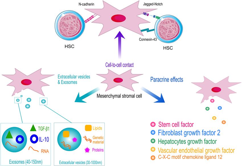
factors, as schematized in Figure 2. Perivascular MSCs express role of B-cell progenitors and its significance in HPSC homing
several Notch ligands, including Jagged-1, Jagged-2, DLL-1, (Greenbaum et al., 2013). CXCL12 selectively activated STAT-
and DLL-4, which are responsible for the activation of Notch 5 signaling pathway in different hematopoietic cells, inducing
signaling in HSPCs, a key pathway controlling HSPC growth cell proliferation (Vila-Coro et al., 1999; Mowafi et al., 2008).
and differentiation during development (Bigas et al., 2010, The activation of STAT5 in HSPCs is accompanied with the
#345; Lampreia et al., 2017). The perturbation of Notch ligand expression of specific microRNAs to guarantee a negative control
expression in MSCs induces premature differentiation of HSPCs for excessive and uncontrolled HSPC proliferation (Haetscher
when ex vivo co-cultured (Corselli et al., 2013). et al., 2015), suggesting a possible role of CXCL12/STAT5 axis
Within the BM niche, the levels of ROS determine the in HSPCs’ survival and proliferation (Janowska-Wieczorek et al.,
balance between quiescent and differentiating HSPCs. Thus, 2001; Aqmasheh et al., 2017).
the fine tuning of ROS level is fundamental to preserve HSPC Mesenchymal stromal cells’ secretion of SCF has been instead
homeostasis (Ludin et al., 2014). In such a context, MSCs associated with appropriate homing of HSPCs (Horwitz et al.,
function as scavenger cells to import ROS from HSPCs avoiding 2011). Mainly secreted by BM adipocytes and Lepr+ stromal
Frontiers in Cell and Developmental Biology | www.frontiersin.org 7 May 2021 | Volume 9 | Article 663316Crippa et al. Hematopoietic Supportive Role of MSCs FIGURE 2 | Schematic representation of the hematopoietic supportive activities of human mesenchymal stromal cells (MSCs). MSCs support hematopoietic stem and progenitor cell (HSPC) homeostasis by cell-contact, through the interaction of specific ligands expressed on MSCs surface with HSPC receptors (N-cadherin and Notch). Adherent junction, such as connexin43, also play a role in the control of HSPC metabolism to protect cells from excessive activation. However, MSCs exert their hematopoietic function mainly through the secretion of supportive factors and the release of extracellular vesicles and exosomes. HSC models are licensed by https://creativecommons.org/licenses/by/3.0/legalcode. cells, studies on mice have proven that SCF is essential for enhancing neuro- and angiogenesis in cases of cerebral damage HSPC regeneration as well as hematopoiesis and cell homeostasis, (Matsuzaki et al., 2001; Svensson et al., 2002; Ford et al., particularly in conditions of obesity and aging (Zhang et al., 2011; Suzuki et al., 2011). VEGF is also a crucial regulator of 2019; Zhou et al., 2017). Inhibition of the appropriate interactions cell differentiation by determining cell fate as well as survival, between SCF receptor and ligand has shown to enhance HSPC migration, and proliferation (Oswald et al., 2004; Li et al., 2011; clearance, confirming the chemokine role in HSPCs’ self-renewal Zaniboni et al., 2015). In terms of hematopoietic support, VEGF (Czechowicz et al., 2007). Evaluation of mutations to the SCF levels in HSPCs tend to raise in response to stimulation by receptor-encoding genes in mice models have been associated cytokines (Bautz et al., 2000). Several subsets of HSPCs present with severe conditions of macrocytic anemia, in the case of loss- VEGF receptor type 2 (VEGFR-2), whose expression has been of-function mutations (Nocka et al., 1989), or erythrocytosis linked to pluripotent stem cell activity (Shalaby et al., 1995; when in the presence of a gain of function (Bosbach et al., 2012). Kabrun et al., 1997). At the end of the last century, early Among the many components of MSCs’ secretome, several development studies observed lethality due to alteration in both were observed to exert important roles in inflammatory reactions, angiogenesis and hematopoiesis, as a consequence of knockout cell homing, and regulation of apoptotic events, such as vascular of either VEGF- and VEGFR-2-encoding genes (Shalaby et al., endothelial growth factor (VEGF), fibroblast growth factor- 1995; Carmeliet et al., 1996; Ferrara et al., 1996). In particular, 2 (FGF-2), angiopoietin (ANGPT), and hepatocyte growth Shalaby et al. (1995) observed Vegfr2−/− mice dying around factor (HGF), all associated with different signaling pathways E8.5. Similarly, knock-in mice with tyrosine residue Y1173 of (Aqmasheh et al., 2017). Vegfr-2 (Y1175 in humans) were observed to die around E8.5– Vascular endothelial growth factor is another factor secreted 9.5, due to scarcity of endothelial cells and HSPCs (Sakurai by MSCs and involved in several immunological responses, et al., 2005). This is due to VEGF ability in recruiting HSPCs Frontiers in Cell and Developmental Biology | www.frontiersin.org 8 May 2021 | Volume 9 | Article 663316
Crippa et al.                                                                                              Hematopoietic Supportive Role of MSCs

and endothelial progenitor cells, which leads to the development         barriers, given their attractive characteristics (Park et al., 2019).
of microvasculature within the BM, essential for appropriate             In hematopoiesis, MSC-EVs are believed to contribute to
hematopoiesis (Gerber et al., 2002; Koch and Claesson-Welsh,             activate HSCs in response to different stimuli, including blood
2012). Also, VEGF-A levels have been shown to increase in                hemorrhage, fluctuations in oxygen concentrations, radiation, or
plasma as a result of ANGPT-1 stimulation of hematopoiesis               chemotherapy (Butler et al., 2018). In vitro, MSC-EVs have been
and mobilization of BM-repopulating stem and progenitor cells            shown to promote the proliferation of umbilical cord-derived
(Hattori et al., 2001; Kopp et al., 2006). Although some of VEGF         HSCs by triggering the Wnt/β-catenin pathway as well as to
abilities to support hematopoiesis have been individuated, the           lower radiation damage to murine HSCs by promoting their
underlying mechanisms that govern the process still need to be           proliferation (Cominal et al., 2019). The translation of such
unveiled, and further studies are required.                              results into clinical practice, however, will still require extensive
    Another example of the regulatory role of MSC’s secretome            additional investigation to address significant limitations
includes the maintenance of HSPCs’ quiescence regulated by               surrounding EVs’ production, quantification, pharmacokinetics,
interaction between tyrosine kinase receptor (Tie2) and ANGPT-           and characterization (Gowen et al., 2020).
1 ligand, through adhesion of MSCs to HSPCs (Blank et al.,                  Among the different kinds of EVs, exosomes are membrane-
2008). Similarly, HSPCs are maintained by MSCs’ production               bound nanoparticles (40–150 nm) released by multivesicular
of the Notch ligands, which are normally involved in regulation          bodies. MSC-derived exosome contains several bioactive
of the proliferation, functioning, and differentiation of T and          molecules, particularly miRNAs of different kinds, plays a critical
B lymphocytes (Kang and Robling, 2014). Production of Notch              role in the control of both physiological and pathological states,
ligands improves survival and proliferation of HSPCs, while              including inflammatory response (Burrello et al., 2016) but also
preventing their differentiation, as demonstrated by Notch               tumorigenesis and progression (Ono et al., 2014).
inhibition leading to greater egress and mobilization of HSPCs              In light of such prominent functions exerted by MSCs in the
(Wang et al., 2015).                                                     context of the hematopoietic process, the different pathways and
    Another significant player in HSPC maintenance is fibroblast         mechanisms are now under critical evaluation in order to better
growth factor 2 (FGF2) that has been demonstrated to support             understand the homeostatic supportive process and potentially
the expansion of stromal cells in mice, consequently raising             exploit certain mechanisms for therapeutic purposes. One of the
SCF and supporting HSPC expansion, which highlighted the                 main settings that could benefit the most is that of hematopoietic
capacity of FGF2 to regulate gene expression and, in turn, support       stem cell transplantation (HSCT).
HSPC proliferation. In mice, FGF2 has been proven to promote
appropriate recovery, following myeloablative treatments, by
supporting HSPC expansion (Itkin et al., 2012; Zhao et al., 2012).       CLINICAL APPLICATIONS EXPLOITING
Nonetheless, FGF2 ability to expand different types of progenitor        THE IMMUNOREGULATORY ACTIVITY
cells may be crucial to boost hematopoiesis after HSCT.                  OF MESENCHYMAL STROMAL CELLS
    Similarly, hepatocyte growth factor (HGF) is known
to stimulate local angiogenesis by stimulating tyrosine                  The paracrine activity of MSCs has rendered these cells an
phosphorylation via the c-MET receptor (Nita et al., 2017).              attractive therapeutic tool for several clinical applications,
HGF is produced within the BM microenvironment and has                   considering the versatile function of MSCs in different
been observed to raise in patients treated with granulocyte              inflammatory and regenerative contexts. It has been a long
colony-stimulating factor (G-CSF). Increased expression of the           time from the first MSC injection in human subjects (Lazarus
c-MET, associated to HGF, is observed in more mobile HSPC,               et al., 2005), demonstrating that the use of MSCs is feasible and
concordant with the previous findings stating HGF/c-MET                  safe. Several clinical trials employing MSCs have been opened
pathways to be associated with MSC mobilization. Hence, HGF              to counteract chronic degeneration, with particular interest for
and G-CSF may both be involved in establishing a proteolytic             neurological disorders, to repair damaged tissues, and to mitigate
microenvironment in the BM, easing the egress of HSPC in                 the inflammatory response in autoimmune diseases and GvHD,
peripheral circulation (Jalili et al., 2010).                            in the context of HSCT.
    Finally, another method through which MSCs interact,                     In addition to the in vitro evidences demonstrating
modify, and respond to the surrounding environment is by                 their immunomodulatory properties, MSCs have also been
releasing extracellular vesicles (EVs). EVs are membrane-bound           employed in vivo to suppress an excessive activation of
particles, ranging between 30 and 1,000 nm of dimension, that            the immune response in different clinical contexts, such as
contain and transport biomolecules such as lipids, proteins,             HSCT, tumor immunity, and autoimmunity, occasionally
and genetic material between cells. When secreted by MSCs                producing conflicting results. Substantial beneficial effects of
(MSC-EV), these particles mediate the cells’ paracrine activity          MSC administration have been obtained in an autoimmune
and have been observed to exert an impactful anti-inflammatory           encephalomyelitis model, an autoimmune inflammatory
effect in a number of conditions, including post-transplant              disease that affects the CNS mediated mainly by T cells and
complications, in addition to control normal and pathological            macrophages (Zappia et al., 2005). Another inflammatory
hematopoiesis (Batsali et al., 2020; Lia et al., 2020). Under a          condition that might take advantage of MSCs infusion is
clinical point of view, EVs appear promising due to their highly         Crohn’s disease (CD). MSCs immunomodulatory properties
safe profile, low immunogenicity, and ability to cross biological        are, in fact, believed to reduce the persistent inflammation

Frontiers in Cell and Developmental Biology | www.frontiersin.org    9                                        May 2021 | Volume 9 | Article 663316
Crippa et al. Hematopoietic Supportive Role of MSCs
of the gastrointestinal tract by secreting anti-inflammatory vesicles (EVs), released by MSCs, has been investigated as
molecules in the target tissues and modulating specific immune potential therapeutic medical product, compared with cell
cells, for example, upregulating Treg cells, a cell population therapy (Wang et al., 2012; Rani et al., 2015). In the clinical
well known to be reduced in CD (Dalal et al., 2012; Carvello setting, MSC-EV infusion has been demonstrated to significantly
et al., 2019). Conversely, in another autoimmune condition, ameliorate GVHD symptoms in a steroid-resistant patient. The
collagen-induced arthritis, MSCs injection, which is supposed to anti-inflammatory molecules, carried by EVs, IL-10, TGFβ,
inhibit T-cell proliferation, did not lead to any significant benefit and HLA-G, might be responsible for the beneficial effects of
on arthritic symptoms. However, in vitro TNFα addition was MSC-EVs as anti-inflammatory mediators (Kordelas et al., 2014).
sufficient to reverse MSC immunosuppressive effects, suggesting In mice, the infusion of MSCs, derived from different sources,
that environmental parameters and the microenvironment, has shown a great immunomodulatory potential to control lethal
in general, may influence and affect the immunomodulatory GvHD (Yanez et al., 2006). However, contradictory results were
properties of MSCs (Djouad et al., 2005). Still, MSC treatment in obtained in another study by Sudres et al. (2006) in which
humans has shown promising outcomes in several conditions, MSC administration did not ameliorate GvHD. Further studies,
including GvHD (Le Blanc et al., 2004) and engraftment aimed at improving the clinical use of MSCs in the context of
promotion (Lazarus et al., 1995; Koc et al., 2000), thanks to their GvHD suggested that multiple MSC injection (Tisato et al., 2007)
potent immunosuppressive role but also in some rare diseases could be necessary to prevent the disease onset in a murine
in which MSCs may be responsible of ameliorating the disease model of aGvHD. This highlights the need to better define the
pathology in specific tissues (Crippa et al., 2019). Therefore, a therapeutic window for MSC administration and, eventually, to
better understanding of MSCs’ immunomodulatory role might determine whether multiple injection of MSCs could improve
be crucial to develop novel and effective therapeutic strategies for their therapeutic efficacy.
a broad range of diseases. In patients, MSCs were successfully employed for the first time
by Le Blanc et al. (2004) to treat GvHD. The intravenous infusion
of maternal BM-derived MSCs was able to control refractory
THE CLINICAL ROLE OF aGvHD manifestations. The same authors reported in a phase
II clinical trial that the infusion of ex vivo expanded BM-MSCs
MESENCHYMAL STROMAL CELL IN significantly improve the disease progression in 55 transplanted
HEMATOPOIETIC STEM CELL patients affected by steroid-resistant aGvHD. Thirty of the treated
TRANSPLANTATION: TREATMENT OF patients showed a complete remission, and in nine patients,
GRAFT-VERSUS-HOST DISEASE symptoms improved, demonstrating that MSC administration
represents a safe and effective treatment option for aGvHD. Any
Considering their immunoregulatory and hematopoietic toxicity related to the injection of MSCs was reported (Le Blanc
supportive function, MSC-based cell therapy has been employed et al., 2008). Several additional clinical studies demonstrated
to ameliorate the outcome of HSCT. the beneficial effect of MSC infusion against steroid-resistant
One of the most severe complications following allogeneic aGvHD (von Bonin et al., 2009) in adults and in pediatric
HSCT, is the development of acute GvHD (aGvHD), contributing patients, with high responses rate in children (Ball et al., 2007;
to the high incidence of transplant-related morbidity [Szyska and Salmenniemi et al., 2017).
Na, 2016 #123]. Although the immunoregulatory effects of MSCs have been
Both acute and chronic GvHD, occurring in the post- well established for the treatment of aGvHD, the efficacy of MSCs
transplant period, may be treated with corticosteroids, in the context of chronic GHvD (cGvHD), occurring 100 days
considered the first choice therapy [Garnett et al., 2013 after allogeneic HSCT and responsible for the late mortality of
#89]. However, as patients may become resistant and may transplanted patients, is poorly characterized. Intra-BM injection
not benefit from this treatment, steroid-resistant GvHD (SR- of BM-derived MSCs were performed in four sclerodermatous
GVHD) still lacks a common standardized treatment (Wolf cGvHD patients showing a gradual symptom improvement,
et al., 2012). Hence, significant efforts toward new therapies, although a complete response was not achieved (Zhou et al.,
addressing this unmet clinical need, may overcome these 2019). However, in a prospective study, Peng et al. reported a
immune-mediated disorders. Second-line treatments for SR- marked amelioration of cGVHD manifestations in 23 patients
aGvHD have been developed by The American Society of Blood after BM-MSC infusion, mainly through an increased number
and Marrow Transplantation and mainly include compounds of IL10-producing CD5+ B-cells (Perez-Simon et al., 2011;
with immunomodulatory activity and monoclonal antibodies Herrmann et al., 2012; Introna et al., 2014; Peng et al., 2015).
(Martin et al., 2012). Among recent treatment strategies, the Despite the infusion of MSCs that has been demonstrated to
better understanding of the immunomodulatory hallmarks be effective in the treatment of GvHD in Phase I/II clinical trials
displayed by MSCs sustains the rationale of MSC-based therapy of allogenic transplants, the use of MSCs as a medical product
for immune-mediated diseases, including aGvHD. As MSCs to ameliorate aGvHD following HSCT remains controversial
might be able to leave the circulation and reach damaged tissues, due to conflicting results achieved in different clinical trials.
they may improve local lesions, caused by aGvHD. Moreover, as A phase III clinical trial carried out in the United States
the paracrine effect of MSCs is one of the central mechanisms failed to match the expected primary endpoint to demonstrate
responsible for their clinical benefits, the use of extracellular the immune suppressive function of MSCs to treat GvHD
Frontiers in Cell and Developmental Biology | www.frontiersin.org 10 May 2021 | Volume 9 | Article 663316You can also read


