Role of Retinoic Acid Signaling, FGF Signaling and Meis Genes in Control of Limb Development
←
→
Page content transcription
If your browser does not render page correctly, please read the page content below
biomolecules

Review
Role of Retinoic Acid Signaling, FGF Signaling and Meis Genes
in Control of Limb Development
Marie Berenguer and Gregg Duester *

                                           Development, Aging, and Regeneration Program, Sanford Burnham Prebys Medical Discovery Institute, 10901 N.
                                           Torrey Pines Road, La Jolla, CA 92037, USA; mberenguer@sbpdiscovery.org
                                           * Correspondence: duester@sbpdiscovery.org

                                           Abstract: The function of retinoic acid (RA) during limb development is still debated, as loss and
                                           gain of function studies led to opposite conclusions. With regard to limb initiation, genetic studies
                                           demonstrated that activation of FGF10 signaling is required for the emergence of limb buds from the
                                           trunk, with Tbx5 and RA signaling acting upstream in the forelimb field, whereas Tbx4 and Pitx1
                                           act upstream in the hindlimb field. Early studies in chick embryos suggested that RA as well as
                                           Meis1 and Meis2 (Meis1/2) are required for subsequent proximodistal patterning of both forelimbs
                                           and hindlimbs, with RA diffusing from the trunk, functioning to activate Meis1/2 specifically in the
                                           proximal limb bud mesoderm. However, genetic loss of RA signaling does not result in loss of limb
                                           Meis1/2 expression and limb patterning is normal, although Meis1/2 expression is reduced in trunk
                                           somitic mesoderm. More recent studies demonstrated that global genetic loss of Meis1/2 results in a
                                           somite defect and failure of limb bud initiation. Other new studies reported that conditional genetic
                                           loss of Meis1/2 in the limb results in proximodistal patterning defects, and distal FGF8 signaling
                                           represses Meis1/2 to constrain its expression to the proximal limb. In this review, we hypothesize
                                           that RA and Meis1/2 both function in the trunk to initiate forelimb bud initiation, but that limb
                                           Meis1/2 expression is activated proximally by a factor other than RA and repressed distally by FGF8
                                 to generate proximodistal patterning.


Citation: Berenguer, M.; Duester, G.       Keywords: retinoic acid; FGF; Meis; limb development
Role of Retinoic Acid Signaling, FGF
Signaling and Meis Genes in Control
of Limb Development. Biomolecules
2021, 11, 80. https://doi.org/             1. Introduction
10.3390/biom11010080                            Investigation of the mechanisms underlying limb development serves as a paradigm
                                           for understanding development in general. Many signaling and transcriptional pathways
Received: 26 November 2020
                                           converge to generate growth and patterning of the limb [1]. Early studies found that
Accepted: 6 January 2021
                                           the emergence of limb buds from the trunk is dependent upon fibroblast growth factor
Published: 9 January 2021
                                           10 (FGF10) signaling [2], whereas subsequent growth of the limb also requires FGF8
                                           signaling [3,4]. During forelimb bud initiation, Tbx5 is required upstream of FGF10 [5–7]
Publisher’s Note: MDPI stays neu-
                                           and retinoic acid (RA) is required upstream of Tbx5 [8]. During hindlimb bud initiation,
tral with regard to jurisdictional clai-
ms in published maps and institutio-
                                           Tbx4 and Pitx1 function upstream of FGF10 [9]. Subsequent proximodistal patterning of
nal affiliations.
                                           both forelimbs and hindlimbs was suggested from studies in chick embryos to require
                                           proximal-specific expression of Meis1 and Meis2 (Meis1/2) as well as RA signaling, which
                                           was proposed to activate Meis1/2 expression proximally [10–12]. However, genetic loss
                                           of RA signaling in mouse embryos using knockout mutations of RA-generating enzymes
Copyright: © 2021 by the authors. Li-      encoded by Aldh1a2 or Rdh10 resulted in no change in proximal limb Meis1/2 expression
censee MDPI, Basel, Switzerland.           and normal limb patterning [8,13,14]. Here, we discuss the mechanisms of limb initiation
This article is an open access article     and proximodistal patterning in the light of recent genetic studies on RA signaling and
distributed under the terms and con-       Meis1/2 function.
ditions of the Creative Commons At-
tribution (CC BY) license (https://
creativecommons.org/licenses/by/
4.0/).

Biomolecules 2021, 11, 80. https://doi.org/10.3390/biom11010080                                       https://www.mdpi.com/journal/biomolecules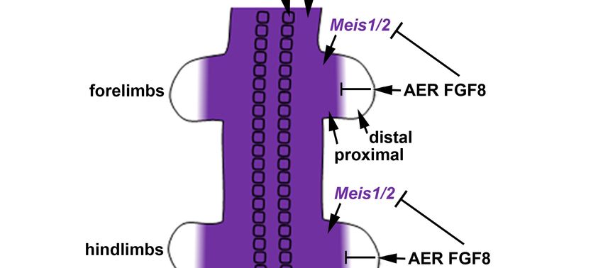
biomolecules 2020, 17, x. https://doi.org/10.3390/biomleculesxxxxx 2 of 11
Biomolecules 2021, 11, 80 2 of 10
2. Requirement of Retinoic Acid for Forelimb Bud Initiation
2. Requirement of Retinoic Acid for Forelimb Bud Initiation
2.1.
2.1. Mechanism
Mechanism of of Retinoic
Retinoic Acid
Acid (RA)
(RA) Signaling
Signaling
During
During early development when limb
early development when limb buds
buds are
are forming,
forming, RA RA is
is generated
generated by bysequen-
sequen-
tial
tial metabolism of retinol (vitamin A) to retinaldehyde by RDH10 [15], followed by
metabolism of retinol (vitamin A) to retinaldehyde by RDH10 [15], followed by the
the
metabolism of retinaldehyde to RA by ALDH1A2 [16]; as Rdh10
metabolism of retinaldehyde to RA by ALDH1A2 [16]; as Rdh10 and Aldh1a2 are expressed and Aldh1a2 are ex-
pressed in the trunk but not the proximal limb, RA enters the limb by
in the trunk but not the proximal limb, RA enters the limb by diffusion from the trunk diffusion from the
trunk
(Figure(Figure
1A). RA1A).canRAbe can be degraded
degraded by CYP26
by CYP26 enzymes
enzymes encoded
encoded by Cyp26a1,
by Cyp26a1, Cyp26b1,
Cyp26b1, or
or Cyp26c1
Cyp26c1 [17];
[17]; Cyp26b1
Cyp26b1 is expressed
is expressed in distal
in distal limblimb
afterafter outgrowth
outgrowth has begun
has begun (Figure
(Figure 1A).
1A). RA controls
RA controls gene gene expression
expression at the at the transcriptional
transcriptional level bylevel by serving
serving as a for
as a ligand ligand for
nuclear
nuclear RA receptors (RARs) that form a heterodimer complex with
RA receptors (RARs) that form a heterodimer complex with retinoid X receptor (RXR) retinoid X receptor
(RXR)
when when
boundbound
to an to RAanresponse
RA response element
element (RARE)(RARE)
[18].[18]. Binding
Binding of RAof RA induces
induces a con-
a confor-
formational
mational shiftshift
in in
RARRAR whichalters
which altersbinding
bindingofofcoactivators
coactivatorsthatthatstimulate
stimulate deposition
deposition of
histone
histone H3
H3 K27
K27 acetylation
acetylation (H3K27ac)
(H3K27ac) marks
marks associated
associated with gene activation, or binding
of
of corepressors
corepressors that
that stimulate
stimulate deposition
deposition of of histone
histone H3 K27 trimethylation (H3K27me3)
(H3K27me3)
marks
marks associated
associated with
with gene repression [19,20] (Figure 1B,C).
Figure
Figure 1.
1. Generation
Generationof ofretinoic
retinoicacid
acid(RA)
(RA)andandmolecular
molecularmechanism
mechanism used
usedbyby
RA RA to to
control
controltranscriptional activation
transcriptional or
activation
repression of target genes. (A) RA is synthesized by sequential conversion of retinol (vitamin A) to retinaldehyde by retinol
or repression of target genes. (A) RA is synthesized by sequential conversion of retinol (vitamin A) to retinaldehyde
dehydrogenase 10 (RDH10), followed by metabolism of retinaldehyde to RA. RA can be degraded by CYP26 enzymes.
by retinol dehydrogenase 10 (RDH10), followed by metabolism of retinaldehyde to RA. RA can be degraded by CYP26
(B,C) The heterodimer formed by nuclear RA receptors (RARs) complexed with retinoid X receptors (RXRs) binds noncod-
enzymes.
ing DNA (B,C) The heterodimer
sequences formed by
called RA response nuclear (RAREs).
elements RA receptors (RARs)
Binding of complexed
RA to RARwith retinoid
creates X receptors changes
conformational (RXRs) binds
that
noncoding DNA sequences called RA response elements (RAREs). Binding of RA to RAR creates
alter the recruitment of nuclear receptor coactivators (NCOAs) or nuclear receptor corepressors (NCORs) leading conformational changes
to a
that alter
change in the
the recruitment
appearance of of epigenetic
nuclear receptor
marks coactivators
on histone H3,(NCOAs) or 27
i.e., lysine nuclear receptor
acetylation corepressors
(K27ac) (NCORs)
associated with geneleading
acti-
to a change
vation in the27appearance
or lysine of epigenetic
trimethylation (K27me3)marks on histone
associated H3, i.e.,
with gene lysine 27 In
repression. acetylation
the presence(K27ac) associated
of RA, with gene
RARE enhancers
stimulate
activationrecruitment
or lysine 27 of NCOAs that(K27me3)
trimethylation result in aassociated
nearby increase
with gene in H3K27ac
repression.and/or
In thedecrease
presenceinofH3K27me3
RA, RARE leading
enhancersto
increased target gene expression
stimulate recruitment of NCOAs such as Meis1
that result in aand Meis2
nearby in embryonic
increase in H3K27actrunkand/or
tissue (B). In theinpresence
decrease H3K27me3 of RA, RARE
leading to
silencers
increasedstimulate recruitment
target gene expressionof NCORs
such as that
Meis1result
and in a nearby
Meis2 decrease in
in embryonic H3K27ac
trunk tissueand/or
(B). Inincrease in H3K27me3
the presence lead-
of RA, RARE
ing to decreased target gene expression such as Fgf8 in embryonic trunk tissue (C).
silencers stimulate recruitment of NCORs that result in a nearby decrease in H3K27ac and/or increase in H3K27me3 leading
to decreased target gene expression such as Fgf8 in embryonic trunk tissue (C).
2.2. Function of RA during Forelimb Budding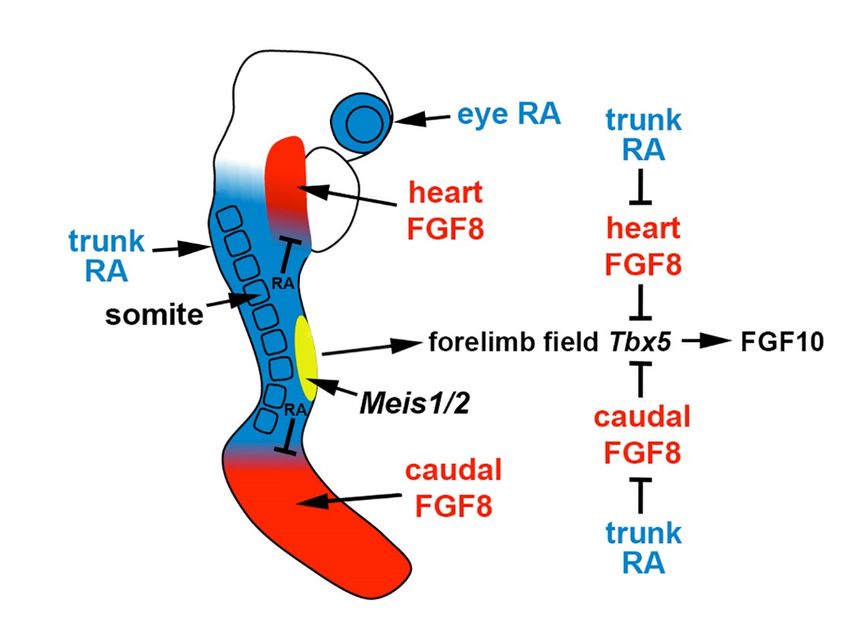
Biomolecules 2021, 11, 80                                                                                          3 of 10

                            2.2. Function of RA during Forelimb Budding
                                  The mechanism through which RA signaling regulates limb development has been
                            controversial as chick limbs treated with RA or RAR antagonists exhibit altered proximodis-
                            tal patterning [10–12], whereas genetic loss of Aldh1a2 or Rdh10 in mice does not alter limb
                            proximodistal patterning [8,13,14]. However, genetic loss of Aldh1a2 or Rdh10 does disrupt
                            initiation of forelimb development in mice or zebrafish, although hindlimb development
                            is not affected [8,13,15,21,22]. Mouse and zebrafish Aldh1a2 mutants lack expression of
                            Tbx5 in trunk lateral plate mesoderm that forms the forelimb field. As Tbx5 is essential for
                            forelimb initiation [5–7] via stimulation of epithelial-to-mesenchymal transition [23] and
                            activation of Fgf10 [2], RA functions upstream of these important regulators of forelimb
                            bud initiation. Other studies that support a function for RA in forelimb bud initiation
                            include the treatment of chick embryos with the RA synthesis inhibitor disulfiram prior
                            to forelimb bud (wing bud) establishment that prevents forelimb bud initiation [24], plus
                            vitamin A-deficient rat embryos that exhibit forelimb hypoplasia [25] similar to Rdh10
                            knockout mice [15]. In addition, in chicken embryos treated with beads containing an
                            RAR antagonist, a phenotype comparable to mouse Rdh10 mutant stunted forelimbs can
                            be observed that leads to a shorter humerus [26].
                                  Comparison of the forelimb fields of mouse Aldh1a2 and Rdh10 mutants revealed that
                            RA activity, normally present throughout the trunk anteroposterior axis, is required to
                            restrict Fgf8 expression to an anterior trunk domain in the heart and a posterior domain
                            (caudal epiblast or tailbud) on either side of the forelimb field. Rdh10 mutants exhibit
                            stunted forelimbs and lose early RA activity in the heart and forelimb domains needed
                            to set the boundary of heart Fgf8 expression, but RA activity still exists caudally which
                            functions to repress caudal Fgf8 and set its caudal expression boundary; this results in an
                            ectopic Fgf8 expression domain that stretches posteriorly from the heart into the forelimb
                            field, resulting in a forelimb field Tbx5 expression domain that is delayed and shortened
                            along the anteroposterior axis [14]. Aldh1a2-/- embryos lack RA activity throughout the
                            entire trunk, resulting in ectopic Fgf8 expression that enters the forelimb field from both
                            the heart and the caudal regions, leading to complete loss of forelimb field Tbx5 expression
                            and no appearance of forelimb buds [8]. These findings, plus the observation that cultured
                            wild-type mouse embryos treated with FGF8 fail to activate Tbx5 in the forelimb field [14],
                            demonstrate that the underlying cause of forelimb bud initiation defects in mutants lack-
                            ing RA synthesis is excessive FGF8 activity in the trunk leading to a disruption of Tbx5
                            activation in the forelimb field (Figure 2). This model, in which RA functions permissively
                            to allow forelimb Tbx5 activation by repressing Fgf8, is also supported by the observation
                            that forelimb bud initiation can be rescued in aldh1a2 mutant zebrafish by introducing a
                            heat-shock-inducible dominant-negative fibroblast growth factor (FGF) receptor [14].
                                  Studies on chick embryos treated with RA combined with mouse enhancer reporter
                            transgene studies suggested that RA signaling may also function instructively to directly
                            activate forelimb Tbx5 via a potential forelimb enhancer located in intron 2 that contains
                            several HOX-binding sites and a RARE [27,28]; however, this was a degenerate RARE with
                            several base pair mismatches to the RARE consensus sequence [18]. When this potential
                            enhancer (including the degenerate RARE) was deleted in mice using CRISPR/Cas9 gene
                            editing, there was no effect on forelimb bud initiation or later forelimb development [29].
                            These deletion studies also demonstrated that enhancer reporter transgenes may not always
                            identify endogenous enhancers, indicating that genetic deletion studies are also needed [30].
                            The challenge of finding limb enhancer regions near Tbx5 with a functional role in vivo
                            is still ongoing and these studies could allow a better understanding of the underlying
                            mechanism of Tbx5 activation. With regard to instructive mechanisms, as genetic studies
                            in zebrafish demonstrate that loss of forelimb bud initiation in RA-deficient embryos
                            can be fully rescued by elimination of excess FGF signaling [14], this finding provides
                            evidence that there is no requirement for RA to function instructively to activate forelimb
                            Tbx5 expression, only a permissive role. Future studies that identify an RA-independent
Biomolecules 2021, 11, 80 4 of 10
biomolecules 2020, 17, x. https://doi.org/10.3390/biomleculesxxxxx 4 of 11
enhancer required in vivo to activate Tbx5 during forelimb initiation would argue in favor
of this hypothesis.
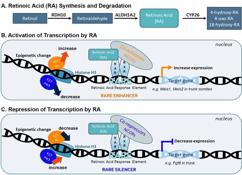
Figure 2. Role of RA signaling, fibroblast growth factor (FGF) signaling, and Meis1/2 genes in forelimb
Figureinitiation.
2. Role of RA In signaling,
the E8.5 fibroblast growth factor
mouse embryo, (FGF) signaling,
the forelimb and Meis1/2
field (yellow) genes
resides in fore-
within an RA-rich trunk
limb initiation. In the E8.5 mouse embryo, the forelimb field (yellow) resides within an RA-rich
domain (blue) where the RA-generating enzymes encoded by Rdh10 and Aldh1a2 are expressed,
trunk domain (blue) where the RA-generating enzymes encoded by Rdh10 and Aldh1a2 are ex-
thus
pressed, positioned
thus positioned between twodomains
between two domains of of FGF8
FGF8 signaling
signaling inheart
in the the heart and caudal
and caudal progeni- progenitors (red).
Trunk
tors (red). RARA
Trunk signaling
signaling repressesFgf8
represses Fgf8 at
at the bordersofofthese
the borders these
twotwo domains
domains to permit
to permit activation of Tbx5
activa-
tion ofin
Tbx5
theinforelimb
the forelimb field,
field, whichthen
which then activates
activates FGF10
FGF10 signaling to stimulate
signaling limb out-
to stimulate limb outgrowth. Thus,
growth. Thus, RA acts permissively to activate forelimb Tbx5 expression. Genetic studies in
RA acts permissively to activate forelimb Tbx5 expression. Genetic studies in zebrafish showing
zebrafish showing that loss of forelimb bud initiation in RA-deficient embryos can be fully rescued
that loss
by reducing FGFof forelimb
signaling bud initiation
provides evidence in RA-deficient
that embryos
RA is not required can beinstructively
to function fully rescued to by reducing FGF
signaling
activate provides
forelimb Tbx5 evidence
expression. thatgenes
Meis1/2 RA isarenot required
required to function
for forelimb instructively
bud initiation, but itto activate forelimb
remains unclear
Tbx5 if they activate
expression. Meis1/2forelimb Tbx5required
genes are or function
forin another bud
forelimb manner.
initiation, but it remains unclear if they
activate forelimb Tbx5 or function in another manner.
Studies on chick embryos treated with RA combined with mouse enhancer reporter
transgene studies
Overall, suggested that RA relying
chick studies signalingonmay also function
treatment withinstructively to directly
signaling agents such as RA may
activate
not reveal normal functions of RA signaling. Similarly, treatment of contains
forelimb Tbx5 via a potential forelimb enhancer located in intron 2 that chick interlimb trunk
several HOX-binding sites and a RARE [27,28]; however, this was a degenerate RARE
(between the forelimb and hindlimb buds) with FGF8 stimulates limb bud initiation [31,32],
with several base pair mismatches to the RARE consensus sequence [18]. When this po-
but it is clear to all in the field from mouse Fgf8 genetic loss-of-function studies that FGF8
tential enhancer (including the degenerate RARE) was deleted in mice using CRISPR/Cas9
is not required
gene editing, there wasfor no limb
effectbud initiation
on forelimb budbut insteadorfor
initiation limb
later outgrowth
forelimb and patterning [3,4];
development
Fgf8 is
[29]. These not expressed
deletion studies alsoin demonstrated
trunk lateral plate mesoderm
that enhancer undergoing
reporter transgenes limb
maybud
not initiation and
alwaysis identify
only expressed
endogenous in the limb after
enhancers, a substantial
indicating bud has
that genetic already
deletion grown.
studies are also
needed [30]. The challenge of finding limb enhancer regions near Tbx5 with a functional
role in2.3. Mechanism
vivo through
is still ongoing andWhich
these RA Represses
studies Fgf8 ina the
could allow Developing
better Trunk of the
understanding
underlyingStudies
mechanism of Tbx5
on the activation.underlying
mechanism With regardRA to instructive
repressionmechanisms,
of trunk Fgf8 as ge-
have shown that
netic studies
Fgf8 is in zebrafish
a direct demonstrate
target that lossthrough
of RA signaling of forelimban bud initiation
upstream RAREin RA-deficient
silencer that was shown
embryos can
to be be fully for
required rescued by elimination
repression of caudalofFgf8
excess FGF signaling
expression in vivo[14], this by
either finding
transgene analysis
provides evidence that there is no requirement for RA to function instructively
or CRISPR/Cas9 deletion of the native RARE [33,34]. ChIP studies on Aldh1a2 knockout to activate
forelimb Tbx5 expression, only a permissive role. Future studies that identify an RA-inde-
embryos suggest that RA-mediated Fgf8 repression caudally involves RA-dependent re-
pendent enhancer required in vivo to activate Tbx5 during forelimb initiation would argue
cruitment of nuclear receptor corepressors (NCOR1 and NCOR2), polycomb repressive
in favor of this hypothesis.
complex-2
Overall, chick(PRC2)
studiesand histone
relying deacetylase-1
on treatment (HDAC1)
with signaling to the
agents such Fgf8 RARE
as RA mayplus
not nearby depo-
reveal normal functions of RA signaling. Similarly, treatment of chick interlimb trunk (be- in trunk cells
sition of the repressive H3K27me3 mark [33,34]. In addition, RA signaling
tweenstimulates
the forelimbmovement
and hindlimb of the Fgf8
buds) chromosomal
with FGF8 stimulates region
limbto budtheinitiation
nuclear[31,32],
periphery, which is
associated with gene repression [35].
Recently, ChIP-seq (H3K27ac and H3K27me3) analysis combined with RNA-seq
analysis performed on wild-type vs. Aldh1a2-/- trunk tissue found that the previously
known RARE upstream of Fgf8 is normally marked by H3K27me3, but this mark is lostBiomolecules 2021, 11, 80 5 of 10
in the RA-deficient mutant [36]. In this study, two additional RAREs near Fgf8 were
also found to be marked by H3K27me3 when RA is present, highlighting the existence
of additional potential Fgf8 RARE silencers that can be further studied to pursue the
mechanism underlying RA repression of Fgf8 in heart tissues compared to caudal tissues
already examined [33,34].
2.4. Loss of Trunk RA Signaling Does Not Affect Hindlimb Bud Initiation
Hindlimb bud initiation is not perturbed by a loss of trunk RA activity in Aldh1a2
and Rdh10 mutants [13,15]. Hindlimb buds do not express Tbx5, but instead use Tbx4 and
Pitx1 to initiate expression of Fgf10 to stimulate outgrowth [9,37,38]. Expression of Tbx4
and Pitx1 is unaffected in the hindlimbs of mouse Aldh1a2 and Rdh10 mutants lacking RA
activity [8,13].
3. Requirement of Meis Genes for Forelimb Bud Initiation
After the Drosophila Meis homolog was shown to be required for proximodistal pat-
terning of fly limbs [39], subsequent studies on vertebrate Meis1 and Meis2 demonstrated
that both are expressed throughout the trunk mesoderm (including lateral plate mesoderm
that gives rise to limb mesoderm) and in the proximal region of both forelimb and hindlimb
buds, suggesting a role in limb proximodistal patterning [10]. Studies on chick limbs
showed that treatment with RA or RAR antagonists alters proximal-specific expression
of Meis1/2, with RA expanding expression into the distal domain, and RAR antagonists
eliminating proximal expression [10–12]. However, genetic loss of RA signaling in mouse
does not alter proximal-specific expression of Meis1/2 despite loss of all RA signaling
activity in the trunk and limb mesoderm [8,13,14]. In addition, no change in Meis2 ex-
pression in the interdigital tissues of the mouse was found in Rdh10 mutants lacking limb
RA signaling [40]. Prior to forelimb bud initiation, RA is also not essential for Meis1/2
expression in trunk lateral plate mesoderm that gives rise to limb buds, but loss of RA does
reduce Meis1/2 expression in somites [14,36].
In order to globally identify RA target genes in the trunk just prior to forelimb bud
initiation, studies were performed that combined epigenetic ChIP-seq (H3K27ac and
H3K27me3) with RNA-seq to compare E8.5 wild-type vs Aldh1a2-/- trunks lacking RA
synthesis. Candidate targets were defined as genes with RA-regulated mRNA abundance
that also have nearby RA-regulated H3K27ac (gene activation) or H3K27me3 (gene repres-
sion) marks associated with conserved RAREs. This approach identified many previously
known RA target genes that control trunk formation, plus additional RA target genes were
identified including Meis1 and Meis2 [36]. Meis1/2 were both found to have nearby RAREs
that are highly conserved from mammals to reptiles or frogs [36]. Together with previous
findings, these new findings suggest that Meis1/2 are normally activated by RA in the trunk
somitic mesoderm but not in the lateral plate mesoderm or limbs, and that the effects of RA
and RAR antagonist treatments on Meis1/2 expression in limb are due to off-target effects in
which high concentrations of these reagents target Meis1/2 RAREs in limb tissue to usurp
control of these genes and override their normal limb activators and repressors.
Genetic studies in mice have been performed to address Meis1/2 function during limb
development. The Meis1 knockout is lethal at E11.5 due to hematopoietic defects, and the
Meis2 knockout is lethal at E14.5 with defects in cardiac and cranial neural crest, but in
both cases no defects in body axis or limb development were observed [41,42]. However,
redundancy between Meis1 and Meis2 may have masked a defect. CRISPR/Cas9 gene
editing was performed to generate Meis1/2 double mutants in mice, and dissection at E10.5
resulted in embryos that exhibit a body axis defect (small somites) with developmental
arrest at E9.5, plus a lack of forelimb buds which are normally easily visible at E9.5;
arrested development prevented analysis of hindlimb buds which develop after forelimb
buds [36]. Thus, global knockout of Meis1/2 unexpectedly resulted in the loss of forelimb
bud initiation, which indicates an important role in limb development, in addition to a role
in somitogenesis (Figure 2).Biomolecules 2021, 11, 80 6 of 10
4. Meis Genes and FGF Signaling Are Required for Limb Proximodistal Patterning but
RA Signaling Is Dispensable
4.1. Chick Studies Support a Two-Signal Model for Limb Proximodistal Patterning
Early studies on chick limbs treated with RA suggested a role for RA in limb antero-
posterior patterning [43]. However, subsequent studies in chicks provided evidence that
RA is not required for limb anteroposterior patterning [44,45], but that sonic hedgehog
(SHH) is the diffusible signaling factor required for limb anteroposterior patterning [46];
this requirement for SHH was confirmed in mouse knockout studies [47]. Other studies
in chicks demonstrated that treatment of limb buds with RA, RAR antagonists, FGF8, or
FGF receptor antagonists alters proximodistal patterning [10–12]. Mouse genetic loss-of-
function studies verified a requirement for Fgf8 (and other Fgf genes) expressed distally in
the apical ectodermal ridge (AER) to control limb proximodistal patterning, including re-
striction of Meis1/2 expression to the proximal limb [48]. However, loss of RA signaling by
knockout of RA-generating enzymes encoded by Aldh1a2 or Rdh10 did not result in loss of
limb proximodistal patterning or loss of proximal-specific expression of Meis1/2 [8,13,14].
Some researchers supporting the two-signal model raised criticisms, summarized in a
review [49], about whether there is a total absence of RA, as measured using the RARE-lacZ
RA-reporter transgene, in limb mesoderm of E9.5-E10.5 Aldh1a2 mutants (that need to be
treated with a small dose of RA at E7.5 in order to survive to E10.5) or Rdh10 mutants (that
survive to E10.5 without RA treatment). Aldh1a2 mutants treated with a very small dose
of RA at E7.5 were shown to activate RARE-lacZ in the trunk mesoderm at E7.5-E8.5 and
survive until E10.5, but at E9.5-E10.5 when limbs form, RARE-lacZ expression was not
detected in the trunk lateral plate mesoderm or limb buds, indicating that the administered
RA had been efficiently cleared [8]. In addition, RA titration studies on cultured Rdh10
mutants that normally have no expression of RARE-lacZ in limb or lateral plate mesoderm
demonstrated that RARE-lacZ expression can be activated by as low as 0.25 nM RA, 100-fold
lower than the normal level of limb RA, which is 25 nM, demonstrating that RARE-lacZ is
a very sensitive RA-reporter [29]. Thus, as RA is reduced by at least 100-fold in limbs and
lateral plate mesoderm of Aldh1a2 and Rdh10 mutants, it is reasonable to conclude that the
concentration would be too low to provide completely normal patterning if RA is required
as suggested by the two-signal model.
With regard to a potential interaction of RA and FGF8 during limb patterning, loss
of limb RA in Rdh10 mutants does not alter the expression of Fgf8 in the AER [13]. This
observation demonstrates that although RA does repress Fgf8 in the body axis to limit its
expression to the heart and caudal regions [33,34], one should not assume that RA represses
Fgf8 in other tissues.
Models based on the treatment of chick embryos with RA and RAR antagonists are
weakened by the possibility of off-target effects. As RAR antagonists function as inverse-
agonists that silence any gene near an RAR-bound RARE [50], they may dominantly
repress genes that have a RARE nearby even though they normally use different regulatory
elements for activation in a particular tissue. In the case of Meis1/2, the ability of RA
and RAR antagonist treatments to effect expression in limb buds, even though loss of
endogenous RA does not, can be rationalized by the recent discovery that these genes do
require RA for full activation in trunk somites and both genes have functional RAREs [36].
Thus, retinoid treatment regimens may force effects on Meis1/2 expression in tissues that
normally do not use RA to regulate Meis1/2.
Despite these conflicting results, a ‘two-signal model’ has been proposed in which RA
generated in the trunk diffuses into the proximal region of the limb to promote proximal
character by activation of Meis1/2 expression, with distal FGF8 signaling promoting distal
character by repressing Meis1/2 expression [10–12]. This model is also supported by the
observation that an RA-degrading enzyme encoded by Cyp26b1 is expressed in the distal
limb under the control of FGF8, with CYP26B1 being required to prevent trunk RA from
diffusing into the distal limb, which was reported to ectopically activate Meis1/2 expression
distally [51,52]. In the Cyp26b1 knockout, Meis1/2 expression expands into the distal limb,Biomolecules 2021, 11, 80 7 of 10
similar to chick RA-treatment studies, but forelimb and hindlimb buds are truncated
along the entire proximodistal axis, which is inconsistent with RA functioning to induce
proximal identity [51]. In addition, in Cyp26b1 knockouts, the presence of endogenous
RA in distal limbs where it should not normally exist results in a phenotype similar
to exogenous RA teratogenesis with increased apoptosis and a block in chondrogenic
differentiation, particularly to form the intricate skeletal structures of distal tissues such as
hand/foot [53,54] or craniofacial structures [55]. Thus, the presence of endogenous RA in
the proximal limb does not necessarily correlate with a designed function in proximodistal
patterning of Meis1/2 expression, but instead may simply indicate diffusion overflow from
the trunk where RA is required for body axis formation and forelimb initiation, with this RA
being neither necessary nor harmful to proximal limb development. Expression of Cyp26b1
in the distal limb may not indicate a role in restricting RA signaling and sharpening an RA
gradient to set the boundary of Meis1/2 expression, but may simply function to eliminate RA
signaling distally where it is harmful to distal limb development. This would be similar to
the function of the related RA-degrading enzyme CYP26A1 in the caudal body axis which
functions to remove RA that interferes with body axis extension [56,57], thus restricting
RA to the trunk/caudal border where RA is required for spinal cord development (mouse
and zebrafish) and somitogenesis (mouse but not zebrafish) [16].
4.2. Mouse Genetic Studies Support a One-Signal Model for Limb Proximodistal Patterning
We previously suggested that the most parsimonious model for limb proximodistal
patterning is a “one-signal model” that is driven by distal FGFs (including FGF8) that
stimulate outgrowth, repress Meis1/2 to generate proximodistal patterning, and activate
Cyp26b1 to remove distal RA from diffusing in from the trunk that would result in limb
teratogenesis [18]. Recent studies reported that conditional genetic loss of Meis1/2 in the
mouse limb results in proximodistal patterning defects [58]. Additionally, it was reported
that restriction of Meis1/2 expression to the proximal limb is controlled by the repressive
biomolecules 2020, 17, x.action of distal FGFs (including FGF8) expressed in8 ofthe
https://doi.org/10.3390/biomleculesxxxxx 11 AER [58]. As the AER does not
form until after the proximal limb is formed, thus delaying production of distal FGFs until
after limb form untilbud initiation
after the proximal limb is[32],
formed,this results
thus delaying in early
production Meis1/2
of distal FGFs until expression throughout the limb.
after limb bud initiation [32], this results in early Meis1/2 expression throughout the limb.
After After thethe AERAER isis established,
established, Meis1/2
Meis1/2 expression expression
is repressed is creating
by distal FGFs, thus repressed by distal FGFs, thus creating
a boundary of Meis1/2 expression that leaves it in a proximal position as the distal limb
a boundary continues toof growMeis1/2
free of Meis1/2expression that leaves it in a proximal position as the distal limb
expression (Figure 3).
continues to grow free of Meis1/2 expression (Figure 3).
Figure 3. Role of Meis1/2 genes and FGF signaling in limb proximodistal patterning. In the E10.5
Figure 3. Role of Meis1/2 genes and FGF signaling in limb proximodistal patterning. In the E10.5 mouse embryo, Meis1/2
expression that already exists in trunk lateral plate mesoderm (LPM) prior to limb bud initiation extends into the proximal
mouse embryo, Meis1/2 expression that already exists in trunk lateral plate mesoderm (LPM) prior to
regions of both forelimbs and hindlimbs as they undergo outgrowth from the trunk. Meis1/2 expression in the trunk LPM
and proximal limb does not require RA signaling; the factor(s) that activate Meis1/2 expression in trunk LPM and limb are
limb bud initiation extends into the proximal regions of both forelimbs and hindlimbs as they undergo
unknown. A boundary of Meis1/2 expression is formed by distal FGF8 (and other FGFs) secreted by the apical ectodermal
ridge (AER) that represses Meis1/2 to limit expression to a proximal limb domain.
outgrowth from the trunk. Meis1/2 expression in the trunk LPM and proximal limb does not require
5. Conclusions and Perspectives
RA signaling; the factor(s) that activate Meis1/2 expression in trunk LPM and limb are unknown. A
New findings have clarified the mechanisms through which RA signaling, FGF sig-
boundary and Meis1/2
naling, of Meis1/2 genesexpression is formed
control limb development. by
We know fromdistal FGF8
recent mouse (and other FGFs) secreted by the apical
genetic
loss-of-function studies that Meis1/2 are required for limb bud initiation (at least forelimb)
ectodermal
as well asridge (AER)
subsequent that represses
proximodistal Meis1/2
patterning [36,58]. Thus, bytocombining
limit expression
all genetic to a proximal limb domain.
studies it can be established that RA signaling functions permissively during forelimb in-
itiation to allow expression of Tbx5 by repressing trunk Fgf8 that represses limb field Tbx5,
but that RA is not required for limb patterning. Furthermore, genetic studies show that
RA control of Meis1/2 occurs in the trunk for somitogenesis but not in limbs, whereas FGF
signaling is required for both limb initiation (FGF10) and patterning (FGF8 and other dis-
tal FGFs), with distal FGFs restricting Meis1/2 expression to the proximal limb. With re-
gard to limb patterning, these new genetic findings do not support a two-signal RA-FGFBiomolecules 2021, 11, 80 8 of 10
5. Conclusions and Perspectives
New findings have clarified the mechanisms through which RA signaling, FGF signal-
ing, and Meis1/2 genes control limb development. We know from recent mouse genetic
loss-of-function studies that Meis1/2 are required for limb bud initiation (at least forelimb)
as well as subsequent proximodistal patterning [36,58]. Thus, by combining all genetic
studies it can be established that RA signaling functions permissively during forelimb
initiation to allow expression of Tbx5 by repressing trunk Fgf8 that represses limb field Tbx5,
but that RA is not required for limb patterning. Furthermore, genetic studies show that
RA control of Meis1/2 occurs in the trunk for somitogenesis but not in limbs, whereas FGF
signaling is required for both limb initiation (FGF10) and patterning (FGF8 and other distal
FGFs), with distal FGFs restricting Meis1/2 expression to the proximal limb. With regard to
limb patterning, these new genetic findings do not support a two-signal RA-FGF model
for proximodistal patterning, but instead support a one-signal model in which a factor
other than RA activates Meis1/2 expression during early limb bud outgrowth followed
by action of distal FGFs to repress Meis1/2. As Meis1/2 is expressed in the trunk lateral
plate mesoderm prior to limb bud outgrowth from this tissue, and as RA is not essential
for Meis1/2 expression in trunk lateral plate mesoderm, it is possible that whatever factor
activates Meis1/2 in the lateral plate mesoderm continues to allow expression as lateral
plate mesodermal cells undergo epithelial-to-mesenchymal transition to form the limb bud.
A final proof that RA is not required for Meis1/2 activation in the proximal limb would
come from the identification of such a factor.
Determining the normal functions of RA during development is difficult as phar-
macological studies and genetic loss-of-function studies often lead to conflicting results.
Similarly, pharmacological studies using treatment with FGF8 led to erroneous conclusions
about FGF8 function in limb bud initiation that were later reversed by mouse genetic stud-
ies. Thus, a greater reliance on genetic studies will provide clarity on the developmental
pathways controlled by RA signaling, FGF signaling, and Meis1/2.
Author Contributions: M.B. and G.D. drafted the manuscript, designed the figures. All authors have
read and agreed to the published version of the manuscript.
Funding: This work was funded by the National Institutes of Health (National Institute of Arthritis
and Musculoskeletal and Skin Diseases) grant R01 AR067731 (G.D.).
Acknowledgments: This work was supported by the Sanford Burnham Prebys Medical
Discovery Institute.
Conflicts of Interest: The authors declare that they have no conflict of interest with the contents of
this article.
References
1. Rabinowitz, A.H.; Vokes, S.A. Integration of the transcriptional networks regulating limb morphogenesis. Dev. Biol. 2012, 368,
165–180. [CrossRef] [PubMed]
2. Sekine, K.; Ohuchi, H.; Fujiwara, M.; Yamasaki, M.; Yoshizawa, T.; Sato, T.; Yagishita, N.; Matsui, D.; Koga, Y.; Itoh, N.; et al. Fgf10
is essential for limb and lung formation. Nat. Genet. 1999, 21, 138–141. [CrossRef] [PubMed]
3. Moon, A.M.; Capecchi, M.R. Fgf8 is required for outgrowth and patterning of the limbs. Nat. Genet. 2000, 26, 455–459.
[CrossRef] [PubMed]
4. Lewandoski, M.; Sun, X.; Martin, G.R. Fgf8 signalling from the AER is essential for normal limb development. Nat. Genet. 2000,
26, 460–463. [CrossRef] [PubMed]
5. Ahn, D.-G.; Kourakis, M.J.; Rohde, L.A.; Silver, L.M.; Ho, R.K. T-box gene Tbx5 is essential for formation of the pectoral limb bud.
Nature 2002, 417, 754–758. [CrossRef] [PubMed]
6. Agarwal, P.; Wylie, J.N.; Galceran, J.; Arkhitko, O.; Li, C.; Deng, C.; Grosschedl, R.; Bruneau, B.G. Tbx5 is essential for forelimb
bud initiation following patterning of the limb field in the mouse embryo. Development 2003, 130, 623–633. [CrossRef]
7. Rallis, C.; Bruneau, B.G.; Del Buono, J.; Seidman, C.E.; Seidman, J.G.; Nissim, S.; Tabin, C.J.; Logan, M.P.O. Tbx5 is required for
forelimb bud formation and continued outgrowth. Development 2003, 130, 2741–2751. [CrossRef]
8. Zhao, X.; Sirbu, I.O.; Mic, F.A.; Molotkova, N.; Molotkov, A.; Kumar, S.; Duester, G. Retinoic acid promotes limb induction
through effects on body axis extension but is unnecessary for limb patterning. Curr. Biol. 2009, 19, 1050–1057. [CrossRef]
9. Logan, M.; Tabin, C.J. Role of Pitx1 upstream of Tbx4 in specification of hindlimb identity. Science 1999, 283, 1736–1739. [CrossRef]Biomolecules 2021, 11, 80 9 of 10
10. Mercader, N.; Leonardo, E.; Piedra, M.E.; Martínez-A, C.; Ros, M.A.; Torres, M. Opposing RA and FGF signals control proxi-
modistal vertebrate limb development through regulation of Meis genes. Development 2000, 127, 3961–3970.
11. Cooper, K.L.; Hu, J.K.; ten Berge, D.; Fernandez-Teran, M.; Ros, M.A.; Tabin, C.J. Initiation of proximal-distal patterning in the
vertebrate limb by signals and growth. Science 2011, 332, 1083–1086. [CrossRef] [PubMed]
12. Rosello-Diez, A.; Ros, M.A.; Torres, M. Diffusible signals, not autonomous mechanisms, determine the main proximodistal limb
subdivision. Science 2011, 332, 1086–1088. [CrossRef] [PubMed]
13. Cunningham, T.J.; Chatzi, C.; Sandell, L.L.; Trainor, P.A.; Duester, G. Rdh10 mutants deficient in limb field retinoic acid signaling
exhibit normal limb patterning but display interdigital webbing. Dev. Dyn. 2011, 240, 1142–1150. [CrossRef] [PubMed]
14. Cunningham, T.J.; Zhao, X.; Sandell, L.L.; Evans, S.M.; Trainor, P.A.; Duester, G. Antagonism between retinoic acid and fibroblast
growth factor signaling during limb development. Cell Rep. 2013, 3, 1503–1511. [CrossRef]
15. Sandell, L.L.; Sanderson, B.W.; Moiseyev, G.; Johnson, T.; Mushegian, A.; Young, K.; Rey, J.P.; Ma, J.X.; Staehling-Hampton,
K.; Trainor, P.A. RDH10 is essential for synthesis of embryonic retinoic acid and is required for limb, craniofacial, and organ
development. Genes Dev. 2007, 21, 1113–1124. [CrossRef]
16. Ghyselinck, N.B.; Duester, G. Retinoic acid signaling pathways. Development 2019, 146, dev167502. [CrossRef]
17. Pennimpede, T.; Cameron, D.A.; MacLean, G.A.; Li, H.; Abu-Abed, S.; Petkovich, M. The role of CYP26 enzymes in defining
appropriate retinoic acid exposure during embryogenesis. Birth Defects Res. 2010, 88, 883–894. [CrossRef]
18. Cunningham, T.J.; Duester, G. Mechanisms of retinoic acid signalling and its roles in organ and limb development. Nat. Rev. Mol.
Cell Biol. 2015, 16, 110–123. [CrossRef]
19. Rada-Iglesias, A.; Bajpai, R.; Swigut, T.; Brugmann, S.A.; Flynn, R.A.; Wysocka, J. A unique chromatin signature uncovers early
developmental enhancers in humans. Nature 2011, 470, 279–283. [CrossRef]
20. Laugesen, A.; Helin, K. Chromatin repressive complexes in stem cells, development, and cancer. Cell Stem Cell 2014, 14,
735–751. [CrossRef]
21. Grandel, H.; Lun, K.; Rauch, G.J.; Rhinn, M.; Piotrowski, T.; Houart, C.; Sordino, P.; Küchler, A.M.; Schulte-Merker, S.; Geisler, R.;
et al. Retinoic acid signalling in the zebrafish embryo is necessary during pre-segmentation stages to pattern the anterior-posterior
axis of the CNS and to induce a pectoral fin bud. Development 2002, 129, 2851–2865. [PubMed]
22. Begemann, G.; Schilling, T.F.; Rauch, G.J.; Geisler, R.; Ingham, P.W. The zebrafish neckless mutation reveals a requirement for
raldh2 in mesodermal signals that pattern the hindbrain. Development 2001, 128, 3081–3094. [PubMed]
23. Gros, J.; Tabin, C.J. Vertebrate limb bud formation is initiated by localized epithelial-to-mesenchymal transition. Science 2014, 343,
1253–1256. [CrossRef] [PubMed]
24. Stratford, T.; Horton, C.; Maden, M. Retinoic acid is required for the initiation of outgrowth in the chick limb bud. Curr. Biol.
1996, 6, 1124–1133. [CrossRef]
25. White, J.C.; Shankar, V.N.; Highland, M.; Epstein, M.L.; DeLuca, P.F.; Clagett-Dame, M. Defects in embryonic hindbrain
development and fetal resorption resulting from vitamin A deficiency in the rat are prevented by feeding pharmacological levels
of all-trans-retinoic acid. Proc. Natl. Acad. Sci. USA 1998, 95, 13459–13464. [CrossRef]
26. Koyama, E.; Golden, E.B.; Kirsch, T.; Adams, S.L.; Chandraratna, R.A.S.; Michaille, J.J.; Pacifici, M. Retinoid signaling is
required for chondrocyte maturation and endochondral bone formation during limb skeletogenesis. Dev. Biol. 1999, 208,
375–391. [CrossRef]
27. Minguillon, C.; Nishimoto, S.; Wood, S.; Vendrell, E.; Gibson-Brown, J.J.; Logan, M.P. Hox genes regulate the onset of Tbx5
expression in the forelimb. Development 2012, 139, 3180–3188. [CrossRef]
28. Nishimoto, S.; Wilde, S.M.; Wood, S.; Logan, M.P. RA acts in a Coherent Feed-Forward Mechanism with Tbx5 to Control Limb
Bud Induction and Initiation. Cell Rep. 2015, 12, 879–891. [CrossRef]
29. Cunningham, T.J.; Lancman, J.J.; Berenguer, M.; Dong, P.D.S.; Duester, G. Genomic knockout of two presumed forelimb Tbx5
enhancers reveals they are nonessential for limb development. Cell Rep. 2018, 23, 3146–3151. [CrossRef]
30. Duester, G. Knocking out enhancers to enhance epigenetic research. Trends Genet. 2019, 35, 89. [CrossRef]
31. Cohn, M.J.; Izpisúa-Belmonte, J.C.; Abud, H.; Heath, J.K.; Tickle, C. Fibroblast growth factors induce additional limb development
from the flank of chick embryos. Cell 1995, 80, 739–746. [CrossRef]
32. Crossley, P.H.; Minowada, G.; MacArthur, C.A.; Martin, G.R. Roles for FGF8 in the induction, initiation, and maintenance of chick
limb development. Cell 1996, 84, 127–136. [CrossRef]
33. Kumar, S.; Duester, G. Retinoic acid controls body axis extension by directly repressing Fgf8 transcription. Development 2014, 141,
2972–2977. [CrossRef] [PubMed]
34. Kumar, S.; Cunningham, T.J.; Duester, G. Nuclear receptor corepressors Ncor1 and Ncor2 (Smrt) are required for retinoic
acid-dependent repression of Fgf8 during somitogenesis. Dev. Biol. 2016, 418, 204–215. [CrossRef] [PubMed]
35. Patel, N.S.; Rhinn, M.; Semprich, C.I.; Halley, P.A.; Dolle, P.; Bickmore, W.A.; Storey, K.G. FGF Signalling Regulates Chromatin
Organisation during Neural Differentiation via Mechanisms that Can Be Uncoupled from Transcription. PLoS Genet. 2013, 9,
e1003614. [CrossRef] [PubMed]
36. Berenguer, M.; Meyer, K.F.; Yin, J.; Duester, G. Discovery of genes required for body axis and limb formation by global
identification of retinoic acid-regulated epigenetic marks. PLoS Biol. 2020, 18, e3000719. [CrossRef] [PubMed]
37. Naiche, L.A.; Papaioannou, V.E. Loss of Tbx4 blocks hindlimb development and affects vascularization and fusion of the allantois.
Development 2003, 130, 2681–2693. [CrossRef]Biomolecules 2021, 11, 80 10 of 10
38. Kawakami, Y.; Marti, M.; Kawakami, H.; Itou, J.; Quach, T.; Johnson, A.; Sahara, S.; O’Leary, D.D.; Nakagawa, Y.; Lewandoski, M.;
et al. Islet1-mediated activation of the beta-catenin pathway is necessary for hindlimb initiation in mice. Development 2011, 138,
4465–4473. [CrossRef]
39. Mercader, N.; Leonardo, E.; Azpiazu, N.; Serrano, A.; Morata, G.; Martinez, C.; Torres, M. Conserved regulation of proximodistal
limb axis development by Meis/Hth. Nature 1999, 402, 425–429. [CrossRef]
40. Mason, M.K.; Hockman, D.; Curry, L.; Cunningham, T.J.; Duester, G.; Logan, M.; Jacobs, D.S.; Illing, N. Retinoic acid-independent
expression of Meis2 during autopod patterning in the developing bat and mouse limb. EvoDevo 2015, 6, 6. [CrossRef]
41. Hisa, T.; Spence, S.E.; Rachel, R.A.; Fujita, M.; Nakamura, T.; Ward, J.M.; Devor-Henneman, D.E.; Saiki, Y.; Kutsuna, H.; Tessarollo,
L.; et al. Hematopoietic, angiogenic and eye defects in Meis1 mutant animals. EMBO J. 2004, 23, 450–459. [CrossRef] [PubMed]
42. Machon, O.; Masek, J.; Machonova, O.; Krauss, S.; Kozmik, Z. Meis2 is essential for cranial and cardiac neural crest development.
BMC Dev. Biol. 2015, 15, 40. [CrossRef] [PubMed]
43. Tickle, C.; Alberts, B.M.; Wolpert, L.; Lee, J. Local application of retinoic acid to the limb bud mimics the action of the polarizing
region. Nature 1982, 296, 564–565. [CrossRef] [PubMed]
44. Wanek, N.; Gardiner, D.M.; Muneoka, K.; Bryant, S.V. Conversion by retinoic acid of anterior cells into ZPA cells in the chick
wing bud. Nature 1991, 350, 81–83. [CrossRef]
45. Noji, S.; Nohno, T.; Koyama, E.; Muto, K.; Ohyama, K.; Aoki, Y.; Tamura, K.; Ohsugi, K.; Ide, H.; Taniguchi, S.; et al. Retinoic acid
induces polarizing activity but is unlikely to be a morphogen in the chick limb bud. Nature 1991, 350, 83–86. [CrossRef]
46. Riddle, R.D.; Johnson, R.L.; Laufer, E.; Tabin, C. Sonic hedgehog mediates the polarizing activity of the ZPA. Cell 1993, 75,
1401–1416. [CrossRef]
47. te Welscher, P.; Fernandez-Teran, M.; Ros, M.A.; Zeller, R. Mutual genetic antagonism involving GLI3 and dHAND prepatterns
the vertebrate limb bud mesenchyme prior to SHH signaling. Genes Dev. 2002, 16, 421–426. [CrossRef] [PubMed]
48. Mariani, F.V.; Ahn, C.P.; Martin, G.R. Genetic evidence that FGFs have an instructive role in limb proximal-distal patterning.
Nature 2008, 453, 401–405. [CrossRef]
49. Lewandoski, M.; Mackem, S. Limb development: The rise and fall of retinoic acid. Curr. Biol. 2009, 19, R558–R561.
[CrossRef] [PubMed]
50. Germain, P.; Iyer, J.; Zechel, C.; Gronemeyer, H. Co-regulator recruitment and the mechanism of retinoic acid receptor synergy.
Nature 2002, 415, 187–192. [CrossRef]
51. Yashiro, K.; Zhao, X.; Uehara, M.; Yamashita, K.; Nishijima, M.; Nishino, J.; Saijoh, Y.; Sakai, Y.; Hamada, H. Regulation of
retinoic acid distribution is required for proximodistal patterning and outgrowth of the developing limb. Dev. Cell 2004, 6,
411–422. [CrossRef]
52. Probst, S.; Kraemer, C.; Demougin, P.; Sheth, R.; Martin, G.R.; Shiratori, H.; Hamada, H.; Iber, D.; Zeller, R.; Zuniga, A. SHH
propagates distal limb bud development by enhancing CYP26B1-mediated retinoic acid clearance via AER-FGF signalling.
Development 2011, 138, 1913–1923. [CrossRef]
53. Pennimpede, T.; Cameron, D.A.; MacLean, G.A.; Petkovich, M. Analysis of Cyp26b1/Rarg compound-null mice reveals two
genetically separable effects of retinoic acid on limb outgrowth. Dev. Biol. 2010, 339, 179–186. [CrossRef] [PubMed]
54. Dranse, H.J.; Sampaio, A.V.; Petkovich, M.; Underhill, T.M. Genetic deletion of Cyp26b1 negatively impacts limb skeletogenesis
by inhibiting chondrogenesis. J. Cell Sci. 2011, 124, 2723–2734. [CrossRef] [PubMed]
55. Berenguer, M.; Darnaudery, M.; Claverol, S.; Bonneu, M.; Lacombe, D.; Rooryck, C. Prenatal retinoic acid exposure reveals
candidate genes for craniofacial disorders. Sci. Rep. 2018, 8, 17492. [CrossRef]
56. Sakai, Y.; Meno, C.; Fujii, H.; Nishino, J.; Shiratori, H.; Saijoh, Y.; Rossant, J.; Hamada, H. The retinoic acid-inactivating enzyme
CYP26 is essential for establishing an uneven distribution of retinoic acid along the anterio-posterior axis within the mouse
embryo. Genes Dev. 2001, 15, 213–225. [CrossRef] [PubMed]
57. Abu-Abed, S.; Dollé, P.; Metzger, D.; Beckett, B.; Chambon, P.; Petkovich, M. The retinoic acid-metabolizing enzyme, CYP26A1,
is essential for normal hindbrain patterning, vertebral identity, and development of posterior structures. Genes Dev. 2001, 15,
226–240. [CrossRef] [PubMed]
58. Delgado, I.; Lopez-Delgado, A.C.; Rosello-Diez, A.; Giovinazzo, G.; Cadenas, V.; Fernandez-de-Manuel, L.; Sanchez-Cabo, F.;
Anderson, M.J.; Lewandoski, M.; Torres, M. Proximo-distal positional information encoded by an Fgf-regulated gradient of
homeodomain transcription factors in the vertebrate limb. Sci. Adv. 2020, 6, eaaz0742. [CrossRef] [PubMed]You can also read