Scrub Typhus: Historic Perspective and Current Status of the Worldwide Presence of Orientia Species - MDPI
←
→
Page content transcription
If your browser does not render page correctly, please read the page content below
Tropical Medicine and
            Infectious Disease

Review
Scrub Typhus: Historic Perspective and Current
Status of the Worldwide Presence of Orientia Species
Allen L. Richards 1, *        and Ju Jiang 2
 1    Department of Preventive Medicine and Biostatistics, Uniformed Services University of the Health Sciences,
      Bethesda, MD 20814, USA
 2    Henry M. Jackson Foundation for the Advancement of Military Medicine, Bethesda, MD 20817, USA;
      jjiang@HJFresearch.org
 *    Correspondence: Allen.Richards@comcast.net

 Received: 1 March 2020; Accepted: 25 March 2020; Published: 1 April 2020

 Abstract: Scrub typhus and its etiological agents, Orientia species, have been around for a very long
 time. Historical reference to the rickettsial disease scrub typhus was first described in China (313 AD)
 by Hong Ge in a clinical manual (Zhouhofang) and in Japan (1810 AD) when Hakuju Hashimoto
 described tsutsuga, a noxious harmful disease in the Niigata prefecture. Other clinicians and scientists
 in Indonesia, Philippines, Taiwan, Australia, Vietnam, Malaysia, and India reported on diseases most
 likely to have been scrub typhus in the early 1900s. All of these initial reports about scrub typhus
 were from an area later designated as the Tsutsugamushi Triangle—an area encompassing Pakistan
 to the northwest, Japan to the northeast and northern Australia to the south. It was not until the 21st
 century that endemic scrub typhus occurring outside of the Tsutsugamushi Triangle was considered
 acceptable. This report describes the early history of scrub typhus, its distribution in and outside the
 Tsutsugamushi Triangle, and current knowledge of the causative agents, Orientia species.

 Keywords: scrub typhus; Orientia species; Tsutsugamushi Triangle

1. Early History of Scrub Typhus and the Etiologic Agents of the Tsutsugamushi Triangle

1.1. Scrub Typhus Disease Presentation and Diagnosis
      Scrub typhus, a febrile disease with mild to life-threatening manifestations, is characterized by
rapid onset of fever, headache, chills, arthralgias and myalgias and often the presentation of eschar
prior to and a macularpapular rash following initiation of disease [1–6]. The illness lasts approximately
3 weeks and ends without sequalae. Rapid response (24–72 h) to antibiotic treatment with tetracyclines,
chloramphenicol, and azithromycin is characteristic and diagnostic [2,3,5,6]. In fatal cases, the disease
is characterized by multi-organ failure, with pathologic lesions in lungs, kidneys, liver, and brain [3,5].
      The lack of scrub typhus-specific signs and symptoms makes the clinical diagnosis very
difficult [2,3,5]. Moreover, laboratory diagnosis at the time of illness is also very difficult, as antibodies
do not reach detectable levels for 5–10 days after disease presentation, and the level of orientiae in the
blood stream demonstrable by molecular methods only reaches detectable levels sporadically during
acute illness and is unapparent after initial treatment with appropriate antibiotic treatment [7]. The
specimen of choice, biopsy of eschar and/or rash, is unfortunately rarely obtained, though the level of
Orientia DNA is in abundance, unaffected by prior antibiotic treatment, and maintained in the lesion
for the life of the lesion [7].

Trop. Med. Infect. Dis. 2020, 5, 49; doi:10.3390/tropicalmed5020049            www.mdpi.com/journal/tropicalmed
Trop. Med. Infect. Dis. 2020, 5, 49 2 of 16
1.2. Early History of Scrub Typhus
China
Historically, scrub typhus has been around for a very long time. Human historical reference to
scrub typhus (Table 1) was first described in China’s Zhouhofang, a clinical manual, in 313 AD [8].
Subsequently, in 610, Yuan-Fang Chao described the epidemiology, clinical course, and treatment of the
disease in a poem, which is considered medically accurate [8] and Shi-Zhen Li, a well-known physician,
described the characteristics of the disease in a book entitled, “Ben Cao, Gang Mu” in 1596 [8]. In 1908,
Ashburn and Craig reported that in China, “shashitsu,” the name for scrub typhus, occurred in old
Chinese writings of more than a thousand years (Table 2). The authors also indicated that the Chinese
recognized the disease as a distinct illness, and it was attributed to the bite of a mite which occurred in
summer in certain districts that had been flooded by spring rains [1]. The red lice (sna ra) or mites had
been associated with illness characterized by fever and a pustule at the site of injury. Moreover, they
recognized that three days after the bite, high fever developed, and a pustule appeared at the site of
the injury [1]. Clearly, the Chinese were well aware of scrub typhus (shashitsu) and for an extensive
period of time.
Table 1. Early History of Scrub Typhus.
Year Location Book or Individual
313 China Zhouhofang by Hong Ge
610 China Yuan-Fang Chao
1596 China Ben Cao Gang Mu by Shi-Zhen Li
1810 Japan Hakuju Hashimoto
1902 Indonesia Wilhelm Schüffner
1908 Philippines P.M. Ashburn and C.F. Craig
1908 Taiwan J. Hatori
1910 Australia O. Smithson
1915 Vietnam F. Noc and P. Gautron
1915 Malaysia A. Kawamura Jr., H. Tanaka, A. Tamura
1932 India C.R. Christian
Table 2. Synonyms for Scrub Typhus.
Synonyms Country
Shashitsu China
Tsutsugamushi Japan
Kedani disease Japan
Japanese river fever Japan
Flood fever Japan
Island fever Japan
Akamushi disease Japan
Shimamushi disease Japan
Pseudotyphoid Indonesia
Chigger-borne rickettsiosis Ubiquitous
Mite-borne typhus Ubiquitous
Mite fever Ubiquitous
Rural or “K” form of tropical typhus Malaysia
Fiévre exanthématique avec ulcére primaire French Indo-China
Indian mite typhus India
Mijtekoorts Indonesia
Mossman fever Australia
Sarina fever Australia
Tropical typhus Malaysia
Rural typhus French Indo-ChinaTrop. Med. Infect. Dis. 2020, 5, 49 3 of 16
Japan
In Japan, in 1810, Hakuju Hashimoto described “tsutsuga”, a noxious harmful disease in the
Niigata prefecture of the main island of Japan [9]. However, according to Tanaka, as described in
Ashburn and Craig [1], the name tsutsugamushi (tsutsuga = disease/illness and mushi (bug/insect)) has
been around since the earliest historical times in Japan. Though reports of tsutsugamushi disease, from
the northwest coast of the main island, Nippon (the two prefectures, Akito and Echigo, later changed
to three prefectures, Akito, Yamagata and Niigata), and research associated with it was published
in Japanese medical and science journals, it was not until Theobald Palm in 1878 [10] and Bälz and
Kawakami in 1879 [11] that tsutsugamushi disease from Japan was reported in European journals.
The Etiology of Scrub Typhus in Japan
Up until the 1920s, the etiology of tsutsugamushi and, therefore, scrub typhus was unknown. In
addition, multiple diseases which were later believed to be synonymous with tsutsugamushi were
known as Japanese river fever, flood fever, island fever, Kedani (mite) disease, akamushi disease,
shimamushi disease yochubio, and shashitsu [1] (Table 2). During the 1920s, the laboratories of Hayashi,
Nagayo, Ogata, and Kawamura were working to discover the causative agent of tsutsugamushi. At
this time, it was postulated that spirits, noxious air, parasites, bacteria, and viruses were possible causes
of tsutsugamushi. In 1920, Hayashi indicated that the causative agent was a protozoan and named
the agent Theileria tsutsugamushi [12]. However, by 1924, Hayashi indicated that the agent was not a
protozoan, but most likely a rickettsia [13], as described by Kawamura et al. [8]. Hayashi, however,
did not give his new agent a binomial name [8]. In the meantime, Nagayo demonstrated the causative
agent of scrub typhus could be maintained in human and dog macrophages and that the agent could
be passed to and cause disease in monkeys by intradermal and intracutaneous inoculations [14],
as described by Kawamura [8]. In 1929, Ogata and Unno used a rabbit intratesticular inoculation
technique to obtain the tsutsugamushi agent from a human blood sample and passed it to other
rabbits (testis) [15]. This was the first time that the scrub typhus agent was isolated from a human
following inoculation of the patient’s blood into rabbit testis and, subsequently, transferring the agent
from that rabbit into another rabbit, showing the ability of the causative agent to be isolated and
transferred [8]. Unfortunately, Ogata and Unno did not provide a binomial name for the agent in their
report. However, they did provide the agent and the methodology to both Nagayo and Kawamura
laboratories [8]. Ogata, subsequently, developed a new rabbit model for tsutsugamushi. This entailed
infecting the anterior chamber of rabbits’ eyes. This proved to be a far more sensitive method of
infection and, subsequently, Nagayo used this tsutsugamushi model in his laboratory [8]. Moreover,
Nagayo utilized the agent from Ogata and the inoculation of the anterior chamber of rabbit eyes to
grow a large number of rickettsiae. These organisms could, subsequently, be maintained in rabbit
Descemet’s membrane cell cultures. The results and the proposed name for the agent of tsutsugamushi,
Rickettsia orientalis, was reported in 1930 in the Japanese Journal of Experimental Medicine [16], as
described by Kawamura et al. [8]. Ogata and Kawamura, also utilizing Ogata’s agent, reported on
the etiology of tsutsugamushi in the German journal Zentralblatt für Bakteriologie in the same issue
in 1931, naming the agent as Rickettsia tsutsugamushi and Rickettsia akamushi, respectively [17,18], as
described by Kawamura et al. [8] (Table 3). In 1932, Hayashi concluded that his agent was the same
as Ogata’s R. tsutsugamushi and Nagayo’s R. orientalis [19]. Further, also in 1932, Ogata reported a
new laboratory animal model for tsutsugamushi—the intraperitoneal (IP) inoculation of mice for the
growth of R. tsutsugamushi [20], as described by Kawamura et al. [8]. This is a scrub typhus laboratory
animal model that is still used today [21]. In the 6th edition of Bergey’s manual, the name of the agent
for scrub typhus was reported as O. tsutsugamushi. Though much controversy was associated with
this name [8], it was not completely resolved until 1995 when R. tsutsugamushi was moved out of the
genus Rickettsia and into its own genus Orientia, with the new species name, Orientia tsutsugamushi [22]
(Table 3).Trop. Med. Infect. Dis. 2020, 5, 49 4 of 16
Table 3. Previous and Current Names of the Scrub Typhus Agent from the Tsutsugamushi Triangle.
Previous Agent Names Country Reference
Theileria tsutsugamushi Japan [12]
Rickettsia orientalis Japan [16]
Rickettsia tsutsugamushi Japan [17]
Rickettsia akamushi Japan [18]
Orientia tsutsugamushi Japan [22]
Indonesia
During the early 1900s, other clinicians and scientists in Asia, Australia, and Islands of the Indian
and Pacific Oceans reported on local diseases most likely to have been scrub typhus (Table 2). Dr.
Schüffner of Deli, Sumatra, Indonesia, described a disease, pseudotyphoid, that resembled scrub typhus
as early as 1902 [23]. Later, he indicated that this disease was similar to Kedani fever (later determined
to be scrub typhus) in Japan [24]. Subsequently, scrub typhus was discovered to be endemic for many
islands throughout the Indonesian archipelago [25–30].
Taiwan
In 1908, Japanese clinicians reported that eastern Taiwan had a febrile disease with a rash that was
reported as a tsutsugamushi disease-like ailment and was confirmed in 1914 to be a tsutsugamushi
disease but with a lower fatality rate (approximately 3%), which was significantly different to the high
mortality rate (~20–40%) seen with tsutsugamushi in Japan at this time [31]. Scrub typhus continues to
be associated with the main island of Taiwan [32,33] as well as the highly endemic Pescadores Islands
of the South China Sea [34–36].
The Philippines
Further, also in 1908, two cases of tsutsugamushi disease were described for two US military
personnel, stationed at Camp Connell, Samar, the Philippines, based upon clinical records [1]. Ashburn
had just returned from Japan where he had seen many cases of tsutsugamushi that Japanese clinicians
had shown him prior to reviewing the clinical records for these two cases and reporting about the
cases and tsutsugamushi disease [1]. During the repatriation of the Philippines in WWII, 284 cases of
scrub typhus occurred in the month of November, 1944 [37,38]. One of the isolates from a US soldier
deployed to the Guinan region of Samar Island, Volner strain, was used to develop a lyophilized rat
lung-spleen vaccine. This was the first US scrub typhus vaccine ever tested in a field trail, that was
unfortunately unsuccessful [39]. Subsequently, studies showed evidence of scrub typhus throughout
the Philippines [37,40].
Australia
In 1910, Mossman fever, later described as endemic glandular fever and, subsequently, determined
to be scrub typhus, was reported in North Queensland, Australia [41–44]. The endemic region of scrub
typhus in Australia now includes Queensland [45,46], the islands of the Torres Strait [46], the Northern
Territory [47], and western Australia [48,49].
Vietnam
In 1915, a fever of unknown etiology among two individuals was reported in Saigon, Vietnam [50]
to be a disease similar to that described in Deli, Sumatra (i.e., pseudotyphoid), that was later
determined to be scrub typhus [24]. Subsequently, the presence of scrub typhus throughout Vietnam
was confirmed [38,51–57].Trop. Med. Infect. Dis. 2020, 5, 49 5 of 16
Korea
In 1915, a “mild” rickettsial disease called paratyphus was reported among 15 patients (no deaths)
in the spring of 1913–1914 from Jemulpo, Incheon, Korea by Weir, a medical missionary [58]. Because
only mild disease presentations with no deaths were associated with paratyphus, it was thought not to
be epidemic typhus, which was endemic for Korea at the time. Moreover, the disease only developed
during March–June and, in retrospect, it was assumed not to be murine typhus (seen year round and
most commonly in the fall) or tsutsugamushi disease (seen in summer and fall). Thus, Chung and
Kang believe it could have been Brill–Zinsser disease and not scrub typhus [59].
During the period of 1910–1945, there was some evidence of endemic scrub typhus in Korea. A
study of mites attached to wild rats collected in Suwon were similar to Trombicula akamshi. In addition,
rickettsial diseases with mild presentations and low OX19 titers may not have been murine typhus but
scrub typhus [59]. During Japanese occupation, the Japanese physicians considered tsutsugamushi
disease a severe disease with a 15–60% mortality rate. Thus, they may have overlooked a milder
form of scrub typhus in Korea. In addition, the Weil–Felix test with OXK was not often utilized [59].
Following 1945, evidence increased for the presence of scrub typhus in Korea [59] and consequently the
endemicity of scrub typhus became obvious by detection of infected mites, rodents and humans [60–63].
In recent years, there has been an ever-increasing number of scrub typhus cases reported in South
Korea (2637 cases in 2001 to 10,485 cases in 2013) [59,64].
Malaysia
In 1900, the Institute of Medical Research (IMR) in Kuala Lumpur, Malaysia, was established as
the Pathological Institute with the aim to promote the health status of the local population. In 1924,
the institute began research on “tropical typhus” in the Federated Malay States. In the annual IMR
Bulletin in 1925, Fletcher and Lesslar described tropical typhus as containing two components—an
urban or shop typhus and a rural or scrub typhus [65].
Due to the fortuitous change in the composition of the Weil–Felix test, “tropical typhus” could be
divided into two unique diseases. The Weil–Felix test initially utilized the strain of Bacillus proteus X.19
(Proteus vulgaris) that was isolated from the urine of a patient with epidemic typhus [66]. The P. vulgaris
agent was not the cause of the disease but was found to be agglutinated by antibodies developed
during epidemic typhus. The cross-reactivity of the antibodies to the P. vulgaris antigens (OX19) has
been successfully used since 1916 to serologically diagnose epidemic typhus and murine typhus.
Subsequently, another strain of P. vulgaris (OX2) was identified that reacted with the sera of spotted
fever patients [7]. These patients also reacted to the OX19 to varying degrees. A third agglutinin (OXK)
was, subsequently, identified in 1926 [67]. It was the Proteus mirabilis Kingsbury strain which reacted
with sera from scrub typhus patients but not with sera from typhus or spotted fever patients [68,69].
With that new development, Fletcher and Lessar concluded that the rural tropical typhus or scrub
typhus was the same as or similar to tsutsugamushi and unique from urban or shop typhus and
other rickettsial diseases [70,71]. Moreover, it was determined that the urban or shop typhus form of
tropical typhus was clinically the same as Brill’s disease and had the same Weil–Felix results. This
disease was later referred to as murine typhus and the causative agent identified as Rickettsia typhi [72].
Throughout the subsequent history of the IMR scrub typhus, research continued with major advances
in O. tsutsugamushi isolations, diagnostics, treatment/prophylaxis, vaccine and immunology research,
and vector and ecology research [38]. Scrub typhus research is not limited to the IMR as indicated by
recent publications [73–78].
India, Burma, Ceylon, and the Maldives
In 1932, Christian reported OXK-positive typhus cases that he believed were due to tick bites [79].
Due to the serologies conducted, they were most likely the first cases of scrub typhus reported from
India. Similarly, in 1934, scrub typhus (OXK+) was reported among personnel from Simla Hills,Trop. Med. Infect. Dis. 2020, 5, 49 6 of 16
India [80]. An investigation by Mehta reported the presence of Trombicula deliensis on rodents and
shrews in the Simla Hills, suggesting that similar to the reports from Malaya, that these mites may
Trop. Med. Infect. Dis. 2020, 5, x FOR PEER REVIEW 6 of 16
be the vectors of scrub typhus [81]. Boyd reported on the presence of typhus among 110 cases in
1935, utilizing
positive cases inclinical presentations
India presented with and Weil–Felix
eschars OXK aserologies
[83]. In 1944, report of two[82].outbreaks
Interestingly, none
of scrub of the
typhus,
OXK-positive cases in India presented with eschars [83]. In 1944, a report
which occurred during the period of 1937–1938 (n = 11) and 1939–1942 (n = 30), that were confirmed of two outbreaks of scrub
typhus,
by whichserology
Weil–Felix occurredalso during the period
indicated of 1937–1938
no presence (n = 11)
of eschars [84].and
One 1939–1942 = 30), 110
case from(nBoyd’s that cases
were
confirmed by Weil–Felix serology also indicated no presence of eschars
was an individual who was OXK+ from Burma [82]. Subsequently, a study by Maitra and Sen Gupta [84]. One case from Boyd’s 110
cases was
showed theanpresence
individual who was
of scrub OXK+
typhus andfrom
murineBurma [82].(OXK
typhus Subsequently,
and OX19a positive,
study by respectively)
Maitra and Sen in
Gupta showed the presence of scrub typhus and murine typhus (OXK
Burma [85]. The famous prototype, O. tsutsugamushi Gilliam, was contracted by Dr. Gilliam and OX19 positive, respectively)
in 1944
in Burma
on [85]. The
the equally famous
famous Stillwell Road,O.
prototype, intsutsugamushi
Burma [67]. Gilliam,
Nicholls was contracted
reported by Dr. Gilliam
OXK-positive cases of in
1944 on the equally
tsutsugamushi (ruralfamous
typhus)Stillwell
in nearby Road,
Ceylon in Burma [67]. Nicholls
[86]. Similarly, reported
in Maldives, OXK-positive
outbreaks of scrubcases
typhus of
tsutsugamushi (rural typhus) in nearby Ceylon [86]. Similarly, in Maldives,
among British troops struck during the period of 1941–1944 [87]. Scrub typhus outbreaks occurred outbreaks of scrub typhus
amonginBritish
again troopsofstruck
the period during
2002–2003 the period
among of 1941–1944
the inhabitants of [87]. Scrub typhus
the Maldives, outbreaks
indicating the occurred
endemic
again inofthe
nature period
this of 2002–2003
disease [88]. This among the inhabitants
is certainly the case for of the Maldives,
India, where indicating
numerous the endemic nature
publications have
of this disease
shown the breadth [88]. of
This
scrubis certainly the case forthe
typhus throughout India, where numerous
subcontinent [89–93].publications have shown the
breadth of scrub typhus throughout the subcontinent [89–93].
1.3. The Tsutsugamushi Triangle
1.3. The Tsutsugamushi Triangle
All of these reports of scrub typhus or diseases very similar to them throughout the Asia–Pacific
All of these reports of scrub typhus or diseases very similar to them throughout the Asia–Pacific
region prior to WWII led to the assumption of a single rickettsial disease for a very large endemic
region prior to WWII led to the assumption of a single rickettsial disease for a very large endemic
region where many people were at risk of disease. Unfortunately, this assumption of a very large
region where many people were at risk of disease. Unfortunately, this assumption of a very large
endemic area of scrub typhus was reinforced during WWII, where approximately 18,000 cases
endemic area of scrub typhus was reinforced during WWII, where approximately 18,000 cases occurred
occurred among the allied forces and a similar number among the Japanese forces in the islands of
among the allied forces and a similar number among the Japanese forces in the islands of Ceylon,
Ceylon, Maldives, New Britain, Goodenough, and the Schouten Islands, and in the countries of
Maldives, New Britain, Goodenough, and the Schouten Islands, and in the countries of China, Thailand,
China, Thailand, Japan, Australia, Lao, Cambodia, Vietnam, and Taiwan [25,37,38,83]. Contemporary
Japan, Australia, Lao, Cambodia, Vietnam, and Taiwan [25,37,38,83]. Contemporary reviews indicated
reviews indicated the extent of scrub typhus distribution throughout the Tsutsugamushi Triangle
the extent of scrub typhus distribution throughout the Tsutsugamushi Triangle [4,6,83,94–98], which
[4,6,83,94–98], which included countries in the west (Pakistan, Afghanistan, Tajikistan, Nepal, India,
included countries in the west (Pakistan, Afghanistan, Tajikistan, Nepal, India, Bangladesh, Sri Lanka,
Bangladesh, Sri Lanka, and Maldives), northeast (China, Russia, Republic of Korea, Japan, and
and Maldives), northeast (China, Russia, Republic of Korea, Japan, and Taiwan), south (Australia,
Taiwan), south (Australia, Papua New Guinea, Indonesia, and the islands of the southwestern
Papua New Guinea, Indonesia, and the islands of the southwestern Pacific), and middle (Myanmar,
Pacific), and middle (Myanmar, Thailand, Laos, Cambodia, Malaysia, Vietnam, and Philippines)
Thailand, Laos, Cambodia, Malaysia, Vietnam, and Philippines) (Figure 1).
(Figure 1).
Tsutsugamushi Triangle
Figure 1.
Figure 1. The
TheTsutsugamushi
TsutsugamushiTriangle: Geographical
Triangle: Distribution
Geographical of Scrub
Distribution Typhus
of Scrub Typhus by Orientia
CausedCaused by
tsutsugamushi.
Orientia tsutsugamushi.
Consistent within the Tsutsugamushi Triangle has been the presence of a single species of
Orientia which was identified within human cases, vector mites and mammalian hosts [8,83,94,98].
This species, O. tsutsugamushi, has a diversity of antigenic phenotypes and genetic genotypes found
not only between countries but within countries [94,99,100]. However, as early as 1951, reports
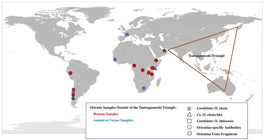
suggesting that scrub typhus occurred outside of the Tsutsugamushi Triangle began to emergeTrop. Med. Infect. Dis. 2020, 5, 49 7 of 16
Consistent within the Tsutsugamushi Triangle has been the presence of a single species of Orientia
which was identified within human cases, vector mites and mammalian hosts [8,83,94,98]. This species,
O. tsutsugamushi, has a diversity of antigenic phenotypes and genetic genotypes found not only between
countries but Dis.
Trop. Med. Infect. within
2020,countries [94,99,100].
5, x FOR PEER REVIEW However, as early as 1951, reports suggesting that 7scrub
of 16
typhus occurred outside of the Tsutsugamushi Triangle began to emerge (Figure 2).
Figure 2.
Figure 2. Geographical Distribution of
Geographical Distribution of Orientia
Orientia spp.
spp. and
and the
the Scrub
Scrub Typhus:
Typhus: A Worldwide Disease.
A Worldwide Disease.
2. Scrub Typhus Outside
Outside the
the Tsutsugamushi
TsutsugamushiTriangle
Triangle
2.1. Case Investigations
2.1. Case Investigations
Africa
Africa
In 1951, Giroud and Jadin published a report that indicated that scrub typhus occurred outside the
In 1951, Giroud and Jadin published a report that indicated that scrub typhus occurred outside
Tsutsugamushi Triangle. This report described an outbreak of a febrile disease among native Africans
the Tsutsugamushi Triangle. This report described an outbreak of a febrile disease among native
from Ruanda-Urundi working on constructing a factory building in Musha Hill, Belgian Congo (now
Africans from Ruanda-Urundi working on constructing a factory building in Musha Hill, Belgian
Rwanda and Burundi) [101]. To investigate the outbreak, the authors utilized tests/reagents available
Congo (now Rwanda and Burundi) [101]. To investigate the outbreak, the authors utilized
at the time to determine whether the illness was due to rickettsiae or coxiella infections. Due to the
tests/reagents available at the time to determine whether the illness was due to rickettsiae or coxiella
existence of R. orientalis (O. tsutsugamushi) antigens, the authors included scrub typhus in the panel of
infections. Due to the existence of R. orientalis (O. tsutsugamushi) antigens, the authors included scrub
rickettsial diseases to assess. Among the ill Africans, several were positive for the various rickettsial
typhus in the panel of rickettsial diseases to assess. Among the ill Africans, several were positive for
antigens, including two individuals who reacted to the R. orientalis antigens. To confirm the skin
the various rickettsial antigens, including two individuals who reacted to the R. orientalis antigens.
tests, blood from the two Orientia-positive patients were tested for complement-fixing antibodies to
To confirm the skin tests, blood from the two Orientia-positive patients were tested for complement-
O. tsutsugamushi and were found to be positive, with titers of 80 and 320. To assess the reactivity to
fixing antibodies to O. tsutsugamushi and were found to be positive, with titers of 80 and 320. To
the scrub typhus assays in other populations who lived closely with native Africans in Musha Hill,
assess the reactivity to the scrub typhus assays in other populations who lived closely with native
healthy individuals, including nine people born in Muscat, Oman, five born in Bombay, India, and two
Africans in Musha Hill, healthy individuals, including nine people born in Muscat, Oman, five born
born in Africa, with parents who were born in Bombay, were tested using the same skin and blood
in Bombay, India, and two born in Africa, with parents who were born in Bombay, were tested using
tests for evidence of previous O. tsutsugamushi infection. The authors considered Muscat and Bombay
the same skin and blood tests for evidence of previous O. tsutsugamushi infection. The authors
as scrub typhus-endemic regions and thought that people from those areas may be antibody positive
considered Muscat and Bombay as scrub typhus-endemic regions and thought that people from those
to O. tsutsugamushi and may, therefore, act as positive controls. Of the nine individuals, originally from
areas may be antibody positive to O. tsutsugamushi and may, therefore, act as positive controls. Of
Muscat, three had strong, weak, and negative skin reactivity and antibodies against O. tsutsugamushi
the nine individuals, originally from Muscat, three had strong, weak, and negative skin reactivity
were detected with titers of 1280 (four individuals) and 640 (three individuals) in eight. Of the five
and antibodies against O. tsutsugamushi were detected with titers of 1280 (four individuals) and 640
people born in Bombay, three displayed positive skin reactivity to O. tsutsugamushi antigens and,
(three individuals) in eight. Of the five people born in Bombay, three displayed positive skin
interestingly, the two people whose parents were from Bombay, but who were born in and never
reactivity to O. tsutsugamushi antigens and, interestingly, the two people whose parents were from
Bombay, but who were born in and never traveled outside of eastern Africa, were also positive. These
results suggested the presence of scrub typhus in eastern Africa. The authors indicated that a similar
study they conducted among natives in western Africa showed that they were negative for evidence
of scrub typhus [101].
In the 1990s, three case reports insinuated, but could not confirm, the presence of scrub typhusTrop. Med. Infect. Dis. 2020, 5, 49 8 of 16
traveled outside of eastern Africa, were also positive. These results suggested the presence of scrub
typhus in eastern Africa. The authors indicated that a similar study they conducted among natives in
western Africa showed that they were negative for evidence of scrub typhus [101].
In the 1990s, three case reports insinuated, but could not confirm, the presence of scrub typhus in
Africa. The first described an individual from Japan visiting the Republic of Congo, who presented
with fever six days after his return from Africa [102]. The disease was identified as scrub typhus,
though the possibility could not be ruled out that the patient had contracted scrub typhus in Japan,
within the Tsutsugamushi Triangle, during the six days prior to disease presentation. The second case
involved a US missionary who visited Cameroon [103]. Within two weeks of his visit, the missionary
noted a lesion on his leg and he had a fever and a rash three days later. Two weeks later, the missionary
returned to the US and was, subsequently, admitted to a hospital in which he was treated for a
rickettsial disease. He recovered within 24 h with doxycycline treatment and he had a four-fold rise in
titer from 256 to 1024 of antibodies against O. tsutsugamushi. Unfortunately, no molecular or culture
evidence confirmed the case of scrub typhus. The third case was of an individual who had visited
Tanzania [104]. She had noted a lesion on her right foot and had a three-day history of fever and
headache after returning to the Netherlands. Her acute and convalescent sera showed a seroconversion
against O. tsutsugamushi antigens fromChile
Subsequently, in 2011, a report of a scrub typhus case in Chile appeared that proved the existence
of scrub typhus worldwide [106].
Unfortunately, an isolate was not recovered, though the limited molecular characterization of
theInfect.
Trop. Med. Orientia DNA
Dis. 2020, indicated, like the Ca. O. chuto, that this Orientia was not O. tsutsugamushi
5, 49 9 of 16or for
that matter Ca. O. chuto [106] (Figure 3).
Figure 3. Phylogenetic Tree. The evolutionary relationships of Orientia species detected outside of the
Figure 3. Triangle
Tsutsugamushi Phylogenetic
were Tree. The evolutionary
compared with Orientiarelationships
tsutsugamushiofstrains,
Orientia speciesspecies
Rickettsia detected
andoutside
other of the
bacteria. The tree was constructed with the 560 bp rrs gene fragments using the Maximum Likelihood and
Tsutsugamushi Triangle were compared with Orientia tsutsugamushi strains, Rickettsia species
methodother
and bacteria. The tree
the Tamura–Nei was in
model constructed
MEGAX. Thewithvalues
the 560 bp bootstrap
of the rrs gene fragments using the were
test (1000 replicates) Maximum
shownLikelihood
next to the method and the Tamura–Nei model in MEGAX. The values of the bootstrap test (1000
branches.
replicates) were shown next to the branches.
Table 5. Current Known and Proposed Agents of Scrub Typhus.
2.2. Serological Evidence of Scrub
AgentTyphus Outside the Tsutsugamushi
Location Triangle
Orientia tsutsugamushi Tsutsugamushi Triangle
Candidatus O. chuto United Arab Emirates
Ca. O. chuto-like Kenya
Candidatus O. chiloensis Chile
Chile
Subsequently, in 2011, a report of a scrub typhus case in Chile appeared that proved the existence
of scrub typhus worldwide [106].
Unfortunately, an isolate was not recovered, though the limited molecular characterization of the
Orientia DNA indicated, like the Ca. O. chuto, that this Orientia was not O. tsutsugamushi or for that
matter Ca. O. chuto [106] (Figure 3).Trop. Med. Infect. Dis. 2020, 5, 49 10 of 16
2.2. Serological Evidence of Scrub Typhus Outside the Tsutsugamushi Triangle
Following these two astounding reports, investigators looked to confirm and expand upon these
results, utilizing current serological and molecular assays for evidence of Orientia spp. infections outside
of the Tsutsugamushi Triangle. Serological assays previously used for detecting rickettsial diseases
outside of the Tsutsugamushi Triangle did not include those for scrub typhus group orientiae (STGO).
Similarly, molecular assays for orientiae were not used to investigate the presence in mammalian
hosts and/or arthropod vectors of orientiae outside of the Tsutsugamushi Triangle. This paradigm
was changed with the two reports of scrub typhus in the United Arab Emirates and Chile. Thus,
scientists included scrub typhus serological and molecular assays to conduct surveillance studies
of rickettsial diseases in areas of Africa, South America, and Europe (Table 4). This resulted in the
substantiation of scrub typhus outside of the Tsutsugamushi Triangle. In 2015, the first of these reports
was of a seroprevalence study of fever patients from hospitals in Kenya. It was determined that 70
of 1401 (5%) patients had antibodies against O. tsutsugamushi that was confirmed by Western blot
assays [107]. In an unrelated investigation conducted in western Kenya among sick children, paired
acute and convalescent serum samples were tested, and it was determined that 15 of 281 patients
(5.8%) had antibodies against O. tsutsugamushi ELISA antigens and 10 of these children seroconverted
to O. tsutsugamushi antigens (3.6%). The seroreactivity was confirmed by Western blot analysis [108].
In Djibouti, a 20 week serosurvey of abattoir workers was conducted to determine their exposure to
infectious disease agents. From multiple serum samples, it was ascertained that 3 of 49 workers had
antibodies against O. tsutsugamushi ELISA antigens and one individual who reported a history of a
febrile disease during the period of the study seroconverted to O. tsutsugamushi antigens by ELISA,
IFA and Western blot tests [109].
2.3. Molecular Evidence of Scrub Typhus Outside the Tsutsugamushi Triangle
In addition to the serological data, molecular evidence for the presence of orientiae in Africa was
conveyed in three separate reports (Figure 3). In 2015, Cosson et al. reported the presence of Orientia
DNA among tissues of rodents from West Africa and Europe [110]. In South Africa, DNA preparation
from the blood of a healthy dog from Mpumalanga Province had a 16S rRNA sequence that was 96.1%
(247/257 bp) similar to that of Orientia spp. [111]. In East Africa, a rodent survey was conducted in a
village where individuals who resided there had tested positive for antibodies against O. tsutusgamushi.
Trombiculid mites were collected from the trapped rodents to assess them for molecular evidence of
Orientia. DNA preparations provided evidence of Orientia species from sequences of gene fragments
of the rrs and htrA that were most closely aligned to but not identical with Ca. O. chuto (Figure 3;
Table 5) [112].
2.4. Endemic Scrub Typhus in South America
Two serological surveys were conducted in South America for rickettsial agents. The first, in
Peru, assessed the role of rickettsial diseases in fever patients in the city of Iquitos on the Amazon
river. It was determined that of 1124 individuals enrolled in the febrile surveillance study, 60 (5.3%)
were seropositive against O. tsutsugamushi ELISA antigens and one person had a four-fold rise in
titer, which suggested that he had scrub typhus. The ELISA results of this sample were confirmed by
IFA [113]. The second survey involved a cross-sectional survey of dogs from Chiloé Island, the initial
scrub typhus focus center in Chile [106,114]. It was revealed that of 202 dogs surveyed, 43 (21.3%) had
immunoglobulin gamma (IgG) antibodies against O. tsutsugamushi antigens, with higher prevalence
levels among dogs from rural areas and older dogs, and it was reported that dogs are a good sentinel
animal for scrub typhus [115].
Since the initial scrub typhus case reported in 2011 [106], additional cases of scrub typhus (n >
40) [116] have been described from Chiloé Island [114,117] and from continental Chile [117,118]. The
agents have been molecularly very much the same (Figure 3) from all cases from Chile [119], exceptTrop. Med. Infect. Dis. 2020, 5, 49 11 of 16
for an imported case from the Republic of Korea, which was determined to be O. tsutsugamushi [120].
This is quite surprising when considering the extreme variation seen among the O. tsutsugamushi
found throughout the Tsutsugamushi Triangle [94,99,100]. Similarly, characterization of orientiae
from trombiculid mites of the genus Herpetacarus from Chiloé Island found the same orientiae as that
associated with scrub typhus cases [121]. Thus, the molecular characterization of the agents both from
human eschar/blood samples and trombiculid mites suggest a new scrub typhus agent, Candidiatus
Orientia chiloensis (Figure 3; Table 5) [119,121].
The conservation of genetic variation in Chile orientiae may be related to the limitations placed
on identifying cases and characterizing the agents—for the most part, utilizing clinical characteristics
to identify cases (e.g., fever, headache, and eschar) and serological and molecular assays based upon O.
tsutsugamushi antigens and sequences. Thus, as we identify more unique cases and develop better
assays that are more sensitive and more generous in recognizing rare antibodies and sequences, with
time, the antigenic and genetic variability of Chile orientiae may be discovered to be greater than
current discoveries.
Author Contributions: Both authors contributed equally to the conceptualization, development and writing of
the manuscript. All authors have read and agree to the published version of the manuscript.
Funding: This research received no external funding.
Acknowledgments: Part of this material was presented at the 2nd Asia Pacific Rickettsia Conference, Chiang Rai,
Thailand, 3–6 November 2019.
Conflicts of Interest: The authors declare no conflict of interest.
References
1. Ashburn, P.M.; Craig, C.F. A comparative study of tsutsugamushi disease and spotted or tick fever of
Montana. Boston Med. Surg. J. 1908, 158, 749–761. [CrossRef]
2. Watt, G.; Kantipong, P. Orientia tsutsugamushi and scrub typhus. In Rickettsial Diseases; Raoult, D., Parola, P.,
Eds.; Informa Healthcare USA, Inc.: New York, NY, USA, 2007; pp. 237–256.
3. Paris, D.H.; Shelite, T.R.; Day, N.P.; Walker, D. Unresolved Problems Related to Scrub Typhus: A Seriously
Neglected Life-Threatening Disease. Am. J. Trop. Med. Hyg. 2013, 89, 301–307. [CrossRef] [PubMed]
4. Kelly, D.J.; Foley, D.; Richards, A.L. A spatiotemporal database to track human scrub typhus using the
VectorMap application. PLoS Neglected Trop. Dis. 2015, 9, e0004161. [CrossRef] [PubMed]
5. Rajapakse, S.; Weeratunga, P.; Sivayoganathan, S.; Fernando, S.D. Clinical manifestations of scrub typhus.
Trans. R. Soc. Trop. Med. Hyg. 2017, 111, 43–54. [CrossRef] [PubMed]
6. Abdad, M.Y.; Abdallah, R.A.; Fournier, P.-E.; Stenos, J.; Vasoo, S. A Concise Review of the Epidemiology
and Diagnostics of Rickettsioses: Rickettsia and Orientia spp. J. Clin. Microbiol. 2018, 56, 1–10. [CrossRef]
[PubMed]
7. Luce-Fedrow, A.; Mullins, K.; Kostik, A.P.; John, H.K.S.; Jiang, J.; Richards, A.L. Strategies for detecting
rickettsiae and diagnosing rickettsial diseases. Future Microbiol. 2015, 10, 537–564. [CrossRef]
8. Kawamura, A., Jr.; Tanaka, H.; Tamura, A. Tsutsugamushi Disease; Univ. Tokyo Press: Tokyo, Japan, 1995.
9. Yoshimoto, T.; Yoshimoto, T. Scrub typhus in Japan. Am. J. Clin. Microbiol. Antimicrobiol. 2019, 2, 1042.
10. Palm, T.A. Some account of a disease called Shima Mushi or Island Insect Diseases by the natives of Japan.
Edin. Med. J. 1878, 24, 128.
11. Bälz, E.; Kawakami. Die Japanische Fluss-oder Ueber-schwemmungsfieber. Virchow’s Arch. 1879, 78, 373.
[CrossRef]
12. Hayashi, N. Etiology of Tsutsugamushi disease. J. Parasitol. 1920, 7, 53–68. [CrossRef]
13. Hayashi, N. On Ricketts’ corpuscles. Trans. Japan. Pathol. Soc. 1924, 22, 569–576. (In Japanese)
14. Nagayo, M.; Miyagawa, Y.; Mitamura, T.; Imamura, A.; Tamiya, T.; Sato, K. On the experimental
Tsutsugamushi disease in monkeys by intracutaneous inoculation of the virus. Sci. Rep. Gov. Inst.
Infect. Dis. 1923, 2, 371–373. (In Japanese)
15. Ogata, N.; Unno, Y. On the transfer of causative agent of Tsutsugamushi disease to the rabbit testis and
microorgansims appearing in the testis. J. Chiba Med. Soc. 1929, 7, 1215–1222. (In Japanese)Trop. Med. Infect. Dis. 2020, 5, 49 12 of 16
16. Nagayo, M.; Tamiya, T.; Mitamura, T.; Sato, K. On the virus of Tsutsugamushi disease and its demonstration
by a new method. Trans. Japan Pathol. Soc. 1930, 20, 556–566.
17. Ogata, N. Aetiology der Tsutsugamushikrankheit: Rickettsia tsutsugamushi. Zbl f Bakt 1931, 122, 249–253.
18. Kawamura, R.; Imagawa, Y. Die Feststellung des Erregers bei der Tsutsugamushikrankheit. Zbl f Bakt 1931,
122, 261.
19. Hayashi, N. On Tsutsugamushi disease. Trans. Japan Pathol. Soc. 1932, 22, 689–690. (In Japanese)
20. Ogata, N.; Nakajima, G.; Kajima, S. Animals employed for experimental infection by pathogenic rickettsiae
in laboratory—Especially recommendation to use mouse for the detection of Rickettsia tsutsugamushi. Tokyo
Med. J. 1932, 2760, 155–160.
21. Luce-Fedrow, A.; Lehman, M.L.; Kelly, D.J.; Mullins, K.; Maina, A.N.; Stewart, R.L.; Ge, H.; John, H.S.;
Jiang, J.; Richards, A.L. A Review of Scrub Typhus (Orientia tsutsugamushi and Related Organisms): Then,
Now, and Tomorrow. Trop. Med. Infect. Dis. 2018, 3, 8. [CrossRef]
22. Tamura, A.; Ohashi, N.; Urakami, H.; Miyamura, S. Classification of Rickettsia tsutsugamushi in a New Genus,
Orientia gen. nov., as Orientia tsutsugamushi comb. nov. Int. J. Syst. Bacteriol. 1995, 45, 589–591. [CrossRef]
23. Schüffner, W. Geneesk. Tijdschr. Med. Indie 1909, 49, 64.
24. Schüffner, W. Pseudotyphoid fever in Deli, Sumatra (a variety of Kedani fever). Philipp. J. Sci. 1915, 10, 345.
25. Logue, J.B. Scrub typhus. Report of epidemic in the Southwest Pacific. US Nav. Med. Bull. 1944, 43, 645–649.
26. Hadi, T.R.; Nalim, S.; Sukaeri, S.; Dennis, D.T. Scrub typhus survey of Biak and Owi islands: ectoparasites
of small mammals and rickettsial isolations. Southeast Asian J. Trop. Med. Public Health 1980, 11, 220–226.
[PubMed]
27. Dennis, D.T.; Hadi, T.R.; Brown, R.J.; Sukaeri, S.; Leksana, B.; Cholid, R. A survey of scrub and murine typhus
in the Ancol section of Jakarta, Indonesia. Southeast Asian J. Trop. Med. Public Health 1981, 12, 574–580.
28. Richards, A.L.; Soeatmandji, D.W.; Widodo, M.A.; Sardjono, T.W.; Yanuwiadi, B.; Hernowati, T.E.;
Baskoro, A.D.; Roebiyoso, E.; Hakim, L.; Soendoro, M. Seroepidemiological evidence for murine and
scrub typhus in Malang, Indonesia. Am. J. Trop. Med. Hyg. 1997, 57, 91–95. [CrossRef]
29. Peterson, R.K.D. The Real Enemy: Scrub Typhus and the Invasion of Sansapor. Am. Entomol. 2009, 55, 91–94.
[CrossRef]
30. Widjaja, S.; Williams, M.; Winoto, I.; Farzeli, A.; Stoops, C.A.; Barbara, K.A.; Richards, A.L.; Blair, P.J.
Geographical Assessment of Rickettsioses in Indonesia. Vector-Borne Zoonotic Dis. 2016, 16, 20–25. [CrossRef]
31. Hatori, J. On the endemic tsutsugamushi disease in Formosa. Ann. Trop. Med. Parasit. 1919, 13, 233–258.
[CrossRef]
32. Gale, J.L.; Irving, G.S.; Wang, H.C.; Lien, J.C.; Chen, W.F.; Cross, J.H. Scrub typhus in Eastern Taiwan, 1970.
Am. J. Trop. Med. Hyg. 1974, 23, 679–684. [CrossRef]
33. Chang, Y.-C.; Kuo, K.-C.; Sun, W.; Lin, J.-N.; Lai, C.-H.; Lee, C.-H. Clinicoepidemiologic characteristics of
scrub typhus and murine typhus: A multi-center study in southern Taiwan. J. Microbiol. Immunol. Infect.
2019, 52, 769–778. [CrossRef]
34. Yamamiya, C.; Honda, S. Observations on tsutsugamushi disease of the Pescadores. J. Formosan Med. Assoc.
1933, 32, 1803–1804.
35. Cooper, W.C.; Chen, W.F.; Lien, J.C.; Hsu, S.H. Scrub Typhus in the Pescadores Islands: An Epidemiologic
and Clinical Study. Am. J. Trop. Med. Hyg. 1964, 13, 833–838. [CrossRef] [PubMed]
36. Olson, J.; Ho, C.; Van Peenen, P.; Santana, F. Isolation of Rickettsia tsutsugamushi from mammals and chiggers
(Fam. Trombiculidae) in the Pescadores Islands, Taiwan. Trans. R. Soc. Trop. Med. Hyg. 1978, 72, 192–194.
[CrossRef]
37. Philip, C.B.; Woodward, T.E.; Sullivan, R.R. Tsutsugamushi Disease (Scrub or Mite-Borne Typhus) in the
Philippine Islands during American Re-Occupation in 1944–45. Am. J. Trop. Med. Hyg. 1946, 26, 229–242.
[CrossRef]
38. Kelly, D.J.; Richards, A.L.; Temenak, J.; Strickman, D.; Dasch, G. The Past and Present Threat of Rickettsial
Diseases to Military Medicine and International Public Health. Clin. Infect. Dis. 2002, 34 (Suppl. 4), s145–s169.
[CrossRef]
39. Berge, T.O.; Gauld, R.L.; Kitaoka, M. A field trial of a vaccine prepared from the Volner strain of Rickettsia
tsutsugamushi. Am. J. Epidemiol. 1949, 50, 337–342. [CrossRef]
40. Cross, J.H.; Basaca-Sevilla, V. Seroepidemiology of scrub typhus and murine typhus in the Philippines.
Philipp. J. Microbiol. Infect. Dis. 1981, 10, 25–34.Trop. Med. Infect. Dis. 2020, 5, 49 13 of 16
41. Smithson, O. Mossman Fever. J. Trop. Med. 1910, 13, 351.
42. Breinl, A.; Priestley, H.; Fielding, J.W. On the occurrence and pathology of endemic glandular fever, a specific
fever, occurring in the Mossman District of North Queensland. Med. J. Aust. 1914, 1, 391–395. [CrossRef]
43. Breinl, A.; Priestley, H.; Fielding, J.W. On the occurrence and pathology of endemic glandular fever, a specific
fever, occurring in the Mossman District of North Queensland. J. Trop. Med. Hyg. 1915, 30–33, Abstracted
from the Med. J. Aust. 1914, 1, 391–395..
44. Langan, A.M.; Mathew, R.Y. The establishment of “Mossman,” “coastal” and other previously unclassified
fevers of north Queensland as endemic typhus. Med. J. Aust. 1935, 2, 145–148. [CrossRef]
45. Carley, J.G.; Doherty, R.L.; Derric, E.H.; Pope, J.H.; Emanuel, M.L.; Ross, C.H. The investigation of fevers in
North Queensland by mouse inoculation, with particular reference to scrub typhus. Aust. Ann. Med. 1955, 4,
91–99. [CrossRef] [PubMed]
46. Faa, A.G.; McBride, W.; Garstone, G.; Thompson, R.E.; Holt, P. Scrub Typhus in the Torres Strait Islands of
North Queensland, Australia. Emerg. Infect. Dis. 2003, 9, 480–482. [CrossRef] [PubMed]
47. Odorico, D.M.; Graves, S.R.; Currie, B.; Catmull, J.; Nack, Z.; Ellis, S.; Wang, L.; Miller, D.J. New Orientia
tsutsugamushii strain from scrub typhus in Australia. Emerg. Infect. Dis. 1998, 4, 641–644. [CrossRef]
[PubMed]
48. Quinlan, M.L.; Chappell, T.; Golledge, C.L. Scrub typhus in Western Australia. Commun. Dis. Intel. 1993, 17,
570–571.
49. Graves, S.; Wang, L.; Nack, Z.; Jones, S. Rickettsia serosurvey in Kimberley, Western Australia. Am. J. Trop.
Med. Hyg. 1999, 60, 786–789. [CrossRef]
50. Noc, F.; Gautron, P. Deux cas de fièvre indéterminée rappelant le pseudo-typhus de Delhi observés à Saigon.
Bull. Soc. Med. Chir d’ Indoch. 1915, 6, 108.
51. Beytout, D. Rickettsioses Diagnostiquées Par Microagglutination De Janvier 1962 A Juin 1963 A Saigon. Bull.
Société Pathol. Exot. 1964, 57, 257–263.
52. Duong, V.; May, T.T.X.; Blasdell, K.; Lo, L.V.; Morvan, C.; Lay, S.; Anukool, W.; Wongprompitak, P.;
Suputtamongkol, Y.; Laurent, D.; et al. Molecular epidemiology of Orientia tsutsugamushi in Cambodia and
Central Vietnam reveals a broad region-wide genetic diversity. Infect. Genet. Evol. 2013, 15, 35–42. [CrossRef]
53. Hamaguchi, S.; Cuong, N.C.; Tra, D.T.; Doan, Y.H.; Shimizu, K.; Tuan, N.Q.; Yoshida, L.M.; Mai, L.Q.;
Duc-Anh, D.; Ando, S.; et al. Clinical and epidemiological characteristics of scrub typhus and murine typhus
among hospitalized patients with acute undifferentiated fever in Northern Vietnam. Am. J. Trop. Med. Hyg.
2015, 92, 972–978. [CrossRef] [PubMed]
54. Nguyen, H.L.; Pham, H.T.; Nguyen, T.V.; Hoang, P.V.; Le, M.T.; Takemura, T.; Hasebe, F.; Hayasaka, D.;
Yamada, A.; Miura, K. The genotypes of Orientia tsutsugamushi, identified in scrub typhus patients in northern
Vietnam. Trans. R. Soc. Trop. Med. Hyg. 2017, 111, 137–139. [CrossRef] [PubMed]
55. Le Viet, N.; Laroche, M.; Pham, H.L.T.; Viet, N.L.; Mediannikov, O.; Raoult, D.; Parola, P. Use of eschar
swabbing for the molecular diagnosis and genotyping of Orientia tsutsugamushi causing scrub typhus in
Quang Nam province, Vietnam. PLoS Negl. Trop. Dis. 2017, 11, e0005397. [CrossRef]
56. Trung, N.V.; Hoi, L.T.; Toan, T.K.; Hoa, T.M.; Fox, A.; Van Kinh, N.; Van Doorn, H.R.; Wertheim, H.F.L.;
Bryant, J.E.; Nadjm, B.; et al. Seroprevalence of Scrub Typhus, Typhus, and Spotted Fever Among Rural and
Urban Populations of Northern Vietnam. Am. J. Trop. Med. Hyg. 2017, 96, 1084–1087. [CrossRef] [PubMed]
57. Trung, N.V.; Hoi, L.T.; Cuong, D.D.; Ha, D.T.; Hoa, T.M.; Lien, V.N.; Hoa, N.T.; Hoa, L.N.M.; Huong, D.T.;
Bich, V.T.N.; et al. Analysis of the 56-kDa type specific antigen gene of Orientia tsutsugamushi from northern
Vietnam. PLoS ONE 2019, 14, e0221588. [CrossRef] [PubMed]
58. Weir, H.H. A continued fever of Korea. China Med. J. 1915, 29, 307–315.
59. Chung, M.H.; Kang, J.S. History of tsutsugamushi disease in Korea. Infect. Chemother. 2019, 51, 196–209.
[CrossRef]
60. Lee, I.Y.; Kim, H.C.; Lee, Y.S.; Seo, J.H.; Lim, J.W.; Yong, T.S.; Klein, T.A.; Lee, W.J. Geographical distribution
and relative abundance of vectors of scrub typhus in the Republic of Korea. Korean J. Parasitol. 2009, 47,
381–386. [CrossRef]
61. O’Guinn, M.L.; Klein, T.A.; Lee, J.S.; Richards, A.L.; Kim, H.-C.; Ha, S.J.; Shim, S.H.; Baek, L.J.; Song, K.-J.;
Chong, S.-T.; et al. Serological Surveillance of Scrub Typhus, Murine Typhus, and Leptospirosis in Small
Mammals Captured at Firing Points 10 and 60, Gyeonggi Province, Republic of Korea, 2001–2005. Vector-Borne
Zoonotic Dis. 2010, 10, 125–133. [CrossRef]Trop. Med. Infect. Dis. 2020, 5, 49 14 of 16
62. Chang, W.H.; Kang, J.S. Isolation of Rickettsia tsutsugamushi from Korean patients. J. Korean Med. Assoc. 1987,
30, 999–1008.
63. Jiang, J.; Myers, T.E.; Rozmajzl, P.J.; Graf, P.C.; Chretien, J.-P.; Gaydos, J.C.; Richards, A.L. Seroconversions
to Rickettsiae in US Military Personnel in South Korea. Emerg. Infect. Dis. 2015, 21, 1073–1074. [CrossRef]
[PubMed]
64. Lee, H.-W.; Cho, P.Y.; Moon, S.-U.; Na, B.-K.; Kang, Y.-J.; Sohn, Y.; Youn, S.-K.; Hong, Y.; Kim, T.-S. Current
situation of scrub typhus in South Korea from 2001−2013. Parasites Vectors 2015, 8, 238. [CrossRef] [PubMed]
65. Fletcher, W.; Lesslar, J.E. “Tropical typhus” in the Federated Malay States; Bulletin Institute of Medical Research:
Kuala Lumpur, Malaysia, 1925.
66. Cruikshank, R. The Weil-Felix reaction in typhus fever. J. Hyg. (Lond.) 1927, 27, 64–69. [CrossRef] [PubMed]
67. Kelly, D.J.; A Fuerst, P.; Richards, A.L. Origins, Importance and Genetic Stability of the Prototype Strains
Gilliam, Karp and Kato of Orientia tsutsugamushi. Trop. Med. Infect. Dis. 2019, 4, 75. [CrossRef] [PubMed]
68. Fletcher, W.; Lessar, J.E. Tropical typhus in the Federated Malay States; Bulletin Institute of Medical Research:
Kuala Lumpur, Malaysia, 1926.
69. Fletcher, W.; Field, J.W. The Tsutsugamushi Disease in the Federated Malay States; Bulletin Institute Medical
Research: Kuala Lumpur, Malaysia, 1927.
70. Fletcher, W.; Lessar, J.E.; Lewthwaite, R. The aetiology of tsutsugamushi disease and tropical typhus in
Federated Malay States: A preliminary note. Part, I. Trans. R. Soc. Trop. Med. Hyg. 1928, 22, 161–174.
[CrossRef]
71. Fletcher, W.; Lessar, J.E.; Lewthwaite, R. The aetiology of the tsutsugamushi disease and tropical typhus in
Federated Malay States. Part II. Trans. R. Soc. Trop. Med. Hyg. 1929, 23, 57–70. [CrossRef]
72. Fletcher, W. Tropical Typhus. BMJ 1932, 2, 1140–1141. [CrossRef]
73. Tee, T.S.; Devi, S.; A Suan, K.; Chun, S.S.; Ming, H.T.; Yasin, R.M.; Kamalanathan, M. Seroepidemiologic
survey of Orientia tsutsugamushi, Rickettsia typhi, and TT118 spotted fever group rickettsiae in rubber estate
workers in Malaysia. Am. J. Trop. Med. Hyg. 1999, 61, 73–77. [CrossRef]
74. Tay, S.T.; Ho, T.M.; Rohani, M.Y.; Shamala, D. Antibody prevalence of Orientia tsutsugamushi, Rickettsia typhi
and TT118 spotted fever group rickettsiae among febrile patients in rural areas of Malaysia. Trans. R. Soc.
Trop. Med. Hyg. 2000, 94, 280–284. [CrossRef]
75. Kwa, B.H. Environmental change, development and vector borne disease: Malaysia’s experience with
filariasis, scrub typhus and dengue. Environ. Dev. Sustain. 2006, 10, 209–217. [CrossRef]
76. Tay, S.T.; Mohamed Zan, H.A.; Lim, Y.A.L.; Ngui, R. Antibody prevalence and factors associated with
exposure to Orientia tsutsugamushi in different aboriginal subgroups in West Malaysia. PLoS Negl. Trop. Dis.
2013, 7, e2341. [CrossRef] [PubMed]
77. Mohamed Zan, H.A.; Ponnampalavanar, S.; Faridah, S.O.S.; Savithiri, P.D.; Lim, Y.A.L.; Tay, S.T. Genetic
variants of Orientia tsutsugamushi identified from scrub typhus cases in Malaysia. Trop. Biomed. 2016, 33,
203–208.
78. Low, V.L.; Tan, T.K.; Khoo, J.J.; Lim, F.S.; Abubakar, S. An overview of rickettsiae in Southeast Asia:
Vector-animal-human interface. Acta Trop. 2020, 202, 105282. [CrossRef] [PubMed]
79. Christian, C.R. A case of typhus due to tick bite. J. R. Army Med. 1932, 59, 445–450.
80. MacNamara, C.V. An epidemic of typhus (vector unknown) in the Simla Hills. J. R. Army Med. Corps 1934,
64, 174–186.
81. Mehta, D.R. Studies on typhus in Simla Hills. VIII. Ectoparasites of rats and shrews with special reference to
their possible role in the transmission of typhus. Indian J. Med. Res. 1936, 25, 353–365.
82. Boyd, J.S.K. Fevers of typhus group in Inda: Analysis of 110 cases reported in 1934. J. R. Army Med. Corps
1935, 65, 289–305.
83. Farner, D.S.; Katsampes, C.P. Tsutsugamushi disease. US Naval Med. Bull. 1944, 43, 800–836.
84. Bardhan, P.N. Typhus in the United Provinces of India. Being a contribution to the study of typhus fever.
Indian Med. Gaz. 1944, 79, 150–154.
85. Maitra, G.C.; Gupta, P.N.S. A Note on Cases of Typhus Fever in Burma and Their Distribution. Indian Med.
Gaz. 1936, 71, 572–574.
86. Nicholls, L. A case of tsutsugamushi (rural typhus) in Ceylon. Br. Med. J. 1940, 2, 490.
87. Audy, J.R. A summary topographical account of scrub typhus 1908–1946. In Bulletins from the Institute for
Medical Research; Federation of Malaya, No.1 of 1949; Government Press: Kuala Lumpur, Malaysia, 1949.Trop. Med. Infect. Dis. 2020, 5, 49 15 of 16
88. Lewis, M.D.; Yousuf, A.A.; Lerdthusnee, K.; Razee, A.; Chandranoi, K.; Jones, J.W. Scrub Typhus Reemergence
in the Maldives. Emerg. Infect. Dis. 2003, 9, 1638–1641. [CrossRef]
89. Varghese, G.M.; Janardhanan, J.; Mahajan, S.K.; Tariang, D.; Trowbridge, P.; Prakash, J.A.J.; David, T.;
Sathendra, S.; Abraham, O.C. Molecular epidemiology and genetic diversity of Orientia tsutsugamushi from
patients with scrub typhus in 3 regions of India. Emerg. Infect. Dis. 2015, 21, 64–69. [CrossRef] [PubMed]
90. Rahi, M.; Gupte, M.; Bhargava, A.; Varghese, G.M.; Arora, R. DHR-ICMR Guidelines for Diagnosis &
Management of Rickettsial Diseases in India. Indian J. Med Res. 2015, 141, 417–422.
91. Khan, S.A.; Bora, T.; Chattopadhyay, S.; Jiang, J.; Richards, A.L.; Dutta, P. Seroepidemiology of rickettsial
infections in Northeast India. Trans. R. Soc. Trop. Med. Hyg. 2016, 110, 487–494. [CrossRef] [PubMed]
92. Sharma, N.; Biswal, M.; Kumar, A.; Zaman, K.; Jain, S.; Bhalla, A. Scrub Typhus in a Tertiary Care Hospital in
North India. Am. J. Trop. Med. Hyg. 2016, 95, 447–451. [CrossRef]
93. Chaudhry, R.; Thakur, C.K.; Gupta, N.; Sagar, T.; Bahadur, T.; Wig, N.; Sood, R.; Misra, M.C. Mortality due to
scrub typhus—Report of five cases. Indian J. Med. Res. 2019, 149, 790–794. [CrossRef]
94. Kelly, D.J.; Fuerst, P.A.; Ching, W.M.; Richards, A.L. Scrub typhus: The geographical distribution of
phenotypic and genotypic variants of Orientia tsutsugamushi. Clin. Infect. Dis. 2009, 48, S203–S230. [CrossRef]
95. Bonell, A.; Lubell, Y.; Newton, P.N.; Crumb, J.A.; Paris, D.H. Estimating the burden of scrub typhus: A
systematic review. PLoS Negl. Trop. Dis. 2017, 11, e0005838. [CrossRef]
96. Xu, G.; Walker, D.H.; Jupiter, D.; Melby, P.C.; Arcari, C.M. A review of the global epidemiology of scrub
typhus. PLoS Negl. Trop. Dis. 2017, 11, e0006062. [CrossRef]
97. Biswal, M.; Zaman, K.; Suri, V.; Rao, H.; Kumar, A.; Kapur, G.; Sharma, N.; Bhalla, A.; Jayashree, M. Use of
eschar for the molecular diagnosis and genotypic characterisation of Orientia tsutsugamushi causing scrub
typhus. Indian J. Med Microbiol. 2018, 36, 422–425. [CrossRef]
98. Elliott, I.; Pearson, I.; Dahal, P.; Thomas, N.V.; Roberts, T.; Newton, P.N. Scrub typhus ecology: a systematic
review of Orientia in vectors and hosts. Parasites Vectors 2019, 12, 513–536. [CrossRef]
99. Batty, E.M.; Chaemchuen, S.; Blacksell, S.D.; Richards, A.L.; Paris, D.; Bowden, R.; Chan, C.; Lachumanan, R.;
Day, N.; Donnelly, P.; et al. Long-read whole genome sequencing and comparative analysis of six strains of
the human pathogen Orientia tsutsugamushi. PLoS Negl. Trop. Dis. 2018, 12, e0006566. [CrossRef] [PubMed]
100. Fleshman, A.C.; Mullins, K.E.; Sahl, J.W.; Hepp, C.M.; Nieto, N.C.; Wiggins, K.B.; Hornstra, H.; Kelly, D.J.;
Chan, T.C.; Phetsouvanh, R.; et al. Comparative pan-genomic analyses of Orientia tsutsugamushi reveal an
exceptional model of bacterial evolution driving genomic diversity. Microb. Genom. 2018, 4, 4. [CrossRef]
101. Giroud, P.; Jadin, J. Presence des anticorps vis-a-vis de Rickettsia orientalis chez les indigenes et des Asiatiques
vivant au Ruanda-urundi (Congo Belge). Bull. Société Pathol. Exotique 1951, 44, 50–51.
102. Osuga, K.; Kimura, M.; Goto, H.; Shimada, K.; Suto, T. A case of tsutsugamushi disease probably contracted
in Africa. Eur. J. Clin. Microbiol. Infect. Dis. 1991, 10, 95–96. [CrossRef]
103. Ghorbani, R.P.; Ghorbani, A.J.; Jain, M.K.; Walker, D.H. A case of scrub typhus probably acquired in Africa.
Clin. Infect. Dis. 1997, 25, 1473–1474. [CrossRef]
104. Groen, J.; Nur, Y.A.; Osterhaus, M.E. Scrub and murine typhus among Dutch travellers. Infection 1999, 27,
291–292.
105. Izzard, L.; Fuller, A.; Blacksell, S.D.; Paris, D.H.; Richards, A.L.; Aukkanit, N.; Nguyen, C.; Jiang, J.; Fenwick, S.;
Day, N.P.J.; et al. Isolation of a novel Orientia species (O. chuto sp. nov.) from a patient infected in Dubai.
Clin. Microbiol. 2010, 48, 4404–4409. [CrossRef]
106. Balcells, M.E.; Rabagliati, R.; García, P.; Poggi, H.; Oddo, D.; Concha, M.; Abarca, K.; Jiang, J.; Kelly, D.J.;
Richards, A.L.; et al. Endemic scrub typhus-like illness, Chile. Emerg. Infect. Dis. 2011, 17, 1659–1663.
[CrossRef]
107. Thiga, J.W.; Mutai, B.; Eyako, W.; Ng’ang’a, Z.; Jiang, J.; Richards, A.L.; Waitumbi, J.N. High sero-prevalence
and IgG titers for spotted fever and scrub typhus in patients with febrile illness in Kenya. Emerg. Infect. Dis.
2015, 21, 688–691. [CrossRef] [PubMed]
108. Maina, A.N.; Farris, C.M.; Odhiambo, A.; Jiang, J.; Laktabai, J.; Armstrong, J.; Holland, T.; Richards, A.L.;
O’Meara, W.P. Q fever, scrub typhus, and rickettsial diseases in children, 2011–2012 Kenya. Emerg. Infect.
Dis. 2016, 22, 883–886. [CrossRef] [PubMed]
109. Horton, K.C.; Jiang, J.; Maina, A.; Dueger, E.; Zayed, A.; Ahmed, A.A.; Pimentel, G.; Richards, A.L. Evidence
of Rickettsia and Orientia infections among abattoir workers in Djibouti. Am. J. Trop. Med. Hyg. 2016, 95,
462–465. [CrossRef]You can also read