Semiconducting polymer nano-PROTACs for activatable photo-immunometabolic cancer therapy - Nature
←
→
Page content transcription
If your browser does not render page correctly, please read the page content below

ARTICLE
                  https://doi.org/10.1038/s41467-021-23194-w                 OPEN

                  Semiconducting polymer nano-PROTACs for
                  activatable photo-immunometabolic cancer
                  therapy
                  Chi Zhang         1,   Ziling Zeng1, Dong Cui1, Shasha He1, Yuyan Jiang1, Jingchao Li1, Jiaguo Huang1 & Kanyi Pu                                    1,2 ✉
1234567890():,;

                  Immunometabolic intervention has been applied to treat cancer via inhibition of certain
                  enzymes associated with intratumoral metabolism. However, small-molecule inhibitors and
                  genetic modification often suffer from insufficiency and off-target side effects. Proteolysis
                  targeting chimeras (PROTACs) provide an alternative way to modulate protein homeostasis
                  for cancer therapy; however, the always-on bioactivity of existing PROTACs potentially leads
                  to uncontrollable protein degradation at non-target sites, limiting their in vivo therapeutic
                  efficacy. We herein report a semiconducting polymer nano-PROTAC (SPNpro) with photo-
                  therapeutic and activatable protein degradation abilities for photo-immunometabolic cancer
                  therapy. SPNpro can remotely generate singlet oxygen (1O2) under NIR photoirradiation to
                  eradicate tumor cells and induce immunogenic cell death (ICD) to enhance tumor immu-
                  nogenicity. Moreover, the PROTAC function of SPNpro is specifically activated by a cancer
                  biomarker (cathepsin B) to trigger targeted proteolysis of immunosuppressive indoleamine
                  2,3-dioxygenase (IDO) in the tumor of living mice. The persistent IDO degradation blocks
                  tryptophan (Trp)-catabolism program and promotes the activation of effector T cells. Such a
                  SPNpro-mediated in-situ immunometabolic intervention synergizes immunogenic photo-
                  therapy to boost the antitumor T-cell immunity, effectively inhibiting tumor growth and
                  metastasis. Thus, this study provides a polymer platform to advance PROTAC in cancer
                  therapy.

                  1 School of Chemical and Biomedical Engineering, Nanyang Technological University, Singapore, Singapore. 2 Division of Chemistry and Biological Chemistry,

                  School of Physical and Mathematical Sciences, Nanyang Technological University, Singapore, Singapore. ✉email: kypu@ntu.edu.sg

                  NATURE COMMUNICATIONS | (2021)12:2934 | https://doi.org/10.1038/s41467-021-23194-w | www.nature.com/naturecommunications                                1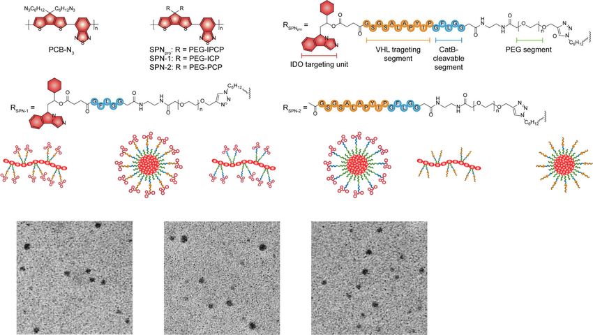
ARTICLE                                                                 NATURE COMMUNICATIONS | https://doi.org/10.1038/s41467-021-23194-w

C
        ancer immunotherapy that leverages the innate and                    chosen as phototheranostic components. Indoleamine 2,3-diox-
        adaptive immune systems to fight against cancer has                   ygenase (IDO) is selected as the POI for PROTAC, because it is a
        revolutionized the treatment of malignancies1. However,              Trp-catabolizing enzyme overexpressed in tumor tissues that
cancer immunotherapy often has low patient response rate and                 converts Trp to kynurenine (Kyn), leading to the dysfunction of
high risk of immune-related adverse events (irAEs, e.g., endo-               dendritic cells (DCs) and the suppression of effector T cells23. The
crinopathy, pneumonitis, hepatitis, nephritis, colitis, etc.), which         IDO-targeting PROTAC peptide (IPP) is made of an IDO-
greatly restrict the clinical applications2. Nanomedicines have              targeting unit (a widely used IDO inhibitor, NLG919) and an E3
shown promise to mitigate both issues in preclinical settings                ubiquitin ligase VHL (the von Hippel Lindau protein)-binding
owing to their ability to modulate the systemic biodistribution              peptide24,25. Cathepsin B (CatB) is chosen as the cancer bio-
and targeted accumulation of administered immunotherapeutic                  marker, which is commonly overexpressed in various cancer cells,
agents3. In particular, activatable immunotherapeutic nanoagents             such as breast cancer, colorectal cancer, melanoma, and prostate
that modulate their immunotherapeutic action in response to                  cancer26.
cancer biomarkers have been developed to further improve the                    The mechanism of SPNpro-mediated activatable photo-
therapeutic specificity for immune checkpoint inhibitors, immu-               immunometabolic cancer therapy is proposed as follows. After
nomodulatory drugs, antibodies, and adjuvants4–6. For example, a             systemic administration, SPNpro passively accumulates in the
pH-sensitive dextran nanoparticle was developed to selectively               tumor of living mice; it generates singlet oxygen (1O2) to elim-
activate anti-PD-1 antibody in mildly acidic tumor micro-                    inate tumor cells and induces tumor-associated antigens release
environment (TME) for checkpoint blockade immunotherapy7,                    and immunogenic cell death (ICD) under NIR photoirradiation.
and a cell surface-conjugated protein nanogel was designed to                These released tumor-associated antigens further induce DC
selectively activate the antitumor immunity of T cells in response           maturation and promote T-cell activation, facilitating antitumor
to the increased reduction potential on the surface of T cells8.             T-cell immune response. Meanwhile, the tumor-overexpressed
These studies clearly validated the feasibility of activatable               CatB cleaves SPNpro and releases IPP in situ. The activated IPP
immunotherapeutic nanoagents to increase therapeutic efficacy                 binds to the immunosuppressive IDO and brings it to the E3
and reduce irAEs.                                                            ubiquitin ligase VHL ligand, leading to the persistent action on
   Immunometabolism that involves a network of intracellular                 IDO degradation via the ubiquitin-proteasome system. The IDO
metabolic pathways such as glycolysis, the tricarboxylic acid cycle,         degradation relieves Trp overconsumption and Kyn accumula-
the pentose phosphate pathway, and amino acid metabolism                     tion, leading to the reversion of immune suppression. As a result,
plays a crucial role in modulating the responses of immune cells9.           SPNpro-mediated activatable photo-immunometabolic therapy
Particularly, the metabolism of amino acids such as tryptophan               exerts a synergetic action to effectively suppress the progression
(Trp), arginine, glutamine, and leucine can affect both tumor                of tumor in mouse model.
progression and proliferation and differentiation of immune
cells10. Thus, immunometabolic cancer therapy has been devel-
oped based on the inhibition of rate-limiting enzymes associated             Results
with the metabolism of these amino acids11. However, small-                  Synthesis and characterization. SPNpro was self-assembled from
molecule inhibitors are generally unable to afford durable                   an amphiphilic semiconducting polymer. First, the carbonylated
response due to the existence of undruggable proteins and drug               NLG919 was prepared by conjugating the hydroxyl group of
resistance12, while genetic methods often suffer from insufficient            NLG919 with succinic anhydride and characterized by ESI-MS
transfection efficiency and off-target side effects13. Thus, alter-           (Supplementary Fig. 1) and 1H NMR (Supplementary Fig. 2). Then
native methods are highly desired to intervene in the metabolism             IPP with CatB-cleavable segment (IPCP) was prepared via the
of amino acids for cancer immunotherapy.                                     standard fluorenylmethyloxycarbonyl (Fmoc) solid-phase peptide
   PROTAC (proteolysis targeting chimera), which comprises two               synthesis (SPPS) method (Supplementary Fig. 3) and conjugated
covalently linked moieties to bind protein of interest (POI) and             with the carbonylated NLG919 (Supplementary Figs. 4, 5)27. IPCP
E3 ligase, respectively, has recently emerged as a powerful tool for         was further reacted with the poly(ethylene glycol) (PEG) chain
targeted posttranslational knockdown of POI14,15. The hetero-                (Mw = 2000) with one terminal of alkynyl group and the other
bifunctional structure of PROTAC allows dictating the POI for                terminal of amine group via an amide condensation to obtain
proteolysis via the ubiquitin-proteasome system16. As PROTAC-                PEG-IPCP conjugate. 1H NMR spectrum of PEG-IPCP exhibited
induced protein degradation is a repetitional process, it can                that the coupled efficiency of IPCP to PEG chain was about 80%.
potentially have more persistent action than inhibitors or genetic           Then, an NIR-absorbing semiconducting polymer (poly(cyclo-
tools, consequently lowering the administration doses. Till now,             pentadithiophene-alt-benzothiadiazole), PCB) with the azide
PROTACs have been applied to modulate the level of essential                 groups (PCB-N3) was prepared through the Suzuki poly-
proteins including the bromodomain and extra-terminal (BET)                  condensation reaction and further conjugated with PEG-IPCP and
family proteins BRD2-417, signal transducer and activator of                 mPEG-alkyne (Mw = 1000) at a molar ratio of 1:4 to afford the
transcription (STAT) family protein STAT318, and cyclin-                     final polymer PCB-PEG-IPCP28. Owing to the hydrophilic PEG
dependent kinases 4 and 6 for cancer therapy19. However,                     brushes and hydrophobic PCB backbone, PCB-PEG-IPCP could
PROTACs have been rarely developed to modulate the proteo-                   self-assemble into SPNpro in aqueous solutions. For comparison,
lysis of immunometabolism-associated proteins for cancer                     two control nanoparticles (SPN-1 and SPN-2) were also prepared
immunometabolic therapy; moreover, no smart activatable                      with the similar method through the self-assembly of the polymer
PROTACs have been developed to minimize the issues of always-                PCB-PEG-ICP and PCB-PEG-PCP, respectively. All the inter-
on bioactivity and off-target side effects.                                  mediates (ICP, PCP, PEG-ICP, PEG-PCP, PCB-PEG-ICP, and
   We herein report the development of a semiconducting poly-                PCB-PEG-PCP) were characterized by ESI-MS and 1H NMR
mer nano-PROTAC (SPNpro) for cancer-activated photo-immu-                    (Supplementary Figs. 6–9).
nometabolic therapy (Fig. 1). SPNpro is composed of a                           The physical and optical properties of the nanoparticles were
semiconducting polymer core conjugated with PROTAC seg-                      studied. The transmission electron microscopy (TEM) images
ments via cancer-biomarker-cleavable peptide (Fig. 2a). Semi-                exhibited a spherical morphology and a uniform size distribution
conducting polymer nanoparticles (SPNs) have good                            of SPNpro, SPN-1, and SPN-2 (Fig. 2b). The dynamic light
biocompatibility and tunable optical properties20–22, and thus are           scattering (DLS) analysis (Fig. 2c and Supplementary Fig. 10)

2                NATURE COMMUNICATIONS | (2021)12:2934 | https://doi.org/10.1038/s41467-021-23194-w | www.nature.com/naturecommunications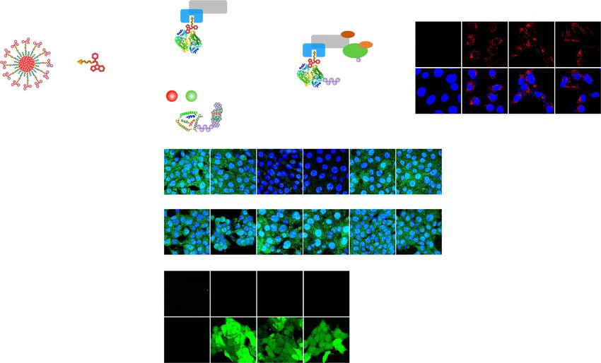
NATURE COMMUNICATIONS | https://doi.org/10.1038/s41467-021-23194-w                                                                         ARTICLE

    a

                                                            PEG           CatB-              VHL binding
                                                          segment       cleavable             segment
                                                                        segment
                                                                                                                             IDO targeting unit

    b
                                            Tumor-associated                                   DC
                                             antigen release                                maturation

        Phototherapy                                                      DCs
                                      ICD                                                                                              Immune
                                                                                                                                      stimulation
                                                                                                           Lymph node

                           SPNpro
                                                                                                                               Teff cells Tumor cells

                                                                   PROTAC-mediated
                                                                    IDO degradation                                                Immunometabolic
           CatB-mediated                                              VHL                                                            intervention
           IPP activation                                                                                    Trp       Kyn
                                                                              Proteasome

                                  IPP            IDO&VHL                                 Trp-metabolism
                                                  targeting                                 inhibition
                                                               IDO

Fig. 1 Schematic illustration of SPNpro-mediated IDO degradation for cancer photo-immunometabolic therapy. a Structure and cathepsin B (CatB)-
specific activation mechanism of SPNpro. b SPNpro-mediated activatable photo-immunometabolic therapy with two processes: (i) a series of cancer immune
responses including immunogenic cell death (ICD), tumor-associated antigen release, DC maturation, and effector T (Teff) cell activation upon NIR
photoirradiation; (ii) SPNpro-mediated immunometabolic intervention processes including CatB-specific activation of IPP, IDO and VHL targeting,
proteasome recruitment, IDO degradation, Trp upregulation and Kyn depletion, and Teff cell activation.

further validated the similar hydrodynamic sizes of SPNpro                      conducted (Fig. 2g) after incubation of the nanoparticle solutions
(28 nm), SPN-1 (32 nm), and SPN-2 (32 nm) and their good                        with CatB. The elution peaks at 26.0, 16.2, and 16.5 min were
stability both in PBS and 10% fetal bovine serum (FBS) solution.                observed for SPNpro, SPN-1, and SPN-2, respectively, which
SPNpro, SPN-1, and SPN-2 had similar UV–vis absorption spectra                  indicated the release of the corresponding chimeric peptides (IPP
with two characteristic absorption peaks from the PCB core at                   for SPNpro, IDO-targeting peptide (IP) for SPN-1, and PROTAC
405 and 680 nm and similar fluorescence emission maximum at                      peptide (PP) for SPN-2) from these nanoparticles (Fig. 3a). In
820 nm (Fig. 2d, e). These data validated that the physical and                 contrast, no such peaks were detected in the absence of CatB,
optical properties of SPNs did not change with the surface                      further confirming the CatB-specific activation of SPNs.
conjugation of various segments.
   Afterwards, the photodynamic properties of SPNpro, SPN-1,                    In vitro studies of photo-immunometabolic therapy. To eval-
and SPN-2 were studied. The generation of 1O2 for these                         uate the cellular uptake of SPNs, 4T1 cancer cells were incubated
nanoparticles under NIR photoirradiation was detected using                     with SPNpro, SPN-1, or SPN-2 for 24 h and then imaged by
singlet oxygen sensor green (SOSG) as a fluorescence indicator.                  confocal fluorescence microscopy. The obvious red fluorescence
The fluorescence intensity of SOSG at 520 nm exhibited a gradual                 signals from the PCB core could be detected in all SPN-incubated
increment for these nanoparticle solutions under NIR photo-                     4T1 cells (Fig. 3b). The relative mean fluorescence intensities
irradiation (0.3 W/cm2 at 808 nm, the maximum permissible                       (MFIs) of SPNpro-, SPN-1-, and SPN-2-incubated 4T1 cells were
exposure for skin), proving the generation of 1O2 (Fig. 2f). After              13.0, 16.1, and 13.6, respectively, demonstrating their similar
photoirradiation for 7 min, the SOSG fluorescence intensity of                   cellular uptake by 4T1 cells (Fig. 3c). Meanwhile, the intracellular
SPNpro, SPN-1, and SPN-2 similarly increased by about 2.4-fold.                 lysosome colocalization analysis exhibited little overlap between
These data indicated that the conjugation of different chimeric                 the fluorescence signals of the lysosome and SPNs, which indi-
peptides (IPCP, PCP, and ICP) did not affect the photodynamic                   cated the effective endosomal escape of SPNs after the cellular
properties of PCB core in SPNpro, SPN-1, and SPN-2.                             internalization (Supplementary Fig. 11).
   To confirm the CatB-specific activation of SPNs, high-                            To evaluate the SPNpro-mediated IDO degradation, the
performance liquid chromatography (HPLC) analysis was                           intracellular IDO expression and Kyn content were detected in

NATURE COMMUNICATIONS | (2021)12:2934 | https://doi.org/10.1038/s41467-021-23194-w | www.nature.com/naturecommunications                                3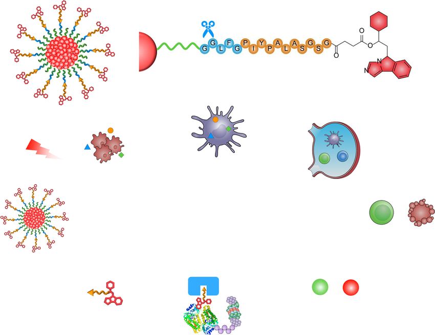
ARTICLE NATURE COMMUNICATIONS | https://doi.org/10.1038/s41467-021-23194-w
a
Self-assembly Self-assembly Self-assembly
PCB-PEG-IPCP PCB-PEG-ICP PCB-PEG-PCP
SPNpro SPN-1 SPN-2
b c
40
SPNpro
SPN-1
30 SPN-2
Number (%)
20
10
100 nm 100 nm 100 nm
0
SPNpro SPN-1 SPN-2 1 10 100 1000
Diameter (nm)
d e4 f g
0.4
Fluorescence intensity (×104 a.u.)
SPNpro
SPNpro SPNpro SPN-1 + CatB
SPN-1 2.5
SPN-1 SPN-1
0.3 3 SPN-2 SPN-1
Absorption (a.u.)
SPN-2 SPN-2
2.0 SPN-2 + CatB
F/F0
0.2 2 SPN-2
1.5 SPNpro + CatB
0.1 1
SPNpro
1.0
0.0 0
300 400 500 600 700 800 900 700 750 800 850 900 0 1 2 3 4 5 6 7 5 10 15 20 25 30
Wavelength (nm) Wavelength (nm) Time (min) Time (min)
Fig. 2 Synthesis and characterization of SPNpro, SPN-1, and SPN-2. a The molecular structures and syntheses of SPNpro, SPN-1, and SPN-2. b TEM images
of SPNpro (left), SPN-1 (middle), and SPN-2 (right). c DLS profiles of SPNpro, SPN-1, and SPN-2 in 1× PBS buffer (pH 7.4). UV/Vis absorption (d) and
fluorescence spectra (e) of SPNpro, SPN-1, and SPN-2 in 1× PBS buffer (pH 7.4) with an excitation wavelength at 650 nm. f The generation of 1O2 in SPNpro,
SPN-1, and SPN-2 in 1× PBS buffer (pH 7.4) ([PCB] = 20 μg/mL) as a function of photoirradiation time. The mean values and SD are presented. Error bars
represent the standard deviation of three separate measurements (n = 3). g HPLC profiles of SPNpro, SPN-1, and SPN-2 ([PCB] = 20 μg/mL) in the
absence or presence of CatB (0.2 U/mL). The experiments in b were repeated independently three times with similar results.
4T1 cells using immunofluorescence staining and HPLC analysis, thus not only validated that SPNpro effectively degraded IDO, but
respectively. 4T1 cells were known to elevate the IDO expression also revealed that the presence of PROTAC unit augmented the
after stimulation with interferon γ (IFN-γ)29, which was also decrease of Kyn contents.
confirmed by elevated MFI of FITC-labeled anti-IDO antibodies The mechanism of SPNpro-mediated IDO degradation was
(10.1-fold) (Fig. 3d, e) and increased Kyn content (15.4-fold) further studied. After stimulation with IFN-γ, 4T1 cells were further
(Fig. 3f) of IFN-γ-stimulated cells relative to the untreated control treated with a CatB inhibitor (CA-074-Me)30, a NEDD8-activating
cells. After incubation of IFN-γ-stimulated cells with IPP or enzyme inhibitor (MLN4924)31, or a 26 S proteasome inhibitor
SPNpro, the MFIs of FITC-labeled anti-IDO antibodies were (epoxomicin)32. MLN4924 was reported to inhibit the neddylation
greatly decreased by 81.8 and 89.3% relative to that for IFN-γ- of E3 ligase (cullin-RING complexes), which needed to be
stimulated cells, respectively. However, incubation of IFN-γ- neddylated in order to be active (Fig. 3a)33. After such treatments,
stimulated cells with NLG919, SPN-1, or SPN-2 did not decrease incubation with SPNpro was no longer able to reduce the green
the green fluorescence signals of FITC-labeled anti-IDO anti- fluorescence signals of FITC-labeled anti-IDO antibodies and the
bodies. This confirmed that the degradation of IDO was induced Kyn contents. Thus, the mechanism of SPNpro-mediated IDO
by the presence of IPP in SPNpro. Meanwhile, incubation of IFN- degradation and Trp-metabolism intervention could be explained as
γ-stimulated cells with IPP or SPNpro led to 62.5 and 71.3% follow: in the presence of CatB, SPNpro was cleaved to induce the
decreases in the relative Kyn content in cells, respectively. release and activation of IPP. The released IPP bound to IDO and
Although the decreases of Kyn contents after incubation of IFN- brought it to the VHL ligand of E3 ligase. Then, E3 ligase was
γ-stimulated cells with NLG919 and SPN-1 were observed owing activated by the NEDD8-activating enzyme that facilitated ubiquitin
to the IDO inhibition of the NLG919 unit, they only had transfer from E2 ligase to IDO, leading to the polyubiquitination of
moderate decreases (50.7% and 38.8%, respectively). These data IDO and subsequent proteasome recruitment. At last, the 26 S
4 NATURE COMMUNICATIONS | (2021)12:2934 | https://doi.org/10.1038/s41467-021-23194-w | www.nature.com/naturecommunications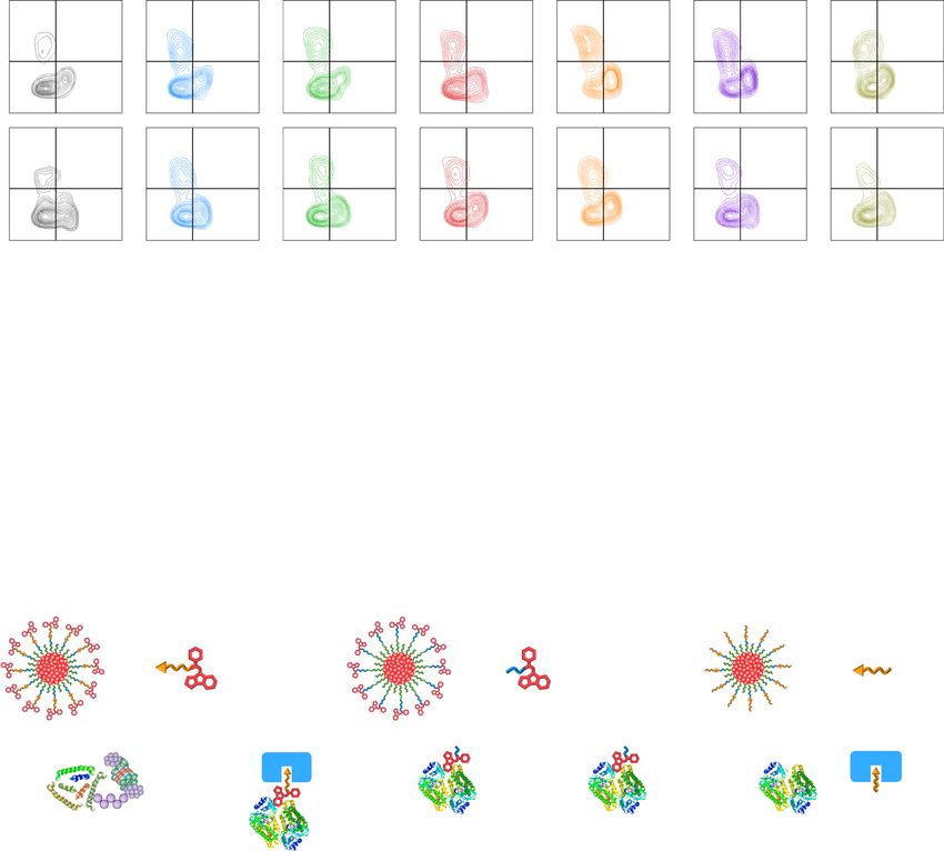
NATURE COMMUNICATIONS | https://doi.org/10.1038/s41467-021-23194-w ARTICLE
a E3 E3 activation and
b
MLN4924
IDO&VHL VHL IDO polyubiquitination Control SPNpro SPN-1 SPN-2
CA-074-Me targeting E3 neddylation
Nanoparticles
E3
CatB IDO VHL NEDD8
E2
cleavage
Ubiquitin
IPP Epoxomicin
Ubiquitin
Merge
transfer
SPNpro Kyn Trp Proteasome
IPP 20 μm
reactivation Proteasome recruitment
and IDO degradation
c d PBS NLG919 IPP SPNpro SPN-1 SPN-2
e
30
+ CA-074-Me + MLN4924 + Epoxomicin
120
*** ** + IFN-γ
100
SPNs MFI (a.u.)
**
IDO MFI (a.u.)
20
80
PBS SPNpro PBS SPNpro PBS SPNpro 60
SPNs
10
+ CA-074-Me + MLN4924 + Epoxomicin 40
20
0 20 μm 0
PBS SPNpro
pro SPN-1 SPN-2
f g Control SPNpro SPN-1 SPN-2 h 200 i
+ CA-074-Me + MLN4924 + Epoxomicin Control
Control
100
120 SPNpro
SPN pro
- laser
+ IFN-γ 150 SPN-1
SPN-1
DCFH MFI (a.u.)
80
Cell Viability (%)
Kyn content (%)
100 SPN-2
SPN-2
80 100 **** SPNpro
60
SPN-1
60
40 SPN-2
50
+ laser
40 SPNpro + laser
SPN-1 + laser
20 1 20
20 μm SPN-2 + laser
0 0 0
- laser + laser 0 10 20 30 40
Concentration (μg/mL)
Fig. 3 In vitro SPNpro-mediated activatable photo-immunometabolic therapy. a Proposed mechanism for CatB-specific activation of PROTAC-mediated
IDO degradation from SPNpro. Confocal fluorescence images (b) and MFI (c) of 4T1 cancer cells after 24 h incubation with SPNpro, SPN-1, and SPN-2
([PCB] = 20 μg/mL) (n = 3). Control versus SPNpro: p = 0.0025; Control versus SPN-1: p = 0.0003; Control versus SPN-2: p = 0.0016. Blue fluorescence
indicated the cell nucleus stained with 4’,6-diamidino-2-phenylindole (DAPI), and red fluorescence showed the signals from the PCB core. Confocal
fluorescence images (d) and MFI (e) of IFN-γ-stimulated 4T1 cells treated with various inhibitors (CA-074-Me, the CatB inhibitor; MLN4924, the
neddylation inhibitor; epoxomicin, the proteasome inhibitor) after 12 h incubation with SPNpro, SPN-1, or SPN-2 ([PCB] = 20 μg/mL), followed by staining
with green fluorescent anti-IDO antibody (n = 3). The cell nucleus was stained with DAPI (blue). f Relative Kyn content in the cell culture medium after
12 h incubation with SPNpro, SPN-1, or SPN-2 ([PCB] = 20 μg/mL) by HPLC assay (n = 3). Confocal fluorescence images (g) and MFI (h) of 4T1 cells after
12 h incubation with SPNpro, SPN-1, or SPN-2 ([PCB] = 20 μg/mL), followed by staining with H2DCFDA with or without NIR photoirradiation (0.3 W/cm2
at 808 nm) for 6 min (n = 3). Control + laser versus SPNpro + laser: p < 0.0001; Control + laser versus SPN-1 laser: p < 0.0001; Control + laser versus
SPN-2 + laser: p < 0.0001. Green fluorescence indicated the signals from DCF. i Relative cell viabilities of 4T1 cells after 12 h incubation with SPNpro, SPN-1,
or SPN-2 at different PCB concentrations with or without NIR photoirradiation (0.3 W/cm2 at 808 nm) for 6 min (n = 3). Statistical significance in c and
h was calculated via one-way ANOVA with a Tukey post-hoc test. **p < 0.0021, ***p < 0.0002, and ****p < 0.0001. The mean values and SD are presented.
The experiments in b and d were repeated independently three times with similar results.
proteasome was recruited to IDO and initiated the degradation of were detected in the nanoparticles-treated or photoirradiated cells
IDO, ultimately resulting in inhibited Trp catabolism and Kyn (Supplementary Fig. 12). Afterwards, the cell viability was studied
depletion. Meanwhile, IPP was released from the ubiquitin- by a 5-(3-carboxymethoxyphenyl)-2-(4,5-dimethylthiazolyl)-3-
proteasome system and reactivated to induce persistent IDO (4-sulfophenyl)-tetrazolium (MTS) assay. After incubation with
degradation in a recycling manner. the nanoparticles at different concentrations, the cell viabilities of
Then, the photoactivity and cytotoxicity of SPNpro, SPN-1, and SPNpro-, SPN-1-, and SPN-2-incubated cells were above 90%,
SPN-2 were studied in vitro. The intracellular 1O2 generation was suggesting the negligible cytotoxicity of these nanoparticles. After
evaluated using 2′,7′-dichlorodihydrofluorescein diacetate NIR photoirradiation at 808 nm with the power intensity of
(H2DCFDA) as the 1O2 fluorescent turn-on indicator. H2DCFDA 0.3 W/cm2 for 6 min, the control cells did not exhibit any obvious
could be rapidly hydrolyzed by the intracellular esterase and changes, while SPNpro-, SPN-1-, and SPN-2-incubated cells
oxidized to fluorescent 2′,7′-dichlorodihydrofluorescein (DCF) in showed increased cytotoxicity in a concentration-dependent
the presence of 1O234. As shown in Fig. 3g, h, obvious green manner (Fig. 3i). At the PCB concentration of 40 μg/mL, the
fluorescence signals of DCF were detected in SPNpro-, SPN-1-, or cell viabilities of SPNpro-, SPN-1-, and SPN-2-incubated cells
SPN-2-incubated 4T1 cells only after NIR photoirradiation, similarly decreased to ~10%. These data confirmed that SPNpro,
which were 83.3-, 79.4-, and 71.6-fold stronger than that for SPN-1, and SPN-2 possess good phototherapeutic abilities.
the untreated cells, respectively. To further evaluate 1O2-mediated
cytotoxicity of these nanoparticles, 4T1 cells were stained with
propidium iodide (PI) to detect the dead cells after NIR In vivo photo-immunometabolic therapy. SPNpro-mediated
photoirradiation. Obvious red fluorescence signals of PI were activatable photo-immunometabolic therapy was further studied
detected in SPNpro-, SPN-1-, or SPN-2-incubated 4T1 cells after in 4T1-tumor-bearing BALB/c mice. 4T1 cells were sub-
NIR photoirradiation, while almost no fluorescence signals of PI cutaneously inoculated in the right flank of mice as the primary
NATURE COMMUNICATIONS | (2021)12:2934 | https://doi.org/10.1038/s41467-021-23194-w | www.nature.com/naturecommunications 5ARTICLE NATURE COMMUNICATIONS | https://doi.org/10.1038/s41467-021-23194-w tumor; 6 days later, the same amount of 4T1 cells was sub- intravenously injected with SPNpro, SPN-1, or SPN-2 and treated cutaneously inoculated in the left flank of mice as the distant with local NIR photoirradiation. After different treatments, the tumor (Fig. 4a). After 2 days, 4T1-tumor-bearing mice were growths of both primary and distant tumors were monitored for Fig. 4 In vivo NIR fluorescence imaging and SPNpro-mediated activatable photo-immunometabolic therapy. a Schematic illustration of the schedule for tumor model implantation and SPNpro-mediated activatable photo-immunometabolic therapy. b NIR fluorescence imaging of 4T1-tumor-bearing BALB/c mice at different timepoints after the intravenous injection of SPNpro, SPN-1, or SPN-2 (200 μL, [PCB] = 200 μg/mL). c Confocal fluorescence images of primary tumors from 4T1-tumor-bearing mice after intravenous injection of SPNpro, SPN-1, or SPN-2. Blue fluorescence showed the cell nucleus stained with DAPI, and red fluorescence showed the signals from the PCB core. d Ex vivo NIR fluorescence images of tumors and major organs in 4T1-tumor- bearing mice at 48 h after intravenous injection of SPNpro, SPN-1, or SPN-2. e Confocal fluorescence images of primary tumor tissues from SPNpro-, SPN-1-, or SPN-2-injected (200 μL, [PCB] = 200 μg/mL) 4T1-tumor-bearing mice with or without NIR photoirradiation (0.3 W/cm2 at 808 nm) for 6 min. Blue fluorescence showed the cell nucleus stained with DAPI, red fluorescence showed the signals from the PCB core, and green fluorescence showed the signals from SOSG. f Quantitative analysis of SOSG MFI in primary tumor tissues of mice after different treatments (n = 5). p < 0.0001. Growth curves of primary tumors (g) and distant tumors (h) in 4T1-tumor-bearing mice after different treatments (n = 5). Saline-treatment versus SPNpro-treatment group: p = 0.0043 for primary tumors (g); p = 0.0192 for distant tumors (h). Quantification of caspase-3 expression in primary (i) and distant (j) tumor tissues of 4T1-tumor-bearing mice (n = 7). p < 0.0001. Statistical significance in f, i, and j was calculated via one-way ANOVA with a Tukey post-hoc test. Statistical significance in g and h was calculated via two-tailed Student’s t-test. *p < 0.032, **p < 0.0021, ***p < 0.0002, and ****p < 0.0001. The mean values and SD are presented. The experiments in c and e were repeated independently three times with similar results. 6 NATURE COMMUNICATIONS | (2021)12:2934 | https://doi.org/10.1038/s41467-021-23194-w | www.nature.com/naturecommunications
NATURE COMMUNICATIONS | https://doi.org/10.1038/s41467-021-23194-w                                                                 ARTICLE

14 days. To confirm the optimal timepoint for NIR photo-                         kidney) in SPNpro-, SPN-1-, or SPN-2-injected and photoirra-
irradiation, the accumulation of nanoparticles in the primary                   diated mice exhibited similar physiological morphologies com-
tumor tissues was evaluated using NIR fluorescence imaging.                      pared to that in saline-injected mice (Supplementary Fig. 16).
After intravenous injection of SPNpro, SPN-1, or SPN-2, the                     These results demonstrated that SPNpro, SPN-1, and SPN-2 had
fluorescence signals in the primary tumor tissues of these groups                high biosafety and biocompatibility.
gradually increased. At 24 h post-injection time, the fluorescence
signals reached a similar maximum MFI, which was about 90-fold
higher than that of the background (Fig. 4b, Supplementary                      In vivo mechanistic studies of photo-immunometabolic therapy.
Fig. 13). These data indicated that these nanoparticles had similar             To study the mechanism of SPNpro-mediated photo-
and effective tumor accumulation abilities owing to their small                 immunometabolic therapy, tumor-infiltrating lymphocytes (TILs)
size (~30 nm) and hydrophilic PEG segment. This was also                        were first detected by collecting the primary and distant tumor
confirmed by the red fluorescence signals from the PCB core in                    tissues and spleens by fluorescence-activated cell sorting (FACS)
the primary tumor tissues by immunofluorescence staining                         analysis. The populations of TILs (CD4+ and CD8+) and cytotoxic
(Fig. 4c, Supplementary Fig. 13). In addition, SPNpro, SPN-1, and               TILs (CD8+granzyme B+) in primary and distant tumor tissues
SPN-2 showed similar tissue distribution in major organs                        and spleen of SPNpro-injected mice were higher than that of SPN-
(Fig. 4d, Supplementary Fig. 13), showing the highest accumu-                   1- or SPN-2-injected mice after NIR photoirradiation (Fig. 5a,
lation in tumor followed by liver.                                              Supplementary Fig. 17). Meanwhile, the population of regulatory
   Photo-immunometabolic therapy was conducted by NIR                           T cells (CD4+Foxp3+) and myeloid-derived suppressor cells
photoirradiation on the primary tumors at the maximum                           (CD11b+Gr-1+) of SPNpro-injected mice were lower than that of
accumulation timepoint (24 h post-injection of nanoparticles).                  SPN-1- or SPN-2-injected mice after NIR photoirradiation (Sup-
Notably, the power intensity of the laser was controlled to ensure              plementary Fig. 18). In addition, the expression of granzyme B in
that the temperature of tumors was below the threshold                          the tumor tissues was detected to examine the tumor killing effect
temperature (43 °C) to minimize photothermal heating effect.                    by cytotoxic TILs. Both the primary and distant tumor tissues of
The 1O2 generation of nanoparticles was studied by detecting the                SPNpro-injected mice exhibited higher MFIs (3.0- and 4.2-fold,
fluorescence signals after local injection of SOSG in tumor tissues.             respectively) of FITC-labelled anti-granzyme B antibodies than
There were obvious green fluorescence signals of SOSG and a                      that for SPN-1- or SPN-2-injected mice (Fig. 5b, c, Supplementary
similar increase (~60-fold) of MFIs in the tumors of SPNpro-,                   Fig. 19). Moreover, we evaluated the antitumor effects of SPNpro in
SPN-1-, or SPN-2-injected mice after photoirradiation compared                  4T1-tumor-bearing immunodeficient NOD-Scid IL2rg−/− (NSG)
to that of the mice treated without photoirradiation. These data                mice, which lack functional lymphocytes (including T cells and NK
confirmed the effective intratumoral 1O2 generation of these                     cells) and are defective in immune functions. Both the primary and
nanoparticles upon NIR photoirradiation (Fig. 4e, f).                           distant tumors exhibited negligible inhibition effects in SPNpro-
   The in vivo cancer therapeutic efficiency was studied by                      injected and photoirradiated NSG mice (Supplementary Fig. 20).
monitoring the growths of primary and distant tumors every                      These data confirmed that SPNpro-mediated therapy was depen-
2 days after NIR photoirradiation for 6 min. After treatments, the              dent on the presence of the immune system and exhibited stronger
primary tumors in SPNpro-, SPN-1-, and SPN-2-injected and                       antitumor T-cell immune responses than SPN-1- or SPN-2-
photoirradiated mice were completely suppressed, whereas the                    mediated therapy.
distant tumors were greatly inhibited only for SPNpro-injected and                 To gain insight into the high antitumor T-cell immunity of
photoirradiated mice, which did not show obvious inhibition for                 SPNpro-mediated therapy, the corresponding immunological
SPN-1- or SPN-2-injected and photoirradiated mice (Fig. 4g, h).                 processes including ICD, DC maturation, and cytokine release
The therapeutic effects were further confirmed by immunofluor-                    of nanoparticle-injected 4T1-tumor-bearing mice were investi-
escence staining and hematoxylin and eosin (H&E) staining. The                  gated and compared. High-mobility group protein B1 (HMGB1)
green fluorescence signals of FITC-labeled anti-caspase-3 anti-                  was chosen to be evaluated as the important marker for ICD35.
bodies were detected in the primary tumor tissues of SPNpro-,                   The MFIs of FITC-labeled anti-HMGB1 antibodies in the
SPN-1-, and SPN-2-injected mice after photoirradiation, which                   primary tumor tissues of SPNpro-, SPN-1-, or SPN-2-injected
were 33.1-, 21.8-, and 24.2-fold higher than the saline-injected                mice were greatly increased by 30.6-, 25.0-, and 26.7-fold relative
mice, respectively (Fig. 4i, Supplementary Fig. 14a). As for the                to that for the saline-injected mice, respectively (Fig. 5d,
distant tumors, the MFIs of FITC-labeled anti-caspase-3 anti-                   Supplementary Fig. 21a). DC maturation was studied by
bodies in SPNpro-injected and photoirradiated mice were                         collecting TDLNs to detect the population of CD11c+CD80+
increased 34.9-fold relative to that in the saline-injected mice,               CD86+ DCs by flow cytometry analysis. The amounts of matured
which were also higher (both 2.2-fold) than that in SPN-1- or                   DCs (CD11c+CD80+CD86+) relative to all DCs (CD11c+) in
SPN-2-injected and photoirradiated mice (Fig. 4j, Supplementary                 TDLNs for SPNpro-, SPN-1-, and SPN-2-injected mice were 30.5,
Fig. 14a). In addition, H&E staining showed obvious dead cells in               24.1, and 26.8%, which were 1.8-, 1.5-, and 1.6-fold higher than
primary and distant tumors of SPNpro-injected and photoirra-                    that of saline-injected mice, respectively (Fig. 5e, Supplementary
diated mice in contrast to the other groups (Supplementary                      Figs. 21b, 22). The serum samples of SPNpro-, SPN-1-, and SPN-
Fig. 14b), which was consistent with the immunofluorescence                      2-injected mice were collected at different timepoints (1, 3, and
staining results. These data suggested that only SPNpro-mediated                5 days) after photoirradiation to detect the cytokines (TNF-α and
therapy effectively eliminated the primary tumors and inhibit the               IL-6) via enzyme-linked immunosorbent assay (ELISA). Both the
growth of the distant tumors, indicating its stronger antitumor                 concentration of TNF-α and IL-6 in SPNpro-, SPN-1-, and SPN-2-
efficacy relative to SPN-1 and SPN-2.                                            injected mice reached maximum at 3 days after photoirradiation
   To evaluate the biosafety of SPNs-mediated photo-immuno-                     (Fig. 5f, g). Moreover, the concentration of TNF-α and IL-6 in
metabolic therapy, body-weight monitoring and histological                      SPNpro-injected mice were 4.7- and 6.4-fold higher than that in
analysis of the major organs were conducted. The body weights                   saline-injected mice, respectively. These ICD, DC maturation,
of saline-, SPNpro-, SPN-1-, and SPN-2-injected mice with or                    and cytokine release profiles confirmed that SPNpro-, SPN-1-, and
without photoirradiation showed no obvious change during 14-                    SPN-2-mediated therapy enhanced tumor immunogenicity to a
day monitoring (Supplementary Fig. 15). Afterwards, H&E                         similar level, which should be ascribed to their similar photo-
staining images of the major organs (heart, liver, lung, and                    therapeutic efficiency.

NATURE COMMUNICATIONS | (2021)12:2934 | https://doi.org/10.1038/s41467-021-23194-w | www.nature.com/naturecommunications                         7
ARTICLE NATURE COMMUNICATIONS | https://doi.org/10.1038/s41467-021-23194-w
a Saline SPNpro SPN-1 SPN-2 SPNpro + Laser SPN-1 + Laser SPN-2 + Laser
10.7 1.63 22.4 1.93 22.2 2.28 18.8 2.12 36.8 2.25 28.4 3.11 23.0 4.79
Primary Tumor
60.1 27.6 51.0 24.7 48.8 26.7 50.0 29.1 19.5 41.4 29.0 39.5 34.9 37.3
CD8
11.4 2.34 19.4 2.61 18.1 3.69 17.0 3.15 28.0 2.68 15.1 1.23 10.8 1.57
Distant Tumor
48.8 37.5 41.1 36.9 39.1 39.1 42.2 37.7 31.3 38.0 45.0 38.7 48.8 38.8
CD4
b 100
100
c 100
100
d 150
150
e 50
f 150
150
Saline SPN-1
SPN-1 Saline SPN-1
SPN-1 Saline SPN-1
SPN-1 Saline
Saline SPN-1
SPN-1 Saline
MFI (a.u.)
MFI (a.u.)
(%)
(a.u.)
(a.u.)
CD86+ DCs (%)
125 ++ laser
SPNpro SPN-2
SPN-2 SPNpro SPN-2
SPN-2 125 SPNpro SPN-2
SPN-2 SPNpro SPN-2
SPN-2 SPNpro
HMGB1 MFI (a.u.)
(a.u)
pro pro pro pro pro
75 75
Primary tumor
tumor
Distant tumor
tumor
**** ****
IL-6 (pg/mL)
**** **** SPN-1
SPN-1
B MFI
B MFI
100
100 **** 100
100
**** *** SPN-2
SPN-2
****
CD80++CD86
50 50 75 25
granzyme B
granzyme B
Primary
Distant
HMGB1
granzyme
granzyme
IL-6
50 50 - laser
25 **** 25
CD80
*** 25
0 0 0 0 0
-- laser ++ laser
laser - laser ++ laser - laser + laser - laser ++ laser 135 135 135 135 135 135 135
Time (d)
(d)
h 125
125
i 150
150
j 125
125
k 80
g 200
200
Saline
Saline SPN-1
SPN-1 Saline
Saline SPN-1
SPN-1 Saline SPN-1
SPN-1 Saline SPN-1
SPN-1 Saline ++ laser
Kyn content (pmol/mg)
(a.u.)
MFI (a.u.)
100
100 SPNpro
pro SPN-2
SPN-2 125
125 SPNpro
pro SPN-2
SPN-2 100
100 SPNpro SPN-2
SPN-2 SPNpro SPN-2
SPN-2 SPNpro
150
TNF-α (pg/mL)
(a.u.)
60 150
(a.u.)
tumor
IDO MFI (a.u.)
IDO MFI (a.u.)
Primary tumor
tumor
****
Distant tumor
100
100 **** **** SPN-1
SPN-1
IDO MFI
75 **** **** **** **** 75 **** SPN-2
SPN-2
** **
75 100
Primary
40 100
TDLN IDO
TNF-α
50 50
IDO
50 - laser
TDLN
20 50
25 25 25
0 0 0 0 0
- laser ++ laser - laser + laser 135 135 135 135 135 135 135
- laser ++ laser - laser + laser
Time (d)
(d)
l
CatB CatB CatB
cleavage cleavage cleavage
IDO&VHL IDO VHL
Reactivation
SPNpro targeting SPN-1 targeting SPN-2 targeting
Recycling
VHL VHL
IDO IDO
degradation inhibition
Trp Kyn Trp Kyn Trp Kyn
Trp normalization & Trp upregulation & Trp overconsumption &
Kyn depletion Kyn downregulation Kyn accumulation
Fig. 5 In vivo SPNpro-mediated activatable photo-immunometabolic therapy. a FACS assay of tumor-infiltrating T lymphocytes (CD8+ and CD4+) of the
primary and distant tumors in 4T1-tumor-bearing mice after different treatments. 4T1-tumor-bearing mice were intravenously injected with saline, SPNpro,
SPN-1, or SPN-2 (200 μL, [PCB] = 200 μg/mL), and the primary tumors were treated with or without NIR photoirradiation (0.3 W/cm2 at 808 nm) for 6
min. Quantification of granzyme B expression in primary (b) and distant (c) tumor tissues of 4T1-tumor-bearing mice (n = 7). p < 0.0001; Saline versus
SPN-2 + laser: p = 0.0004 for primary tumors (b). p < 0.0001 for distant tumors (c). d Quantification of HMGB1 expression in the tumor tissues of 4T1-
tumor-bearing mice by confocal fluorescence images after different treatments (n = 6). p < 0.0001. e Quantification of matured DCs (CD80+ and CD86+)
from the tumor-draining lymph nodes (TDLNs) in 4T1-tumor-bearing mice after different treatments (n = 3). p < 0.0001; Saline versus SPN-1 + laser: p =
0.0002. In vivo cytokine detection of TNF-α (f) and IL-6 (g) in sera from mice after different treatments at different timepoints (1, 3, and 5 days) (n = 3).
h–j Quantification of IDO expression in primary tumors (h), distant tumors (i), and TDLNs (j) of 4T1-tumor-bearing mice by immunofluorescence staining
after different treatments (n = 7). p < 0.0001. k The Kyn content in primary tumors of 4T1-tumor-bearing mice after different treatments (n = 3). p <
0.0001; SPNpro versus SPN-1: p = 0.0038; SPNpro + laser versus SPN-1 + laser: p = 0.0041. l Schematic illustration of the mechanisms of SPNpro-, SPN-1-,
and SPN-2-mediated IDO degradation and Trp-metabolism intervention. Statistical significance in b, c, d, e, h, i, j, and k was calculated via one-way
ANOVA with a Tukey post-hoc test. *p < 0.032, **p < 0.0021, ***p < 0.0002, and ****p < 0.0001. The mean values and SD are presented.
To further verify the enhanced antitumor T-cell immune mice, respectively. In addition, the Kyn content in the primary
response of SPNpro relative to other SPNs, the IDO expression tumor tissues of SPNpro-injected mice had obvious decreases
and Kyn content in the primary and distant tumor tissues and before (79.2%) or after (77.2%) photoirradiation relative to that for
TDLNs were investigated. Obvious green fluorescence signals of saline-injected mice (Fig. 5k). Although SPN-1 possessed the
FITC-labelled anti-IDO antibodies were detected in the primary ability for IDO inhibition owing to the presence of NLG919 unit,
and distant tumor tissues and TDLNs of treated mice (Fig. 5h–j, SPNpro-injected mice still showed higher decreases of the Kyn
Supplementary Fig. 23). The MFIs of FITC-labelled anti-IDO contents before (59.6%) or after (57.1%) photoirradiation relative
antibodies in the primary and distant tumor tissues and TDLNs of to that for SPN-1-injected mice. These data further confirmed the
SPNpro-injected and photoirradiated mice were greatly decreased different mechanisms of SPNpro and SPN-1 for IDO inhibition and
by 10.9, 12.3, and 3.4 times relative to that of the saline-injected subsequent Kyn depletion (Fig. 5l). SPN-1 can inhibit the activity
8 NATURE COMMUNICATIONS | (2021)12:2934 | https://doi.org/10.1038/s41467-021-23194-w | www.nature.com/naturecommunicationsNATURE COMMUNICATIONS | https://doi.org/10.1038/s41467-021-23194-w ARTICLE
of IDO via occupancy-driven pharmacology, which requires Gly-OH, Fmoc-Ser(tBu)-OH, Fmoc-Ala-OH, Fmoc-Pro-OH, Fmoc-Tyr(tBu)-OH,
persistent drug binding to the active site of IDO in order to Fmoc-Ile-OH, Fmoc-Phe-OH, and Fmoc-Leu-OH) were purchased from GL
Biochem. Ltd. (Shanghai, China) and used as received without any purification.
inhibit protein activity. In contrast, SPNpro can directly induce the SOSG was purchased from Molecular Probes Inc. (Carlsbad, CA, USA). 3-(4,5-
degradation of IDO in a sustainable manner due to the reactivation Dimethylthiazol-2-yl)-5-(3-carboxymethoxyphenyl)-2-(4-sulfophenyl)-2H-tetra-
and reuse of the active IPP. Thereby, SPNpro mediated more zolium, inner salt (MTS) solution was purchased from Promega Corp. (Madison,
effective IDO degradation and Kyn depletion than SPN-1 and WI, USA). Dulbecco’s modified eagle medium (DMEM) with L-glutamine was
SPN-2 did, leading to better immunometabolic reprogramming purchased from Lonza. Trypsin–EDTA (0.05%), penicillin–streptomycin (10,000
U/mL), fetal bovine serum (FBS), ACK lysis, type I collagenase, and type IV
and higher antitumor immune response. collagenase were purchased from Gibco. MLN4924, IDO antibody (Catalog no.
ab106134, dilution: 1:50), granzyme B antibody (ab255598, dilution: 1:200), and
secondary antibody Alexa Fluor 488 conjugated goat anti-rabbit IgG H&L
Discussion (ab150077, dilution: 1:500) were purchased from Abcam Inc. (Cambridge, CA,
Existing PROTAC technology mainly utilizes heterobifunctional USA). HMGB1 antibody (Catalog no. 3935 S, dilution: 1:100) and cleaved caspase-
binding ligands to induce targeted proteolysis of undruggable 3 antibody (Catalog no. 9661 L, dilution: 1:500) were purchased from Cell Signaling
oncoproteins for cancer therapy36–38, which has not been Technology. ELISA kits for TNF-α and IL-6 detection, PE-CD11b antibody
(Catalog no. 101208, dilution: 1:80), BV605-Gr-1 antibody (Catalog no. 108440,
designed with a biomarker-activatable action. In contrast, SPNpro dilution: 1:40), AF647-Foxp3 antibody (Catalog no. 126408, dilution: 1:50), APC
only activates its PROTAC function in response to a specific anti-mouse CD11c (Catalog no. 117310, dilution: 1:80), FITC anti-mouse CD80
cancer biomarker (CatB), realizing targeted proteolysis of an (Catalog no. 104706, dilution: 1:50), PE anti-mouse CD86 (Catalog no. 105008,
immunometabolism-associated enzyme (IDO). The molecular dilution: 1:20), FITC anti-mouse CD3 (Catalog no. 100204, dilution: 1:50), APC
anti-mouse CD8a (Catalog no. 100712, dilution: 1:80), PE anti-mouse CD4 (Cat-
mechanism of SPNpro-mediated IDO degradation was further alog no. 130310, dilution: 1:80), and purified anti-mouse CD16/32 (Catalog no.
confirmed using various signaling pathway inhibitors (Fig. 3d–f), 156604, dilution: 1:200) were purchased from Biolegend. HO-PEG2000-COOH was
showing its high specificity. With its high tumor accumulation purchased from JenKem Technology USA Inc. NLG919 was purchased from D&C
and ideal biosafety (Fig. 4b–d, Supplementary Figs. 13, 15, 16), Chemicals.
SPNpro enabled targeted degradation of IDO in the tumor of
living mice, leading to localized immunometabolism intervention. Material characterization. Proton nuclear magnetic resonance (1H NMR) spectra
Thus, SPNpro represents the first smart PROTAC system with were recorded on a Bruker Avance II 300 MHz system (Bruker Physik AG, Ger-
high disease specificity to minimize off-target side effects for many). Electrospray ionization-mass spectrometry (ESI-MS) spectra were con-
ducted with a Thermo Finnigan Polaris Q quadrupole ion trap mass spectrometer
cancer therapy. (Thermo Fisher Corporation) equipped with a standard ESI source. Absorption
Mechanistically different from inhibitor and genetic approa- and fluorescence spectra were measured on a UV-2450 spectrophotometer (Shi-
ches, SPNpro reprograms immunometabolic pathways through madzu, Japan) and a Fluorolog 3-TCSPC spectrofluorometer (Horiba Jobin Yvon),
direct degradation of IDO in a persistent manner (Figs. 3a, 5l), as respectively. DLS measurements were performed on a Malvern Nano-ZS Particle
Sizer (Malvern Instruments, Southborough, UK). TEM images were captured using
the activated PROTAC (IPP) can be recycled for repeated pro- a JEM 1400 transmission electron microscope (JEOL, Tokyo, Japan). HPLC ana-
teolysis. It has been validated that SPNpro had the higher IDO lyses and purification were performed on an Agilent 1260 system using methanol
degradation (ca. 90%) and Kyn depletion (ca. 70%) efficiencies in (MeOH)/water (H2O) as the eluent. Confocal images were captured using a
IFN-γ-stimulated 4T1 cells (Fig. 3d–f) relative to those for free LSM800 confocal laser scanning microscope (Carl Zeiss, Germany). Flow cyto-
metry assay was performed on Fortessa X20 (BD Biosciences). In vivo animal
NLG919 (ca. 10% and ca. 50%) and SPN-1 (ca. 10% and ca. 40%) fluorescence images were captured using an IVIS imaging system (IVIS-CT
without PROTAC segment at the same concentration of NLG919 machine, PerkinElmer). Tissues were cut into sections using a cryostat (Leica). The
(1 μM). Notably, this Kyn depletion efficiency was also higher tissue sections were examined on a Nikon ECLIPSE 80i microscope (Nikon
than the reported studies on various NLG919-encapsulated Instruments). NMR spectra were analyzed using Mestre Nova LITE v5.2.5-
nanosystems (ca. 50%) at the same concentration of NLG919 4119 software (Mestre lab Research S.L.).
(1 μM) in vitro39,40. Moreover, the higher IDO degradation of
SPNpro relative to other controls was observed in the tumor of Synthesis of carbonylated NLG919. A mixture of NLG919 (564 mg, 2 mmol),
living mice (Fig. 5h–k, Supplementary Fig. 23). Thus, although all succinic anhydride (240 mg, 2.4 mmol), and 4-dimethylaminopyridine (24.4 mg,
0.2 mmol) in anhydrous dichloromethane (DCM, 20 mL) was stirred at room
three SPNs enhanced tumor immunogenicity to the similar level temperature for 48 h. After the reaction mixture was concentrated under reduced
after NIR phototherapy, SPNpro had the strongest antitumor T- pressure, it was washed with ammonium chloride solution and extracted by DCM.
cell immune response (Fig. 5a–c), leading to the most effective The organic layer was dried with anhydrous sodium sulphate and concentrated
inhibition of tumor growth and complete prevention of metas- under vacuum to afford carbonylated NLG919 as a white crystalline solid. TLC
(silica gel, DCM), Rf = 0.5. 1H NMR (300 MHz, CDCl3): δ 0.85–1.79 (m, 10H),
tasis (Fig. 4g–j). 2.36 (m, 1H), 2.72 (m, 4H), 3.46 (s, 1H), 5.32 (m, 2H), 6.46 (m, 1H), 7.08–8.15 (m,
In summary, we report a nano-PROTAC (SPNpro) that 4H). ESI-MS (m/z): calc: 382.2, found [M + H]+: 383.2.
synergizes phototherapeutic function with biomarker-activated
protein degradation for photo-immunometabolic cancer therapy. Synthesis of IPCP. 2-Chlorotrityl chloride resin (1.14 mmol/g) was soaked in
SPNpro represents a unique type of immunotherapeutic nanoa- anhydrous dimethylformamide (DMF) for 1 h. Then a mixture of Fmoc-Gly-OMe (4
gents that specifically block the immunosuppressive metabolism equiv.) and diisopropylethylamine (DIEA, 10 equiv.) was dissolved in DMF and
via targeted proteolysis of catabolizing enzyme by the ubiquitin- added into the resins for stirring 3 h under a N2 atmosphere. After being washed
proteasome system. Such PROTAC design can be generalized for thrice with DMF to remove the unreacted mixture solution, the resins were reacted
with a mixture of MeOH and DIEA in DMF (v/v/v = 1:2:7) for 30 min to cap the
biomarker-activated proteolysis of many other unreacted groups. Then, the resins were incubated with 20% piperidine in DMF (v/v)
immunometabolism-associated proteins (such as glutaminase, for 15 min twice to remove the Fmoc protecting groups for further amide con-
arginase, fatty acid synthase, lactate dehydrogenase, and acetyl- densation reaction. The next amino acid couplings were conducted by reacting a
CoA acetyltransferase) by conjugating the corresponding target- mixture of Fmoc-protected amino acids (3 equiv.), HBTU (3.6 equiv.), HOBT (3.6
equiv.), and DIEA (7.5 equiv.) with the resins for 2 h. Kaiser reagent was further used
ing moiety onto the polymer10. Thus, our study not only provides to confirm the coupling efficiency. After repeated amide condensation reaction with
a new combinational therapeutic modality, but also opens new Fmoc-protected amino acids, the resins were reacted with a mixture of carbonylated
opportunities to advance PROTAC in cancer therapy. NLG919 (2 equiv.), HBTU (2.4 equiv.), HOBT (2.4 equiv.), and DIEA (5 equiv.)
overnight. After being washed with DMF (4 times), MeOH (4 times), and DCM (4
times), the resins were dried under vacuum for 30 min. Finally, the chimeric peptide
Methods IPCP was cleaved from the resins with a mixture of trifluoroacetic acid (TFA)/H2O/
Chemicals. All the chemicals were purchased from Sigma–Aldrich unless other- 1,2-ethanedithiol (v/v/v = 95:2.5:2.5) for 1.5 h at room temperature. After being
wise stated. 2-Chlorotrityl chloride polystyrene resin, o-benzotriazole-N,N,N’,N’- collected and concentrated under reduced pressure, the filtrate was dropped into cold
tetramethyluroniumhexafluoro-phosphate (HBTU), 1-hydroxybenzotriazole anhydrous ether to obtain IPCP as precipitates. 1H NMR (300 MHz, CD3OD): δ 0.88
(HOBt), N-fluorenyl-9-methoxycarbonyl (Fmoc)-protected L-amino acids (Fmoc- (m, 24H), 1.22–1.72 (m, 23H), 1.81–2.30 (m, 12H), 2.83–3.18 (m, 6H), 3.43–4.04 (m,
NATURE COMMUNICATIONS | (2021)12:2934 | https://doi.org/10.1038/s41467-021-23194-w | www.nature.com/naturecommunications 9ARTICLE NATURE COMMUNICATIONS | https://doi.org/10.1038/s41467-021-23194-w
21H), 4.21–4.67 (m, 13H), 6.61–7.65 (m, 15H). ESI-MS (m/z): calc: 1826.9, found In vitro photodynamic studies. PBS solutions (1 mL) containing SPNpro, SPN-1,
[M + 2H]2+: 914.8. or SPN-2 ([PCB] = 20 μg/mL) were mixed with 1 μL SOSG probe (500 μM). Then
For comparison, the control peptides ICP and PCP were also prepared in a the solutions were irradiated with NIR laser (0.3 W/cm2 at 808 nm) for 7 min. The
similar method. ICP was synthesized without the PROTAC segment. 1H NMR fluorescence intensities of various samples at 520 nm were recorded every 1 min
(300 MHz, CD3OD): δ 0.84–1.37 (m, 19H), 2.30–3.01 (m, 8H), 3.40–3.98 (m, 10H), during NIR photoirradiation using a Fluorolog 3-TCSPC spectrofluorometer
7.04–7.92 (m, 11H). ESI-MS (m/z): calc: 813.4, found [M + H]+: 814.4. PCP was termed as F. The fluorescence intensities of the samples at 520 nm without NIR
synthesized without the NLG919 unit. 1H NMR (300 MHz, CD3OD): δ 0.70–1.34 photoirradiation were also recorded as F0. At last, the enhancement of fluorescence
(m, 24H), 1.37–2.17 (m, 20H), 2.82–3.24 (m, 5H), 3.41–3.69 (m, 3H), 4.08–3.69 (m, intensities of each sample was calculated as F/F0, indicating the generation of 1O2.
13H), 4.12–4.72 (m, 11H), 6.55–7.38 (m, 9H). ESI-MS (m/z): calc: 1504.7, found
[M + 2H]2+: 753.7.
In vitro CatB-specific activation studies. The buffer solutions (200 μL) containing
SPNpro, SPN-1, or SPN-2 ([PCB] = 40 μg/mL) were incubated with CatB (0.2 U/mL)
Synthesis of PEG-IPCP. Amino-PEG-alkyne (Mw = 2000) was synthesized for 6 h. Then the solutions with or without CatB incubation were purified by fil-
according to a previous study24. HO-PEG-COOH (Mw = 2000, 2 g, 1 mmol) and tration through a 0.22 μm polyvinylidene fluoride syringe driven filter (Millipore),
NaH (100 mg, 4.2 mmol) were dissolved in dry tetrahydrofuran (THF) and stirred followed by HPLC analysis.
at 0 °C for 1 h. Then, propargyl bromide (120 μL, 1.52 mmol) and NaH (50 mg, 2.1
mmol) were added and stirred overnight. The reaction solution was filtered to In vitro cellular uptake assay. 4T1 cancer cells were purchased from American
remove precipitates, and the filtrate was concentrated via rotary evaporation. Type Culture Collection (ATCC) and cultured in DMEM cell culture medium
Afterwards, the crude product was precipitated in cold diethyl ether and collected supplemented with 10% FBS and 1% antibiotics (penicillin−streptomycin, 10,000
via centrifugation, followed by dialysis and lyophilization to obtain carboxyl-PEG- U/mL) at 37 °C and 5% CO2. The cells were seeded into confocal cell culture dishes
alkyne. Carboxyl-PEG-alkyne (500 mg, 0.25 mmol), EDC (120 mg, 0.625 mmol), at a density of 5 × 10⁴ cells/dish and cultured overnight for cell attachment. Then
and NHS (72 mg, 0.625 mmol) were dissolved in anhydrous THF, and then stirred the cells were incubated with SPNpro, SPN-1, or SPN-2 ([PCB] = 20 μg/mL) for 24
at room temperature for 1 h. Afterwards, ethylenediamine (160 μL, 2.5 mmol) was h. After washed three times with PBS to remove free nanoparticles, the cells were
added and then stirred for 2 days. After reaction, THF was removed by rotary stained with DAPI, and the fluorescence images of cells were captured using a
evaporation, and the residues were purified by dialysis and lyophilization to obtain LSM800 confocal laser scanning microscope (Carl Zeiss, Germany).
amino-PEG-alkyne. 1H NMR (300 MHz, CDCl3): δ 4.19 (s, 2H), 4.04 (s, 2H), 3.87
(m, 2H), 3.63 (m, 176H), 2.44 (m, 3H).
A mixture of IPCP (50 mg, 0.03 mmol), EDC (14 mg, 0.075 mmol), and NHS (8 In vitro lysosome colocalization assay. 4T1 cancer cells were cultured in DMEM
mg, 0.075) was dissolved in 10 mL THF and stirred at room temperature for 1 h. cell culture medium supplemented with 10% FBS and 1% antibiotics (penicillin
Then, the solution was added into 5 mL THF solution containing amino-PEG- −streptomycin, 10,000 U/mL) at 37 °C and 5% CO2. The cells were seeded into
alkyne (100 mg, 0.05 mmol) and stirred at room temperature for another 48 h. The confocal cell culture dishes at a density of 5 × 10⁴ cells/dish and cultured overnight
obtained PEG-IPCP was further dialyzed against H2O for 3 days using a dialysis for cell attachment. Then the cells were incubated with SPNpro, SPN-1, or SPN-2
membrane with MWCO of 2000 and then lyophilized in vacuum. 1H NMR (300 ([PCB] = 20 μg/mL) for 24 h. After washed three times with PBS to remove free
MHz, CDCl3): δ 0.60–1.41 (m, 19H), 1.46–2.74 (m, 23H), 3.28–3.52 (m, 3H), 3.65 nanoparticles, the cells were stained with DAPI and the lysosome tracker (Green
(m, 180H), 3.79–4.42 (m, 10H), 6.49–7.22 (m, 2H), 7.32–8.81 (m, 4H). DND-26), and the fluorescence images of cells were captured using a LSM800
For comparison, the control polymers PEG-ICP and PEG-PCP were also confocal laser scanning microscope (Carl Zeiss, Germany).
prepared in a similar method. PEG-ICP was synthesized by reacting amino-PEG-
alkyne with ICP. 1H NMR (300 MHz, CDCl3): δ 0.74–0.98 (m, 6H), 0.99–1.79 (m, In vitro IDO expression assay. 4T1 cancer cells were seeded into confocal cell
17H), 1.84–3.19 (m, 17H), 3.26–3.48 (m, 3H), 3.65 (m, 180H), 3.83–4.35 (m, 5H). culture dishes at a density of 5 × 10⁴ cells/dish and cultured overnight for cell
PEG-PCP was synthesized by reacting amino-PEG-alkyne with PCP. 1H NMR attachment. Then the cells were stimulated with IFN-γ for 12 h and incubated with
(300 MHz, CDCl3): δ 0.66–1.06 (m, 3H), 1.08–1.40 (m, 3H), 1.39–2.27 (m, 4H), NLG919, IPP, SPNpro, SPN-1, or SPN-2 ([PCB] = 20 μg/mL, [NLG919] = 1 μM)
2.28–2.51 (m, 2H), 2.53–3.50 (m, 10H), 3.65 (m, 180H), 3.81–4.62 (m, 11H), for 12 h. After washed three times with PBS to remove free samples, the cells were
6.62–7.05 (m, 1H), 7.42–8.45 (m, 3H). stained with DAPI and anti-IDO antibodies, and the fluorescence images of cells
were captured using a LSM800 confocal laser scanning microscope (Carl Zeiss,
Synthesis of PCB-PEG-IPCP. PCB-N3 and mPEG-alkyne (Mw = 1000) were Germany).
synthesized according to the previous study28. mPEG-OH (Mw = 1000, 1 g, 1 For the mechanistic study of PROTAC-mediated IDO degradation, IFN-γ-
mmol) and NaH (100 mg, 4.2 mmol) were dissolved in dry tetrahydrofuran (THF) stimulated 4T1 cancer cells were further treated with CA-074-Me, MLN4924, or
and stirred at 0 °C for 1 h. Then, propargyl bromide (120 μL, 1.52 mmol) and NaH epoxomicin for 6 h. Then the cells were incubated with or without SPNpro
(50 mg, 2.1 mmol) were added and stirred overnight. The reaction solution was ([PCB] = 20 μg/mL) for 12 h. After washed three times with PBS to remove free
filtered to remove precipitates and the filtrate was concentrated via rotary eva- samples, the cells were stained with DAPI and anti-IDO antibodies, and the
poration. Afterwards, the crude product was precipitated in cold diethyl ether and fluorescence images of cells were captured using a LSM800 confocal laser scanning
collected via centrifugation, followed by dialysis and lyophilization to obtain microscope (Carl Zeiss, Germany).
mPEG-alkyne. 1H NMR (300 MHz, CDCl3): δ 4.21 (s, 2H), 3.65 (m, 88H), 3.38 (s,
3H), 2.45 (s, 1H). In vitro Kyn content measurement. 4T1 cancer cells were seeded in 6-well cell
2,6-Dibromo-4,4-bis(6-bromohexyl)-4H-cyclopenta[2,1-b:3,4-b’]dithiophene culture plates at a density of 3 × 10⁵ cells/well and then cultured overnight for cell
(50 mg, 0.075 mmol), 4,7-bis(4,4,5,5-tetramethyl-1,3,2-dioxaborolan-2-yl)benzo[c] attachment. Then the cells were stimulated with IFN-γ for 12 h and further treated
[1,2,5]thiadiazole (29 mg, 0.075 mg), Pd(PPh3)4 (5.5 mg, 0.052 mmol), and K2CO3 with or without CA-074-Me, MLN4924, or epoxomicin for 6 h, followed by
(103.7 mg, 0.75 mmol) were added to a 50-mL Schlenk tube. Then, 2.5 mL toluene incubation with NLG919, IPP, SPNpro, SPN-1, or SPN-2 ([PCB] = 20 μg/mL,
with methyltrioctylammonium chloride (1 mg) and 1.25 mL water were injected [NLG919] = 1 μM) for 12 h. After that, 1 mL of the supernatant in each well was
into the Schlenk tube. The mixture was degassed 3 times with freeze-pump-thaw collected and incubated with 100 μL of 30% trichloroacetic acid at 50 °C for 30 min.
circles and then stirred at 100 °C under nitrogen atmosphere for 1 h. Thereafter, the Then the supernatant in each well was collected for HPLC analysis to quantify the
solvent was removed via rotary evaporation, and the precipitates were redissolved Kyn contents.
in dichloromethane, followed by three times of washing with water. Subsequently,
the obtained PCB-Br was precipitated in methanol, washed 3 times with methanol
Intracellular ROS generation measurement. 4T1 cancer cells were seeded in
and dried under vacuum. PCB-Br (12 mg) dissolved in 4.8 mL THF was added into
confocal cell culture dishes at a density of 5 × 10⁴ cells/dish and then cultured
2.4 mL DMF containing sodium azide (4.8 mg). Then, the mixture was stirred at
overnight for cell attachment. Then the cells were incubated with SPNpro, SPN-1, or
room temperature for 12 h. After that, the solvent was removed under reduced
SPN-2 ([PCB] = 20 μg/mL) for 12 h. Afterwards, the cells were added with
pressure and the obtained PCB-N3 was redissolved in dichloromethane, washed
H2DCFDA probe and cultured for 30 min. The treated cells were irradiated with
with water for three times. The organic phase was concentrated and precipitated in
NIR laser (0.3 W/cm2 at 808 nm) for 6 min. The cells were washed three times with
methanol, followed by three times of washing with methanol and dried under
PBS, and the fluorescence images of cells were captured using a LSM800 confocal
vacuum.
laser scanning microscope (Carl Zeiss, Germany).
A mixture of N,N,N’,N”,N”’-pentamethyldiethylenetriamine (PMDETA, 13.4
mg), PCB-N3 (2 mg), CuBr (2.2 mg), mPEG-alkyne (12 mg), and PEG-IPCP (12
mg) in 3 mL THF was stirred at room temperature under N2 atmosphere for 48 h. In vitro cell viability assay. 4T1 cancer cells were seeded in 96-well cell culture
Then the solvent was evaporated under reduced pressure, and the obtained PCB- plates at a density of 1 × 10⁴ cells/well and then cultured overnight for cell attach-
PEG-IPCP was redissolved in water and dialyzed against water using a dialysis ment. Then the cells were incubated with SPNpro, SPN-1, or SPN-2 at different
membrane with MWCO of 10,000 for 3 days. concentrations ([PCB] = 10, 20, 30, and 40 μg/mL) for 12 h. The cells without
For comparison, the control polymers PCB-PEG-ICP and PCB-PEG-PCP were nanoparticle incubation were used as control. Then the cells were irradiated with
also prepared in a similar method. PCB-PEG-ICP was synthesized by reacting NIR laser (0.3 W/cm2 at 808 nm) for 6 min. The cells with or without photo-
PCB-N3 with PEG-ICP. PCB-PEG-PCP was synthesized by reacting PCB-N3 with irradiation were further cultured for 12 h and then incubated with MTS in DMEM
PEG-PCP. cell culture medium for 4 h. The absorbance at 490 nm (A) of each well was
10 NATURE COMMUNICATIONS | (2021)12:2934 | https://doi.org/10.1038/s41467-021-23194-w | www.nature.com/naturecommunicationsYou can also read