Sequestration of Polo kinase to microtubules by phosphopriming-independent binding to Map205 is relieved by phosphorylation at a CDK site in mitosis
←
→
Page content transcription
If your browser does not render page correctly, please read the page content below
Downloaded from genesdev.cshlp.org on March 24, 2015 - Published by Cold Spring Harbor Laboratory Press

Sequestration of Polo kinase
to microtubules by
phosphopriming-independent binding
to Map205 is relieved by phosphorylation
at a CDK site in mitosis
Vincent Archambault,1,3 Pier Paolo D’Avino,1 Michael J. Deery,2 Kathryn S. Lilley,2
and David M. Glover1,4
1
 Department of Genetics, University of Cambridge, Cambridge, CB2 3EH, United Kingdom; 2Department of Biochemistry,
University of Cambridge, Cambridge, CB2 1GA, United Kingdom

The conserved Polo kinase controls multiple events in mitosis and cytokinesis. Although Polo-like kinases are
regulated by phosphorylation and proteolysis, control of subcellular localization plays a major role in
coordinating their mitotic functions. This is achieved largely by the Polo-Box Domain, which binds
prephosphorylated targets. However, it remains unclear whether and how Polo might interact with partner
proteins when priming mitotic kinases are inactive. Here we show that Polo associates with microtubules in
interphase and cytokinesis, through a strong interaction with the microtubule-associated protein Map205.
Surprisingly, this interaction does not require priming phosphorylation of Map205, and the Polo-Box Domain
of Polo is required but not sufficient for this interaction. Moreover, phosphorylation of Map205 at a CDK site
relieves this interaction. Map205 can stabilize Polo and inhibit its cellular activity in vivo. In syncytial
embryos, the centrosome defects observed in polo hypomorphs are enhanced by overexpression of Map205 and
suppressed by its deletion. We propose that Map205-dependent targeting of Polo to microtubules provides a
stable reservoir of Polo that can be rapidly mobilized by the activity of Cdk1 at mitotic entry.
[Keywords: Polo; cell cycle; Drosophila; kinase; Map205; mitosis]
Supplemental material is available at http://www.genesdev.org.
Received May 9, 2008; revised version accepted August 7, 2008.

Control of the eukaryotic cell division cycle requires the                 al. 2005); centrosome separation and maturation (Sunkel
regulated activity of several kinases, including cyclin-                   and Glover 1988; Lane and Nigg 1996; Casenghi et al.
dependent kinases (Cdks) and Polo-like kinases (Plks).                     2003; Oshimori et al. 2006); establishment of the bipolar
Drosophila Polo (Sunkel and Glover 1988; Llamazares et                     spindle (Sunkel and Glover 1988; Sumara et al. 2004; van
al. 1991) is the founding member of the Plk family. Of                     Vugt et al. 2004); tension sensing at the kinetochore in
the four human Plks (Plk1-4), Plk1 is the closest ortholog                 the spindle attachment checkpoint (Ahonen et al. 2005;
of Polo in both sequence and function and the best char-                   Wong and Fang 2005, 2007); and cytokinesis (Ohkura et
acterized of the vertebrate Plks (Barr et al. 2004; van de                 al. 1995; Carmena et al. 1998; Seong et al. 2002; Neef et
Weerdt and Medema 2006). Polo and its orthologs have                       al. 2003, 2007; Burkard et al. 2007; Petronczki et al.
been implicated in an extensive list of functions in cell                  2007).
division (Barr et al. 2004; Glover 2005; van de Weerdt and                    This multiplicity of functions requires Polo kinases to
Medema 2006) that include enabling mitotic entry by                        be precisely regulated in space and time. Indeed, the cel-
phosphorylating the Cdc25C phosphatase (Kumagai and                        lular localization of Polo kinases changes drastically as
Dunphy 1996); facilitating loss of sister chromatid cohe-                  the cell cycle progresses (Llamazares et al. 1991; Gol-
sion (Alexandru et al. 2001; Sumara et al. 2002; Clarke et                 steyn et al. 1995; Moutinho-Santos et al. 1999). To this
                                                                           end, Polo kinases possess a C-terminal Polo-Box Domain
                                                                           (PBD) (Elia et al. 2003a,b; Lowery et al. 2004) that func-
Corresponding authors.                                                     tions in targeting Polo kinases to centrosomes, mitotic
3
 E-MAIL va228@cam.ac.uk; FAX 44-0-1223-333992.
4
 E-MAIL dmg25@hermes.cam.ac.uk; FAX 44-0-1223-333992.                      kinetochores, and the cytokinetic midbody (Seong et al.
Article is online at http://www.genesdev.org/cgi/doi/10.1101/gad.486808.   2002; Park et al. 2004). The PBD alone is able to interact

         GENES & DEVELOPMENT 22:2707–2720 © 2008 by Cold Spring Harbor Laboratory Press ISSN 0890-9369/08; www.genesdev.org      2707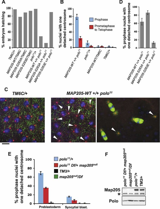
Downloaded from genesdev.cshlp.org on March 24, 2015 - Published by Cold Spring Harbor Laboratory Press

Archambault et al.

with a plethora of cellular phosphoproteins (Lowery et              mediated through a strong interaction between Polo and
al. 2007), and many interactions require the binding of             Map205, a MT-associated protein. Although this inter-
the PBD to a phospho-serine or -theronine of the motif              action depends on a functional PBD, it does not require
S-pS or S-pT (Elia et al. 2003b; van de Weerdt and                  priming phosphorylation of Map205, demonstrating the
Medema 2006). Cyclin B–Cdk1 kinase activity is often                ability of Plks to engage in phosphorylation-independent
responsible for priming these Polo docking sites. Phos-             interactions. We show that phosphorylation of Map205
phorylation of INCENP (Goto et al. 2006) and BubR1                  at a Cdk1 site releases Polo from MTs and that Map205
(Wong and Fang 2007) by Cdk1 promotes the binding of                can stabilize Polo and inhibit its cellular activity. We
hPlk1 to those targets at the kinetochore. In other cases,          propose that Cdk1 phosphorylation of Map205 at mitotic
Polo kinase primes its own binding. Thus, phosphoryla-              entry releases Polo from its inhibitory sequestration to
tion of PIBP1 by hPlk1 promotes a stable interaction be-            MTs to promote its cellular activities in mitosis. This
tween the two proteins, also at the kinetochore (Kang et            mechanism provides a novel layer of spatio-temporal
al. 2006). hPlk1 also primes its own binding to the motor           control of Polo in the cell cycle.
protein MKlp2 on the central spindle in anaphase/telo-
phase (Neef et al. 2003). Recently, it has been shown that
Cdk1 phosphorylation of the central spindle protein                 Results
PRC1 in early mitosis prevents its binding to hPlk1,
                                                                    Polo localizes to MTs in interphase and cytokinesis
while hPlk1 primes its own binding to PRC1 in ana-
phase. It was proposed that inactivation of cyclin                  To study Polo’s dynamic localization, we generated a
B–Cdk1 at the metaphase–anaphase transition controls                cell line stably expressing Polo-GFP (D’Avino et al.
the translocation of hPlk1 from centrosomes and kineto-             2007). Consistent with previous studies in the whole or-
chores to the central spindle and midbody, where it is              ganism, this cell line revealed the dynamic association of
required for cytokinesis (Neef et al. 2007). Polo is also           Polo with mitotic kinetochores and centrosomes and
targeted to the central spindle by Feo, the Drosophila              with the cleavage furrow and midbody in cytokinesis
counterpart of PRC1 (D’Avino et al. 2007). However, the             (Fig. 1A,B; Logarinho and Sunkel 1998). In addition, we
PRC1 residues targeted by Plk1 are not conserved in Feo,            found that Polo strongly localized to interphase and cen-
suggesting an alternative mechanism for these proteins              tral spindle MTs (Fig. 1A–C). The interphase colocaliza-
to interact. Localization of hPlk1 to centrosomes was               tion of Polo-GFP with ␣-tubulin was particularly clear in
also reported to depend on a PBD capable of binding                 cells showing a cortical array of MTs (Fig. 1B) (typical of
phosphorylated targets, and that hPlk1 binding to                   a large proportion of D-Mel cells). This localization of
Cdc25C depends on its phosphorylation (Elia et al.                  Polo-GFP to MTs in interphase cells was confirmed by
2003b). However, recent results suggest that hPlk1 does             plating cells on a concanavalin A-coated surface, allow-
not require a functional PBD to localize to centrosomes             ing them to spread and promote the formation of long
and that the PBD can interact with a Cdc25C peptide in              MTs (Fig. 1C; Rogers et al. 2002). Time-lapse analysis
vitro regardless of its phosphorylation state (Garcia-Al-           (Supplemental Movie S1) showed that Polo-GFP local-
varez et al. 2007). Together these data indicate that al-           ized on MTs emanating from centrosomes in prophase
though priming phosphorylation strongly promotes                    (Fig. 1A, T0, top left cell) and became almost invisible on
docking of Plks to their targets, it may not be necessary           MTs from prometaphase to anaphase (Fig. 1A,
for all interactions.                                               8:40–36:40, top left cell). Polo-GFP progressively reap-
   Polo kinases are also regulated by phosphorylation and           peared on MTs in telophase (Fig. 1A, 45:40–50:00, top
proteolysis, and their activity peaks in mitosis (Fenton            left cell) and became strongly localized to the central
and Glover 1993; Golsteyn et al. 1995). Human Plk1 and              spindle in cytokinesis (Fig. 1A, T0–8:40, bottom right
Xenopus Plx1 have both been shown to be activated in                cell). Fixed cells displayed a very similar Polo distribu-
mitosis by phosphorylation at a threonine residue (T210             tion pattern (Fig. 1B; Supplemental Fig. S1). To verify if
in hPlk1) that is conserved in Drosophila Polo and in               Polo also localizes to MTs in the whole animal, we ex-
other Plks (Qian et al. 1999; Kelm et al. 2002). In human           amined embryos from a transgenic Drosophila line ex-
cells, this activating phosphorylation is mediated by Au-           pressing GFP-Polo (Moutinho-Santos et al. 1999). Like in
rora A and its adaptor protein Bora (Macurek et al. 2008;           cultured cells, GFP-Polo localized to centrosomes, ki-
Seki et al. 2008). hPlk1 levels are cell cycle–regulated by         netochores, and the central spindle, but also strongly ac-
an APC-dependent ubiquitin–proteasome pathway (Fer-                 cumulated to the nuclear envelope in this tissue (Fig. 1D;
ris et al. 1998; Lindon and Pines 2004), peaking in early           Moutinho-Santos et al. 1999). Because the nuclear enve-
mitosis and reaching its lowest levels in G1 (Lindon and            lope fenestrates but remains around the spindle during
Pines 2004). This degradation requires a destruction box            early mitosis in Drosophila embryos and since GFP-Polo
that is conserved in Drosophila Polo (Lindon and Pines              is enriched at the nuclear envelope, it is difficult to as-
2004). However, degradation of Plk1 is relatively ineffi-           sess its localization to MTs in early mitosis. However,
cient, and substantial levels of Plk1 remain in interphase          GFP-Polo was clearly visible on MTs of the central
(Lindon and Pines 2004). What happens to the interphase             spindle during karyokinesis in the syncytial embryo (Fig
pool of Polo or Plk1 is unknown.                                    1D, arrows). Therefore, Polo kinase is recruited onto
   Here we show that Drosophila Polo is targeted to mi-             MTs in interphase and cytokinesis in cultured cells and
crotubules (MTs) in cytokinesis and interphase. This is             during karyokinesis in embryos.

2708      GENES & DEVELOPMENT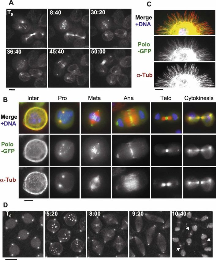
Downloaded from genesdev.cshlp.org on March 24, 2015 - Published by Cold Spring Harbor Laboratory Press
Polo control by Map205 and Cdk1
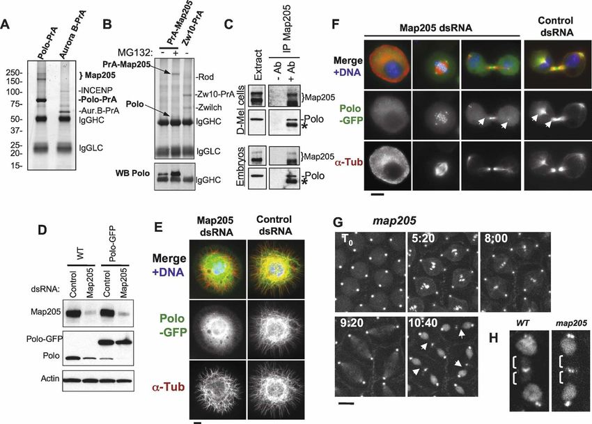
Figure 1. Polo localizes to MTs in a cell
cycle-dependent manner. In addition to cen-
trosomes, kinetochores, and the midbody,
Polo-GFP localizes to MTs in cytokinesis
and interphase. (A) Polo-GFP dynamics in D-
Mel cells by time lapse (time is shown in
minutes:seconds). Bar, 5 µm. See Supplemen-
tal Movie 1 and the text for details. (B) Polo-
GFP colocalizes with MTs during interphase
and cytokinesis. Cells were fixed with form-
aldehyde. Polo-GFP appears in green, ␣-Tu-
bulin is stained in red, and DNA is DAPI-
stained in blue. Bar, 5 µm. (C) Polo-GFP co-
localizes with MTs in a cell spreading on a
concanavalin-A-coated surface and fixed
with methanol/formaldehyde (only a portion
of the cell is shown for clarity). Stainings are
anti-GFP (green), ␣-Tubulin (red), and DNA
(blue). Bar, 5 µm. (D) GFP-Polo dynamics in a
transgenic syncytial embryo (cycle 13) by
time lapse (time is shown in minutes:sec-
onds). Note that GFP-Polo localizes to the
MTs of the central spindle during karyokine-
sis (arrows). Bar, 10 µm.
Polo’s targeting to MTs requires an interaction with Maiato et al. 2004). Its orthologs in human (Map4), Xeno-
Map205 pus (XMap230), and budding yeast (Mhp1) are all re-
quired for normal MT dynamics in interphase and mito-
To identify binding partners of Polo, we stably expressed sis (Goldstein et al. 1986; Cha et al. 1999; Andersen
Polo in fusion with two IgG-binding domains of the Pro- 2000; Maiato et al. 2004). Since Map205 localizes to
tein A from Staphylococcus aureus (Polo-PrA) and car- MTs throughout the cell cycle (Goldstein et al. 1986) and
ried out purifications by affinity of the PrA tag for rabbit Polo localizes to MTs in interphase and cytokinesis (Fig.
IgG. We then identified the associated proteins by mass 1), it is not surprising that we found that the two co-
spectrometry. The strongest band was identified as the localized in interphase and cytokinesis (Supplemental
MT-associated protein Map205 (Fig. 2A). This interac- Fig. S2).
tion was specific since Map205 was not isolated in pu- We hypothesized that Map205 could be required for
rifications of unrelated proteins in parallel and under the the recruitment of Polo to MTs. To test this, we depleted
same conditions. Furthermore, the apparent stoichiom- Map205 by RNAi in cultured cells (Fig. 2D,E). Interest-
etry and the absence of other bands of similar intensity ingly, Polo levels were reduced following depletion of
strongly suggested a direct interaction with Polo (Fig. Map205, suggesting that the interaction with Map205
2A). Reciprocally, endogenous Polo specifically copuri- may stabilize Polo. Consistent with this, we found that
fied with PrA-Map205 (Fig. 2B). Direct immunoprecipi- RNAi depletion of Map205 altered the cell cycle profile
tation of endogenous Map205 confirmed its interaction as revealed by FACS and led to an increase in mitotic
with endogenous Polo, in both D-Mel cells and embryos index accompanied by an accumulation of cells in pro-
(Fig. 2C). Cofractionation of Polo with Map205 was also metaphase (Supplemental Fig. S3). We verified that Polo
recently shown in a sucrose gradient analysis of Dro- destabilization following Map205 depletion was caused
sophila MT-binding proteins prepared from embryos (So- by the loss of interaction between Polo and Map205 and
rokina and Kashina 2005). Map205 is a functionally con- not from other perturbations arising from the loss of
served MT-associated protein (Goldstein et al. 1986; Map205, by replacing the endogenous Map205 with a
GENES & DEVELOPMENT 2709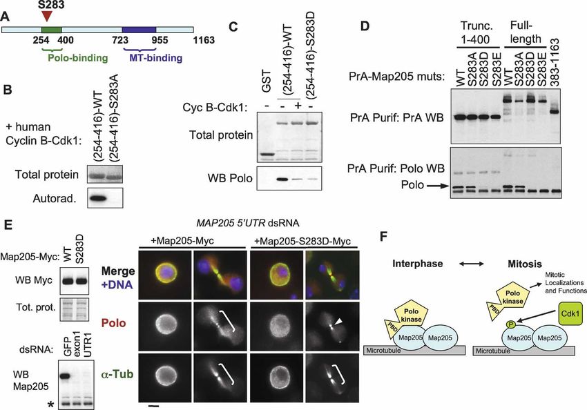
Downloaded from genesdev.cshlp.org on March 24, 2015 - Published by Cold Spring Harbor Laboratory Press

Archambault et al.

Figure 2. Polo interacts with Map205, and this is required for its localization to MTs. (A) Polo specifically interacts with Map205.
Polo-PrA and Aurora B-PrA (control) were purified from stably expressing cell lines, and purified proteins were resolved on SDS-PAGE
and stained with Coomassie Blue. The indicated proteins were identified by mass spectrometry. (IgGHC) IgG heavy chain; (IgHLC) IgG
light chain. (Left) Molecular mass markers are in kilodaltons (kDa). (B) Reciprocal purification. (Top) PrA-Map205 and Zw10-PrA
(control) were purified from stably expressing cell lines, and products were analyzed as in A. In one sample, cells were treated overnight
with 25 µM MG132 to inhibit the proteasome, which results in a stabilization of Map205 (but does not lead to an arrest of the majority
of the cells in mitosis). (Bottom) The same purification products were probed for Polo by Western blot. (C) Endogenous Polo and
Map205 interact in D-Mel cells and embryos. Polo was coimmunoprecipitated with Map205 using anti-Map205 antibodies from D-Mel
cells and from syncytial embryos. As a control, antibodies were not added. (*) Antibodies from the IP cross-reacting in the Polo Western
blot. (D) Depletion of Map205 by RNAi. D-Mel cells, wild-type or stably expressing Polo-GFP, were treated with dsRNA against
Map205 or the bacterial kanamycin-resistance gene (control). Four days later, cells were analyzed by Western blotting against Map205,
Polo, and actin (loading control). Note that in addition to the depletion of Map205, Polo levels are also reduced. (E,F) Polo requires
Map205 for its localization to MTs. (E) Polo-GFP cells were treated with Map205 dsRNA or control dsRNA (as in D), spread on
concanavalin-A, fixed with methanol/formaldehyde, and stained for ␣-tubulin (red) GFP (anti-GFP; green), and DNA (blue). Bar, 5 µm.
(F) Polo-GFP cells were treated with Map205 or control dsRNA (as in D and E), fixed with formaldehyde, and stained for ␣-tubulin (red)
and DNA (blue). (Right) Note that unlike in the control, Polo-GFP fails to colocalize with MTs in cytokinesis after Map205 RNAi
(arrows). Also compare with cells in Figure 1B. Bar, 5 µm. (G) GFP-Polo dynamics in a transgenic syncytial embryo laid by a map205
homozygous null mother (cycle 13) by time lapse (time is shown in minutes:seconds). Note that, unlike in map205+ embryos (Fig. 1D),
GFP-Polo fails to localize to the MTs of the central spindle during karyokinesis in map205-null embryos (arrows). Bar, 10 µm. (H)
Magnified view of karyokinetic nuclei from wild-type (WT) or map205-null embryos expressing GFP-Polo (taken from G and Fig. 1D).
Note that unlike in the wild-type control, GFP-Polo fails to localize to central spindle MTs in the map205-null embryo.

variant that cannot bind Polo, Map205-S283E (see below)                 GFP-Polo was no longer visible on the MTs of the central
(Supplemental Fig. S4). Strikingly, depletion of Map205                 spindle in karyokinesis (cf. Figs. 2G and 1D, 2H). How-
also resulted in the loss of either Polo-GFP (Fig. 2E-F) or             ever, Polo-GFP was still able to localize to centrosomes,
endogenous Polo (Supplemental Fig. S1) at MTs. MTs                      kinetochores, and the spindle midzone in the absence of
also appeared more fragmented and disorganized follow-                  Map205 (Fig. 2F–H). Polo localization to the cytokinetic
ing Map205 RNAi (Fig. 2E,F). To verify that Map205 was                  midbody has been previously shown to depend on an-
also required to localize Polo on MTs in the fly, we in-                other MT-associated protein, Feo/PRC1 (D’Avino et al.
troduced a GFP-Polo transgene in map205-null flies. In                  2007; Neef et al. 2007), and is therefore distinct from the
embryos laid by map205 homozygous null mothers,                         pool of Polo bound to Map205. Together these results

2710      GENES & DEVELOPMENT
Downloaded from genesdev.cshlp.org on March 24, 2015 - Published by Cold Spring Harbor Laboratory Press
Polo control by Map205 and Cdk1
demonstrate that Polo’s recruitment to MTs in inter- (amino acids 298–576) was not sufficient for this inter-
phase and cytokinesis is strongly dependent on an inter- action (Supplemental Fig. S5). Altogether, these results
action with Map205. strongly suggest that the interaction of Polo with
Map205 requires the PBD as well as some structural el-
ements of the kinase domain of Polo.
The Polo-Box Domain is required but not sufficient
for Polo to interact with Map205
Polo binding does not require phosphopriming
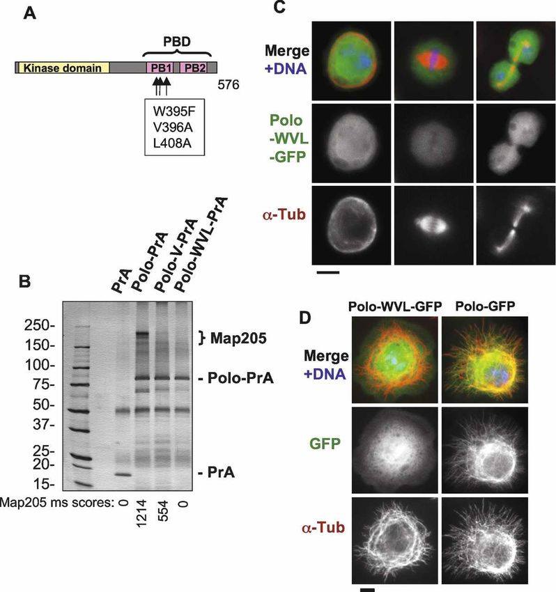
Plks stably interact with their targets via the Polo-Box
of Map205
Domain (Elia et al. 2003a,b; Lowery et al. 2004). To test
if the PBD of Polo was required for its interaction with In order to map the binding site of Polo in Map205, we
Map205 and localization to MTs, we expressed PBD-mu- first generated truncations of Map205 fused with PrA
tant forms of Polo in fusion with either PrA or GFP. and used them in a copurification assay probing for Polo.
Amino acid substitutions in conserved residues have The robust copurification of PrA-Map205 with endog-
been shown to disable the PBD of hPlk1 (Seong et al. enous Polo (Fig. 2B) was disrupted by truncations remov-
2002). We introduced the equivalent mutations in Dro- ing a region comprised between amino acid residues 254
sophila Polo (W395F, V396A, and L408A = Polo-WVL) and 400 of Map205. Conversely, residues 254–416 were
(Fig. 3A). This mutant form of Polo (or a truncated form sufficient to interact strongly with Polo (Fig. 4A–C). Sev-
of Polo removing the PBD) failed to copurify Map205 eral reports suggested that Plks require the PBD to bind
(Fig. 3B; data not shown) and to localize to centrosomes, to a S-(pS/pT) motif on their targets (see Introduction).
kinetochores, the central spindle, or interphase MTs Residues 254–416 contain only one such potential SS or
(Fig. 3C,D; data not shown). Even the V396A substitu- ST motif (S381–S382) (Fig. 4B), which if phosphorylated
tion alone largely abolished the interaction of Polo with at the second residue would correspond to the known
Map205 (Fig. 3B) and its localization to MTs (data not Polo kinase-binding motif. We therefore replaced the ser-
shown). Therefore, a functional PBD is required for Polo ine residue with an alanine residue (S382A). Surpris-
to interact with Map205 and for MT localization. How- ingly, PrA-Map205(254–416)–S382A was still able to
ever, in a copurification test performed using D-Mel cells bind Polo just as strongly as the wild-type equivalent
expressing tagged proteins, we found that the PBD along (Fig. 4C). To our knowledge, this is the first demonstra-
with the linker separating it from the kinase domain tion of an interaction involving Polo (or any Plk) that
Figure 3. Polo requires a functional Polo-Box
Domain to interact with Map205 and to localize
to MTs. (A) Amino acid substitutions introduced
in Polo to disrupt the Polo-Box Domain (PBD).
The kinase domain is in yellow, and the two Polo
Boxes (PB1 and PB2) are in pink. (B) A functional
Polo-Box Domain is required for Polo to interact
with Map205. Stable cell lines expressing Polo-
wt, Polo-V (V396A), Polo-WVL (W395F, V396A,
and L408A) in fusion with PrA, or PrA alone were
used in affinity purifications. Purified proteins
are shown on a Coomassie Blue-stained gel. Each
purification product was assayed for the presence
of Map205 in the area defined by the bracket.
Mascot scores (see Materials and Methods) for
Map205 are shown under the gel image. (IgGHC)
IgG heavy chain; (IgHLC) IgG light chain. (Left)
Molecular mass markers are in kilodaltons (kDa).
(C,D) A functional Polo-Box Domain is required
for Polo to localize to MTs. (C) Cells stably ex-
pressing Polo-WVL in fusion with GFP (green)
were fixed with formaldehyde and stained for ␣-
tubulin (red) and DNA (blue). Compare with Fig-
ure 1B. Note that Polo-WVL-GFP fails to localize
to MTs, kinetochores, centrosomes, and the mid-
body. Bar, 5 µm. (D) Cells stably expressing Polo-
GFP or Polo-WVL-GFP were plated on concanav-
alin-A, fixed with methanol/formaldehyde, and
stained for GFP (anti-GFP; green), ␣-tubulin (red),
and DNA (blue). Bar, 5 µm.
GENES & DEVELOPMENT 2711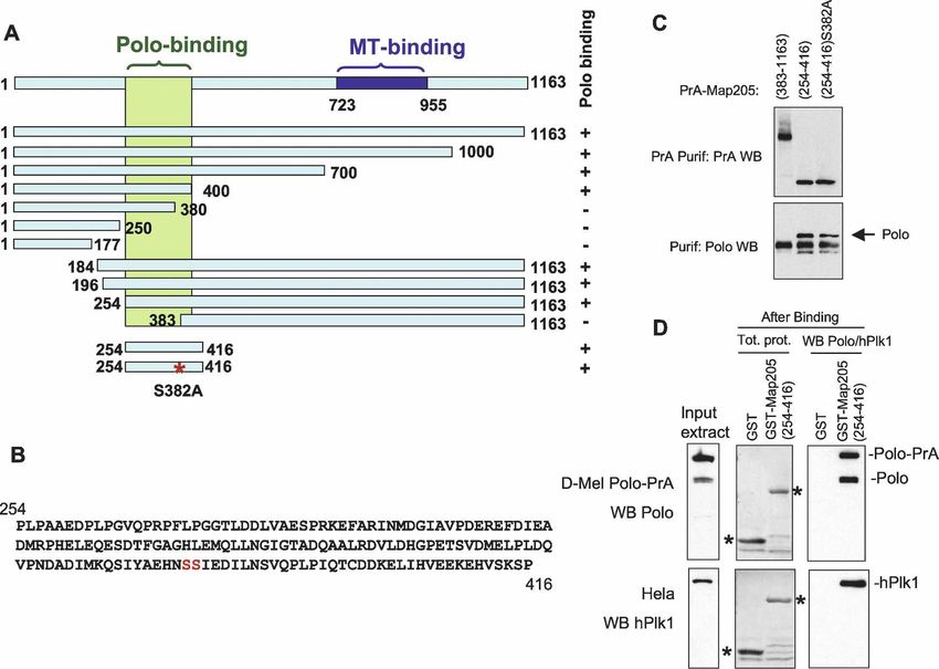
Downloaded from genesdev.cshlp.org on March 24, 2015 - Published by Cold Spring Harbor Laboratory Press

Archambault et al.

Figure 4. Polo binding to Map205 does not require phosphopriming. (A) Summary of a truncation analysis defining a small region of
Map205 necessary and sufficient for Polo binding. The assay used is as in C and Figure 2B. Polo binds to the region in green. The
MT-binding region (blue) was defined elsewhere (Irminger-Finger et al. 1990). (B) Sequence of a small segment of Map205 (amino acids
254–416) sufficient for binding Polo. Note that it contains only one potential canonical PBD-binding motif (SS or ST) at S381–S382.
(C) Map205 does not require phosphopriming at a canonical PBD-binding motif (S-pS or S-pT) for Polo binding. Cells transiently
expressing the indicated Map205 mutants in N-terminal fusion with Protein A were used in Protein A affinity purification, and the
products were probed for Protein A and Polo. Note that Map205(254–416)–S382A can still interact with Polo. (D) Map205 does not
require phosphorylation to interact with Drosophila Polo or human Plk1. GST-Map205(254–416) or GST alone (control) were ex-
pressed in bacteria, purified (asterisks), and subjected to a Polo or hPlk1-binding assay using D-Mel (stably expressing Polo-PrA) or
HeLa cell extracts, respectively. Purification products were analyzed by Western blotting for Polo (top) or hPlk1 (bottom).

does not require a S-(pS/pT) phosophopriming motif in                the central spindle during karyokinesis when heterolo-
vivo. However, the possibility remained that phos-                   gously expressed in Drosophila syncytial embryos (Pear-
phopriming for Polo binding could still occur at a non-              son et al. 2006). In the case of Drosophila GFP-Polo, this
canonical site on Map205. To investigate this possibil-              localization requires Map205 (Fig. 2G,H), and it seems
ity, we expressed Map205(254–416) in fusion with GST                 reasonable to assume that the localization of hPlk1 to
in Escherichia coli. Because bacteria possess little kinase          the central spindle observed in Drosophila embryos is
activity, and as their kinases differ strongly from those of         also mediated by its interaction with Map205. The phos-
eukaryotic cells, Map205(254–416) expressed in bacteria              phopriming-independent interaction between Polo or
should not be phosphoprimed for Polo binding. We incu-               hPlk1 and Map205 reinforces the emerging idea that Plks
bated GST-Map205(254–416) beads with a Drosophila                    can engage in such interactions (Garcia-Alvarez et al.
cell extract at 4°C and probed for Polo binding by West-             2007).
ern blotting. Strong and specific binding of Polo was de-
tected in this assay (Fig. 4D). Taken together, these re-
                                                                     Phosphorylation of Map205 by Cdk1 disrupts
sults indicate that the interaction between Polo and
                                                                     its interaction with Polo
Map205 does not require any phosphopriming.
   We also found that hPlk1 was able to interact strongly            Because we initially suspected that Polo’s interaction
with bacterially expressed Map205 in the same assay,                 with Map205 would require phosphopriming, we
using a HeLa cell extract, and independently of priming              mapped phosphorylation sites on Map205 by mass spec-
phosphorylation (Fig. 4D). This is consistent with the               trometry (Supplemental Fig. S6). We identified four sites:
previously reported ability of GFP-hPlk1 to localize to              S283, S679, S690, and S852. These are novel sites that

2712      GENES & DEVELOPMENT
Downloaded from genesdev.cshlp.org on March 24, 2015 - Published by Cold Spring Harbor Laboratory Press

                                                                                                        Polo control by Map205 and Cdk1

were not reported in a previous study that identified                 (Fig. 5C). The phospho-mimic mutations S283D and
phosphorylation sites in the Drosophila proteome, in-                 S283E also abolished the binding of Polo to Map205 in
cluding other sites in Map205 (Bodenmiller et al. 2007).              vivo, while the S283A mutation had no detectable effect
One of the sites we identified, S283 is located in the                (Fig. 5D). To test the effect of phosphorylation on Polo’s
region of Map205 that mediates Polo binding (Figs. 4A,                localization in vivo, we made stable cell lines expressing
5A). We noticed that S283 lies in a perfect consensus                 either Map205-Myc or the phospho-mimic mutants
motif for phosphorylation by cyclin-dependent kinases                 Map205-S283D-Myc and Map205-S283E-Myc. All fusion
(SPRK; the ideal motif is S/T-P-X-K/R) (Nigg 1993). In-               proteins were still able to localize to MTs (Supplemental
deed, human cyclin B–Cdk1 was able to strongly phos-                  Fig. S7). We then depleted the endogenous Map205 in
phorylate Map205(254–416) in vitro, but not the equiva-               those cell lines using dsRNA against an untranslated re-
lent S283A mutant form (Fig. 5B). Phosphorylation of                  gion (UTR) of the transcript (Fig. 5E). While expression of
Map205(254–416) by cyclin B–Cdk1 or substitution at                   wild-type Map205-Myc did not affect the colocalization
S283 with a negatively charged, phospho-mimic aspartic                of Polo with ␣-tubulin in interphase and cytokinesis, the
acid residue (S283D) reduced its affinity for Polo in vitro           S283D mutant disrupted this localization, leaving Polo

Figure 5. Map205 phosphorylation at Cdk1 site Ser 283 prevents interaction with Polo. (A) Map205 is phosphorylated at Ser 283,
inside the Polo-binding region in vivo (for details, see Supplemental Figure S6). (B) Ser 283 of Map205 is a target of cyclin B–Cdk1 in
vitro. Map205(254–416)-wt or S283A was generated using bacterial expression and subjected to a kinase assay using human cyclin
B–Cdk1. The total protein (amido black staining) and the autoradiograph are shown. (C) Phosphorylation of Map205(254–416) by cyclin
B–Cdk1 or phosphomimicking at Ser 283 inhibits the interaction with Polo in vitro. The indicated proteins were treated or not with
cyclin B–Cdk1 before being subjected to a Polo-binding assay as in Figure 4D. (D) Mimicking phosphorylation at Ser 283 of Map205
abolishes its interaction with Polo in vivo. Cells transiently expressing the indicated proteins (full-length or truncated Map205) were
used in Protein A affinity purification, and the products were probed for PrA and Polo. Note that the phosphomimicking S283D and
S283E mutations abolish the interaction with Polo. (E) Phosphomimicking phosphorylation at S283 disrupts the localization of Polo
on MTs. (Top left) Stable cell lines expressing Map205-Myc (wild type and S283D) were created and expressed at similar levels. RNAi
targeting an untranslated region of the endogenous MAP205 transcript was used to deplete endogenous Map205. (Bottom left) The
Western blot shown is from cells not transfected with Map205 cDNA, for clarity. Cells were then stained for Polo (red), ␣-Tubulin
(green), and DNA (blue). Note that Polo is present on MTs (brackets) when Map205-Myc wild type, but not S283D is expressed. Polo
is sometimes detected at the midbody (arrow) even when it does not localize to MTs. Bar, 5 µm. (F) Model for the regulation of Polo
localization by Map205 and Cdk1. In interphase, Polo is sequestered to MTs via an interaction with Map205 that depends on both the
PBD and the kinase domain of Polo, but that does not require priming phosphorylation. At mitotic entry, Cdk1 phosphorylates
Map205, which releases Polo from Map205 and MTs. Polo is then free to translocate to its mitotic localizations, where it functions
to promote proper mitosis. In late mitosis, Cdk1 becomes inactivated, and Polo returns to MTs in cytokinesis.

                                                                                                   GENES & DEVELOPMENT            2713
Downloaded from genesdev.cshlp.org on March 24, 2015 - Published by Cold Spring Harbor Laboratory Press
Archambault et al.
only in the cytoplasm and on the midbody (Fig. 5E; mid- This study indicated that Greatwall could act to down-
body staining is not always observed). Similar results regulate Polo or a Polo-dependent pathway required for
were obtained when the S283E mutant was expressed the cohesion between centrosomes and the nuclear en-
(data not shown). As shown above, Polo localization to velope (Archambault et al. 2007). In both cases, detached
MTs is strong in interphase and cytokinesis, when cyclin centrosomes were also detected in later mitotic stages,
B–Cdk1 activity is low, and is almost lost completely in leading to mono-astral spindles (Fig. 6B,C; Archambault
early to mid-mitosis, when cyclin B–Cdk1 is most ac- et al. 2007). However, the percentage of nuclei showing
tive. Thus, together, our results strongly suggest that centrosome detachment in prometaphase to telophase
cyclin B–Cdk1 phosphorylation of Map205 in mitosis was lower than in prophase (Fig. 6B). This is consistent
disrupts the interaction between Polo and Map205 until with time-lapse studies showing that detached centro-
Cdk1 inactivation at anaphase onset. Therefore, Cdk1 somes can be recaptured by growing spindles in prometa-
activation at mitotic entry is poised to promote the re- phase (Archambault et al. 2007). As expected, the cen-
lease of Polo from Map205 (Fig. 5F), enabling Polo to trosome detachment defects were also dependent on
concentrate on kinetochores, centrosomes, and centro- the ability of Map205 to interact with Polo (Fig. 6D);
meres, where it can conduct its essential mitotic func- overexpression of Map205-S283A induced more centro-
tions (van de Weerdt and Medema 2006). Inactivation of some detachment than Map205-wt (86% vs. 64%, re-
cyclin B–Cdk1 in late mitosis would allow Polo to bind spectively), while overexpression of Map205-S283E in-
Map205 again. duced considerably fewer defects (13%, similar to
polo11/+ alone). Our results strongly suggest that
Map205 is able to inhibit Polo’s cellular activity in vivo
Map205 contributes to the regulation of Polo in vivo
by direct binding. We note that such an inhibition does
To explore the function of Polo interaction with Map205 not necessarily require direct inhibition of the kinase
in vivo, we generated transgenic fly lines expressing domain. This presumably imposes a requirement for
Myc-tagged Map205, either wild type or mutated at S283 Cdk1-dependent release of Polo from Map205 at mitotic
(the Cdk1 phosphorylation site), under the control of the entry.
UASp promoter. We induced moderate overexpression of In order to assess if Map205 contributes to regulate
the Map205 transgenes in the female germline using the Polo in vivo, we tested if the removal of Map205 would
maternal ␣-Tubulin Gal-4 driver, allowing for a mater- rescue or enhance phenotypes observed in the context of
nal contribution to the early embryo. Females overex- a partial loss of Polo function. Although polo heterozy-
pressing wild-type Map205 and heterozygous for the gous females laid embryos that develop until adulthood,
polo11-null allele (Archambault et al. 2007) or a small these embryos show a low but significant frequency of
deficiency uncovering polo, laid embryos, but a majority single centrosome detachment in prophase nuclei of the
of them failed to hatch (Fig. 6A; Supplemental Fig. S8). syncytial blastoderm (Fig. 6B). By scanning deeper into
This effect was dependent on the ability of Map205 to early syncytial embryos, we were able to examine the
bind Polo because overexpression of the Map205-S283E preblastoderm nuclei in cycles 8–9 and found that polo
mutant, which fails to interact with Polo (Fig. 5), did not heterozygous-derived embryos had a much higher inci-
cause a failure of embryos to hatch. As expected, the dence of single centrosome detachment in prophase at
S283A mutant, which presumably retains the ability to that stage, which was almost never observed in wild-
bind Polo even when Cdk1 is active (Fig. 5), caused an type-derived embryos (Fig. 6E; Supplemental Fig. S9).
even more severe hatching failure than Map205-wt (Fig. This single centrosome detachment did not cause em-
6A; Supplemental Fig. S8). Moreover, this effect was de- bryonic lethality because nuclei kept the ability to mi-
pendent on a reduced gene dosage of Polo because em- grate to the cortex, probably because they always re-
bryos derived from polo+ females could hatch normally mained attached to one centrosome (we observed no
even when Map205 was overexpressed (Fig. 6A). To in- gross increase in the number of yolk nuclei) (data not
vestigate potential effects on mitotic divisions, we ex- shown) and because the detached centrosome could be
amined syncytial embryos at the early blastoderm stage recaptured. We tested how this polo-dependent defect
(cycles 10–13) by immunofluorescence. Embryos pro- was affected by deletion of map205. Western blot analy-
duced by polo heterozygous females overexpressing sis revealed that deletion of map205 did not destabilize
Map205 displayed a very high percentage of mitoses in Polo in the embryo (Fig. 6F) as it did in cultured cells (Fig.
which a single centrosome detached from the nuclear 2D); Polo degradation likely requires Fizzy related,
envelope in prophase (Fig. 6B,C). This defect is extremely which is absent in embryos (see Discussion). Strikingly,
rare in wild-type embryos but was, instead, significantly the frequency of single centrosome detachment observed
increased in syncytial blastoderm embryos derived from in heterozygous polo-derived embryos was suppressed
polo11 heterozygous females (Fig. 6B; Archambault et al. following deletion of map205 (Fig. 6E; sample images are
2007). Penetrant single centrosome detachment from nu- shown in Supplemental Fig. S9). This rescue was ob-
clei in prophase in early blastoderm embryos was re- served in both preblastoderm (from 69% to 36% nuclei
cently found to be the prevalent and initial defect occur- with centrosome detachment) and syncytial blastoderm
ring when the ratio of Greatwall:Polo activity was in- embryos (from 16% to 7.5%). We therefore conclude that
creased, and there too this was associated with a failure endogenous Map205 partially restrains the cellular ac-
in embryonic development (Archambault et al. 2007). tivity of Polo in syncytial embryos.
2714 GENES & DEVELOPMENTDownloaded from genesdev.cshlp.org on March 24, 2015 - Published by Cold Spring Harbor Laboratory Press
Polo control by Map205 and Cdk1
Figure 6. Map205 contributes to the regu-
lation of Polo in vivo. (A–C) Overexpression
of Map205 enhances Polo-dependent de-
fects. (A) Percentage of hatching embryos
produced by females of the indicated geno-
types. All genotypes included one copy of
the Maternal ␣-Tubulin Gal4 driver (not
shown). TM6C is a balancer chromosome.
Map205 transgenes were under the control
of the UASp promoter. Transgenic lines
were selected for equal and moderate over-
expression levels of Map205 variants
(Supplemental Fig. S8). At least 1000 em-
bryos laid by 12 females over 3 d were
scored for each genotype. (B) Percentage of
nuclei showing single centrosome detach-
ment in syncytial blastoderm embryos
(cycles 10–13) produced by females of the
indicated genotypes (as in A, separate ex-
periment). Between five and 10 embryos
were scored for each entry (±SEM). (C, right)
Examples of single centrosome detachment
(arrows) observed in prophase and in meta-
phase (genotypes are as in A and B). The
metaphase spindles on the bottom and on
the right show typical single centrosome de-
tachment, while the spindle on the top ap-
pears normal. (Left) Image taken from a con-
trol embryo where both centrosomes re-
main normally tethered to the nuclear
envelope in prophase. Stainings are ␣-Tubu-
lin (green), ␥-Tubulin (red), and DNA (blue).
Bar, 10 µm. (D) Percentage of prophase nu-
clei showing single centrosome detachment
in syncytial blastoderm embryos (cycles 10–
13) produced by females of the indicated ge-
notype (as in A and B, separate experiment).
Ten embryos were scored for each entry
(±SEM). (E) Deletion of Map205 partially
rescues the Polo-dependent centrosome de-
tachment. Percentage of prophase nuclei
showing single centrosome detachment in
preblastoderm (cycles 8–9) and early blasto-
derm (cycles 10–11) embryos produced by females of the indicated genotypes. Note that single centrosome detachment is frequent in
embryos laid by polo11/+ females and that this defect is partially rescued by deletion of map205. Between five and 10 embryos were
scored for each entry (±SEM). Refer to Supplemental Figure S9 for sample images. (F) Western blot from embryos laid by females of the
indicated genotypes (0–3 h collection; as in E). (Top) Map205 detection. (*) Cross-reacting band serving as a loading control. (Bottom)
Polo detection. Note that the absence of Map205 does not correlate with a lower amount of Polo in embryos (unlike in cultured cells).
Discussion mains in G1 (Lindon and Pines 2004). Second, human
Plk1 and Xenopus Plx1 are activated in mitosis by
How Plks are controlled in space and time during the cell phosphorylation at a threonine residue in the T-loop
cycle is not completely understood, despite three known (T210 in hPlk1, T201 in Plx1) (Qian et al. 1999; Kelm et
levels of control. First, hPlk1 has been shown to be ubiq- al. 2002). As this site is conserved, this likely acts as a
uitinated and degraded by the proteasome in mammalian universal activation mechanism. In human cells, Aurora
cells (Ferris et al. 1998; Lindon and Pines 2004). How- A and its adaptor protein Bora have been shown recently
ever, the importance and extent of conservation of this to activate hPlk1 by phosphorylation at T210 (Macurek
mechanism are still to be determined. For example, al- et al. 2008; Seki et al. 2008). A third control mechanism
though the destruction box identified in hPlk1 appears to lies in docking of Plks to prephosphorylated targets using
be conserved in Drosophila Polo (Lindon and Pines the PBD. This provides an efficient way to target Plks to
2004), Polo does not disappear (or even decrease signifi- their mitotic targets (Elia et al. 2003b; Lowery et al.
cantly in abundance) between mitosis and interphase 2004). It has also been recently shown that PBD-depen-
(Fig. 1; Supplemental Fig. S1). Moreover, even in mam- dent sequestration of Polo to a strong binding partner, a
malian cells, a substantial fraction of the hPlk1 pool re- female germline-specific protein named Matrimony
GENES & DEVELOPMENT 2715Downloaded from genesdev.cshlp.org on March 24, 2015 - Published by Cold Spring Harbor Laboratory Press Archambault et al. (Mtrm), can keep this kinase inactive in meiotic pro- whether or not Polo’s kinase activity is inhibited at the phase I (Xiang et al. 2007). Polo later overcomes this structural level. Our antibodies against Polo and Map205 inhibition, and is thought to trigger germinal vesicle did not allow visualization of the endogenous proteins in breakdown (Xiang et al. 2007). This interaction depends the embryo for technical reasons, but the genetic, func- on phosphopriming of Mtrm. In contrast, we now de- tional, and biochemical results obtained in this system scribe a mechanism for regulating Polo through its cell support the model presented here. Exploring what other cycle-regulated sequestration to MTs by strong binding tissues rely on this Polo-control pathway should be the to a MT-associated protein in a manner that is not subject of future studies. primed by phosphorylation but nonetheless requires the In addition, the interaction with Map205 appears to PBD. stabilize Polo (see below). In this way, the Map205–Polo Our results shed new light on how Drosophila Polo is complex on MTs (or even if partly present as a cytosolic regulated at mitotic entry and exit. Based on the results pool) could constitute a reservoir of Polo, making Polo shown above, we suggest a model whereby Polo is se- available for rapid, Cdk1-triggered mobilization at mi- questered on MTs (and can be stabilized) in interphase by totic entry, and resequestration at mitotic exit. The pre- interacting with Map205, and where this interaction is vention of hPlk1 from binding PRC-1 by Cdk1-depen- negatively regulated by Cdk1 activity in early mitosis dent phosphorylation was recently identified as a mecha- (Fig. 5F). Although Polo kinases have been reported to nism for timing hPlk1 localization to the midbody (Neef interact with tubulins in vitro (Feng et al. 1999; Neef et et al. 2007). Our findings augment the general model al. 2003), this does not appear to provide an efficient whereby the choice of Polo kinase binding partners is mechanism of Polo targeting to MTs, which is instead controlled by Cdk1 activity during the cell cycle (Neef et strongly dependent on Map205. The cellular concentra- al. 2007). Our model does not exclude the possibility that tion of Map205 is very similar or possibly higher than Polo is targeted to MTs for another function. For ex- that of Polo (Supplemental Fig. S10), consistent with a ample, Polo could regulate Map205. Indeed, we found role for Map205 in sequestering Polo. Such sequestration that Polo can phosphorylate Map205 in vitro (Supple- of Polo onto MTs could serve to store and stabilize the mental Fig. S11), and the physiological relevance of this kinase in an innocuous location, until needed at mitotic phosphorylation remains to be investigated. Addition- entry. ally, targeting of Polo to MTs could facilitate its regula- While we show that phosphorylation of Map205 at tion of motor proteins (Neef et al. 2003) or other MT- S283 is sufficient to block its interaction with Polo and associated proteins with roles in interphase or cytokine- the localization of Polo to mitotic MTs, we do not know sis. if this phosphorylation event is necessary for inhibiting We identified orthologs of Map205 in the 12 sequenced the targeting of Polo to MTs. For example, phosphoryla- Drosophila genomes. An alignment between these pro- tion of Map205 at other sites may also contribute to tein sequences (Supplemental Fig. S12) shows that the negatively regulate the interaction. We have not yet rig- N-terminal Polo-binding region of Map205 (within orously tested this possibility. However, the fact that amino acids 254–416 in Drosophila melanogaster) is overexpression of Map205-S283A in embryos enhances very well conserved. Importantly, the Cdk1 phosphory- polo-dependent defects more strongly than overexpres- lation site (S283) and the sequence immediately sur- sion of Map205-wt suggests that phosphorylation of rounding it (VAESPRK) are perfectly conserved between Map205 at S283 is necessary for full inhibition of the all 12 species. In fact, the Polo-binding region appears to Map205–Polo interaction in vivo. be even better conserved than the C-terminal MT-bind- We showed that the single centrosome detachment ing region (amino acids 723–955 in D. melanogaster). from prophase nuclei observed in polo mutants is en- This suggests to us that the Polo-binding function of hanced by overexpression of Map205 and suppressed by Map205 may be at least as important as its MT-binding the loss of Map205 function. Centrosome detachment function, and that the molecular mechanisms reported appeared more sensitive to a reduction in Polo activity in here are likely to be conserved between species. the preblastoderm than in the early blastoderm embryo. RNAi depletion of Map205 in cultured Drosophila We speculate that this may be due to a difference in the cells leads to perturbations in the cell cycle profile, in- forces that the centrosome–nuclear envelope linkage cluding an increase in the percentage of cells in mitosis, must withstand at those different stages. The organismal and more specifically in prometaphase. This may be a and cellular phenotypes observed in polo mutants after consequence of the Polo destabilization (Fig. 2D; Supple- Map205 overexpression were dependent on the ability of mental Fig. S4), since partial loss of Polo function also Map205 to interact with Polo and identical to those results in an accumulation of cells in prometaphase. By caused by an increase in the activity of Greatwall, a re- replacing endogenous Map205 with a form of Map205 cently identified antagonist of Polo (Archambault et al. that cannot bind Polo (Map205-S283E), we verified that 2007). Importantly, the suppression of the polo-depen- the destabilization of Polo following Map205 depletion dent centrosome-detachment phenotype by deletion of in D-Mel cells results from the loss of interaction be- map205 is consistent with the notion that endogenous tween Polo and Map205 and not from other perturba- Map205 functions to regulate Polo in vivo. Sequestering tions arising from the loss of Map205 (Supplemental Fig. of Polo by Map205 could prevent it from accessing its S4). In contrast, removal of Map205 from the early em- targets prematurely on centrosomes and kinetochores, bryo did not result in a destabilization of Polo (Fig. 6F). 2716 GENES & DEVELOPMENT
Downloaded from genesdev.cshlp.org on March 24, 2015 - Published by Cold Spring Harbor Laboratory Press
Polo control by Map205 and Cdk1
Budding yeast Cdc5 and human Plk1, both orthologs of sequence conservation between Map205 and its human
Polo, undergo cell cycle-regulated degradation that is ortholog, Map4, (our unpublished observations) but this
thought to depend on Cdh1 (Lindon and Pines 2004; Vis- is not unusual among MT-associated proteins. The rela-
intin et al. 2008). If Drosophila Polo is degraded by the tive importance of the different mechanisms for regulat-
same pathway, our results can be explained by the ab- ing the location and activity of Plks—activating phos-
sence of Fizzy related (Drosophila Cdh1) in the early em- phorylation, proteolysis, and protein binding—will al-
bryo (Sigrist and Lehner 1997). most certainly depend on the organism and may even
Our experiments reveal that Map205 can strongly alter vary between cell and tissue types. As different types of
the subcellular localization of Polo (by targeting Polo to cancer cells may undergo deregulation of different hPlk1
MTs), stabilize Polo in D-Mel cells, and inhibit Polo’s control mechanisms, it will be of considerable interest to
cellular activity in the embryo (probably by sequestering further explore the relative contributions of these alter-
of the enzyme away from its substrates). Yet, despite the native mechanisms for regulating hPlk1 in different dis-
ability of Map205 depletion to delay progression through ease situations.
prometaphase in cultured cells, map205-null flies are vi-
able and fertile (Pereira et al. 1992), and we did not detect
obvious mitotic defects in their embryos or dividing neu- Materials and methods
roblasts (data not shown). Because of the existence of at DNA constructs
least three more, partially overlapping levels of control
of Polo activity—proteolysis, activating phosphoryla- Drosophila expression vectors were made using the Gateway
technology (Invitrogen). The Polo, Aurora B, Zw10, and Map205
tion, phosphopriming of Plk docking—it is difficult to
genes (as well as truncations of Map205 and Polo) were cloned
assess the consequences of a total failure to down-regu- by recombination of PCR products into the pDONR221 entry
late and stabilize Plks in interphase. Other mechanisms vector (Invitrogen). The same genes were then recombined into
regulating Polo may compensate for the loss of the the relevant destination vectors for expression in fusion with
Map205-dependent pathway in most tissues. Indeed, one Protein A, GFP, or Myc, at the N or C terminus, and under the
would need to combine disruptions of the multiple regu- control of the Actin 5C, UASp, or MT (Metallothionein) pro-
latory mechanisms for Polo or hPlk1 to determine the moter. Amino acid substitution mutants were made using
effects of a full failure to down-regulate or up-regulate QuickChange (Stratagene). Bacterial expression constructs for
these kinases in the cell cycle, and these experiments Map205 fragments in N-terminal fusion with GST-TEV were
may become possible as we continue to dissect the path- made by inserting MAP205 cDNA into pGEX4T-GST-TEV.
ways controlling these kinases. The existence of mul-
tiple, partially redundant control systems is seen for Cell culture, cell lines, and RNAi
other cell cycle regulators with functions at key stages of D-Mel cells (Invitrogen) were cultured in SFM medium (Invit-
cell cycle progression (for example, Cdk1, the APC, or rogen) supplemented with 1 mM glutamine, penicillin, and
the prereplicative complex). Such redundancy in regula- streptomycin. Transfections and stable cell line selection were
tory mechanisms can provide robustness to the system done as described (D’Avino et al. 2006). Transiently transfected
that can become crucial in situations of stress. cells were analyzed 3 d later. For RNAi, 3 × 10⁶ cells were trans-
We showed that the Polo–Map205 interaction does not fected with 30 µg of dsRNA and Transfast (Promega) and al-
lowed to grow in SFM for 3-4 d before being assayed. dsRNAs
depend on priming phosphorylation. Interestingly, the
were synthesized from PCR products; oligonucleotide se-
PBD is essential for this interaction, but it is not suffi-
quences are available upon request.
cient, and therefore some elements of the kinase domain
appear to be also needed. This is the first time that such
Fly genetics
requirements have been demonstrated for a Plk interac-
tion in vivo. This phosphorylation-independent mode of The GFP-Polo X-chromosome insertion used (Moutinho-Santos
interaction is well suited to control Polo in interphase, et al. 1999) was expressed from the Polo promoter, at levels very
when cellular kinase activities are typically low. Since similar to endogenous Polo (data not shown). The map205-null
third chromosome used was documented elsewhere (Pereira et
hPlk1, like Drosophila Polo, can also bind Map205, this
al. 1992) and consisted in a small deletion removing the genes
mode of interaction is likely to be conserved for other
map205 and modulo, and where modulo was reinserted as a
Plks and could potentially involve other substrates. It transgene. Transgenic flies for expression of UASp-Map205-
was recently shown that the PBD of hPlk1 is capable of Myc (wild type and mutants) were created by injections of our
interacting with Cdc25C peptides with similar affinities plasmids by BestGene, Inc. Insertions on the third chromosome
for the phospho- and nonphospho forms in vitro (Garcia- allowing very similar levels of expression were selected for ex-
Alvarez et al. 2007). The full biological significance of periments (Supplemental Fig. S8). The deficiencies used were
these modes of interaction and how they affect the ac- the DrosDel Df(3R)ED6362 (from John Roote) uncovering
tivity of the kinase should become clearer as the molecu- map205 and Df(3L,77A1-77D1) (from Adelaide Carpenter), un-
lar details of more Plk–target interactions are investi- covering polo.
gated.
The strong interaction between hPlk1 and Map205 in Protein affinity purification and immunoprecipitation
vitro also raises the possibility that the type of regulatory Large-scale Protein A-affinity purifications (Figs. 2A,B, 3B) were
mechanism identified here may be conserved beyond the performed as described (Chen et al. 2007). Small-scale Protein
Drosophila genus. We note that there is relatively poor A-affinity purifications (Figs. 4A,C, 5D; Supplemental Figs. S5,
GENES & DEVELOPMENT 2717Downloaded from genesdev.cshlp.org on March 24, 2015 - Published by Cold Spring Harbor Laboratory Press
Archambault et al.
S11) followed a similar protocol. Briefly, between 3 × 10⁷ and electron transfer dissociation [ETD] [Syka et al. 2004] EI/CI
1 × 10⁸ cells stably or transiently expressing the Protein A fu- source) instruments (ThermoFisher). The LTQ was operated in
sion were used as starting materials. Cells were lysed by four a data-dependent neutral loss mode, in which peptide ions were
cycles of freeze–thaw in lysis buffer (75 mM K-HEPES at pH 7.5, subjected to collision-induced dissociation (CID) to generate se-
150 mM KCl, 2 mM MgCl2, 2 mM EGTA, 5% glycerol, 0.1% quence-specific fragment ions. The LTQ XL was operated to
NP-40, Complete Protease Inhibitors [Roche]). Lysates were switch between standard CID and ETD modes in a data-depen-
clarified by centrifuging at 20,000g for 30 min in a tabletop dent manner. For identification of proteins, the MS/MS data
centrifuge at 4°C. Supernatants were incubated for 1–2 h with were used to search the mouse NCBI database using the Mascot
Dynabeads M270 Epoxy (Invitrogen) conjugated to whole rabbit (Matrix Science) or SEQUEST (ThermoFisher) search engines.
IgG (MP Biochemicals). Beads were washed five times with 1
mL of lysis buffer for 5–10 min. Proteins were eluted from the
Kinase assays
beads with 0.5 M NH4OH, 0.5 mM EDTA, and desiccated before
being redissolved in Laemmli buffer for SDS-PAGE (Sigma). For For the kinase assay shown in Figure 5B, GST-TEV-
immunoprecipitations (Fig. 2C) of Map205 from D-Mel cells, Map205(254-416)-wt or S283A was expressed in bacteria and
extracts were prepared as above, but without glycerol in the purified using Sepharose-4B (Amersham). Proteins were eluted
lysis buffer. For immunoprecipitations of Map205 from em- with the TEV protease (Invitrogen). Elution proteins were sub-
bryos (Fig. 2C), embryos from 0–3-h collections were dechori- jected to a kinase reaction using human cyclin B–Cdk1 (Invit-
onated in 50% bleach, crushed in lysis buffer (without glycerol), rogen) in 20 mM K-HEPES (pH 7.5), 2 mM MgCl2, 1 mM DTT,
and clarified as above. In both cases, clarified lysates were in- 500 µM ATP, [␥-32P]ATP, incubating for 20 min at 30°C. Reac-
cubated with Map205 antibodies for 2 h and with Protein A- tions were stopped by heating in Laemmli SDS-PAGE sample
conjugated DynaBeads (Invitrogen) for a further 2 h, before being buffer (Sigma), and reaction products were resolved by SDS-
washed in lysis buffer as above and eluted in 100 mM glycine PAGE and transferred to nitrocellulose, and membranes were
(pH 2.5). analyzed by autoradiography. For the kinase assay shown in
Supplemental Figure S11, protein complexes on beads were di-
rectly incubated in 20 mM K-HEPES (pH 7.5), 2 mM MgCl2, 1
Immunofluorescence and Western blotting
mM DTT, 500 µM ATP, and [␥-32P]ATP and analyzed as above.
For the experiments presented in Figures 1B, 2F, 3C, and 5E and
Supplemental Figures S1, S2, and S3, cells were fixed with form-
aldehyde and stained as described (Bettencourt-Dias et al. 2004), In vitro binding assay
and the antibodies were rat anti-␣-Tubulin YL1/2 (Sigma), Bacterially expressed and purified GST-fusion proteins (as
mouse monoclonal anti-Polo M294, rabbit anti-Map205 (gift above) were kept on beads and assayed for in vitro binding to
from Andrea Pereira), rabbit anti-phospho-H3 (Upstate Biotech- Polo or hPlk1 by incubating with D-Mel or HeLa cell extracts in
nologies), anti-rat-FITC (Jackson), anti-rat-Rhodamine (Jack- lysis buffer (75 mM K-HEPES at pH 7.5, 150 mM KCl, 2 mM
son), anti-mouse-Rhodamine (Jackson), anti-mouse-FITC, and MgCl2, 2 mM EGTA, 5% glycerol, 0.1% NP-40, 0.5% Triton
anti-rabbit-Rhodamine (Jackson). For the experiments presented X-100, Complete Protease Inhibitors [Roche]) for 30 min at 4°C,
in Figures 1C, 2E, and 3D and Supplemental Figure S7, cells followed by five washes (5–10 min each) in lysis buffer. Beads
were plated on concanavalin-A (Rogers et al. 2002) and fixed were then heated in Laemmli SDS-PAGE sample buffer and
with 90% methanol, 3.3% formaldehyde, 5 mM NaHCO3 for 15 analyzed by anti-Polo Western blot.
min on dry ice, and then for 12 min at room temperature. Cells
were then washed with PBS and stained as in Bettencourt-Dias
et al. (2004); the antibodies were rabbit anti-GFP A6455 (Invit- Microscopy and flow cytometry
rogen), mouse monoclonal anti-␣-Tubulin DM1A (Sigma), rab- Fixed cells stained by immunofluorescence were visualized on a
bit anti-Myc (Santa Cruz Biotechnologies), anti-rabbit-Alexa488 Zeiss Axiovert 200 fluorescence microscope with Metamorph
(Invitrogen), anti-mouse-Alexa594 (Invitrogen), and anti-rabbit- software. Movies of Polo-GFP D-Mel cells and GFP-Polo em-
Alexa488 (Invitrogen). Embryos were stained as described (Ar- bryos were made on a Zeiss Axiovert 200 fluorescence micro-
chambault et al. 2007). Western blotting was done using a stan- scope equipped with the PerkinElmer UltraVIEW confocal scan-
dard method, and the antibodies were Protein A: peroxidase- ner and software. Fixed embryos were examined on a Nikon
conjugated rabbit IgG (Jackson 011-030-003); Polo: M294 Optiphot fluorescence microscope equipped with a Bio-Rad
monoclonal; Map205 (gift from Andrea Pereira). MRC1024 confocal scanner and software. For flow cytometry,
D-Mel cells were fixed in 70% ethanol, RNase-treated, and
Protein identification and phosphorylation site mapping stained with propidium iodide. Results were analyzed with
by mass spectrometry Summit (Dako Cytommation) and Multicycle (Phoenix Flow
Systems).
Protein identification was performed as described (Chen et al.
2007).
For phosphorylation site mapping, Bands corresponding to po-
Acknowledgments
tential phosphoproteins were excised from the gel, cut into
squares, and subjected to reduction and alkylation with DTT We thank Michaela Scigelova and Gary Woffendin from Ther-
and iodacetamide, respectively. Proteins were digested over- moFisher for performing the ETD mass spectrometry experi-
night with trypsin. Reduction, alkylation, and digestion were ments for phosphorylation site mapping and Svenja Hester for
performed using a MassPrep station (Waters Corporation). Sepa- protein identification. We thank Andrea Pereira for his generous
ration of peptides before mass spectrometry was performed by gift of anti-Map205 antibodies, Hiro Ohkura for the map205-
reverse-phase chromatography using an Eksigent NanoLC-1D null fly line, Matthew Savoian for precious help with the mi-
Plus (Eksigent Technologies) and an LC-Packings (Dionex) C18, crocopy, and members of the Glover laboratory for useful advice
75 µM PepMap column. All mass spectrometry experiments and discussions. V.A. was supported by long-term fellowships
were performed using either LTQ or LTQ XL (fitted with an from the European Molecular Biology Organization and from
2718 GENES & DEVELOPMENTYou can also read