Serum and glucocorticoid-inducible kinase (SGK) is a target of the PI 3-kinase-stimulated signaling pathway
←
→
Page content transcription
If your browser does not render page correctly, please read the page content below
The EMBO Journal Vol.18 No.11 pp.3024–3033, 1999

Serum and glucocorticoid-inducible kinase (SGK) is
a target of the PI 3-kinase-stimulated signaling
pathway

Jongsun Park, Meredith L.L.Leong1,                                Con8.hd6 rat mammary tumor cell line (Webster et al.,
Patricia Buse1, Anita C.Maiyar1,                                  1993a). In contrast to most characterized protein kinases,
Gary L.Firestone1 and Brian A.Hemmings2                           an unusual property of SGK is its acute transcriptional
                                                                  control by several distinct signal transduction pathways.
Friedrich Miescher-Institut, Maulbeerstrasse 66, CH-4056 Basel,   The SGK promoter contains a functional glucocorticoid
Switzerland and 1Department of Molecular and Cell Biology,        response element, which accounts for its glucocorticoid
University of California, Berkeley, CA 94720, USA
                                                                  inducibility (Maiyar et al., 1997), and a SP-1 regulatory
2Corresponding author
                                                                  element that mediates its induction by follicle stimulating
e-mail: Hemmings@fmi.ch
                                                                  hormone/forskolin in ovarian cells (Alliston et al., 1997),
                                                                  and is a transcriptional target of the p53 tumor suppressor
Serum and glucocorticoid-inducible kinase (SGK) is a              protein (Maiyar et al., 1996). The stimulated transcription
novel member of the serine/threonine protein kinase               of SGK is also an immediate early response to serum
family that is transcriptionally regulated. In this study,        (Webster et al., 1993b). Depending on the tissue type,
we have investigated the regulatory mechanisms that
                                                                  SGK expression has been shown to be induced by injury
control SGK activity. We have established a peptide
                                                                  or cell volume changes, and more recently by aldosterone
kinase assay for SGK and present evidence demonstrat-
                                                                  in kidney cells (Imaizumi et al., 1994; Hollister et al.,
ing that SGK is a component of the phosphoinositide 3
                                                                  1997; Waldegger et al., 1997; Chen et al., 1999), or
(PI 3)-kinase signaling pathway. Treatment of human
                                                                  suppressed by heparin which is known to inhibit vascular
embryo kidney 293 cells with insulin, IGF-1 or pervana-
                                                                  smooth muscle cell proliferation (Delmolino and
date induced a 3- to 12-fold activation of ectopically
                                                                  Castellot, 1997). It is likely that these responses also
expressed SGK. Activation was completely abolished
                                                                  involve the regulation of SGK promoter activity. SGK
by pretreatment of cells with the PI 3-kinase inhibitor,
                                                                  can also be regulated at the post-translational level because
LY294002. Treatment of activated SGK with protein
                                                                  the cytoplasmic–nuclear shuttling of this protein has
phosphatase 2A in vitro led to kinase inactivation.
Consistent with the similarity of SGK to other second-            recently been shown to proceed in synchrony with the
messenger regulated kinases, mutation of putative                 cell cycle (Buse et al., 1999). Thus, SGK is probably a
phosphorylation sites at Thr256 and Ser422 inhibited              functional convergence point between several types of cell
SGK activation. Cotransfection of PDK1 with SGK                   signaling pathways and cellular phosphorylation cascades.
caused a 6-fold activation of SGK activity, whereas               However, the precise role of SGK in the coordination of
kinase-dead PDK1 caused no activation. GST-pulldown               these processes is only poorly understood because the
assays revealed a direct interaction between PDK1                 regulation of its kinase activity has not been studied
and the catalytic domain of SGK. Treatment of rat                 previously.
mammary tumor cells with serum caused hyperphos-                     SGK displays similarity (45–55% sequence identity)
phorylation of endogenous SGK, and promoted trans-                throughout its catalytic domain with protein kinase B
location to the nucleus. Both hyperphosphorylation                (PKB/cAKT), protein kinase C, ribosomal protein S6
and nuclear translocation could be inhibited by wort-             kinase and cyclic AMP-dependent protein kinase
mannin, but not by rapamycin.                                     (Webster et al., 1993a) (see Figure 1). Recently, it was
Keywords: insulin/phosphoinositide 3-kinase/                      reported that the activation loop (A-loop) site in the
3-phosphoinositide-dependent protein kinase 1/SGK                 second-messenger regulated protein kinase family is
protein kinase/signal transduction                                phosphorylated by 3-phosphoinositide-dependent protein
                                                                  kinase 1 (PDK1), first identified as one of the upstream
                                                                  kinases activating PKB (Alessi et al., 1997a; Stokoe
                                                                  et al., 1997). Thus, PDK1 phosphorylates PKB, which is
Introduction                                                      implicated in glucose metabolism, transcriptional control
A diverse set of environmental cues utilize intracellular         and in the regulation of apoptosis in many different
protein phosphorylation–dephosphorylation cascades to             cell types (reviewed in Galetić et al., 1999), within the
rapidly and reversibly transduce their signals from their         A-loop on Thr308 (Alessi et al., 1997b; Stokoe et al.,
plasma membrane receptors to the cytoplasm and the                1997). PDK1 has also been shown to phosphorylate
nucleus. The regulation of individual protein components          p70S6K (Alessi et al., 1998; Pullen et al., 1998), a key
within these cascades provides the biological specificity         molecule in the control of protein synthesis, at the
and flexibility that allows cells to respond quickly to           analogous A-loop site, Thr229. Further studies have
extracellular stimuli in a physiologically appropriate            demonstrated PDK1 phosphorylation at Thr197 in protein
manner. The serum and glucocorticoid-inducible kinase             kinase A (PKA) (Cheng et al., 1998) and Thr410 in PKCζ
(SGK) is a novel member of the serine/threonine protein           (Chou et al., 1998; Le Good et al., 1998).
kinase gene family that was first identified from the                Here we report the development of a kinase assay

3024                                                                               © European Molecular Biology Organization
Regulation of SGK protein kinase activity
Fig. 1. Schematic presentation of SGK structure and comparison with Fig. 2. Development of an SGK protein kinase assay. Immuno-
other second-messenger regulated kinases. Putative sites of SGK precipitated HA-SGK was prepared from HEK 293 cells. Peptides
phosphorylation within the activation loop, Thr256, and in the were tested for phosphorylation by HA-SGK as described in Materials
C-terminus, Ser422, are compared with analogous sites in other and methods. The extent of phosphorylation of each peptide is
second-messenger regulated kinases. Threonine residues known to be expressed as a percentage relative to the optimal substrate peptide,
phosphorylated by PDK1 are indicated (highlighted). Amino acid Sgktide (KKRNRRLSVA). The serine phosphoacceptor (position 0) is
sequences surrounding sites of phosphorylation in the various kinases shown in bold.
are shown below. PH, pleckstrin homology domain; C1,
diacylglycerol/phorbol ester binding domain; C2, Ca21/TPA binding
domain.
increased ~13-fold by treating cells with pervanadate,
permitting biochemical analysis of SGK regulation, which is a well characterized inhibitor of tyrosine phos-
function and substrate specificity. We demonstrate that phatases and mimics insulin signaling (Posner et al., 1994;
SGK is a downstream target of phosphoinositide 3-kinase Andjelković et al., 1996). In contrast, HA-SGK activation
(PI 3-kinase)-stimulated growth factor signaling, and that was completely abolished by pretreatment of cells with the
PDK1 can phosphorylate the A-loop site of SGK in vivo specific inhibitor of PI 3-kinase, LY294002. Furthermore,
and in vitro, leading to activation of SGK. transfection of a kinase-dead form of SGK with a methion-
ine substitution at lysine 127 of the ATP binding site
Results failed to phosphorylate the Sgktide substrate (Figure 3A).
Thus, SGK is responsible for all of the activity present in
Establishment of an SGK protein kinase assay the HA-SGK immunoprecipitates. The HA-SGK protein
SGK has been shown to be transcriptionally regulated from untreated cells migrated as a doublet during SDS–
by several specific hormonal and environmental signals. polyacrylamide gel electrophoresis (PAGE) and Western
However, until now, studies demonstrating enzyme blot analysis (Figure 3A, lane 1). However, a shift in
activity for SGK have been unsuccessful because SGK electrophoretic mobility is seen following pervanadate
fails to transphosphorylate traditional substrates, such as treatment probably due to a change in phosphorylation
histone H1, myelin basic protein and casein (Webster state. These slower migrating forms of SGK were previ-
et al., 1993a). To develop a novel in vitro kinase assay ously shown to be phosphorylated by their incorporation
for SGK, we prepared lysates from human embryo kidney of 32P-phosphate (Buse et al., 1999). The mobility shift
293 (HEK 293) cells expressing wild-type hemagglutinin in pervanadate-treated cells was also observed for the
(HA)-tagged SGK, and screened a library composed of kinase-deficient form, HA-SGK-K127M, even though
synthetic peptides (sequences available upon request) for kinase activity was undetectable (Figure 3A, lower panel).
a potential SGK substrate. Several peptides were identified Activation occurred within 5 min, kinase activity
as SGK substrates, and a selection of these are shown in remained high for at least 15 min (Figure 3B) and
Figure 2. Of the peptides tested, the best substrate for correlated with decreased mobility of HA-SGK on SDS–
SGK was KKRNRRLSVA, derived from the optimal PAGE. These results suggested that SGK is a component
peptide substrate for p90 ribosomal protein S6 kinase of the PI 3-kinase signaling pathway and SGK activity is
(Leighton et al., 1995). This peptide was named modulated by reversible phosphorylation.
Sgktide. By comparing the phosphorylation of Sgktide In order to investigate this possible regulation by
with the other peptides, we concluded that the arginines phosphorylation, we tested the in vivo effects of a serine/
at the –2/–3 and –5/–6 positions are required for SGK threonine protein phosphatase inhibitor on HA-SGK from
activity. In addition, basic residues on the C-terminal side HEK 293 cells. Strikingly, treatment of the cells with
of the phosphorylation site appear to be inhibitory. In calyculin-A, a specific inhibitor of protein phosphatase
subsequent experiments, Sgktide was used as an SGK 2A (PP2A) and protein phosphatase 1 (PP1), induced an
substrate and basic parameters for the kinase assay were ~17-fold increase in HA-SGK activity and concomitant
established (immunoprecipitation of HA-tagged, wild- change in electrophoretic mobility, as expected for the
type SGK from 400 µg of cell extracts and 60 min of hyperphosphorylated form of SGK (Figure 3C, lane 4).
incubation for the kinase assay). Similar results in terms The addition of H2O2 (0.2 mM) or 12-O-tetradecanoyl-
of substrate specificity were obtained using HA-SGK from phorbol 13-acetate (TPA) (100 ng/ml) to transfected cells
pervanadate-treated HEK 293 cells (see below). did not lead to significant increase of HA-SGK activity
HEK 293 cells were used to investigate the possible (Figure 3C). However, it should be noted that higher
involvement of SGK in the PI 3-kinase signaling pathway. concentrations of H2O2 (5 mM) were found to promote
Immunoprecipitated HA-tagged SGK activity was robust kinase activation (data not shown).
3025J.Park et al.
Fig. 3. Regulation of exogenous SGK activity by pervanadate through the PI 3-kinase pathway in HEK 293 cells. (A) HEK 293 cells overexpressing
SGK were either treated with 0.1 mM pervanadate for 15 min or pretreated with 50 µM LY 294002 for 15 min, followed by 0.1 mM pervanadate
for 15 min. HA-SGK was immunoprecipitated from cell extracts with 12CA5 monoclonal and the immune complexes were assayed for kinase
activity using Sgktide as substrate. Kinase activity is the average 6 standard deviation (SD) of three independent experiments. Activity of SGK
immunoprecipitated from quiescent cells was taken as 1. SGK migration following the above treatments and detected by immunoblot analysis is
shown (lower panel). (B) Time course of SGK activation following pervanadate stimulation of HEK 293 cells. Kinase activity is a typical result of
three independent experiments. Activity of SGK immunoprecipitated from unstimulated cells was taken as 1. Immunoblot analysis of SGK (insert).
(C) HEK 293 cells overexpressing SGK were treated with 0.1 mM pervanadate, 0.2 mM H2O2, 100 nM calyculin-A or 100 ng/ml TPA for 10 min.
Immunoprecipitated HA-SGK was assayed for kinase activity using Sgktide as substrate. Kinase activity is the average (6SD) of three experiments
with duplicate immunoprecipitates. Activity of SGK immunoprecipitated from untreated cells was taken as 1. SGK immunoblot analysis is shown in
the lower panel.
SGK activity is regulated by reversible protein
phosphorylation
The mobility shift detected by SDS–PAGE during
pervanadate treatment of cells and during activation by
calyculin-A suggested that SGK activity is modulated by
reversible protein phosphorylation. To test this possibility,
we investigated the in vitro effects of the PP2A on
HA-SGK kinase immunoprecipitated from pervanadate-
stimulated HEK 293 cells. Immunoprecipitated HA-SGK
was incubated with PP2A catalytic subunit purified from
the baculovirus system (T.Myles and B.A.Hemmings,
unpublished data). Dephosphorylation of the activated
SGK in vitro by PP2A resulted in an ~96% reduction of
kinase activity and corresponding increase in electro-
phoretic mobility (Figure 4). Inhibition of PP2A in vitro Fig. 4. Inactivation of SGK by PP2Ac in vitro. Cells were stimulated
by 100 nM okadaic acid (OA) prevented SGK inactivation with 100 µM pervanadate for 15 min and HA-SGK was
and the increase in electrophoretic mobility. These results immunoprecipitated from extracts. The immune complexes were
confirm that SGK activity is regulated by reversible incubated with PP2Ac in the presence or absence of okadaic acid or
phosphorylation. buffer alone for 30 min, then SGK activity was measured with Sgktide
as substrate. Relative kinase activity is the average (6SD) of two
experiments with duplicate immunoprecipitates. Activity of stimulated
Insulin and IGF-1 promote SGK activation SGK was taken as 1.
PI 3-kinase has been implicated in several cellular
responses resulting from the stimulation of growth factor
receptors, including the promotion of cell survival, and concomitant gel shift promoted by insulin or IGF-1
cytoskeletal rearrangements and vesicular trafficking was inhibited by pretreatment of cells with LY294002.
(Carpenter and Cantley, 1996). Also, SGK mRNA These results indicate that SGK is a downstream target of
synthesis is known to be stimulated by glucocorticoids the insulin/IGF-1 receptors with activation being mediated
and serum within 30 min (Webster et al., 1993a). To by lipid second messengers generated by PI 3-kinase.
address whether SGK is involved in insulin or IGF-1 To test further whether SGK activation is dependent
signaling, we examined the effects of these agonists on on PI 3-kinase, HA-SGK was cotransfected with a constitu-
SGK activity in vivo. Treatment of HEK 293 cells with tively activated, membrane-targeted form of PI 3-kinase
insulin or IGF-1 induced a 4.5- or 3.5-fold increase in (p110-CAAX) (Didichenko et al., 1996). Transient trans-
HA-SGK activity, respectively, and an electrophoretic fection of HEK 293 cells followed by kinase assays
mobility shift similar to that observed during pervanadate revealed that cotransfection of SGK with wild-type p110-
treatment of cells (Figure 5). This activation of HA-SGK CAAX caused an 8-fold activation of HA-SGK accom-
3026Regulation of SGK protein kinase activity
Fig. 5. The effects of insulin and IGF-1 on SGK activation. HEK 293
cells transiently transfected with pCMV4. HA-SGK were treated for
15 min with 50 µM LY294002 or drug vehicle prior to 15 min
stimulation with either 50 ng/ml IGF-1 or 100 nM insulin. HA-SGK
was immunoprecipitated from cell extracts with the 12CA5 antibody
and the immune complexes were assayed for kinase activity. Kinase
activity is a typical result of six independent experiments with
duplicate immunoprecipitates. SGK activity immunoprecipitated from
untreated cells was taken as 1. The expression level of HA-SGK in
HEK 293 cells was determined by immunoblot analysis using the
12CA5 monoclonal antibody (lower panel).
Fig. 7. Effect of SGK phosphorylation mutants on activation by
pervanadate. HEK 293 cells were transiently transfected with wild-
type HA-SGK or the mutants [(A) HA-SGK K127M, HA-SGK
T256A, HA-SGK S422A, (B) HA-SGK T256D, HA-SGK S422D or
HA-SGK DD]. Cells were pretreated with (B) or without (A) 50 µM
LY294002 for 15 min followed by 15 min treatment with 0.1 mM
pervanadate or buffer. Immunoprecipitated SGK from the extracts was
assayed for kinase activity, after correcting SGK expression levels for
each DNA construct. The results are expressed as fold activation
relative to the specific activity of wild-type HA-SGK from
unstimulated HEK 293 cells. Equal amounts of extracts were
immunoblotted with 12CA5 monoclonal to monitor expression levels
of different SGK mutants in HEK 293 cells (lower panel).
sites in kinase regulation by carrying out an extensive
mutational analysis of these sites. Therefore, Thr256 and
Fig. 6. Effect of membrane-targeted PI 3-kinase on SGK activity. Ser422 were mutated to Ala or Asp to investigate the role
Wild-type HA-p110-CAAX, kinase-deficient HA-p110-KD-CAAX or
pcDNA3 vector were transiently cotransfected into HEK 293 cells of these sites in kinase regulation. The results of this
with HA-SGK. HA-SGK was immunoprecipitated from the extracts analysis are shown in Figure 7. For these experiments we
with 12CA5 antibody, and immunoprecipitated HA-SGK activity was stimulated the cells with pervanadate (prepared according
assayed in the presence of LY294002 to inhibit the PI 3-kinase activity to the methods of Posner et al., 1994) because it is the
using Sgktide as substrate. Kinase activity is the average (6SD) of
three experiments with duplicate immunoprecipitates. Activity of SGK
most potent activator of SGK activity. We have obtained
immunoprecipitated from pcDNA3-vector transfected cells was taken similar results using insulin and IGF-1 (data not shown).
as 1. The expression from each construct was observed to be equal by As found earlier (cf. Figure 3A), HA-SGK activity is
immunoblot analysis (lower panel). elevated 10-fold by pervanadate treatment, concomitant
with a change in electrophoretic mobility (Figure 7A).
panied by mobility shift (Figure 6). However, a kinase- The kinase-dead HA-SGK K127M also shows the same
deficient p110-CAAX control caused no activation or mobility shift without increase in activity. Conversion of
mobility shift for immunoprecipitated HA-SGK (Figure Thr256 to Ala almost completely blocked kinase activa-
6). These results provide further evidence that SGK is tion, as did the mutation of Ser422 to Ala (Figure
downstream of PI 3-kinase. 7A). Significantly, the single phosphorylation site mutants
still showed a mobility shift on SDS–PAGE, suggesting
Mutation of the putative phosphorylation sites of that the SGK protein is being phosphorylated at other
SGK abolishes its activation in HEK 293 cells phosphorylation sites. In the case of PKB, a single
According to the sequence alignment of SGK with other phosphorylation site mutation (Thr308 to Ala or Ser473
second-messenger regulated kinases (Figure 1), putative to Ala) did not block the phosphorylation of the
regulatory phosphorylation sites in SGK are Thr256 in second regulation site (Alessi et al., 1996), indicating
the A-loop and Ser422 in the C-terminal domain. In view independent regulation of these two sites. From this
of the fact that SGK is regulated in a manner very similar analysis we conclude that both Thr256 and Ser422 are
to PKB, we decided to investigate the role of these required for complete activation of SGK. At this juncture
3027J.Park et al.
we cannot exclude that other sites in SGK are constitu-
tively phosphorylated (modified in unstimulated cells) or
become phosphorylated following stimulation of cells.
Significantly, there are several Ser/ThrPro (S/TP) sites in
the N- and C-terminal regions of the kinase outside of the
catalytic domain.
In order to extend these studies, we mutated the Thr256
and Ser422 to an acid residue (Asp) to mimic the effect
of phosphorylation (Figure 7B). These mutant derivatives
of SGK were analyzed in the HEK 293 cells using
pervanadate stimulation. Mutation of Thr256 to Asp led
to a modest activation of kinase in unstimulated cells
(found in two experiments), but apparently blocked further
activation following pervanadate treatment. Significantly,
the migration of this mutant in SDS–PAGE was also
decreased, and this was inhibited by LY294002, suggesting
that phosphorylation of Ser422 takes place following
pervanadate treatment. Conversion of Ser422 to Asp led
to a significant increase (8-fold) of kinase activity that
could be enhanced with pervanadate treatment (Figure 7B)
concomitant with a dramatic reduction of electrophoretic
mobility, suggesting that Thr256 becomes phosphorylated
during activation. Contrary to our expectations, the double
acidic mutant (T256D/S422D) is not constitutively active.
In the case of PKB, conversion of the two major regulatory
sites (Thr308 and Ser 473) to acidic residues converts the
kinase to a partially constitutively active form (Alessi
et al., 1996). At present we do not have an explanation
Fig. 8. PDK1 activates SGK in vivo and in vitro. (A) Wild-type Myc-
for the behavior of SGK. Unexpectedly, we find that the PDK1, kinase-deficient Myc-PDK1-KD or pCMV5 vector constructs
migration of this double acidic mutant on SDS–PAGE were transiently cotransfected with HA-SGK or HA-SGK T256A into
following pervanadate treatment is decreased (Figure 7B). HEK 293 cells. Immunoprecipitated HA-SGK and HA-SGK T256A
This result indicates that other sites in SGK are also were assayed for kinase activity using Sgktide. Kinase activity is the
average (6SD) of three experiments with duplicate immuno-
phosphorylated during cell stimulation. Mutation of precipitates. SGK activity immunoprecipitated from pCMV5-vector
Ser78 (an SP site) to a non-phosphorylated residue suggest transfected cells was taken as 1. Expression of each construct was
that this is a phosphorylation site during serum stimulation equal as determined by immunoblot analysis (lower panel). (B) HA-
in Con8.hd6 cells (data not shown). Taken together, SGK (control), HA-SGK T256A, Myc-PDK1 or Myc-PDK1-KD were
our results show that Thr256 and Ser422 are important independently expressed in HEK 293 cells. Extracts from cells
expressing all four were immunoprecipitated with monoclonal anti-HA
regulatory phosphorylation sites in SGK, and other sites (12CA5) or anti-Myc (9E10) antibodies. HA-SGK and HA-SGK
in this kinase are also modified during stimulation of HEK T256A were eluted from immune complexes with HA epitope peptide
293 cells. Currently, we are mapping the phosphorylation in kinase buffer, and a coupled kinase assay was performed as
sites using mass spectrometry to obtain a complete picture described in the Materials and methods. Kinase activity is the average
(6SD) of three experiments with duplicate eluted HA-SGK. Activity
of kinase modification during cell stimulation. of SGK from buffer-treated cells was taken as 1.
In vivo activation and in vitro phosphorylation of
SGK by PDK1 in this coexpression experiment, the mobility shift of SGK
Phosphorylation appears to be a regulatory mechanism occurred concomitantly with the change in SGK activation.
for SGK activation. SGK contains putative phosphoryla- As a control experiment, HA-tagged PKB lacking the PH
tion sites conserved among other members of second- domain (∆PH-PKBα), which is a substrate for PDK1 in
messenger regulated protein kinases (Figure 1). One of the absence of phospholipids (Alessi et al., 1997b), was
these sites is Thr256, analogous to Thr308 of PKB, and cotransfected with PDK1 or PDK1-KD and effects on
is phosphorylated by PDK1). We therefore tested if human activity were determined. A 28-fold activation of ∆PH-
PDK1 (Pullen et al., 1998) phosphorylates Thr256 of PKBα was observed following the cotransfection with
SGK and activates the kinase. For in vivo experiments, PDK1 (data not shown).
wild-type Myc-tagged PDK1 or kinase-deficient Myc- For the in vitro experiments, HA-SGK, HA-SGK
PDK1-KD constructs were transiently transfected into T256A, Myc-PDK1 or Myc-PDK1-KD were independ-
HEK 293 cells and the effect on coexpressed HA-SGK or ently expressed in HEK 293 cells. Extracts from cells
HA-SGK-T256A activity was determined. Kinase assays expressing HA-SGK, HA-SGK-T256A, Myc-PDK1 or
revealed that cotransfection with wild-type PDK1 caused Myc-PDK1-KD were immunoprecipitated with mono-
a 6-fold activation of SGK, whereas kinase-dead PDK1 clonal anti-HA (12CA5) or anti-Myc (9E10) antibodies,
(PDK1-KD) had no effect (Figure 8A). In addition, HA- respectively. HA-SGK and HA-SGK T256A were eluted
SGK T256A activity was not affected by coexpressed from immune complexes with HA epitope peptide in
PDK1. These results indicate that Thr256 in the A-loop kinase buffer, and a coupled-kinase assay was used to
of SGK is the target site for activation by PDK1. Moreover, determine the activity of eluted HA-SGK activity in the
3028Regulation of SGK protein kinase activity
Fig. 9. Interaction between PDK1 and SGK in vitro. (A) cDNA for full-length wild-type SGK was transcribed and translated in vitro. The
[35S]methionine-labeled in vitro translation (IVT) product was incubated with GST alone (lane 3) or GST–PDK1 (lane 4), as indicated. Following
recovery of fusion protein on glutathione–Sepharose beads, the bound fraction was analyzed by SDS–PAGE and visualized by autoradiography. The
unprogrammed lysate is shown in lane 1, and 10% of the IVT product is represented in lane 2. (B) The IVT product of either full-length SGK, JNK
or PKCζ was incubated with GST–PDK1 (lanes 2, 4 and 6) and proteins bound to the beads resolved by SDS–PAGE as described in (A). The IVT
designated as input in lanes 1, 3 and 5 show 10% of the labeled proteins used in the binding assays. (C) Various fragments of SGK comprised of
wild-type full-length (WT-SGK,1–431 aa), kinase-dead (K127M-SGK, 1–431 aa), N-terminal deleted SGK (∆N-SGK, 60–431 aa), C-terminal deleted
SGK (∆C-SGK, 1–355 aa) and catalytic domain only SGK (Cat.-SGK, 60–355 aa) were synthesized as [35S]methionine-labeled products as in (A),
and incubated with either GST alone (lower panel, lanes 1, 3, 5, 7 and 9) or GST–PDK1 (lanes 2, 4, 6, 8 and 10) and samples processed as in (A).
The lanes spanning 1–5 (middle panel) depict 10% of the IVT products included in the binding reactions. Molecular weight markers are shown on
the far left side of each panel in (A), (B) and (C). Similar results were obtained in three separate experiments.
presence or absence of PDK1, as described in the Materials [35S]methionine-labeled JNK or PKCζ, which is known
and methods. The HA-SGK activity was increased 7-fold to be phosphorylated by PDK1 (Chou et al., 1998; Le
upon incubation with PDK1, but activity of HA-SGK Good et al., 1998), and binding compared with that
T256A was unaffected. Moreover, HA-SGK activity of in vitro translated [35S]methionine-labeled SGK.
was not affected in the presence of Myc-PDK1-KD Strikingly, no binding was detectable between GST–
(Figure 8B). In parallel, the reactions were performed with PDK1 and JNK, while under the same conditions,
[γ-32P]ATP to monitor the phosphorylation of HA-SGK specific interaction between GST–PDK1 and SGK was
by PDK1. Consistent with in vivo activation of SGK readily apparent (Figure 9B, compare lanes 2 and 4).
by PDK1, the incubation of Myc-PDK1 with HA-SGK As expected, GST–PDK1 and PKCζ interact in vitro,
led to a robust increase in the amount of phosphate consistent with previous reports of PKCζ being a direct
incorporated into HA-SGK; the signal was not detectable target of PDK1 (Chou et al., 1998; Le Good et al.,
with HA-SGK T256A (data not shown). 1998). None of the in vitro translated products bound
GST alone (data not shown).
In vitro association of PDK1 with SGK The specific domains within SGK involved in binding
In order to confirm the interaction of between PDK1 and to PDK1 were characterized further by incubating GST–
SGK, in vitro glutathione S-transferase (GST)-pull down PDK1 with various SGK truncations, and the proteins
assays were performed. PDK1 was expressed as a GST retained on the beads were analyzed by SDS–PAGE
fusion protein and tested for its ability to bind specific- as described previously. The different SGK fragments
ally to [35S]methionine-labeled in vitro translation product included full-length wild-type SGK (WT-SGK, 1–431 aa),
of full-length SGK. Incubation of GST–PDK1 with kinase-dead SGK (K127M-SGK, 1–431 aa), N-terminal
[35S]methionine-labeled SGK followed by recovery of deletion mutant of SGK which lacks the first 60 amino
bound SGK on glutathione–Sepharose beads revealed that acids (aa) (∆N-SGK, 60–431 aa), C-terminal deletion
GST–PDK1 and SGK associate in vitro (Figure 9A, mutant of SGK that is devoid of 76 aa at the carboxy end
lane 4), whereas in contrast, SGK displayed negligible (∆C-SGK, 1–355 aa) and SGK sequences encompassing
binding with GST alone (Figure 9A, lane 3). To evaluate the catalytic domain (Cat.-SGK, 60–355 aa). Specific
the specificity of interaction between SGK and PDK1, binding between GST–PDK1 and SGK was noted with
the GST–PDK1 fusion protein was incubated with all the fragments tested (Figure 9C, lower panel, compare
3029J.Park et al.
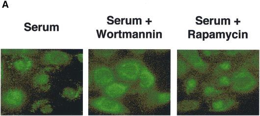
Fig. 10. Intracellular localization and mobility-shift assay of endogenous SGK. Con8.hd6 mammary tumor cells were serum starved for 48 h. During
the last 24 h, the cells were pretreated with 100 nM wortmannin or 20 µM rapamycin on cover-slips. After starvation the cells were stimulated for
3 h with 10% FCS/DMEM-F12 in the contained presence of the appropriate inhibitor. (A) The cells were stained with affinity-purified rabbit
polyclonal anti-SGK antibody, followed by anti-rabbit FITC-conjugated secondary antibody to reveal the intracellular location of the SGK protein.
(B) After the treatment described in (A), cells were lysed and equal amounts of extracts were electrophoretically fractionated and immunoblotted
with the affinity-purified rabbit polyclonal anti-SGK antibody.
lanes 1, 3, 5, 7 and 9 with 2, 4, 6, 8 and 10, respectively), phosphorylated forms of SGK protein (Figure 10B).
thereby defining the catalytic domain as the region within Consistent with the localization data, treatment of serum-
SGK responsible for mediating interaction with GST– stimulated cells with rapamycin had no effect in the
PDK1. The middle panel in Figure 9C indicates 10% of electrophoretic mobility of SGK. Short-term stimulation
the in vitro translated products of the SGK deletions of transfected HEK 293 or COS-1 cells revealed that SGK
used for the binding assays. Taken together, these data translocated to the nucleus following agonist treatment
demonstrate direct association between the catalytic (data not shown). Taken together, these data establish that
domain of SGK with PDK1. endogenous SGK lies downstream of PI 3-kinase signaling,
but is not on the same pathway as p70S6K.
Inhibition of PI 3-kinase signaling disrupts the
serum-dependent localization and electrophoretic
Discussion
mobility of endogenous SGK
In serum-stimulated mammary epithelial cells, a hyper- In the present study we have demonstrated that SGK, a
phosphorylated form of SGK localizes to the nucleus novel member of the second-messenger family of serine/
(Buse et al., 1999). Therefore, to determine whether PI threonine protein kinases, is regulated by reversible
3-kinase signaling affects the subcellular distribution of phosphorylation, apparently mediated by the PI 3-kinase
endogenous SGK, serum-stimulated Con8.hd6 rat signaling pathway. Furthermore, our findings suggest that
mammary tumor cells were treated either with wort- PDK1 phosphorylates Thr256 in the activation loop of
mannin, a selective inhibitor of PI 3-kinase, or as a control SGK leading to activation of the kinase both in vivo
with rapamycin, an inhibitor of p70S6K activity. After and in vitro. Significantly, this work also reveals that SGK
serum stimulation, SGK localization was examined by is a serine/threonine-specific protein kinase which is
indirect immunofluorescence using affinity-purified SGK activated in response to insulin/IGF-1 stimulation.
antibodies. As shown in Figure 10A, exposure to We propose that the following events are required for
wortmannin prevented the serum-dependent nuclear the rapid and efficient activation of SGK. Growth factor
localization of SGK, which resulted in a cytoplasmically binding to their cognate receptors prompts PI 3-kinase
localized form of SGK of this protein kinase. In contrast, activity, thereby generating 3-phosphorylated phospho-
treatment with rapamycin did not alter SGK localization inositides leading to the activation of upstream kinase(s),
compared with the untreated serum-stimulated cells, with which in turn activates SGK. Furthermore, we propose
~90% of SGK remaining nuclear under both conditions. that PDK1 is one of the immediate upstream regulators
Characterization of the electrophoretic mobility of endo- of SGK activity as judged by GST-pulldown experiments
genous SGK protein revealed that treatment with either of and phosphorylation data. In this scheme, PP2A would
the two PI 3-kinase inhibitors, wortmannin or LY294002, act as a negative regulator of the activating phosphoryla-
inhibited the serum-dependent induction of the hyper- tion. The data obtained by in vitro phosphatase treatment
3030Regulation of SGK protein kinase activity
or in vivo calyculin-A treatment are consistent with PP2A signal transduction pathway. Interestingly, the accumulated
being a primary negative regulator of SGK activity data on SGK transcription in different cell type and stimuli
(Figures 3 and 4). Therefore, the balance between PDK1 (Imaizumi et al., 1994; Maiyar et al., 1996; Alliston et al.,
and PP2A activity determines the phosphorylation state 1997; Delmolino and Castellot, 1997; Hollister et al.,
and thus activity of SGK. Consistent with this possibility, 1997; Waldegger et al., 1997) indicate that SGK is
pervanadate, a broad spectrum phosphatase inhibitor, implicated in two opposite pathways of proliferation
induces SGK activation and the presence of the hyper- and anti-proliferation, respectively. Additionally, recent
phosphorylated form of SGK. The activation of SGK by reports show that the cytoplasmic-nuclear distribution
pervanadate was sensitive to LY294002, implying that of SGK is differentially regulated depending on the
SGK is a cellular target of receptor-activated PI 3-kinase. proliferative state of the cells (Buse et al., 1999). It is
Furthermore, our results show that serum, insulin or IGF- tempting to speculate that steroid or growth factor regula-
1 activates SGK through a LY294002- or wortmannin- tion of protein kinase transcription could be a general
sensitive mechanism. Activation by these growth factors mechanism for regulating specific phosphorylation net-
suggests that SGK is an important part of the insulin and works, involving cross-talk between a membrane-linked
IGF-1 signaling network along with PKB family, p70S6K signaling pathway and intracellular steroid hormone
and some PKCs. Therefore, any physiological response activation of gene transcription (Webster et al., 1993a).
involving insulin signaling should include SGK as part of Furthermore, the cytoplasmic-nuclear shuttling of SGK is
this signaling cascade. regulated in synchrony with the cell cycle (Buse et al.,
Identification of the phosphorylation sites, the kinases 1999), suggesting a central role for SGK in proliferation
responsible for modifying them and their association control. We hypothesize that like other early growth-
with SGK is of considerable importance in gaining a response gene products, SGK plays a role in amplifying
detailed understanding of the activating mechanism and the mitogenic signal. However, an intriguing question
delineation of the signaling pathway. Based on comparison remains as to why this kinase is also partially transcrip-
with the sequences of PKB, PKC, p70S6K and PKA (Figure tionally regulated (by mRNA induction) rather than being
1), Thr256 and Ser422 of SGK were predicted to be modulated solely post-translationally (by phosphorylation–
potential phosphorylation sites involved in the regulation dephosphorylation) by second messengers and other
of its kinase activity. Indeed, the mutations of these sites kinases.
to alanine (T256A, S422A) caused significant decreases
in the kinase activity (Figure 7A). The current data indicate
that SGK is one of the protein kinases that is apparently
Materials and methods
robustly activated following insulin or growth factor Construction of expression vectors
treatment. Several second-messenger protein kinases are HA epitope-tagged constructs (HA-SGK) were prepared by amplifying
targets of PDK1, and these fall into two classes: inducible the rat SGK cDNA in-frame with the initiator methionine (Webster
et al., 1993a) with primers 59-CCC GGT ACC ACC ATG GCT TAC
or constitutive phosphorylation (for a review see Belham CCA TAC GAT GTT CCA GAT TAC GCT TCG ACC GTC AAA ACC
et al., 1999). The identity of the kinase responsible for GAG GCT GCT CGA and 39-CCC GGT ACC ACC ATG GCT TAC
modifying Ser422 remains to be established. Recent data CCA TAC GAT GTT CCA GAT. The polymerase chain reaction (PCR)
(P.Buse, S.Tran, M.L.L.Leong, J.Park, B.A.Hemmings and products were subcloned between KpnI and XbaI sites of the mammalian
expression vector pCMV4. The mutants at Lys127 (kinase-deficient
G.L.Firestone, in preparation) showed that SGK associates HA-SGK K127M), Thr256 (HA-SGK T256A and HA-SGK T256D),
with extracellular signal-related kinase/mitogen-activated Ser422 (HA-SGK S422A and HA-SGK S422D) or both Thr256 and
protein kinase (ERK/MAPK) following serum stimulation Ser422 (HA-SGK DD) were created using the Quickchange kit
of mammary tumor cells. These results suggest that (Stratagene) as described by the manufacturer, with pCMV4 HA-SGK
ERK/MAPK could be responsible for phosphorylating as template. The N- and C-terminal deletions of SGK were constructed
in pcDNA3 expression vector using standard PCR-cloning strategies.
S/TP sites in the N- and C-terminal domains. The Myc-tagged PDK1 and Myc-PDK1-KD, the kinase-deficient mutant
None of the endogenous downstream targets of SGK of PDK1, were the same as reported previously (Pullen et al., 1998).
have been identified so far. The identification of the The GST–PDK1 fusion protein was constructed in pGEX-4T1 bacterial
upstream components between PI 3-kinase and SGK and expression vector using standard PCR cloning techniques. Membrane-
targeted p110 constructs (HA-p110 CAAX and HA-p110 KD CAAX)
downstream targets will be crucial for understanding this were the same as reported previously (Didichenko et al., 1996). All
signaling pathway. Moreover, the regulation of SGK constructs were confirmed by automated DNA sequencing. Sequences
activity by PI 3-kinase pathway further implicates SGK of the mutagenic oligonucleotides are available upon request.
in cell survival mechanisms. In this regard we have
recently shown that osmotic shock stimulates both SGK Cell culture and stimulation
HEK 293 cells were maintained in Dulbecco’s modified Eagle’s medium
expression and enzymatic activity (L.Bell, M.L.L.Leong, (DMEM) supplemented with 10% fetal calf serum (FCS; Life Techno-
J.Park, B.A.Hemmings and G.L.Firestone, unpublished logies) at 37°C, in an atmosphere containing 5% CO2. HEK 293 cells
data). Furthermore, recent work has shown that SGK seeded at 106/10-cm dish and 0.53106/5-cm dish, respectively, were
regulates the activity of the epithelial sodium channel in transfected the following day by a modified calcium phosphate
method (Chen and Okayama, 1988), with 1–2 µg/ml plasmid DNA.
coinjected Xenopus laevis oocytes, suggesting a role for The transfection mixture was removed after 16 h incubation, and cells
SGK in the control of cell volume and sodium homeostasis were serum starved for 24 h before stimulation for 15 min with 100 nM
(Chen et al., 1999). insulin (Boehringer Mannheim), 50 ng/ml IGF-1 (Life Technologies),
SGK was originally identified as a gene which is induced 100 nM calyculin-A (Alexis), 100 ng/ml TPA (Life Technologies),
by serum and glucocorticoids, respectively (Webster et al., 0.2 mM H2O2 or 0.1 mM pervanadate prepared with 0.2 mM H2O2
(Posner et al., 1994). Pretreatment with 50 µM LY294002 (Alexis) was
1993a). These effects of serum and glucocorticoids on done for 15 min prior to cell stimulation.
SGK expression are additive (Webster et al., 1993a,b), The Con8.hd6 mammary epithelial tumor cells were grown to 30%
suggesting that each modulator acts through a distinct confluency in 6-well tissue culture plates in DMEM-F12 medium with
3031J.Park et al.
10% calf serum (Biowhittaker). Cells were serum starved for 48 h and using the TNT-coupled rabbit reticulocyte kit (Promega) according to
treated with 100 nM wortmannin (Calbiochem), 50 µM LY29004 the manufacturer’s instructions. The expression plasmids encoding JNK
(Calbiochem) or 50 µM rapamycin (generous gift from B.Webb and protein and PKCζ were kindly provided by J.S.Gutkind (Molecular
G.Steven Martin, University of California at Berkeley, CA) for 24 h. signaling unit, National Institute of Dental Research NIH, Bethesda,
Serum was added in the presence or absence of respective inhibitors for MD) and have been described previously (Crespo et al., 1995).
3 h.
Expression of GST fusion protein in bacterial system
Immunoprecipitation and in vitro kinase assay The GST–PDK1 fusion protein was isolated from AB1899 cells trans-
Cells were placed on ice and extracted with lysis buffer containing formed with GST–PDK1 expression plasmid. Briefly, bacteria were
50 mM Tris–HCl pH 7.5, 1% w/v Nonidet P-40 (NP-40), 120 mM initially grown at 37°C for 2 h (OD600 5 0.5–0.7) and subsequently
NaCl, 25 mM NaF, 40 mM β-glycerol phosphate, 0.1 mM sodium induced with 0.5 mM isopropyl-thio-β-D-galactopyranoside (IPTG) for
orthopervanadate, 1 mM phenylmethylsulfonyl fluoride (PMSF), 1 mM 16 h at 30°C. Cells were lysed using the French Press (three times) in
benzamidine and 2 µM microcystin-LR. Lysates were centrifuged for lysis buffer (PBS containing 0.05% Tween-20, 2 mM EDTA, 1 mM
15 min at 12 000 g, and the HA-SGK protein was immunoprecipitated DTT and 0.1% 2-mercaptoethanol). The GST–PDK1 fusion protein
from 400 µg of cell-free extracts with the anti-HA epitope 12CA5 was purified on glutathione–agarose beads (Pharmacia) as described
monoclonal antibody coupled to protein A–Sepharose (Pharmacia previously (Chakraborty et al., 1991).
Biotech.). The immune complexes were washed once with lysis buffer
Binding of GST–PDK-1 and in vitro translated SGK
containing 0.5 M NaCl, followed by washing with lysis buffer and
In order to examine the interaction between PDK1 and SGK in vitro,
finally with kinase assay buffer (50 mM Tris–HCl pH 7.5, 0.1% v/v
25 µg GST–PDK1 immobilized on glutathione–Sepharose beads were
2-mercaptoethanol). In vitro kinase assays were performed for 60 min
incubated with 5 µl of [35S]methionine-labeled SGK in vitro transla-
at 30°C in 50 µl of reaction volume containing 30 µl of immunoprecipitate
tion product in 180 µl of binding buffer (20 mM HEPES–KOH pH 7.9,
in kinase buffer, 1 mM Sgktide (KKRNRRLSVA) as substrate, 10 mM
50 mM KCl, 2.5 mM MgCl2, 10% glycerol, 1 mM DTT, 0.2% NP-40,
MgCl2, 1 µM PKA inhibitor peptide (Bachem) and 100 µM [γ-32P]ATP
1.5 mM PMSF, and 3 µl of normal goat serum/180 µl binding buffer).
(1000–2000 c.p.m./pmol; Amersham).
The slurry was incubated overnight at 4°C on a nutator, following which
For the coupled kinase assay, HA-SGK and Myc-PDK1 were
the beads were washed five times in wash buffer (200 mM NaCl, 0.2%
independently expressed in HEK 293 cells. Extracts from cells expressing
Tween 20, 10 mM Tris pH 7.5 and 0.5% non-fat dry milk). After
HA-SGK or Myc-PDK1 were immunoprecipitated with the anti-HA
removing the supernatant in the final wash, samples were resuspended
(12CA5) or anti-Myc (9E10) monoclonal antibody coupled to protein
in 25 µl of 23 SDS sample buffer, boiled for 5 min and the proteins
A–Sepharose or protein G–Sepharose (Pharmacia Biotech.), respectively.
retained on the beads resolved by SDS–PAGE. Binding was compared
HA-SGK was eluted from immune complexes with HA epitope peptide
with that of 10% of the in vitro translated products added to the binding
(1 mg/ml) in kinase buffer. The immunoprecipitated Myc-PDK1 was
reactions. Gels were dried at 60°C and autoradiography carried out
then mixed with eluted HA-SGK in the presence of 10 mM MgCl2 and
at –70°C.
100 µM ATP. After incubation for 30 min at 30°C, the protein-G beads
were removed by centrifugation, and 1 mM Sgktide and [γ-32P]ATP Indirect immunofluorescence microscopy
(1000–2000 c.p.m./pmol) were added. The reaction mixtures were further Con8.dh6 mammary tumor cells were cultured on 8-well Lab-Tek
incubated for 60 min at 30°C to measure the SGK activity. All reactions Permanox slides (Nalgene) and grown to 30% confluency before the
were stopped by adding 50 µM EDTA and processed as described indicated combinations of serum and/or wortmannin or rapamycin were
previously (Andjelkovi et al., 1996). Protein concentrations were deter- added for 24 h. Cell confluency prior to fixation did not exceed 60%.
mined by the method of Bradford (1976) using bovine serum albumin Cells were washed with PBS, fixed for 15 min in 3.7% formaldehyde/
(BSA) as a standard. 0.1% glutaraldehyde, rinsed with PBS and permeabilized with 50%
methanol/50% acetone for 1 min. Following a rinse in PBS, the cells
Protein phosphatase 2A (PP2A) treatment were preabsorbed for 5 min in PBS containing 4% normal goat serum
Immunoprecipitated HA-SGK was incubated with 10 ng of purified (Jackson Immuno Research Laboratories). The cells were incubated in
recombinant PP2A catalytic subunit (T.Myles and B.A.Hemmings, a 1:300 dilution of affinity-purified rabbit polyclonal anti-SGK antibody
unpublished data) in 45 µl buffer containing 50 mM Tris–HCl pH 7.5, for 1–2 h at 25°C. After five washes with PBS, cells were treated for
1% 2-mercaptoethanol, 1 mM MnCl2, 1 mM benzamidine, 0.5 mM 5 min with PBS containing 4% normal goat serum. The cells were
PMSF for 30 min at 30°C. The reactions were stopped by addition of incubated with a 1:300 dilution of anti-rabbit FITC-conjugated secondary
1 µM okadaic acid. The immune complexes were washed with 50 mM antibody (Cappel Research Products) in PBS and then incubated for
Tris–HCl pH 7.5, 1 mM benzamidine, 0.5 mM PMSF and 1 µM okadaic 30 min at 25°C. Cells were washed 5 times with PBS and mounted with
acid (Alexis), and SGK was assayed as described above. 50% glycerol, 50 mM Tris–HCl pH 8.0 containing 4 mg/ml N-propyl
gallate, and examined under a Nikon Optiphot fluorescence microscope.
Immunoblot analysis Images were captured using Adobe Photoshop 3.0.5 (Adobe Systems)
HEK 293 cell extracts and immunoprecipitates were resolved by 10% and a Sony DKC-5000 digital camera. Non-specific fluorescence was
SDS–PAGE and transferred to Immobilon P membranes (Millipore). determined by incubation with the secondary antibody alone and shown
The filters were blocked for 30 min in 13 phosphate-buffered saline to be negligible.
(PBS) containing 5% skimmed milk, 0.5% Triton X-100 and 0.5%
Tween-20, followed by a 2 h incubation with the anti-HA epitope 12CA5 Acknowledgements
monoclonal antibody diluted 1000-fold in the same blocking solution.
The secondary antibody was alkaline phosphatase-conjugated anti-rabbit We would like to thank the following people (all F.M.I. except where
IgG (Sigma) diluted 2500-fold in the blocking buffer. The detection and stated) for providing essential reagents and technical support. Peter Cron
quantitation of SGK expression was carried out by using the AP color for preparing HA-SGK, Dr M.Thelen (University of Bern, Switzerland)
development reagents from Bio-Rad. for p110-CAAX, Dr T.Millward for assembling the kinase peptide
Soluble whole-cell extracts from Con8.hd6 mammary epithelial cells library, Dr T.Myles for preparing baculovirus expression constructs for the
were grown under the conditions indicated and resolved by SDS– expression of HA-tagged catalytic subunit of PP2A, Dr M.Andjelković for
PAGE. Proteins were transferred to Nytran membranes (Schleicher & help with PP2A treatment, P.Müller for synthesis of oligonucleotides
Schuell). The membrane was probed with a 1:2500 dilution of polyclonal and Dr H.Angliker for DNA sequence analysis. Drs R.Meier, D.Brodbeck
anti-SGK antibody (Webster et al., 1993a) in 50 mM Tris–HCl pH 8.0, and T.Millward are acknowledged for comments on the manuscript. J.P.
150 mM NaCl, 0.05% Tween-20 with 1% non-fat dry milk. The especially thanks David R.H.Evans for tutelage in writing and critical
secondary antibody was a goat anti-rabbit IgG HRP-conjugated antibody reading of this manuscript. This work was partially supported by Public
(Bio-Rad). The Western blot was developed by using the Renaissance Health Service grant CA-71514 awarded to G.L.F. from the National
developing kit (NEN, Boston, MA) and exposure to film. Cancer Institute (USA).
In vitro transcription, and translation of SGK References
In vitro transcription and translation of full-length wild-type SGK
(WT-SGK, 1–431 aa), kinase-dead SGK (K127M-SGK, 1–431 aa), Alessi,D.R., Andjelković,M., Caudwell,B., Cron,P., Morrice,N., Cohen,P.
N- and C-terminal deleted SGK (∆N-SGK, 60–431 aa; ∆C-SGK, 1–355 and Hemmings,B.A. (1996) Mechanism of activation of protein kinase
aa) and catalytic domain only SGK (Cat.-SGK, 60–355 aa) was performed B by insulin and IGF-1. EMBO J., 15, 6541–6551.
3032Regulation of SGK protein kinase activity
Alessi,D.R. et al. (1997a) 3-Phosphoinositide-dependent protein phosphoinositide 3-kinase through the protein kinase PDK1. Science,
kinase-1 (PDK1): structural and functional homology with the 281, 2042–2045.
Drosophila DSTPK61 kinase. Curr. Biol., 7, 776–789. Leighton,I.A., Dalby,K.N., Caudwell,F.B., Cohen,P.T.W. and Cohen,P.
Alessi,D.R., James,S.R., Downes,C.P., Holmes,A.B., Gaffney,P.R., Reese, (1995) Comparison of the specificities of p70 S6 kinase and MAPKAP
C.B. and Cohen,P. (1997b) Characterization of a 3-phosphoinositide- kinase-1 identifies a relatively specific substrate for p70 S6 kinase:
dependent protein kinase which phosphorylates and activates protein the N-terminal kinase domain of MAPKAP kinase-1 is essential for
kinase Bα. Curr. Biol., 7, 261–269. peptide phosphorylation. FEBS Lett., 375, 289–293.
Alessi,D.R., Kozlowski,M.T., Weng,Q.P., Morrice,N. and Avruch,J. Maiyar,A.C., Huang,A.J., Phu,P.T., Cha,H.H. and Firestone,G.L. (1996)
(1998) 3-Phosphoinositide-dependent protein kinase 1 (PDK1) p53 stimulates promoter activity of the sgk serum/glucocorticoid-
phosphorylates and activates the p70 S6 kinase in vivo and in vitro. inducible serine/threonine protein kinase gene in rodent mammary
Curr. Biol., 8, 69–81. epithelial cells. J. Biol. Chem., 271, 12414–12422.
Alliston,T.N., Maiyar,A.C., Buse,P., Firestone,G.L. and Richards,J.S. Maiyer,A.C., Phu,P.T., Huang,A.J. and Firestone,G.L. (1997) Repression
(1997) Follicle stimulating hormone-regulated expression of serum/ of glucocorticoid receptor transactivation and DNA binding of a
glucocorticoid-inducible kinase in rat ovarian granulosa cells: A glucocorticoid response element within the serum/glucocorticoid-
functional role for the Sp1 family in promoter activity. Mol. inducible protein kinase (sgk) gene promoter by the p53 tumor
Endocrinol., 11, 934–949. suppressor protein. Mol. Endocrinol., 11, 312–329.
Andjelković,M., Jakubowicz,T., Cron,P., Ming,X.F., Han,J.W. and Posner,B.I. et al. (1994) Peroxovanadium compounds. A new class
Hemmings,B.A. (1996) Activation and phosphorylation of a pleckstrin of potent phosphotyrosine phosphatase inhibitors which are insulin
homology domain containing protein kinase (RAC-PK/PKB) promoted mimetics. J. Biol. Chem., 269, 4596–4604.
by serum and protein phosphatase inhibitors. Proc. Natl Acad. Sci. Pullen,N., Dennis,P.B., Andjelković,M., Dufner,A., Kozma,S.C.,
USA, 93, 5699–5704. Hemmings,B.A. and Thomas,G. (1998) Phosphorylation and activation
Belham,C., Wu,S. and Avruch,J. (1999) PDK1—a kinase at the hub of of p70S6K by PDK1. Science, 279, 707–710.
things. Curr. Biol., 9, R93–R96. Stokoe,D., Stephens,L.R., Copeland,T., Gaffney,P.R., Reese,C.B.,
Bradford,M.M. (1976) A rapid and sensitive method for the quantitation Painter,G.F., Holmes,A.B., McCormick,F. and Hawkins,P.T. (1997)
of microgram quantities of protein utilizing the principle of protein- Dual role of phosphatidylinositol-3,4,5-trisphosphate in the activation
dye binding. Anal. Biochem., 72, 248–254. of protein kinase B. Science, 277, 567–570.
Buse,P., Tran,S.H., Luther,E., Phu,P.T., Aponte,G.W. and Firestone,G.L. Waldegger,S., Barth,P., Raber,G. and Lang,F. (1997) Cloning and
(1999) Cell cycle and hormonal control of nuclear-cytoplasmic characterization of a putative human serine/threonine protein kinase
localization of the serum and glucocorticoid inducible protein kinase, transcriptionally modified during anisotonic and isotonic alterations
Sgk, in mammary tumor cells: A novel convergence point of anti- of cell volume. Proc. Natl Acad. Sci. USA, 94, 4440–4445.
proliferative and proliferative cell signaling pathways. J. Biol. Chem., Webster,M.K., Goya,L., Ge,Y., Maiyar,A.C. and Firestone,G.L. (1993a)
274, 7253–7263. Characterization of sgk, a novel member of the serine/threonine
Carpenter,C.L. and Cantley,L.C. (1996) Phosphoinositide kinases. Curr. protein kinase gene family which is transcriptionally induced by
Opin. Cell Biol., 8, 253–258. glucocorticoids and serum. Mol. Cell. Biol., 13, 2031–2040.
Chakraborty,T., Brennan,T.J., Li,L., Edmondson,D. and Olson,E.N. Webster,M.K., Goya,L. and Firestone,G.L. (1993b) Immediate-early
(1991) Inefficient homooligomerization contributes to the dependence transcriptional regulation and rapid mRNA turnover of a putative
of myogenin on E2A products for efficient DNA binding. Mol. Cell. serine/threonine protein kinase. J. Biol. Chem., 268, 11482–11485.
Biol., 11, 3633–3641.
Chen,C.A. and Okayama,H. (1988) Calcium phosphate-mediated gene Received March 19, 1999; revised and accepted April 14, 1999
transfer: a highly efficient transfection system for stably transforming
cells with plasmid DNA. Biotechniques, 6, 632–638.
Chen,S., Bhargava,A., Mastroberardino,L., Meijer,O.C., Wang,J.,
Buse,P., Firestone,G.L., Verrey,F. and Pearce,D. (1999) Epithelial
sodium channel regulated by aldosterone-induced protein sgk. Proc.
Natl Acad. Sci. USA, 96, 2514–2519.
Cheng,X., Ma,Y., Moore,M., Hemmings,B.A. and Taylor,S.S. (1998)
Phosphorylation and activation of cAMP-dependent protein kinase by
phosphoinositide-dependent protein kinase. Proc. Natl Acad. Sci. USA,
95, 9849–9854.
Chou,M.M., Hou,W., Johnson,J., Graham,L.K., Lee,M.H., Chen,C.S.,
Newton,A.C., Schaffhausen,B.S. and Toker,A. (1998) Regulation of
protein kinase Cζ by PI 3-kinase and PDK-1. Curr. Biol., 8, 1069–1077.
Crespo,P., Mischak,H. and Gutkind,J.S. (1995) Overexpression of
mammalian protein kinase Cζ does not affect the growth characteristics
of NIH 3T3 cells. Biochem. Biophys. Res. Commun., 213, 2266–2272.
Delmolino,L.M. and Castellot,J.J.,Jr (1997) Heparin suppresses sgk, an
early response gene in proliferating vascular smooth muscle cells. J.
Cell. Physiol., 73, 371–379.
Didichenko,S.A., Tilton,B., Hemmings,B.A., Ballmer-Hofer,K. and
Thelen,M. (1996) Constitutive activation of protein kinase B and
phosphorylation of p47phox by a membrane-targeted phosphoinositide
3-kinase. Curr. Biol., 6, 1271–1278.
Galetić,I., Andjelković,M., Meier,R., Brodbeck,D., Park,J. and
Hemmings,B.A. (1999) Mechanism of protein kinase B activation by
insulin/IGF1 revealed by specific inhibitors of phosphoinositide 3-
kinase : significance for diabetes and cancer. Pharmacol. Ther., in press.
Hollister,R.D., Page,K.J. and Hyman,B.T. (1997) Distribution of the
messenger RNA for the extracellularly regulated kinases 1, 2 and 3
in rat brain: effects of excitotoxic hippocampal lesions. Neuroscience,
79, 1111–1119.
Imaizumi,K., Tsuda,M., Wanaka,A., Tohyama,M. and Takagi,T. (1994)
Differential expression of sgk mRNA, a member of the Ser/Thr protein
kinase gene family, in rat brain after CNS injury. Brain Res. Mol.
Brain Res., 26, 189–196.
Le Good,J.A., Ziegler,W.H., Parekh,D.B., Alessi,D.R., Cohen,P. and
Parker,P.J. (1998) Protein kinase C isotypes controlled by
3033You can also read