Simple hypertrophic tonsils have more active innate immune and inflammatory responses than hypertrophic tonsils with recurrent inflammation in ...
←
→
Page content transcription
If your browser does not render page correctly, please read the page content below
Huang et al. Journal of Otolaryngology - Head and Neck Surgery                    (2020) 49:35
https://doi.org/10.1186/s40463-020-00428-3

 ORIGINAL RESEARCH ARTICLE                                                                                                                           Open Access

Simple hypertrophic tonsils have more
active innate immune and inflammatory
responses than hypertrophic tonsils with
recurrent inflammation in children
Qun Huang1†, Hu Hua2†, Wei Li1, Xi Chen3 and Lei Cheng3*

  Abstract
  Background: Tonsil hypertrophy has negative impact on children’s health, but its pathogenesis remains obscure
  despite the fact that numerous bacteriological studies have been carried out. Understanding the innate immune
  and inflammatory states of hypertrophic tonsils with different clinical manifestations is of great significance for
  defining the pathogenesis of tonsil hypertrophy and establishing treatment strategies. The present study was
  undertaken to examine the characteristics of innate immunity and inflammation in children with hypertrophic
  palatine tonsils and different clinical manifestations.
  Methods: Tonsil tissues were surgically removed from the patients and classified based on the patients’ clinical
  manifestations. The patients were divided into three groups: 1) Control group; 2) Tonsil Hypertrophy (TH) group;
  and 3) Tonsil Hypertrophy combined with Recurrent Infection (TH + RI) group. The immune and inflammatory
  statuses of these tissues were characterized using qRT-PCR and ELISA methods.
  Results: Viral protein 1 (VP1) was highly expressed in TH group, but not in TH + RI group. In TH group, elevated
  expression was observed in the innate immune mediators, including retinoic acid-inducible gene I (RIG-I), interferon
  alpha (IFN-α), mitochondrial antiviral-signaling protein (MAVS), NLR family pyrin domain containing 3 (NLRP3), toll-
  like receptor (TLR) 4 and TLR7. Consistent with the innate immune profile, the expression of inflammatory markers
  (IL-1β, NF-κB and IL-7) was also significantly elevated in TH group. Meanwhile, the COX-2/PGE2/EP4 signaling
  pathway was found to be involved in the inflammatory response and the formation of fibroblasts.
  Conclusions: Innate immune and inflammatory responses are more active in simple hypertrophic tonsils, rather
  than hypertrophic tonsils with recurrent inflammation. A local relative immune deficiency in the hypertrophic
  tonsils may be a causative factor for recurrent tonsillitis in TH + RI. These differences, together with the patient’s
  clinical manifestations, suggest that tonsillar hypertrophy might be regulated by diverse immune and/or
  inflammatory mechanism through which novel therapeutic strategies might be created.
  Keywords: Palatine tonsil, Hypertrophy, Tonsillitis, Innate immunity, Inflammation, Infection

* Correspondence: chenglei@jsph.org.cn
†
 Qun Huang and Hu Hua contributed equally to this work.
3
 Department of Otorhinolaryngology, The First Affiliated Hospital, Nanjing
Medical University, 300 Guangzhou Road, Nanjing 210029, Jiangsu, China
Full list of author information is available at the end of the article

                                         © The Author(s). 2020 Open Access This article is licensed under a Creative Commons Attribution 4.0 International License,
                                         which permits use, sharing, adaptation, distribution and reproduction in any medium or format, as long as you give
                                         appropriate credit to the original author(s) and the source, provide a link to the Creative Commons licence, and indicate if
                                         changes were made. The images or other third party material in this article are included in the article's Creative Commons
                                         licence, unless indicated otherwise in a credit line to the material. If material is not included in the article's Creative Commons
                                         licence and your intended use is not permitted by statutory regulation or exceeds the permitted use, you will need to obtain
                                         permission directly from the copyright holder. To view a copy of this licence, visit http://creativecommons.org/licenses/by/4.0/.
                                         The Creative Commons Public Domain Dedication waiver (http://creativecommons.org/publicdomain/zero/1.0/) applies to the
                                         data made available in this article, unless otherwise stated in a credit line to the data.
Huang et al. Journal of Otolaryngology - Head and Neck Surgery (2020) 49:35 Page 2 of 9 Background patient had a history of tonsillitis and sore throat (≥3 in- The tonsil is an independent organ constructed by fections per year in 3 years) that failed to respond to an- mucosal-associated lymphoid tissue (MALT). As the first tibiotics. The degree of tonsil hypertrophy was scored stronghold against foreign pathogens, the tonsil can se- from 1+ to 4+ according to the percentage of decreased lectively regulate immune response in the internal envir- pharyngeal lumen diameter [12–14]: 1+, 0–25%; 2+, 26– onment [1, 2]. Simple tonsil hypertrophy refers to the 50%; 3+, 51–75% and 4+, 76–100%. Tonsil hypertrophy enlargement of the tonsil uncomplicated with inflamma- was scored 3–4+ preoperatively [15]. tion. Lymphoid tissue degenerates in adulthood. There- Patients advised to undergo tonsillectomy (TE) in ac- fore, tonsil hypertrophy is much more prevalent, cordance with the AAO-HNS tonsillectomy guidelines characteristic [3–5], and causative to obstructive sleep- [10], were recruited into the study. Consecutive patients disordered breathing (SDB) in children [6]. Also in chil- were divided into three groups according to the clinical dren, the tonsil can kindle infections that may give rise manifestations of their tonsils: 1) control group; 2) tonsil to acute or chronic tonsillitis [7]. Symptoms of tonsillitis hypertrophy (TH) group; and 3) tonsil hypertrophy com- include sore throat, fever, enlargement of the tonsil, dif- bined with recurrent infection (TH + RI) group. Included ficult swallowing, and swollen lymph nodes around the were those [1]: aged 2 to 13 years [2]; exhibiting significant neck. Recurrent tonsillitis (RI) is diagnosed when tonsil- symptoms of snoring that disrupted the quality of sleep or litis relapses as sympomized by ≥7 episodes of throat in- daily activities; and [3] who planned to undergo adenoi- fection (well-documented, clinically important, dectomy and tonsillectomy. Excluded were those who dis- adequately treated) in the preceding year, or ≥ 5 episodes played [1]: symptoms of acute infection at the time of the in each of the preceding 2 years, or ≥ 3 episodes in each surgery, and received antibiotic therapy within the previ- of the preceding 3 years [8]. ous 4 weeks [2]; congenital anomalies including clef palate, Tonsillar disease is routinely treated with antibiotics Down syndrome, congenital heart disease, and craniofacial or tonsillectomy [9]. Tonsillectomy is most recom- anomalies [3]; major systemic disease, such as diabetes, mended for children who meet the standards of the nephrotic disease, autoimmune disorders, immunodefi- AAO-HNS tonsillectomy guidelines [10], but contro- ciency, malignancy, and other chronic illnesses; or [4] versy still exists in the indications and the benefits/ known allergic conditions such as asthma, allergic rhinitis, harms after eliminating this immune organ [11]. Al- or history of allergies. As we defined that tonsils with though numerous studies have been carried out to ex- hypertrophy score < 3 were not hypertrophic, the patients plore the bacteriology in the tonsil, the underlying with SDB and adenoid hypertrophy, and no tonsillar pathogenic mechanism of tonsil hypertrophy has not hypertrophy or inflammation were collected as controls. A been fully understood. total of 45 patients were included in our study, including Tonsil hypertrophy exhibits varying clinical manifesta- 20 patients in control group, 10 patients in TH group, and tions, either as simple hypertrophy or hypertrophy ac- 15 patients in TH + RI group. companied with recurrent inflammation. These different TE was conducted using plasma wand with a high- clinical manifestations suggest that tonsillar hypertrophy frequency electrosurgery system (Coblator II, Arthro- might be regulated by diverse immune and/or inflamma- Care Corporation, Austin, TX, USA). TE procedures tory mechanisms. But so far, there have been few reports were performed by surgeons with ≥5 years’ operating ex- analyzing these differences. This study, therefore, aimed perience. The tonsils were completely removed during to demonstrate the differences in innate immune and in- TE. The tissues that were not required for pathological flammatory responses in tonsils with various clinical examination were immediately collected and delivered manifestations, which will refresh our understanding of into icy phosphate buffered saline (PBS) plus antibiotics the pathology of tonsil hypertrophy and improve related in a − 70 °C refrigerator within half an hour after exci- clinical prevention and treatment. sion under aseptic conditions. Methods Histopathological examination Human tissue samples Tonsil tissues were fixed in 4% paraformaldehyde (PFA) This study was reviewed and approved by the institu- for 16 h, then paraffin-embedded. The sections (5 μm tional review board. Informed consent was obtained thickness) were prepared and stained with Masson or from the legal caregiver of each participant. Before sur- hematoxylin-eosin (H&E) and analyzed under the light gery, the children who underwent tonsillectomy for ei- microscope. ther SDB or RI were identified. The diagnosis of SDB was established by a spectrum of breathing disorders Real time quantitative PCR (qRT-PCR) during sleep, habitual and loud snoring, and upper air- Total RNA was isolated from the tonsil using the Trizol re- way obstruction [6]. RI was clinically diagnosed if the agent (Takara Bio Inc., Japan) following the manufacturer’s
Huang et al. Journal of Otolaryngology - Head and Neck Surgery (2020) 49:35 Page 3 of 9
instructions, and reverse-transcribed using reverse tran- including one-way analysis of variance (ANOVA) for
scription kit (Takara Bio Inc., Japan). qRT-PCR was per- multiple comparisons. An unpaired Student’s t-test
formed using SYBR Green master mix (Vazyme) and 7500 was also performed to determine any significant dif-
Real-Time PCR System (Applied Biosystems, Foster City, ference between two groups. P < 0.05 was considered
CA, USA). Primers used for qRT-PCR are shown in Table 1. statistically significant.
Cycling conditions were 95 °C for 10 min, followed by 40
cycles of 95 °C for 15 s and 60 °C for 1 min. The amount of
mRNA for each gene was standardized with the internal Results
control GAPDH, and calculated using the comparative Pathological features of tonsils in each group
cycle threshold (ΔΔCt) method. H&E staining showed that the tonsils in the control
group were composed of lymphoid tissues, superficial
ELISA assay squamous epithelium, and some lymphoid nodules. The
The PGE2 levels in tonsils were determined by the tonsils in TH group showed lymphoid hyperplasia, in-
ELISA kit (Elabscience, Wuhan, China) according to the creased lymphoid follicles, and enlarged germinal cen-
manufacturer’s instructions. ters. Besides these three changes, the tonsils in TH + RI
group showed the epithelium with papillary hyperplasia
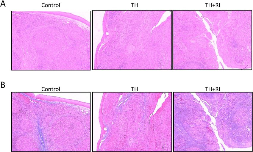
Statistical analysis or high-level infiltration of inflammatory cells (Fig. 1a).
All the data were presented as mean ± SEM. Graph- Masson staining showed that the tonsils in TH + RI
Pad Prism version 5.0 (GraphPad Software, La Jolla, group had developed more significant interstitial fibrosis,
CA, USA) software was used for statistical analyses, compared with the other two groups (Fig. 1b).
Table 1 Primers used for qRT-PCR
Primer Forward (5′ → 3′) Reverse(5′ → 3′)
VP1 AGTCCTCCCACGTCACTCAC TGATCACTAGGGTGGCATCA
IFN-α GCCTCGCCCTTTGCTTTACT CTGTGGGTCTCAGGGAGATCA
RIG-I CTGGACCCTACCTACATCCTG GGCATCCAAAAAGCCACGG
MAVS CAGGCCGAGCCTATCATCTG GGGCTTTGAGCTAGTTGGCA
NLRP3 CGTGAGTCCCATTAAGATGGAGT CCCGACAGTGGATATAGAACAGA
TLR1 TGAACCTCAAGCACTTGGACC CCCATAAGTCTCTCCTAAGACCA
TLR2 ATCCTCCAATCAGGCTTCTCT GGACAGGTCAAGGCTTTTTACA
TLR3 TTGCCTTGTATCTACTTTTGGGG TCAACACTGTTATGTTTGTGGGT
TLR4 AGACCTGTCCCTGAACCCTAT CGATGGACTTCTAAACCAGCCA
TLR5 TCCCTGAACTCACGAGTCTTT GGTTGTCAAGTCCGTAAAATGC
TLR7 TCGTGGACTGCACAGACAAG GGTATGTGGTTAATGGTGAGGGT
TNF-α CCTCTCTCTAATCAGCCCTCTG GAGGACCTGGGAGTAGATGAG
IL-6 ACTCACCTCTTCAGAACGAATTG CCATCTTTGGAAGGTTCAGGTTG
IL-1β ATGATGGCTTATTACAGTGGCAA GTCGGAGATTCGTAGCTGGA
NF-κB AACAGAGAGGATTTCGTTTCCG TTTGACCTGAGGGTAAGACTTCT
IL-7 TTCCTCCCCTGATCCTTGTTC CTTGCGAGCAGCACGGAATA
IL-10 GACTTTAAGGGTTACCTGGGTTG TCACATGCGCCTTGATGTCTG
IL-12A CCTTGCACTTCTGAAGAGATTGA ACAGGGCCATCATAAAAGAGGT
IL-17A AGATTACTACAACCGATCCACCT GGGGACAGAGTTCATGTGGTA
IL-18 TCTTCATTGACCAAGGAAATCGG TCCGGGGTGCATTATCTCTAC
COX2 GAATCATTCACCAGGCAAATT TCTGTACTGCGGGTGGAACA
EP1 ACCTTCTTTGGCGGCTCTC CCAACACCAGCATTGGGCT
EP2 GCTCCTTGCCTTTCACGATTT AGGATGGCAAAGACCCAAGG
EP3 AGCTTATGGGGATCATGTGC TTTCTGCTTCTCCGTGTGTG
EP4 CGCTCGTGGTGCGAGTATT AGGGGTCTAGGATGGGGTTC
GAPDH CGCTCTCTGCTCCTCCTGTT CATGGGTGGAATCATATTGGHuang et al. Journal of Otolaryngology - Head and Neck Surgery (2020) 49:35 Page 4 of 9 Fig. 1 Pathological features of tonsils in each group. (a) Tonsil sections were stained with H&E and evaluated by microscope (× 200). (b) Tonsil sections were stained with Masson and evaluated by microscope (× 200) Viral infection and innate immune response in each group that the mRNA levels of RIG-I and MAVS in TH group With qRT-PCR, we found that the expression levels of were significantly higher than that in the control group, VP1 and IFN-α in TH group were significantly higher while the mRNA levels of RIG-I and MAVS in TH + RI than that in the control group, but that in TH + RI group were lower than that in TH group (Fig. 2c&d). group was significantly lower than that in the TH group The expression profiles of RIG-I and MAVS were con- (Fig. 2a&b). We next examined the pathways involved in sistent with that of VP1 and TNF-α. NLRP3, a compo- innate immune response caused by the virus. We found nent of the innate immune system, was also detected in Fig. 2 Viral infection and innate immune response in each group. (a-e) mRNA expressions of VP1, IFN-α, RIG-I, MAVS and NLRP3 in the tonsils were determined by qRT-PCR. Data were expressed as mean ± SEM. n = 20 in control group; n = 10 in TH group; n = 15 in TH + RI group.*P < 0.05 compared to the control group; **P < 0.01 compared to the control group; ##P < 0.01compared to the TH group; ns (no significance) compared to the control group
Huang et al. Journal of Otolaryngology - Head and Neck Surgery       (2020) 49:35                                                     Page 5 of 9

each group. The expression of NLRP3 in TH group was                        this increase was less obvious than that in the TH group.
up-regulated, compared with the control group, while                       In particular, the mRNA levels of IL-1β and NF-κB were
the expression level of NLRP3 in TH + RI group signifi-                    significantly lower than those in TH group (Fig. 4a-d).
cantly decreased (Fig. 2e). These results indicate that the                These results suggested that the inflammatory response
innate immune response in the tonsil was more active in                    in TH group was stronger than that in TH + RI group.
TH group than in TH + RI group.                                            We also tested the mRNA levels of IL-7, IL-10, IL-12A,
                                                                           IL-17A and IL-18. The results showed that IL-7 and IL-
Expression of toll-like receptors (TLRs) in each group                     10 levels in TH group had an increasing trend, com-
TLRs and their signaling play a crucial role in innate im-                 pared with the control group, and the levels in TH + RI
mune response [16]. Therefore, we examined the expres-                     group were significantly lower than those in TH group
sion of TLRs. Compared with the control group, the                         (Fig. 5a&b). The levels of IL-12A, IL-17A and IL-18 in
expression levels of TLR1, TLR2, TLR4 and TLR7 in TH                       TH group were not significantly different from those in
group were significantly up-regulated (Fig. 3a, b, d&f).                   the control group. IL-12A and IL-17A levels in TH + RI
TLR3 and TLR5 in TH group did not change significantly                     group were not significantly different from those in TH
(Fig. 3c&e). The expression levels in TLRs (especially                     group (Fig. 5c-e).
TLR4 and TLR7) in TH + RI group were significantly de-
creased, compared with TH group (Fig. 3d&f). These re-                     Expression of COX-2, PGE2 and its receptors in each
sults indicate that TLR1, TLR2, TLR4, TLR7 may be                          group
involved in the activation of the innate immune response                   Cyclooxygenase (COX) catalyzes Cox (bis-oxygenase) re-
in TH group.                                                               action in which arachidonic acid is converted into PGG2
                                                                           and then PGG2 is reduced by two electrons into PGH2,
Inflammatory response in each group of tonsils                             a precursor to all prostanoids, including PGE2, PGD2,
To assess the inflammatory response, we examined the                       PGF2, PGI2, and thromboxane. Prostaglandins are crit-
mRNA levels of TNF-α, IL-6, IL-1β and NF-κB. The re-                       ical mediators in inflammation [17]. Through assessing
sults showed that the expression levels of these four in-                  the expression of COX-2, PGE2 and its receptors, we
flammatory factors in TH group were significantly                          teased out the prostaglandin-related signaling pathways
higher than those in the control group, while these in-                    in the tonsil inflammatory response. The results of qRT-
flammatory factors in TH + RI group were increased to                      PCR showed that the expression of COX-2 in TH group
varying degrees, compared with the control group, but                      was significantly higher than that in the control group.

 Fig. 3 Expression of TLRs in each group. (a-f) mRNA expressions of TLR1, TLR2, TLR3, TLR4, TLR5 and TLR7 in the tonsils were determined by qRT-
 PCR. Data were expressed as mean ± SEM. n = 20 in control group; n = 10 in TH group; n = 15 in TH + RI group. *P < 0.05 compared to the control
 group; **P < 0.01 compared to the control group; ***P < 0.001 compared to the control group; #P < 0.05 compared to the TH group; ###P < 0.001
 compared to the TH group; ns (no significance) compared to the control group or TH group
Huang et al. Journal of Otolaryngology - Head and Neck Surgery (2020) 49:35 Page 6 of 9 Fig. 4 Inflammatory response in each group. (a-d) mRNA expressions of TNF-α, IL-6, IL-1β and NF-κB in the tonsils were determined by qRT-PCR. Data were expressed as mean ± SEM. n = 20 in control group; n = 10 in TH group; n = 15 in TH + RI group. **P < 0.01 compared to the control group; ***P < 0.001 compared to the control group; #P < 0.05 compared to the TH group; ns (no significance) compared to the TH group Fig. 5 Expression of some other inflammatory factors in each group. (a-e) mRNA expressions of IL-7, IL-10, IL-12A, IL-17A and IL-18 in the tonsils were determined by qRT-PCR. Data were expressed as mean ± SEM. n = 20 in control group; n = 10 in TH group; n = 15 in TH + RI group. *P < 0.05 compared to the control group; #P < 0.05compared to the TH group; ###P < 0.001 compared to the TH group; ns (no significance) compared to the control group or TH group
Huang et al. Journal of Otolaryngology - Head and Neck Surgery       (2020) 49:35                                                     Page 7 of 9

The expression level of COX-2 in TH + RI group was                        The etiology of hypertrophy in tonsillar lymphoid tissue
not significantly higher than that in the control group,                  remains unknown.
but significantly lower than that in TH group                               In the present study, the high expression of VP1 in the
(Fig. 6a). The ELISA results also showed that the                         simple hypertrophic tonsils indicated that tonsil hyper-
level of PGE2 in TH group was significantly up-                           trophy may be caused by viral infection. The innate im-
regulated and higher than that in TH + RI group                           mune system serves as the first line of defense against
(Fig. 6b). We then tested the receptors for PGE2, in-                     various microorganisms. Recent studies have shown
cluding EP1, EP2, EP3 and EP4, by qPCR. The re-                           various molecules playing critical roles in innate antiviral
sults showed that only the expression of EP4 was                          immune response [19–21]. The RIG-I-MAVS-mediated
significantly changed in the three groups. The expression                 antiviral immune response is triggered in the host upon
of EP4 in TH group was significantly higher than that in                  the invasion of pathogens. RIG-I recognizes viral RNA,
the control group, while that in TH + RI group was close                  undergoes conformational changes, and releases its
to that in the control group (Fig. 6c-f). These results sug-              CARDs domain. E3 ubiquitin ligase TRIM25 is recruited
gest that the COX-2/PGE2/EP4 axis may be involved in                      to catalyze the ubiquitination of K63 in RIG-I, and then
the tonsillar inflammatory response.                                      binds to the downstream linker protein MAVS to trans-
                                                                          mit anti-transfer virus signal [22]. NLRP3 is an innate
Discussion                                                                immune signaling receptor. Once activated, it initiates
Tonsil hypertrophy is a common otolaryngological dis-                     caspase1-mediated proteolytic activation of the IL-1β
ease, affecting 5–27% of children with primary snoring                    family of cytokines, and induces an inflammatory re-
[18] and 10–12% of SDB children aged 2–8 years [6].                       sponse [23–25]. The expression of these innate immune

 Fig. 6 The changes of COX-2, PGE2 and its receptors in each group. (a) mRNA expression of COX-2 in the tonsil was determined by qRT-PCR. (b)
 PGE2 content in the tonsil was determined by ELISA. (c-f) mRNA expression of EP1, EP2, EP3 and EP4 in the tonsil was determined by qRT-PCR.
 Data were expressed as mean ± SEM. n = 20 in control group; n = 10 in TH group; n = 15 in TH + RI group. *P < 0.05 compared to the control
 group; **P < 0.01 compared to the control group; #P < 0.05 compared to the TH group; ##P < 0.01 compared to the TH group; ns (no significance)
 compared to the control group or TH group
Huang et al. Journal of Otolaryngology - Head and Neck Surgery (2020) 49:35 Page 8 of 9
factors, including RIG-I, MAVS and NLRP3, may be up- In summary, our results suggest that innate immune
regulated by viral infections in simple hypertrophic and inflammatory responses are more active in simple
tonsils. hypertrophic tonsils, rather than hypertrophic tonsils
The innate immune system relies on TLRs to recognize with recurrent inflammation. On the other hand, a local
and bind pathogen-associated molecules. It has been dem- relative immune deficiency in the hypertrophic tonsils
onstrated that TLRs recognize viruses and initiate a series may be a causative factor for recurrent tonsillitis in TH +
of cellular antiviral responses via intracellular signaling RI. These differences, together with the patient’s clinical
pathways [26, 27]. Our results showed that TLR4 and manifestations, suggest that tonsillar hypertrophy might
TLR7 were involved in virus-geared innate immune re- be regulated by diverse immune and/or inflammatory
sponse in simple hypertrophic tonsils. The activation of mechanism. Our findings provided a new understanding
TLR initiates downstream signaling cascades (like MAPK on the pathogenesis of tonsil hypertrophy, through
or NF-κB) and consequent proinflammatory response [28, which novel therapeutic strategies might be created.
29]. TLR7 can activate NF-κB to mediate the immune in-
Acknowledgements
flammatory response. Consistent with their performance in We thank associate professor Yong-Ke Cao at the College of Foreign Lan-
innate immune response, IFN-α, NF-κB, TNF-α, IL-1β and guages of Nanjing Medical University for professional English-language
IL-7 showed up-regulated expression in the simple hyper- proofreading of the manuscript. We also thank Chun-Li Wang, MSc and Jiao-
Jiao Xing, MSc at the Nanjing Key Laboratory of Pediatrics, Children’s Hospital
trophic tonsils. The high levels of TNF-α and IL-6 in TH of Nanjing Medical University for delivering and preserving samples.
group reflected that monocyte-macrophage transformation
was enhanced, thus inducing the activation and prolifera- Authors’ contributions
tion of endothelial cells and fibroblasts. In this process, im- Q.H., H.H. and W.L. performed the experimental study and data collection.
H.H. analyzed and interpreted the data. Q.H. and H.H. were major
munologically active tissue can be gradually replaced by contributors in writing the manuscript. Q.H. X.C. and L.C. revised the
fibrotic tissue [30]. This may be the reason why TH group manuscript and gave the material support. Q.H. and L.C. conceived and
exhibited stronger inflammation than TH + RI group. supervised the whole project. All authors read and approved the final
manuscript.
Over activation of TLR4 signaling pathway can
cause over expression of various inflammatory media- Funding
tors, including COX-2 [31]. Recent studies have veri- This work was supported by grants from Nanjing General Project of Medical
Science and Technology Development (YKK16175), People’s Republic of
fied the contribution of inducible COX-2 and its China.
downstream prostaglandin signaling pathways in
modulating inflammatory responses [32–34]. PGE2 is Availability of data and materials
The datasets used and/or analyzed during the current study are available
generated through a sequential enzyme cascade of from the corresponding author on reasonable request.
COX/PGE2 synthases (PGES) [35], and mediated via
four different G-protein-coupled PGE receptors (e.g. Ethics approval and consent to participate
EP1, EP2, EP3, and EP4) [36]. PGE2 appears to facili- This study was approved by the ethics committee of Children’s Hospital of
Nanjing Medical University (Approval number: 2016010003–1.1). Informed
tate the function of helper T cells mainly via EP4 consent was obtained from the legal caregiver of each participant.
in vivo, and modulate the pathogenesis of various
chronic inflammation and autoimmune diseases [37]. Consent for publication
Not applicable.
In the simple hypertrophic tonsils, the up-regulation
of COX2, PGE2 and EP4 indicates that this pathway Competing interests
may be responsible for the inflammatory response The authors declare that they have no competing interests.
and formation of fibroblasts. Whether this pathway Author details
can be used as a target in the treatment of tonsil 1
Department of Otorhinolaryngology, Children’s Hospital of Nanjing Medical
hypertrophy requires further research. University, Nanjing, China. 2Nanjing Key Laboratory of Pediatrics, Children’s
Hospital of Nanjing Medical University, Nanjing, China. 3Department of
Also, RI may lead to fibrosis of the tonsils and to fix-
Otorhinolaryngology, The First Affiliated Hospital, Nanjing Medical University,
ation of the tonsil in its bed. The volume of the tonsils 300 Guangzhou Road, Nanjing 210029, Jiangsu, China.
cannot be relied on to establish the diagnosis of tonsil-
Received: 12 January 2020 Accepted: 18 May 2020
litis, but to define its symptoms, such as SDB [38]. Dif-
ferent production of inflammatory and innate immune
mediators suggested that tonsillar tissue hypertrophy References
may be regulated by divergent mechanisms and have dif- 1. El Hennawi DED, Geneid A, Zaher S, Ahmed MR. Management of recurrent
tonsillitis in children. Am J Otolaryngol. 2017;38:371–4.
ferent immunophenotype. Detecting these differences 2. Kamekura R, Imai R, Takano K, et al. Expression and localization of human
may help otolaryngologists to administer target treat- defensins in palatine tonsils. Adv Otorhinolaryngol. 2016;77:112–8.
ments for hypertrophic tonsils. However, lacking data of 3. Friedman NR, Prager JD, Ruiz AG, Kezirian EJ. A pediatric grading scale for
lingual tonsil hypertrophy. Otolaryngol Head Neck Surg. 2016;154:171–4.
RI is a limitation of our study, which should be resolved 4. Prosser JD, Shott SR, Rodriguez O, Simakajornboon N, Meinzen-Derr J,
in our future work. Ishman SL. Polysomnographic outcomes following lingual tonsillectomy forHuang et al. Journal of Otolaryngology - Head and Neck Surgery (2020) 49:35 Page 9 of 9
persistent obstructive sleep apnea in Down syndrome. Laryngoscope. 2017; 29. Medvedev AE, Lentschat A, Wahl LM, Golenbock DT, Vogel SN.
127:520–4. Dysregulation of LPS-induced toll-like receptor 4-MyD88 complex formation
5. May JG, Shah P, Lemonnier L, Bhatti G, Koscica J, Coticchia JM. Systematic and IL-1 receptor-associated kinase 1 activation in endotoxin-tolerant cells. J
review of endoscopic airway findings in children with gastroesophageal Immunol. 2002;169:5209–16.
reflux disease. Ann Otol Rhinol Laryngol. 2011;120:116–22. 30. Serpero LD, Kheirandish-Gozal L, Dayyat E, Goldman JL, Kim J, Gozal D. A
6. Benninger M, Walner D. Obstructive sleep-disordered breathing in children. mixed cell culture model for assessment of proliferation in tonsillar tissues
Clin Cornerstone. 2007;9(Suppl 1):S6–12. from children with obstructive sleep apnea or recurrent tonsillitis.
7. Silva J, Almeida ECS, Sousa JC, Reis LGV, Sousa JB, Etchebehere RM. Tonsillar Laryngoscope. 2009;119:1005–10.
hyperplasia and recurrent acute tonsillitis in children: Immunohistochemical 31. Fukata M, Chen A, Klepper A, et al. Cox-2 is regulated by toll-like receptor-4
evaluation of the lymphatic tissue. Int J Pediatr Otorhinolaryngol. 2019;121: (TLR4) signaling: role in proliferation and apoptosis in the intestine.
15–9. Gastroenterology. 2006;131:862–77.
8. Paradise JL, Bluestone CD, Colborn DK, Bernard BS, Rockette HE, Kurs-Lasky 32. Pradhan SS, Salinas K, Garduno AC, et al. Anti-inflammatory and
M. Tonsillectomy and adenotonsillectomy for recurrent throat infection in neuroprotective effects of PGE2 EP4 signaling in models of Parkinson's
moderately affected children. Pediatrics. 2002;110:7–15. disease. J Neuroimmune Pharmacol. 2017;12:292–304.
9. Gao Y, Mi J, Chen F, et al. Detection of GSK-3beta activation index in 33. Shi J, Johansson J, Woodling NS, Wang Q, Montine TJ, Andreasson K. The
pediatric chronic tonsillitis is an indicator for chronic recurrent prostaglandin E2 E-prostanoid 4 receptor exerts anti-inflammatory effects in
inflammation. Am J Otolaryngol. 2018;39:277–81. brain innate immunity. J Immunol. 2010;184:7207–18.
10. Randel A. AAO-HNS guidelines for tonsillectomy in children and 34. Woodling NS, Wang Q, Priyam PG, et al. Suppression of Alzheimer-
adolescents. Am Fam Physician. 2011;84:566–73. associated inflammation by microglial prostaglandin-E2 EP4 receptor
11. Isaiah A, Hamdan H, Johnson RF, Naqvi K, Mitchell RB. Very severe signaling. J Neurosci. 2014;34:5882–94.
obstructive sleep apnea in children: outcomes of Adenotonsillectomy and 35. Murakami M, Naraba H, Tanioka T, et al. Regulation of prostaglandin E2
risk factors for persistence. Otolaryngol Head Neck Surg. 2017;157:128–34. biosynthesis by inducible membrane-associated prostaglandin E2 synthase
12. Zonato AI, Bittencourt LR, Martinho FL, Junior JF, Gregorio LC, Tufik S. that acts in concert with cyclooxygenase-2. J Biol Chem. 2000;275:32783–92.
Association of systematic head and neck physical examination with severity 36. Cho JS, Han IH, Lee HR, Lee HM. Prostaglandin E2 induces IL-6 and IL-8
of obstructive sleep apnea-hypopnea syndrome. Laryngoscope. 2003;113: production by the EP receptors/Akt/NF-kappaB pathways in nasal polyp-
973–80. derived fibroblasts. Allergy Asthma Immunol Res. 2014;6:449–57.
13. Friedman M, Tanyeri H, La Rosa M, et al. Clinical predictors of obstructive 37. Kawahara K, Hohjoh H, Inazumi T, Tsuchiya S, Sugimoto Y. Prostaglandin E2-
sleep apnea. Laryngoscope. 1999;109:1901–7. induced inflammation: relevance of prostaglandin E receptors. Biochim
14. Zonato AI, Martinho FL, Bittencourt LR, de Oliveira Campones Brasil O, Biophys Acta. 2015;1851:414–21.
Gregorio LC, Tufik S. Head and neck physical examination: comparison 38. Windfuhr JP, Toepfner N, Steffen G, Waldfahrer F, Berner R. Clinical practice
between nonapneic and obstructive sleep apnea patients. Laryngoscope. guideline: tonsillitis I. diagnostics and nonsurgical management. Eur Arch
2005;115:1030–4. Otorhinolaryngol. 2016;273:973–87.
15. Bicknell PG. Role of adenotonsillectomy in the management of pediatric
ear, nose and throat infections. Pediatr Infect Dis J. 1994;13:S75–8; Publisher’s Note
discussion S78–9. Springer Nature remains neutral with regard to jurisdictional claims in
16. Takeda K, Akira S. Toll-like receptors in innate immunity. Int Immunol. 2005; published maps and institutional affiliations.
17:1–14.
17. Dilek FH, Sahin O, Tokyol C, Mazlum M, Aycicek A. Expression of
cyclooxygenase-1 and 2 in chronic tonsillitis. Indian J Pathol Microbiol. 2010;
53:451–4.
18. Brown KA. Outcome, risk, and error and the child with obstructive sleep
apnea. Paediatr Anaesth. 2011;21:771–80.
19. Xiang Y, Tang JJ, Tao W, Cao X, Song BL, Zhong J. Identification of
cholesterol 25-hydroxylase as a novel host restriction factor and a part of
the primary innate immune responses against hepatitis C virus infection. J
Virol. 2015;89:6805–16.
20. West AP, Khoury-Hanold W, Staron M, et al. Mitochondrial DNA stress
primes the antiviral innate immune response. Nature. 2015;520:553–7.
21. Moskwa S, Piotrowski W, Marczak J, et al. Innate immune response to viral
infections in primary bronchial epithelial cells is modified by the atopic
status of asthmatic patients. Allergy Asthma Immunol Res. 2018;10:144–54.
22. Hao Q, Jiao S, Shi Z, et al. A non-canonical role of the p97 complex in RIG-I
antiviral signaling. EMBO J. 2015;34:2903–20.
23. Mangan MSJ, Olhava EJ, Roush WR, Seidel HM, Glick GD, Latz E. Targeting
the NLRP3 inflammasome in inflammatory diseases. Nat Rev Drug Discov.
2018;17:588–606.
24. Kim BG, Lee PH, Lee SH, Park MK, Jang AS. Effect of TiO (2) nanoparticles on
Inflammasome-mediated airway inflammation and responsiveness. Allergy
Asthma Immunol Res. 2017;9:257–64.
25. Halfmann P, Hill-Batorski L, Kawaoka Y. The induction of IL-1beta secretion
through the NLRP3 Inflammasome during Ebola virus infection. J Infect Dis.
2018;218:S504–7.
26. Kurt-Jones EA, Popova L, Kwinn L, et al. Pattern recognition receptors TLR4
and CD14 mediate response to respiratory syncytial virus. Nat Immunol.
2000;1:398–401.
27. Bowie A, Kiss-Toth E, Symons JA, Smith GL, Dower SK, O'Neill LA. A46R and
A52R from vaccinia virus are antagonists of host IL-1 and toll-like receptor
signaling. Proc Natl Acad Sci U S A. 2000;97:10162–7.
28. Kawai T, Akira S. Toll-like receptors and their crosstalk with other innate
receptors in infection and immunity. Immunity. 2011;34:637–50.You can also read