SirA enforces diploidy by inhibiting the replication initiator
←
→
Page content transcription
If your browser does not render page correctly, please read the page content below
Molecular Microbiology (2009) 䊏                                                              doi:10.1111/j.1365-2958.2009.06825.x

SirA enforces diploidy by inhibiting the replication initiator
DnaA during spore formation in Bacillus subtilis                                                   mmi_6825 1..12

Jennifer K. Wagner, Kathleen A. Marquis and                       diploidy. Instead, it specifically activates a developmental
David Z. Rudner*                                                  regulator that blocks new rounds of replication by inhibit-
Department of Microbiology and Molecular Genetics,                ing the replication initiation protein DnaA.
Harvard Medical School, 200 Longwood Ave., Boston,                   Upon entry into sporulation, B. subtilis first remodels its
MA 02115, USA.                                                    replicated chromosomes into a serpentine structure
                                                                  (called the axial filament) that extends from one cell pole
                                                                  to the other (Ryter et al., 1966; Ben-Yehuda et al., 2003)
Summary                                                           (Fig. 1A). Then, an asymmetric cell division generates two
How cells maintain their ploidy is relevant to cellular           cellular compartments: the larger is referred to as the
development and disease. Here, we investigate the                 mother cell and the smaller cell is the prospective spore
mechanism by which the bacterium Bacillus subtilis                (called the forespore). The polar division plane traps
enforces diploidy as it differentiates into a dormant             approximately a quarter of one chromosome in the fore-
spore. We demonstrate that a sporulation-induced                  spore compartment (Wu and Errington, 1994; Sullivan
protein SirA (originally annotated YneE) blocks new               et al., 2009). The remaining three quarters are pumped
rounds of replication by targeting the highly con-                from the mother cell into the forespore by a DNA translo-
served replication initiation factor DnaA. We show                case (Wu and Errington, 1994). The second chromosome
that SirA interacts with DnaA and displaces it from the           is retained in the mother cell. Next, mother cell and
replication origin. As a result, expression of SirA               forespore-specific gene expression drive the engulfment
during growth rapidly blocks replication and causes               of the forespore in a phagocytic-like process that gener-
cell death in a DnaA-dependent manner. Finally, cells             ates a cell within a cell. At this late stage, the mother
lacking SirA over-replicate during sporulation. These             packages the spore in a protective coat while the spore
results support a model in which induction of SirA                prepares for dormancy. Once the stress-resistant spore is
enforces diploidy by inhibiting replication initiation as         mature, it is released into the environment through lysis of
B. subtilis cells develop into spores.                            the mother cell (reviewed in Stragier and Losick, 1996;
                                                                  Errington, 2003; Hilbert and Piggot, 2004).
                                                                     The decision to sporulate is largely a function of the
Introduction                                                      master regulator Spo0A. In response to starvation
                                                                  signals, sensor kinases phosphorylate and activate this
In eukaryotes, replication origins are licensed to fire once      response-regulator transcription factor (Burbulys et al.,
and only once per cell cycle regardless of growth rate. By        1991). Spo0A, in turn, activates the expression of several
contrast, in bacteria, origin licensing is tightly linked to      key genes that set the developmental pathway of sporu-
nutritional status. Fast-growing bacteria like Escherichia        lation into motion (Fig. 1A) (reviewed in Stragier and
coli and Bacillus subtilis have a generation time of              Losick, 1996; Errington, 2003; Hilbert and Piggot, 2004).
20–30 min but require 50–60 min to duplicate their DNA.           Specifically, Spo0A activates genes required for the
Therefore, during rapid growth, replication initiation            switch from medial to polar division (Levin and Losick,
occurs more than once per cell division, resulting in partial     1996; Khvorova et al., 1998; Ben-Yehuda and Losick,
diploidy and tetraploidy. B. subtilis responds to nutrient        2002), and the reorganization of the replicated chromo-
depletion by entering the developmental process of                somes into the axial filament (Ryter et al., 1966; Ben-
sporulation, during which it maintains two and only two           Yehuda et al., 2003). In addition, it turns on the genes
copies of the chromosome. One chromosome is destined              encoding the first compartment-specific transcription
for the dormant spore, the other for the ‘mother’ cell that       factors and their regulators (Jonas et al., 1988; Schmidt
prepares it for dormancy. Here we show that the sporu-            et al., 1990).
lating cell does not rely on nutritional status to enforce           Here, we show that the master regulator for entry into
Accepted 22 July, 2009. *For correspondence. E-mail rudner@hms.   sporulation induces an additional key regulator (SirA)
harvard.edu; Tel. (+1) 617 432 4455; Fax (+1) 617 738 7664.       whose role is to maintain a diploid state during
© 2009 The Authors
Journal compilation © 2009 Blackwell Publishing Ltd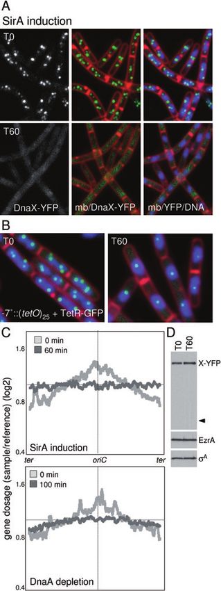
2 J. K. Wagner, K. A. Marquis and D. Z. Rudner 䊏

                                                                           over-replicate during sporulation. These results support a
                                                                           model in which SirA enforces diploidy by inhibiting DnaA
                                                                           after cells have entered the sporulation pathway.

                                                                           Results
                                                                           SirA (YneE) expression is lethal to vegetatively
                                                                           growing cells

                                                                           In a screen for factors that are required for proper chro-
                                                                           mosome organization during sporulation, we identified a
                                                                           Spo0A-regulated gene (Fawcett et al., 2000; Molle et al.,
                                                                           2003; Fujita et al., 2005) of unknown function yneE (sirA)
                                                                           that when deleted had a subtle defect in the organization
                                                                           of the axial filament (L. Mulcahy and D.Z. Rudner,
                                                                           unpublished). As a first step in the characterization of sirA,
                                                                           we investigated the consequences of artificial expression
                                                                           during vegetative growth. We placed sirA under the
                                                                           control of an IPTG-inducible promoter (Phyperspank), and
                                                                           examined the cells pre and post induction. Strikingly,
                                                                           induction of SirA resulted in the inability to produce colo-
                                                                           nies on agar plates (Fig. 2A). Analysis of cells expressing
                                                                           SirA in liquid culture revealed a dramatic change in
                                                                           the appearance of the chromosome mass (called the
                                                                           nucleoid). After 40–60 min of induction, the nucleoids
                                                                           rounded up and the spacing between them increased
                                                                           (Fig. 2B and Fig. S1). As cell division continued unabated,
                                                                           SirA induction eventually led to the production of many
                                                                           anucleate cells (Fig. 2B and Fig. S1). In addition, some of
Fig. 1. A. The Spo0A transcription factor activates the key                the cells contained nucleoids guillotined by division septa.
regulators that set the developmental pathway of sporulation into
motion. These include SpoIIE, required for asymmetric cell division;
                                                                           These phenotypes were strikingly similar to cells depleted
RacA, responsible for reorganization of the replicated                     of the DNA replication initiator protein, DnaA (Simmons
chromosomes into an axial filament; the first forespore (sF) and           et al., 2007 and Fig. 2B).
mother cell (sE) transcription factors and their regulators; and SirA,
required to maintain diploidy. Representative sporulating cells are
shown. The DNA was stained with DAPI (blue), the membranes
with FM4-64 (red) or TMA-DPH (white). A forespore promoter                 SirA inhibits DNA replication at the initiation step
(PspoIIQ) or a mother-cell promoter (PspoIIIA) fused to the gene
encoding GFP (green) shows compartment-specific transcription              SirA induction during vegetative growth closely phenocop-
factor activity. TetR–GFP (green) bound to a tetO array inserted           ied DnaA depletion; the nucleoid rounded up and cells
adjacent to oriC shows that the sporulating cell has only two
origins, one in each compartment (B) SirA enforces diploidy in
                                                                           continued to divide even though the chromosomes
sporulating cells by inhibiting DnaA, thereby preventing new rounds        appeared to have stopped replicating. To directly test
of initiation at oriC.                                                     whether ongoing replication had ceased, we used a
                                                                           marker for replication: a fusion of the tau subunit of DNA
development. We show that SirA (for sporulation inhibitor                  polymerase to the yellow fluorescent protein (DnaX–
of replication A) inhibits new rounds of replication by tar-               YFP). Cells harbouring DnaX–YFP that are actively rep-
geting the replication initiator DnaA. Induction of SirA                   licating their DNA contain fluorescent foci that colocalize
during vegetative growth rapidly blocks replication initia-                with the nucleoid (Fig. 3A) (Lemon and Grossman,
tion and phenocopies cellular depletion of DnaA. More-                     1998). Consistent with the idea that SirA blocks replica-
over, a strain that does not require DnaA to initiate                      tion, the number of DnaX–YFP foci diminished over
replication is immune to SirA. We further show that SirA                   an induction time-course and by 60 min, all cells in the
interacts directly with DnaA and that SirA disrupts DnaA                   field had diffuse DnaX–YFP fluorescence (Fig. 3A and
localization at the replication origin (oriC). Low levels of               Fig. S2). Immunoblot analysis indicates that the diffuse
SirA form faint foci at oriC that are themselves lost upon                 fluorescent signal was not a consequence of proteolytic
depletion of DnaA, suggesting that SirA acts on DnaA                       release of YFP (Fig. 3D). Replisome foci were similarly
bound to oriC. Finally, we show that cells lacking SirA                    lost after 100 min of depletion of DnaA (data not shown).

                                                                                                                      © 2009 The Authors
                                                               Journal compilation © 2009 Blackwell Publishing Ltd, Molecular Microbiology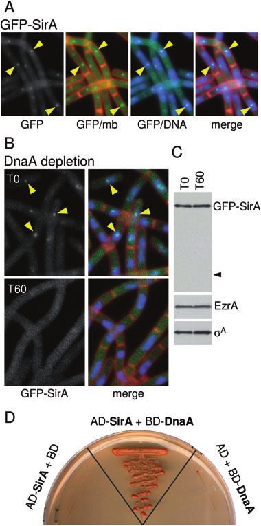
SirA inhibits replication initiation 3

                                                                        to four TetR–GFP foci per cell, representing two to four
                                                                        copies of the -7° locus per nucleoid (Fig. 3B). This
                                                                        number is consistent with the chromosomal content of
                                                                        B. subtilis grown under our conditions, where cell-
                                                                        doubling time is ~30 min (Haeusser and Levin, 2008). In
                                                                        support of the idea that SirA inhibits replication initiation,
                                                                        following 60 min of induction, the number of foci was
                                                                        reduced to one per DNA mass (Fig. 3B and Fig. S3B).
                                                                        Similar results were obtained in cells depleted of DnaA
                                                                        (data not shown). To rule out the possibility that the origins
                                                                        had duplicated but could not be resolved by fluorescence
                                                                        microscopy, we used a strain in which the tetO array was
                                                                        inserted at -35° (406 kb away from oriC). After 60 min of
                                                                        SirA induction, a single TetR–GFP focus was associated
                                                                        with each DNA mass (data not shown). These results are
                                                                        consistent with the idea that SirA inhibits replication
                                                                        initiation.
                                                                           To directly assess whether replication was inhibited at
                                                                        the initiation step following SirA induction, we performed
                                                                        genomic microarray analysis. The genomic DNA isolated
                                                                        from cells (before and after SirA induction) was normal-
                                                                        ized to a reference sample in which the ratio of origin to
                                                                        terminus DNA (oriC/ter) was 1.0, and was then used
                                                                        to probe a B. subtilis microarray (see Experimental
                                                                        procedures). Prior to the induction of SirA, the microarray
                                                                        profile was similar to what was observed previously for
                                                                        vegetatively growing cells (Wang et al., 2007) and the
                                                                        oriC/ter ratio was 1.6 (Fig. 3C). Following 60 min of SirA
                                                                        induction, the oriC/ter ratio was reduced to 1.02. As a
                                                                        control, we examined the oriC/ter ratio of cells depleted of
                                                                        DnaA. Following 100 min of depletion, the profiles looked
                                                                        similar to the SirA induction (Fig. 3C) and the oriC/ter ratio
                                                                        was 1.1. Collectively, these results are most consistent
Fig. 2. SirA expression is lethal to vegetatively growing cells.
A. Cells harbouring an IPTG-inducible promoter (Phyperspank) fused to   with SirA inhibiting the initiation of DNA replication.
sirA (Phy-sirA; strain BJW38) or gfp (Phy-gfp; strain BDR1003)
were streaked on LB agar plates with and without IPTG.
B. Phenotypic consequences of SirA induction (strain BJW38) and         SirA inhibits DnaA-dependent replication
DnaA depletion (strain LAS223) in liquid CH medium. Time
(in minutes) after induction or depletion is indicated. Membranes       We hypothesized that SirA might inhibit initiation by dis-
were stained with the fluorescent dye FM4-64 (red) and DNA was
                                                                        rupting the activity of the initiator protein DnaA. If SirA is
stained with DAPI (false-coloured green). Examples of anucleate
cells (white carets) and guillotined nucleoids (yellow carets) are      acting on DnaA, then a DnaA-independent strain should
indicated.                                                              be immune to the growth-inhibitory (or lethal) effects of
                                                                        SirA induction. To test this, we took advantage of a strain
Similar results were obtained with another marker for                   in which a plasmid origin (oriN) that does not require DnaA
ongoing replication: a GFP fusion to the single-strand                  to initiate replication was inserted into the B. subtilis
binding protein (SSB–GFP) (Fig. S3A).                                   chromosome. This strain can support growth and replica-
  As an independent assay to examine the replication                    tion in the absence of oriC and DnaA. (Hassan et al.,
status of the chromosome, we labelled a chromosomal                     1997). The plasmid origin was inserted at 359° (~11 kb
locus near the origin of replication (-7° on the genetic                from the native origin) (Berkmen and Grossman, 2007) in
map; 78 kb from oriC) by introducing a tandem array of 25               a strain that contains a deletion in the oriC region, which
tet operators (tetO) and the tet repressor fused to GFP                 disrupts DnaA-based replication. Accordingly, the
(TetR–GFP) (Michael, 2001; Burton et al., 2007). We then                matched control strain contained oriC inserted at this
examined the number of TetR–GFP foci within the cell                    same position. Consistent with the idea that SirA inhibits
before and after induction of SirA (Fig. 3B, Figs S2 and                DnaA-dependent replication, the cells utilizing oriN for
S3B). Prior to SirA induction, cells typically contained two            replication initiation were immune to SirA induction and

© 2009 The Authors
Journal compilation © 2009 Blackwell Publishing Ltd, Molecular Microbiology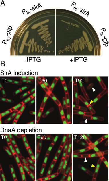
4 J. K. Wagner, K. A. Marquis and D. Z. Rudner 䊏

                                                               Fig. 3. SirA inhibits DNA replication at the initiation step.
                                                               A. Cells were analysed for ongoing replication using a DnaX–YFP
                                                               fusion. Images show cells (strain BJW84) before and after SirA
                                                               induction. Time (in min) after the addition of IPTG is indicated. Prior
                                                               to SirA induction, DnaX–YFP (false-coloured green) is present in
                                                               replisome foci. After SirA induction, DnaX–YFP foci are lost.
                                                               Membranes (mb) were stained with FM4-64 (red) and DNA was
                                                               stained with DAPI (blue).
                                                               B. Cells were analysed for origin content using TetR–GFP bound to
                                                               a tetO array adjacent to oriC. Images show cells (strain BJW141)
                                                               before and after SirA induction. Prior to SirA induction, most cells
                                                               have two or four origin foci per DNA mass. After SirA induction,
                                                               cells have a single origin focus per nucleoid.
                                                               C. Genomic DNA microarray analysis following SirA induction and
                                                               DnaA depletion. The top graph shows the average gene dosage
                                                               before (light grey) and after (dark grey) 60 min of SirA induction
                                                               (strain BJW38). The bottom graph shows the average gene dosage
                                                               before (light grey) and after (dark grey) 100 min of DnaA depletion
                                                               (strain LAS223). All the probed genes in the B. subtilis
                                                               chromosome arranged from -172° to +172° (ter-oriC-ter) are
                                                               represented on the x-axis. The y-axis represents the gene dosage
                                                               (log2) relative to a reference DNA with an oriC/ter ratio of 1 (see
                                                               Experimental procedures). The smoothed line was generated by
                                                               plotting the average gene dosage of the 25 genes before and 25
                                                               genes after each gene probed.
                                                               D. Immunoblot analysis shows that DnaX–YFP remains intact after
                                                               SirA induction. DnaX–YFP was analysed using anti-GFP antibodies
                                                               and the caret identifies the predicted size of free GFP. EzrA and sA
                                                               were used to control for loading.

                                                               formed colonies in the presence of IPTG (Fig. 4). By con-
                                                               trast, the matched control strain could only grow in the
                                                               absence of inducer. Analysis of the oriN-containing strain
                                                               by fluorescence microscopy revealed no difference in
                                                               nucleoid morphology in the presence or absence of SirA
                                                               (data not shown). These results support the idea that SirA
                                                               acts on oriC and/or DnaA.

                                                               SirA disrupts DnaA–GFP localization
                                                               If SirA is acting on DnaA, then expression of SirA might
                                                               disrupt DnaA localization at oriC. To investigate this pos-
                                                               sibility, we used a strain harbouring wild-type DnaA and a

                                                               Fig. 4. Cells that initiate replication in a DnaA-independent
                                                               manner are immune to SirA induction. Cells harbouring a
                                                               DnaA-independent origin of replication (oriN, strain BJW173) or a
                                                               DnaA-dependent origin (oriC, strain BJW174) were streaked on
                                                               plates with and without IPTG. Both strains contain an
                                                               IPTG-inducible promoter (Phyperspank) fused to sirA.

                                                                                                          © 2009 The Authors
                                                   Journal compilation © 2009 Blackwell Publishing Ltd, Molecular Microbiology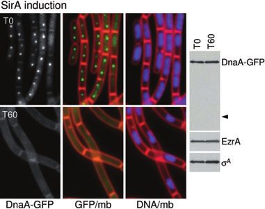
SirA inhibits replication initiation 5

                                                                      growth (Fig. S4), but analysis by fluorescence microscopy
                                                                      revealed rounded-up DNA and anucleate cell phenotypes
                                                                      (Fig. S5A) similar to the untagged fusion. Examination of
                                                                      GFP–SirA revealed faint foci early during an induction
                                                                      time-course, but within 20 min the fusion appeared as a
                                                                      diffuse haze (Fig. S5A). As the kinetics of foci loss were
                                                                      similar to what we observed for DnaA–GFP following SirA
                                                                      induction, we wondered whether GFP–SirA first localized
                                                                      to DnaA bound to oriC, and then became diffuse as DnaA
                                                                      was displaced from this site. To investigate this, we gen-
                                                                      erated a xylose-inducible promoter fusion to gfp–sirA that
                                                                      produces approximately 10-fold less protein than the
                                                                      IPTG-inducible version (Fig. S5B). At this low level of
                                                                      GFP–SirA, the cells were not growth-inhibited although
                                                                      nucleoid morphology was somewhat aberrant (Fig. 6A).
                                                                      Importantly, 10–20% of the cells contained one or two
Fig. 5. SirA disrupts DnaA–GFP localization.                          faint foci. These foci colocalized with the DNA mass
A. Localization of DnaA–GFP (green) before and after SirA
                                                                      (Fig. 6A and Fig. S5A) and an origin marker (Fig. S6).
induction (strain BJW218). Time (in min) after the addition of IPTG
is indicated. Membranes (mb) were stained with FM4-64 (red) and       Upon depletion of DnaA, the GFP–SirA foci were lost
DNA was stained with DAPI (blue). Immunoblot analysis shows that      (Fig. 6B). Immunoblot analysis shows that the diffuse
DnaA–GFP remains intact after SirA induction. DnaA–GFP was
                                                                      signal was not a result of degradation of SirA and release
analysed using anti-GFP antibodies and the caret identifies the
predicted size of free GFP. EzrA and sA were used to control for      of free GFP (Fig. 6C). These results indicate that GFP–
loading. The apparent septal membrane association of DnaA–GFP         SirA localization requires DnaA and suggest that SirA acts
is due to bleed through from the membrane dye.
                                                                      on DnaA.

DnaA–GFP fusion inserted at an ectopic chromosomal
                                                                      SirA and DnaA interact directly
locus. Cells expressing this fusion contained two DnaA–
GFP foci per DNA mass, one near each cell quarter, or a               We found that SirA disrupts DnaA foci, and that SirA foci
diffuse fluorescent haze that appeared to colocalize with             are dependent on DnaA. A simple model incorporating
the DAPI-stained DNA (Fig. 5). DnaA–YFP exhibited a                   these reciprocal results is that SirA acts on DnaA bound to
similar localization pattern as DnaA–GFP, and the foci                oriC, releasing it from the origin, in turn preventing repli-
colocalized with an origin proximal marker (data not                  cation initiation. To determine if SirA and DnaA interact
shown), suggesting that these fluorescent foci reflect                directly, we performed a yeast two-hybrid assay. SirA was
DnaA bound to oriC. When SirA was induced, the DnaA–                  fused to the DNA binding domain of GAL4, and DnaA was
GFP foci became diffuse after 15 min of induction and                 fused to the GAL4 activation domain. When both fusions
were completely lost after 60 min. At this time, the DNA              were expressed in yeast a target gene (ADE2) was acti-
had taken on its characteristic rounded-up appearance                 vated resulting in the ability to grow in the absence of
(Fig. 5). Immunoblot analysis indicates that the diffuse              adenine (Fig. 6D). Importantly, when the GAL4–SirA
fluorescent signal was not a consequence of proteolytic               fusion was coexpressed with the empty GAL4 activation
release of GFP (Fig. 5). Recent studies from Graumann                 domain or the empty GAL4 DNA binding domain was
and colleagues suggest that DnaA tracks with the repli-               coexpressed with the GAL4–DnaA fusion, neither was
some (Soufo et al., 2008). Although our results suggest               able to activate expression of the target gene and restore
that DnaA–GFP foci colocalize with oriC, our data do not              adenine prototrophy (Fig. 6D). Attempts to detect an inter-
rule out the possibility that some of the DnaA–GFP is                 action between GFP–SirA and DnaA by immuno-affinity
replisome-associated. In either case, our results indicate            purification from B. subtilis lysates were unsuccessful,
that SirA disrupts DnaA–GFP foci and suggest that SirA                suggesting that the interaction between these proteins is
acts on DnaA.                                                         weak or transient.

DnaA is required for SirA localization                                SirA inhibits replication during sporulation

We hypothesized that if SirA targets DnaA, then SirA                  Collectively, our data indicate that SirA disrupts the inter-
localization would be dependent on DnaA. We first exam-               action between DnaA and the origin of replication, and
ined the localization of an IPTG-inducible GFP–SirA                   that this leads to an inhibition of DNA replication.
fusion. The GFP fusion was a less potent inhibitor of cell            However, the replication inhibition phenotypes we

© 2009 The Authors
Journal compilation © 2009 Blackwell Publishing Ltd, Molecular Microbiology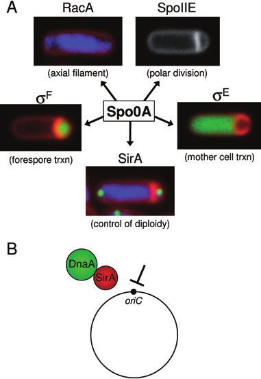
6 J. K. Wagner, K. A. Marquis and D. Z. Rudner 䊏
Fig. 6. DnaA is required for GFP–SirA localization.
A. Localization of GFP–SirA (green) produced at low levels during
vegetative growth (strain BJW196). Membranes (mb) were stained
with FM4-64 (red) and DNA was stained with DAPI (blue). Yellow
carets highlight nucleoid-associated GFP–SirA foci.
B. GFP–SirA localization before and after depletion of DnaA (strain
BJW197). Time (in min) after the removal of IPTG (to deplete
DnaA) is indicated.
C. Immunoblot analysis shows that GFP–SirA remains intact after
DnaA depletion. GFP–SirA was analysed using anti-GFP antibodies
and the caret identifies the predicted size of free GFP. EzrA and sA
were used to control for loading.
D. SirA and DnaA interact in the yeast two-hybrid assay. A yeast
strain harbouring the Gal4 activation domain fused to SirA
(AD–SirA) and the Gal4 DNA binding domain fused to DnaA
(BD–DnaA) was able to activate transcription of the GAL2 promoter
fused to the ADE2 gene. Growth on medium lacking adenine
indicates a positive interaction between the hybrid proteins. Strains
containing the AD–SirA fusion and the unfused DNA binding
domain (BD) or the BD–DnaA fusion and the unfused activation
domain (AD) did not activate transcription and were unable to grow
in the absence of adenine.
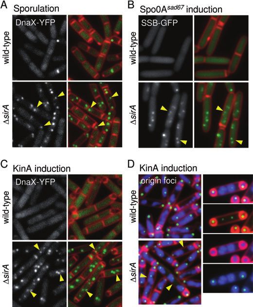
more DnaX–YFP foci (Fig. 7A). By contrast, 31%
(n = 202) of the SirA mutant had DnaX–YFP foci at this
time point (Fig. 7A). Thirty minutes later, at hour two,
most of the wild-type cells had completed replication as
evidenced by the loss of DnaX–YFP foci. At this time
point only 0.6% (n = 344) of the sporulating cells had
replisome foci. However, 12.2% (n = 500) of the SirA
mutant cells still had ongoing replication. Strikingly, even
SirA mutant cells that had completed engulfment (a late
stage in the developmental pathway) contained replica-
tion foci. At this stage, all wild-type sporulating cells had
diffuse DnaX–YFP signal. These results indicate that
SirA contributes to the inhibition of replication during
sporulation.
Although it would be inconsistent with the vegetative
results, it was possible that the DnaX–YFP foci in the SirA
mutant represent replisomes that were stalled during
sporulation rather than an actual increase in replication
initiation. To determine if the SirA mutant cells have an
increased frequency of initiation relative to wild type, we
labelled an origin-proximal region of the chromosome
using a tetO array and TetR–GFP, and quantified the
observed resulted from the artificial induction of a number of foci per sporulating cell. In wild-type cells at
sporulation-specific protein during vegetative growth. To hour two of sporulation, two copies of the -7° region were
determine whether SirA functions to inhibit DNA replica- present in 98.2% (n = 393) of the sporulating cells, while
tion during sporulation, we assayed the replication status only 1.3% possessed more than two foci. By contrast, in
of wild type and the SirA mutant after the cells had com- the SirA mutant 4.5-fold more sporulating cells had
mitted to sporulation. As a marker for replication, we greater than two foci (5.8%, n = 380). We could not detect
again utilized DnaX–YFP. Cells were induced to sporu- a substantial increase in the oriC/ter ratio during sporula-
late by resuspension in a nutrient-poor medium and tion using genomic microarrays (data not shown); this
analysed after 1.5 and 2 h of sporulation. In both wild result was not surprising, as only a subset of cells in the
type and the SirA mutant, DnaX–YFP foci were present population appear to over-replicate under these sporula-
in all cells at the time of resuspension (not shown). At tion conditions. In sum, these results are consistent with
hour 1.5, 6.9% (n = 303) of the wild-type sporulating cells the idea that SirA helps maintain diploidy during
(defined by the presence of a polar septum) had one or sporulation.
© 2009 The Authors
Journal compilation © 2009 Blackwell Publishing Ltd, Molecular MicrobiologySirA inhibits replication initiation 7
Fig. 7. Sporulating cells over-replicate in the
absence of SirA.
A. Cells induced to sporulate by resuspension
in defined minimal medium were analysed for
ongoing replication using a DnaX–YFP fusion.
The images show wild type (strain BKM1585)
and the SirA mutant (strain BJW91) at hour
1.5 of sporulation. DnaX–YFP (false-coloured
green) and membranes stained with
TMA-DPH (false-coloured red) are shown.
Yellow carets highlight sporulating cells with
replisome foci.
B. Cells expressing a constitutively active
allele of Spo0A (Spo0Asad67) for 60 min were
analysed for ongoing replication using an
SSB–GFP fusion. The images show wild type
(strain BJW271) and the SirA mutant (strain
BJW275). Yellow carets highlight cells with
ongoing replication as assessed by SSB–GFP
foci.
C. Cells sporulated in rich medium by
induction of the sensor kinase KinA were
analysed for ongoing replication using a
DnaX–YFP fusion. The images show wild type
(strain BJW230) and the SirA mutant
(strain BJW232) after 2 h of KinA induction.
Yellow carets highlight sporulating cells with
replisome foci.
D. Cells sporulated in rich medium by
induction of the sensor kinase KinA were
analysed for origin content using TetR–GFP
bound to a tetO array adjacent to oriC. The
images show wild type (strain BJW279) and
the SirA mutant (strain BJW279) after 2 h of
KinA induction. Yellow carets highlight cells
with more than two TetR–GFP (green) foci.
Membranes were stained with FM4-64 (red)
and DNA was stained with DAPI (blue). An
example of a mother cell (with two
chromosomes) and twin forespores after 2.5 h
of KinA induction is shown on the right.
Spo0A inhibits replication in a SirA-dependent manner fusion of the single-strand binding protein to GFP (SSB–
GFP) (for reasons that we were unable to determine, the
Our data indicate that only a subset of sporulating cells spo0A sad67 allele was unstable in the presence of the
initiate new rounds of replication in the absence of SirA. DnaX–YFP fusion.) Prior to induction of Spo0Asad67, all cells
This suggests that additional factors help enforce diploidy were actively replicating their chromosomes as evidenced
as cells initiate sporulation. A good candidate is Spo0A by the presence of SSB–GFP foci (for example, see
itself, which binds near oriC and prevents the formation of Fig. S3A). However, within 60 min of induction, 81%
open replication complexes in vitro (Castilla-Llorente et al., (n = 336) of the cells had diffuse SSB–GFP signal
2006). To investigate whether Spo0A directly inhibits rep- (Fig. 7B). Those with active replisomes (19%) usually had
lication in vivo, we took advantage of a constitutively active a single focus. This result indicates that either Spo0A or
allele of Spo0A (called Spo0Asad67) (Ireton et al., 1993). genes under its control can efficiently inhibit replication. To
It has been shown previously that Spo0Asad67 binds to oriC determine the contribution of SirA to this replication block,
in vivo (Molle et al., 2003) and is able to prevent replica- we examined SSB–GFP foci upon induction of Spo0Asad67
tion in vitro (Castilla-Llorente et al., 2006). Induction of in the SirA mutant. Strikingly, 94% (n = 361) of the cells had
Spo0Asad67 during growth results in the activation of the foci after 60 min of Spo0Asad67 induction (Fig. 7B). These
Spo0A regulon; the terminal phenotype is cell lysis. results indicate that activated Spo0A is incapable of
However, at early times after induction, it is possible to directly inhibiting replication initiation or does so ineffi-
study cell physiology (Levin and Losick, 1996). To monitor ciently. It further suggests that SirA is the principal regulator
ongoing replication upon Spo0Asad67 induction, we used a of replication initiation under Spo0A control.
© 2009 The Authors
Journal compilation © 2009 Blackwell Publishing Ltd, Molecular Microbiology8 J. K. Wagner, K. A. Marquis and D. Z. Rudner 䊏
SirA plays a more critical role in replication inhibition ling of the replicated chromosomes into an axial filament
during fast growth (Ben-Yehuda et al., 2003), and forespore- and mother-
specific gene transcription (Jonas et al., 1988; Schmidt
We observed that, at most, 31% of SirA mutant cells have
et al., 1990). Here we have shown that Spo0A also
an over-replication phenotype during sporulation. These
enforces diploidy on the sporulating cell. Spo0A appears
results indicate that SirA cannot be the only factor main-
to accomplish this primarily via the induction of the regu-
taining diploidy during sporulation. When bacteria enter
latory protein SirA, rather than through direct binding to
stationary phase or slow down growth, they co-ordinately
the origin of replication. Our data indicate that SirA main-
reduce replication (Haeusser and Levin, 2008). Accord-
tains diploidy by inhibiting new rounds of replication, spe-
ingly, we wondered whether the conditions in which we
cifically by targeting the initiator protein DnaA (Fig. 1B).
induce sporulation in the laboratory (resuspension in
The interaction between SirA and DnaA could prevent
minimal medium) might result in reduced replication, thus
binding to oriC. Alternatively, SirA could function like
masking the importance of SirA. To test this, we took
E. coli Hda (Su’etsugu et al., 2008), by stimulating the
advantage of a strain in which efficient sporulation can be
nucleotide hydrolysis of ATP bound to DnaA. SirA has no
induced during vegetative growth. Fujita and Losick have
apparent sequence motifs (and is only found in endospore
shown that a small increase in expression of the sensor
formers) providing no clues to its function. Experiments
kinase KinA leads to a gradual accumulation of active
aimed at directly testing these models in vitro have been
Spo0A and efficient entry into sporulation (Fujita and
hampered by the insolubility of recombinant SirA. That
Losick, 2005). We induced low levels of KinA in wild-type
SirA is an inhibitor of DNA replication was independently
or SirA mutant cells in early exponential growth and moni-
discovered by R. Losick and colleagues (pers. comm.)
tored ongoing replication using the DnaX–YFP fusion.
(Rahn-Lee et al., 2009).
After 1.5 h of induction, virtually all cells had initiated
sporulation as evidenced by the formation of polar septa.
Under these conditions, 11% (n = 607) of the wild-type SirA and nutrient status enforce diploidy
sporulating cells had DnaX–YFP foci (compared with
Our results indicate that SirA inhibition of DnaA is not
6.9% of the cells that had replisome foci when sporulation
the sole mechanism for preventing new rounds of DNA
was induced by resuspension in minimal medium). By
replication as cells enter sporulation. SirA plays a more
contrast, in cells lacking SirA, 73% (n = 532) of sporulat-
significant role in enforcing diploidy when cells are engi-
ing cells had replisome foci (Fig. 7C). Similar results were
neered to sporulate in the presence of nutrients than
obtained monitoring TetR–GFP bound to a tetO array at
under nutrient-depleted conditions. Accordingly, these
-7° (Fig. 7D and Fig. S7A). Specifically, 11% (n = 290) of
data are most consistent with the idea that nutrient
wild-type sporulating cells had more than two origin foci,
status (and co-ordinately controlled replication) serves
while 54% (n = 396) had more than two foci in the SirA
as the other mechanism that maintains diploidy. It is
mutant. Over-replication in the sporulating cells lacking
likely that B. subtilis typically sporulates under conditions
SirA could be observed in both the mother cell and fore-
of nutrient deprivation and slow growth, where the rate
spore (Fig. 7C and D and Fig. S7A). Moreover, in a subset
of replication initiation is also co-ordinately slow. Nutrient
of cells (~2%) we could detect ‘twin spores’ (P. Piggot and
status should therefore dictate that most cells do not
M. Elowitz, pers. comm.). These sporulating cells have a
initiate new rounds of replication. Why then does the
single mother with one or two chromosomes and two
sporulating cell also induce a protein that specifically
spores, one at each pole (Fig. 7D and Fig. S7). These
inhibits replication initiation? Differentiation into a
results indicate that the sporulation conditions can have a
dormant spore is an energy-intensive process, requiring
major impact on the importance of SirA in preventing new
the synthesis of hundreds of sporulation-specific pro-
rounds of replication. They further suggest that nutrient
teins (Eichenberger et al., 2004; Wang et al., 2006).
status and SirA are the major enforcers of diploidy during
Therefore, cells must enter this developmental pathway
spore formation.
prior to the complete exhaustion of nutrients. The induc-
tion of SirA as cells commence spore formation may
serve as a fail-safe mechanism, ensuring that nutrient
Discussion reserves for spore maturation are not further depleted by
The Spo0A transcription factor is the master regulator new rounds of inappropriate DNA replication. It is note-
governing entry into sporulation (Fig. 1A) (reviewed in worthy that cells lacking SirA have only a modest defect
Stragier and Losick, 1996; Errington, 2003; Hilbert and in sporulation efficiency (15% reduction). However, in
Piggot, 2004). Spo0A controls the genes responsible for the context of the environment, it is likely that the strin-
the switch from medial to polar cell division (Khvorova gent control of energy and cellular metabolites is critical
et al., 2000; Ben-Yehuda and Losick, 2002), the remodel- to overall fitness.
© 2009 The Authors
Journal compilation © 2009 Blackwell Publishing Ltd, Molecular MicrobiologySirA inhibits replication initiation 9
An additional role for SirA could be to help commit the scription of the sda gene is regulated by DnaA; however,
sporulating cell to its developmental fate. After polar divi- it is unclear how it carries out this function (Burkholder
sion and the activation of the first mother cell and fore- et al., 2001). If the accumulation of DnaA–ATP promotes
spore transcription factors, the sporulating cell becomes the expression of sda (and inhibition of sporulation), con-
irreversibly committed to sporulation (Dworkin and Losick, ditions that reduce DnaA–ATP levels (such as initiation
2005). Even in the face of nutrient-rich conditions, the itself) would lead to the loss of sda gene expression and
mother and forespore continue down their respective ultimately to entry into sporulation.
developmental pathways. It is possible that the inhibition
of replication by SirA in both differentiating cell types is
one arm of the control mechanism that prevents return to Twin spores
vegetative growth. Finally, we have found that one consequence of a failure
It had been previously proposed that the binding of to enforce diploidy during sporulation is the formation of
active Spo0A to the sequences within oriC could serve to ‘twins’, a phenotype identified by P. Piggot and M. Elowitz
inhibit replication initiation (Castilla-Llorente et al., 2006). (pers. comm.) (Eldar et al., 2009) in which a single mother
Our data indicate that a constitutively active allele of (with one or two chromosomes) nurtures two spores, one
Spo0A is not sufficient to inhibit replication in the absence at each cell pole. These spores appear to develop nor-
of SirA, suggesting that Spo0A is not likely to play a major mally as judged by their grey appearance by phase-
role in enforcing diploidy. However, these results do not contrast microscopy (Fig. S7B) (Piggot and Coote, 1976).
exclude the possibility that Spo0A binding to oriC could Symmetrical spore formation has been described previ-
function in tandem with SirA to help prevent replication ously in a cousin of B. subtilis, Metabacterium polyspora
initiation. Interestingly, the master transcriptional regulator (Angert and Losick, 1998). Remarkably, these bacteria
(CtrA) in the bacterium Caulobacter crescentus has also produce spores as part of their cell cycle, and the fore-
been suggested to control the initiation of DNA replication spores themselves have been observed replicating DNA
(Quon et al., 1998). In its absence, cells over-replicate. and undergoing division (Ward and Angert, 2008). We
CtrA, like Spo0A, binds to sites around oriC and it has speculate that the energetic cost of raising two spores
been proposed that this binding occludes DnaA (Quon instead of one has been selected against in asymmetrical
et al., 1998). However, mutations in the five CtrA binding spore formers like B. subtilis. Indeed, B. subtilis has
sites in C. crescentus oriC did not measurably perturb evolved elaborate regulatory pathways that normally
replication control (Bastedo and Marczynski, 2009). By ensure a one mother one spore family (Khvorova et al.,
analogy to SirA, we hypothesize that a CtrA-controlled 1998; Eichenberger et al., 2001; Zupancic et al., 2001).
protein inhibits replication initiation, perhaps by directly
targeting DnaA.
Experimental procedures
What prevents haploid cells from entering sporulation? General methods
We have argued that nutrient status and SirA maintain Unless otherwise noted, B. subtilis strains were derived from
and enforce diploidy during sporulation. What then pre- the prototrophic strain PY79 (Youngman et al., 1983). E. coli
strains were TG1 and DH5a. To visualize the localization of
vents slow-growing cells with only one chromosome from
fluorescent fusions during vegetative growth, strains were
prematurely entering this developmental pathway? A
grown in CH medium (Harwood and Cutting, 1990) at 37°C.
good candidate for this function is the checkpoint protein Sporulation was induced by resuspension at 37°C according
Sda. In response to blocks in replication initiation and/or to the method of Sterlini-Mandelstam (Harwood and Cutting,
elongation, Sda inhibits the initiation of sporulation by 1990). A description of strains (Table S1) and plasmids
preventing the activation of Spo0A (Burkholder et al., (Table S2) and oligonucleotide primer sequences (Table S3)
2001). In addition to inhibiting sporulation when cells are can be found in supplemental material.
experiencing replication stress, one of Sda’s functions
could be to prevent haploid cells from entering sporulation Induction and depletion conditions
prior to a final round of DNA replication (Michael, 2001).
Mechanistically, one way this regulation could occur is Unless otherwise indicated, fluorescence microscopy was
through the normal cycling of ATP- and ADP-bound DnaA. performed on exponentially growing cells (OD600 = 0.3–0.8).
SirA was induced with 1.0 mM IPTG or 10 mM xylose in liquid
In E. coli (and presumably in B. subtilis) DnaA–ATP (the
cultures and on LB agar plates. KinA and Spo0Asad67 were
form of DnaA that promotes origin melting) accumulates
induced as described previously (Fujita and Losick, 2005)
during the cell cycle. Once DnaA–ATP achieves a critical with 20 mM and 200 mM IPTG respectively. SSB–GFP and
concentration, it triggers the initiation of replication DnaA–GFP were induced with 10 mM xylose. The DnaA–
(Kurokawa et al., 1999), resulting in ATP hydrolysis. Tran- GFP fusion was synthesized at levels below those required to
© 2009 The Authors
Journal compilation © 2009 Blackwell Publishing Ltd, Molecular Microbiology10 J. K. Wagner, K. A. Marquis and D. Z. Rudner 䊏
support growth in the absence of DnaA. For DnaA depletions, alized with a phase contrast objective UplanFLN 100¥ and
exponentially growing cells in CH medium supplemented with captured with a monochrome CoolSnapHQ digital camera
0.5 mM IPTG and 10 mg ml-1 tetracycline were washed three (Photometrics) using Metamorph software version 6.1 (Uni-
times with CH medium supplemented with 10 mg ml-1 tetra- versal Imaging). Exposure times were typically 500–1000 ms
cycline but lacking IPTG. The washed cells were resus- for GFP and YFP protein fusions. Membranes were stained
pended in CH supplemented with 10 mg ml-1 tetracycline at with either TMA-DPH or FM4-64 (Molecular Probes), at a final
an OD600 = 0.1 to begin the depletion time-course. concentration of 0.02 mM and 3 mg ml-1 respectively, and
imaged with exposure times of 200 ms. DNA was stained with
DAPI (Molecular Probes), at a final concentration of 2 mg ml-1
Genomic microarray
and imaged with a typical exposure time of 200 ms. Fluores-
Genomic microarray analysis was carried as described pre- cence images were analysed, adjusted and cropped using
viously (Wang et al., 2007). Briefly, genomic DNA was iso- Metamorph v 6.1 software (Molecular Devices).
lated from the strains of interest and cleaved to completion
with HaeIII. Reference DNA was from a temperature-
sensitive replication initiation mutant (dnaB134) grown at the Acknowledgements
non-permissive temperature (42°C) for one hour to ensure
that all rounds of replication were complete. The HaeIII- We thank members of the Rudner laboratory past and
cleaved reference DNA was labelled with Cy3 (GE Health- present, Michael Laub and Bill Burkholder for valuable dis-
care) and the experimental samples were labelled with Cy5 cussions. We thank Larry Mulcahy for the original identifi-
(GE Healthcare). The labelled DNA was hybridized to an cation of yneE; Geng Li and Danesh Moazed for help with
oligonucleotide microarray containing a 70mer for each gene the two-hybrid assay. We acknowledge Alan Grossman,
in the B. subtilis genome. Scanning was carried out on a Masaya Fujita, Rich Losick, Lyle Simmons, Heath Murray
Genepix 4000B scanner (Software v. 5.1), and data were and Petra Levin for generously providing strains and anti-
analysed in Microsoft Excel. Intensities from the 532 nm bodies. We thank Patrick Piggot and Michael Elowitz for
channel were normalized to the reference (635 nm channel) communicating results prior to publication. Support for this
prior to subsequent analysis. Each data point on the smooth work comes in part from the National Institute of Health
line in Fig. 3C represents an average of the normalized ratios Grant GM073831-01A1, the Giovanni Armenise-Harvard
of the 25 genes on either side of each locus. The oriC/ter Foundation and the Hellman Family Faculty Fund. D.Z.R. is
ratios were calculated by dividing the average normalized supported by the Damon Runyon Cancer Research Foun-
intensities of the 25 genes on either side of dnaA (oriC) by the dation (DRS-44-05). J.K.W. dedicates this paper to the
normalized intensities of 25 genes on either side of rtp (ter). memory of her brother.
Immunoblot analysis References
During logarithmic growth the OD600 was measured (for Angert, E.R., and Losick, R.M. (1998) Propagation by sporu-
equivalent loading) and samples were collected by lation in the guinea pig symbiont Metabacterium polyspora.
centrifugation. Whole-cell extracts were prepared by resus- Proc Natl Acad Sci USA 95: 10218–10223.
pension of cell pellets in 50 ml lysis buffer (20 mM Tris pH 7.0, Bastedo, D.P., and Marczynski, G.T. (2009) CtrA response
10 mM MgCl2, 1 mM EDTA, 1 mg ml-1 lysozyme, 10 mg ml-1 regulator binding to the Caulobacter chromosome replica-
DNase I, 100 mg ml-1 RNase A, with protease inhibitors: tion origin is required during nutrient and antibiotic stress
1 mM PMSF, 1 mg ml-1 leupeptin, 1 mg ml-1 pepstatin) and as well as during cell cycle progression. Mol Microbiol 72:
incubation at 37°C for 10 min followed by addition of 50 ml 139–154.
sodium dodecyl sulphate (SDS) sample buffer (0.25 M Tris Ben-Yehuda, S., and Losick, R. (2002) Asymmetric cell divi-
pH 6.8, 4% SDS, 20% glycerol, 10 mM EDTA) containing sion in B. subtilis involves a spiral-like intermediate of the
10% 2-Mercaptoethanol. Samples were heated for 5 min at cytokinetic protein FtsZ. Cell 109: 257–266.
80°C prior to loading. Proteins were separated by SDS-PAGE Ben-Yehuda, S., Rudner, D.Z., and Losick, R. (2003) RacA,
on 15% polyacrylamide gels, electroblotted onto Immobilon-P a bacterial protein that anchors chromosomes to the cell
membrane (Millipore) and blocked in 5% non-fat milk in poles. Science 299: 532–536.
phosphate-buffered saline-0.5% Tween-20. The blocked Berkmen, M.B., and Grossman, A.D. (2007) Subcellular posi-
membrane was probed with anti-GFP (Rudner and Losick, tioning of the origin region of the Bacillus subtilis chromo-
2002), anti-EzrA (Levin et al., 1999) and anti-sA (Fujita, 2000) some is independent of sequences within oriC, the site of
antibodies diluted into 3% BSA in phosphate-buffered saline- replication initiation, and the replication initiator DnaA.
0.05% Tween-20. The primary antibodies were detected Mol Microbiol 63: 150–165.
using horseradish peroxidase-conjugated goat, anti-rabbit Burbulys, D., Trach, K.A., and Hoch, J.A. (1991) Initiation of
immunoglobulin G (Bio-Rad) with the Supersignal Substrate sporulation in B. subtilis is controlled by a multicomponent
as described by the manufacturer (Perkin Elmer). phosphorelay. Cell 64: 545–552.
Burkholder, W.F., Kurtser, I., and Grossman, A.D. (2001)
Fluorescence microscopy Replication initiation proteins regulate a developmental
checkpoint in Bacillus subtilis. Cell 104: 269–279.
Fluorescence microscopy was performed as previously Burton, B.M., Marquis, K.A., Sullivan, N.L., Rapoport, T.A.,
described (Doan et al., 2005). Fluorescent signals were visu- and Rudner, D.Z. (2007) The ATPase SpoIIIE transports
© 2009 The Authors
Journal compilation © 2009 Blackwell Publishing Ltd, Molecular MicrobiologySirA inhibits replication initiation 11 DNA across fused septal membranes during sporulation in (1998) The spoIIE locus is involved in the Spo0A- Bacillus subtilis. Cell 131: 1301–1312. dependent switch in the location of FtsZ rings in Bacillus Castilla-Llorente, V., Munoz-Espin, D., Villar, L., Salas, M., subtilis. J Bacteriol 180: 1256–1260. and Meijer, W.J. (2006) Spo0A, the key transcriptional Khvorova, A., Chary, V.K., Hilbert, D.W., and Piggot, P.J. regulator for entrance into sporulation, is an inhibitor of (2000) The chromosomal location of the Bacillus subtilis DNA replication. EMBO J 25: 3890–3899. sporulation gene spoIIR is important for its function. Doan, T., Marquis, K.A., and Rudner, D.Z. (2005) Subcellular J Bacteriol 182: 4425–4429. localization of a sporulation membrane protein is achieved Kurokawa, K., Nishida, S., Emoto, A., Sekimizu, K., and through a network of interactions along and across the Katayama, T. (1999) Replication cycle-coordinated change septum. Mol Microbiol 55: 1767–1781. of the adenine nucleotide-bound forms of DnaA protein in Dworkin, J., and Losick, R. (2005) Developmental commit- Escherichia coli. EMBO J 18: 6642–6652. ment in a bacterium. Cell 121: 401–409. Lemon, K.P., and Grossman, A.D. (1998) Localization of Eichenberger, P., Fawcett, P., and Losick, R. (2001) A three- bacterial DNA polymerase: evidence for a factory model of protein inhibitor of polar septation during sporulation in replication. Science 282: 1516–1519. Bacillus subtilis. Mol Microbiol 42: 1147–1162. Levin, P.A., and Losick, R. (1996) Transcription factor Spo0A Eichenberger, P., Fujita, M., Jensen, S.T., Conlon, E.M., switches the localization of the cell division protein FtsZ Rudner, D.Z., Wang, S.T., et al. (2004) The program of from a medial to a bipolar pattern in Bacillus subtilis. Genes gene transcription for a single differentiating cell type Dev 10: 478–488. during sporulation in Bacillus subtilis. PLoS Biol 2: e328. Levin, P.A., Kurtser, I.G., and Grossman, A.D. (1999) Identi- Eldar, A., Chary, V.K., Xenopoulos, P., Fontes, M.E., Loson, fication and characterization of a negative regulator of FtsZ O.C., Dworkin, J., et al. (2009) Partial penetrance ring formation in Bacillus subtilis. Proc Natl Acad Sci USA facilitates developmental evolution in bacteria. Nature 96: 9642–9647. 460: 510–514. Michael, W.M. (2001) Cell cycle: connecting DNA replication Errington, J. (2003) Regulation of endospore formation in to sporulation in Bacillus. Curr Biol 11: R443–R445. Bacillus subtilis. Nat Rev Microbiol 1: 117–126. Molle, V., Fujita, M., Jensen, S.T., Eichenberger, P., Fawcett, P., Eichenberger, P., Losick, R., and Youngman, P. Gonzalez-Pastor, J.E., Liu, J.S., and Losick, R. (2003) The (2000) The transcriptional profile of early to middle sporu- Spo0A regulon of Bacillus subtilis. Mol Microbiol 50: 1683– lation in Bacillus subtilis. Proc Natl Acad Sci USA 97: 1701. 8063–8068. Piggot, P.J., and Coote, J.G. (1976) Genetic aspects of bac- Fujita, M. (2000) Temporal and selective association of terial endospore formation. Bacteriol Rev 40: 908–962. multiple sigma factors with RNA polymerase during sporu- Quon, K.C., Yang, B., Domian, I.J., Shapiro, L., and Marc- lation in Bacillus subtilis. Genes Cells 5: 79–88. zynski, G.T. (1998) Negative control of bacterial DNA Fujita, M., and Losick, R. (2005) Evidence that entry into replication by a cell cycle regulatory protein that binds at sporulation in Bacillus subtilis is governed by a gradual the chromosome origin. Proc Natl Acad Sci USA 95: 120– increase in the level and activity of the master regulator 125. Spo0A. Genes Dev 19: 2236–2244. Rahn-Lee, L., Gorbatyuk, B., Skovgaard, O., and Losick, R. Fujita, M., Gonzalez-Pastor, J.E., and Losick, R. (2005) High- (2009) The conserved sporulation protein YneE inhibits and low-threshold genes in the Spo0A regulon of Bacillus DNA replication in Bacillus subtilis. J Bacteriol 191: 3736– subtilis. J Bacteriol 187: 1357–1368. 3739. Haeusser, D.P., and Levin, P.A. (2008) The great divide: Rudner, D.Z., and Losick, R. (2002) A sporulation membrane coordinating cell cycle events during bacterial growth and protein tethers the pro-sigmaK processing enzyme to its division. Curr Opin Microbiol 11: 94–99. inhibitor and dictates its subcellular localization. Genes Harwood, C.R., and Cutting, S.M. (1990) Molecular Biological Dev 16: 1007–1018. Methods for Bacillus. New York: Wiley. Ryter, A., Schaeffer, P., and Ionesco, H. (1966) [Cytologic Hassan, A.K., Moriya, S., Ogura, M., Tanaka, T., Kawamura, classification, by their blockage stage, of sporulation F., and Ogasawara, N. (1997) Suppression of initiation mutants of Bacillus subtilis Marburg]. Ann Inst Pasteur defects of chromosome replication in Bacillus subtilis dnaA (Paris) 110: 305–315. and oriC-deleted mutants by integration of a plasmid rep- Schmidt, R., Margolis, P., Duncan, L., Coppolecchia, R., licon into the chromosomes. J Bacteriol 179: 2494–2502. Moran, C.P., Jr and Losick, R. (1990) Control of develop- Hilbert, D.W., and Piggot, P.J. (2004) Compartmentalization mental transcription factor sigma F by sporulation regula- of gene expression during Bacillus subtilis spore formation. tory proteins SpoIIAA and SpoIIAB in Bacillus subtilis. Proc Microbiol Mol Biol Rev 68: 234–262. Natl Acad Sci USA 87: 9221–9225. Ireton, K., Rudner, D.Z., Siranosian, K.J., and Grossman, Simmons, L.A., Grossman, A.D., and Walker, G.C. (2007) A.D. (1993) Integration of multiple developmental signals in Replication is required for the RecA localization response Bacillus subtilis through the Spo0A transcription factor. to DNA damage in Bacillus subtilis. Proc Natl Acad Sci USA Genes Dev 7: 283–294. 104: 1360–1365. Jonas, R.M., Weaver, E.A., Kenney, T.J., Moran, C.P., Jr and Soufo, C.D., Soufo, H.J., Noirot-Gros, M.F., Steindorf, A., Haldenwang, W.G. (1988) The Bacillus subtilis spoIIG Noirot, P., and Graumann, P.L. (2008) Cell-cycle- operon encodes both sigma E and a gene necessary for dependent spatial sequestration of the DnaA replication sigma E activation. J Bacteriol 170: 507–511. initiator protein in Bacillus subtilis. Dev Cell 15: 935–941. Khvorova, A., Zhang, L., Higgins, M.L., and Piggot, P.J. Stragier, P., and Losick, R. (1996) Molecular genetics of © 2009 The Authors Journal compilation © 2009 Blackwell Publishing Ltd, Molecular Microbiology
12 J. K. Wagner, K. A. Marquis and D. Z. Rudner 䊏

  sporulation in Bacillus subtilis. Annu Rev Genet 30: 297–           protein required for DNA segregation during asymmetric
  341.                                                                cell division. Science 264: 572–575.
Su’etsugu, M., Nakamura, K., Keyamura, K., Kudo, Y., and            Youngman, P.J., Perkins, J.B., and Losick, R. (1983) Genetic
  Katayama, T. (2008) Hda monomerization by ADP binding               transposition and insertional mutagenesis in Bacillus sub-
  promotes replicase clamp-mediated DnaA-ATP hydrolysis.              tilis with Streptococcus faecalis transposon Tn917. Proc
  J Biol Chem 283: 36118–36131.                                       Natl Acad Sci USA 80: 2305–2309.
Sullivan, N.L., Marquis, K.A., and Rudner, D.Z. (2009)              Zupancic, M.L., Tran, H., and Hofmeister, A.E. (2001) Chro-
  Recruitment of SMC by ParB-parS organizes the origin                mosomal organization governs the timing of cell type-
  region and promotes efficient chromosome segregation.               specific gene expression required for spore formation in
  Cell 137: 697–707.                                                  Bacillus subtilis. Mol Microbiol 39: 1471–1481.
Wang, J.D., Berkmen, M.B., and Grossman, A.D. (2007)
  Genome-wide coorientation of replication and transcription
  reduces adverse effects on replication in Bacillus subtilis.
  Proc Natl Acad Sci USA 104: 5608–5613.                            Supporting information
Wang, S.T., Setlow, B., Conlon, E.M., Lyon, J.L., Imamura,
                                                                    Additional supporting information may be found in the online
  D., Sato, T., et al. (2006) The forespore line of gene
                                                                    version of this article.
  expression in Bacillus subtilis. J Mol Biol 358: 16–37.
Ward, R.J., and Angert, E.R. (2008) DNA replication during          Please note: Wiley-Blackwell are not responsible for the
  endospore development in Metabacterium polyspora. Mol             content or functionality of any supporting materials supplied
  Microbiol 67: 1360–1370.                                          by the authors. Any queries (other than missing material)
Wu, L.J., and Errington, J. (1994) Bacillus subtilis SpoIIIE        should be directed to the corresponding author for the article.

                                                                                                               © 2009 The Authors
                                                        Journal compilation © 2009 Blackwell Publishing Ltd, Molecular Microbiology
You can also read


