Skin and wound care in - EPIDERMOLYSIS BULLOSA Best Practice Guidelines - ERN Skin
←
→
Page content transcription
If your browser does not render page correctly, please read the page content below
INTERNATIONAL CO NS ENSU S

Best Practice Guidelines

Skin and wound care in
EPIDERMOLYSIS BULLOSA

    An expert working group consensus
PUBLISHED BY:
Wounds International
                                                                 GUIDELINE DEVELOPMENT TEAM
1.01 Cargo Works
                                                                 Authors
1–2 Hatfields
                                                                 Jacqueline Denyer, EB Senior Clinical Nurse Specialist, Great Ormond
London SE1 9PG, UK
                                                                 St Hospital for Children NHS Foundation Trust, London and DEBRA UK
Tel: + 44 (0)20 3735 8244
                                                                 (Retired)
www.woundsinternational.com
                                                                 Elizabeth Pillay, EB Advanced Nurse Practitioner, Guy’s and St Thomas’ NHS
                                                                 Foundation Trust Hospital, London and DEBRA UK (EB research)
© Wounds International 2017
                                                                 Jane Clapham Lead EB CNS, Adults, Guy’s and St Thomas’ NHS Foundation
                                                                 Trust Hospital, London and DEBRA UK

This document has been developed by
Wounds International and supported by
an unrestricted educational grant from                           EXPERT REVIEWERS AND CLINICAL TEAMS
the Activa Healthcare, Coloplast,                                Reviewers
Ferris/PolyMem, H&R Healthcare                                   Magnus Agren, Professor, Department of Surgery and Copenhagen Wound
and medi UK                                                      Healing Center, Bispebjerg Hospital, Copenhagen, Denmark

The views expressed are those of the                             Jo-David Fine, Professor of Medicine (Dermatology) and Professor of
expert working group and review panel                            Paediatrics, Vanderbilt University School of Medicine, Nashville, Tennesse,
and may not reflect those of Activa                              USA
Healthcare, Coloplast, Ferris/PolyMem,                           Ravi Hiremagalore, Paediatric Dermatologist, Dr Malathi Manipal Hospital,
H&R Healthcare and medi UK                                       Banaglore, India
                                                                 Avril Keenan, Research Manager, DEBRA Ireland
                                                                 Anna Martinez, Consultant, Paediatric Dermatology, Great Ormond Street
How to cite this document:
                                                                 Hospital for Children, London, UK
Denyer J, Pillay E, Clapham J. Best practice guidelines
for skin and wound care in epidermolysis bullosa. An             Kattya Mayre-Chilton, Clinical Practice Guideline Coordinator/Psychosocial
International Consensus. Wounds International, 2017.             CPG Project Manager and Research Dietician, Guy's and Thomas' NHS
                                                                 Foundation Trust, London, UK
                                                                 Jemima Mellerio, Professor, Consultant Dermatologist, St John's Institute of
                                                                 Dermatology, Guy's and Thomas' NHS Foundation Trust and Great Ormond
                                                                 Street Hospital NHS Trust, London, UK
                                                                 Elizabeth Orrin, EB Clinical Research Fellow, Guy's and Thomas' NHS
Disclaimer
                                                                 Foundation Trust and Great Ormond Street Hospital NHS Trust, London, UK
This document does not seek to be prescriptive, but it
provides a framework for practice. It is not intended to
replace clinical judgement and in each situation the clinician
                                                                 REVIEW BY EB TEAMS
must use their own judgement about their patient and their
particular wounds. In addition, manufacturers’ instructions      Great Ormond Street Hospital, London
for product usage should also be noted.                          St Thomas’ Hospital, London
Conflict of interest                                             Birmingham Children’s Hospital
None of the authors declared a conflict of interest. The         Heartlands Hospital, Birmingham
original guidelines were funded by an educational award          DEBRA Ireland and DEBRA International.
from the Urgo Foundation; there was no influence on the
content or process of developing the guidelines. Publication
of this updated document is funded by Activa Healthcare,
Coloplast, Ferris/PolyMem, H&R Healthcare and Medi UK
and again there was no influence on the content or process
of developing the guidelines.

All rights reserved. © 2017
No reproduction, copy or transmission of this publication
may be made without written permission. No paragraph of
this publication may be reproduced, copied or transmitted
save with written permission or in accordance with the
provisions of the Copyright, Designs and Patents Act 1988
or under the terms of any license permitting limited copying
issued by the copyright licensing agency, 90 Tottenham
Court Road, London W1P 0LP.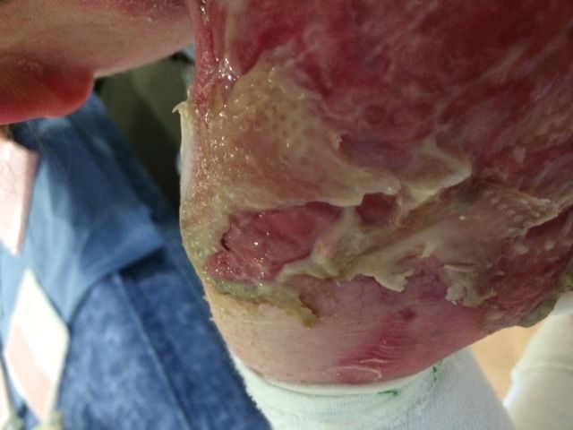
Introduction

What is this Scottish        PURPOSE AND SCOPE
 Intercollegiate             These guidelines have been developed to aid all clinicians who manage the skin and wound care of patients
Guidelines Network           with the genetic skin fragility disorder epidermolysis bullosa (EB). Management strategies for wounds or
(SIGN)?                      wound complications are suggested for patients of any age diagnosed with any form of this genetically
                             inherited disorder. It is a tool that can be used globally and includes advice for practitioners who have limited
n The Scottish               access to wound care materials. A variety of options for managing EB wounds will be presented.
  Intercollegiate
  Guidelines Network
  (SIGN) develops            ABOUT THIS DOCUMENT
  evidence-based clinical    This document was developed using a survey of clinicians from different countries who work with
  practice guidelines        the condition and who were prepared to share their knowledge of EB wounds and their management.
  for the National           A systematic literature search (described overleaf) was undertaken to provide further evidence for
  Health Service (NHS)       recommendations. However, as EB is a rare condition with small patient numbers, the literature is
  in Scotland. SIGN          predominantly made up of non-analytic studies or expert opinion (Scottish Intercollegiate Guidelines
  guidelines are derived     Network Level 3–4 or D). A review of dressings conducted by Ly (Ly and Su, 2008) noted the difficulties
  from a systematic          in evaluating wound care options in EB where patient numbers are small and there is inconsistency in the
  review of the scientific   outcome measures used.
  literature and are         The information was supplemented by day-to-day experience of people living with EB and their carers’
  designed as a vehicle      testimonials. This was gathered informally at home visits and clinic attendances by EB nursing teams.
  for accelerating the
  translation of new         Cost is always a factor to be considered in any healthcare recommendations and this is particularly relevant
  knowledge into action      for EB treatments where vast quantities of expensive dressings can be used over a lifetime (Kirkorian, Weitz
  to meet our aim of         et al, 2014; Angelis, Kanavos et al, 2016). We have recommended only products that we have experience of
  reducing variations in     using over many years and where we are confident of the results they can achieve.
  practice, and improving
  patient-important          HOW THE GUIDELINES WERE DEVELOPED
  outcomes.                  The initial work was carried out in workshops in 2012; opinions were gathered from clinicians working with
                             patients with EB, both in the UK and worldwide.
                             As part of an advanced course on EB management, nurses and doctors working with EB patients were asked
                             to complete a questionnaire relating to the management of a range of EB wounds. These wounds ranged from
                             chronic ulcerated areas seen in the more severe forms of EB, to new blister sites; they were chosen by the
                             authors as they represent the most common wound types seen in all forms of EB, or a particular problem area.
                             The group was supplied with photographs of both typical and atypical wounds and asked which primary
                             and secondary dressings, the preferred method of retention and any topical treatments they would use in
                             managing the wound. They were asked to give a range of options for each category.

                                BEST PRACTICE GUIDELINES FOR SKIN AND WOUND CARE IN EPIDERMOLYSIS BULLOSA | 3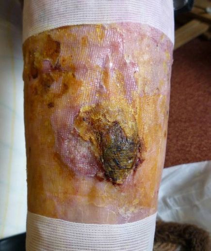
There was a wide range of experience of wound management for EB within the group: some clinicians
                                   had large caseloads having worked solely with EB for many years; others had only experienced one or two
                                   cases. Some of the group worked as individuals while others worked as part of a team, largely reflecting
                                   their common working practices. In addition, some participants had limited access to modern wound
                                   management products (see Table 17, page 38).
                                   The results of the surveys were drawn together to supply evidence for the guidelines. Opinions were given
                                   from clinicians from different countries. The draft guidelines were then subject to international peer review
                                   by recognised experts in the field of EB, and modifications were made accordingly. The guidelines were then
                                   reviewed by a small group of patients and carers, and their feedback was used to make further modifications.
                                   In order to develop the 2017 update, we conducted a more comprehensive literature review and used
                                   the results as a basis for recommendations. The 2012 guidelines search was limited to papers published
                                   between 2000 and 2011: the search years have been extended and the search methodology improved, both
                                   of which are detailed below. In addition, new wound management products, which have been used and
                                   evaluated by the guideline group and other EB professionals, have been included.

                                   PLANS FOR UPDATING THE GUIDELINES
                                   The guidelines will be reviewed and updated in three years time following a further literature review.

                                   LITERATURE REVIEW
                                   Search methodology
                                   A systematic literature search was undertaken concluding in July 2016. The databases searched were
                                   Medline, Embase, British Nursing Index and CINAHL. The search limits were papers published from 1980 to
                                   July 2016, papers published in English and involving humans. As wound management is a rapidly evolving
                                   field, it was felt that papers published prior to 1980 were unlikely to yield information that is appropriate
                                   to today. This latter point was proven as many papers published in the earlier decades of our search
                                   recommended out-dated strategies such as the use of continuous topical antibiotics; a measure now known
                                   to lead to bacterial resistance (Moy, Caldwell-Brown et al, 1990; Amirthalingam, Yi et al, 2015).
                                   In order to be thorough the initial search term used was ‘epidermolysis bullosa’ followed by separate
                                   searches on ‘wound’, ‘erosion’, ‘dressing’, ‘exudate’, ‘pruritus’, ‘itch’, ‘odour’, ‘pain’, ‘cancer’, ‘malignancy’,
                                   ‘carcinoma’, ‘wound dressing’, ‘wound care’, ‘wound pain’, ‘wound management’. The search terms were
                                   then individually combined with ‘epidermolysis bullosa’ using the Boolean operator ‘and’.

                                   SEARCH RESULTS
                                   The papers were then appraised and graded by the reviewers as per the SIGN guidelines and a synopsis
                                   made of the information they contained. SIGN now uses a new methodology for grading; however, the older
                                   system was chosen both because it is familiar to the guideline development group and because it had been
                                   used in all previous EB guidelines.

Initial results were screened at the abstract stage                                    After reading the full papers
1,342 abstracts were retrieved                                                         70 were identified to be included in the review
422 were duplicates                                                                    112 were excluded as not relevant or contained
                                                                                       information that was deemed potentially harmful,
920 unique results                                                                     such as the use of topical antibiotics as cited above
102 further duplicates were removed manually
818 abstracts to review
636 abstracts rejected as not relevant; these were excluded because they did not
relate to the topic (e.g. papers discussing EB acquisita, surgical management or
related purely to non-clinical issues)
182 were identified for reading of the full papers

4 | INTERNATIONAL CONSENSUS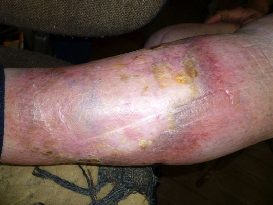
The majority of the papers were graded level 3, being small-scale case studies with many others being
                            level 4 i.e. expert opinions. Given the rarity of EB and the many compounding factors that impact healing,
                            it is difficult to conduct statistically valid studies to provide evidence to support the efficacy of any
                            particular wound management strategy.
                            There is variation in study methodology and outcome measures, as noted previously Ly (Ly and Su, 2008)
                            in a review of EB blister management. Additionally and importantly Petrof (Petrof, Martinez-Queipo et al,
                            2013), while investigating the use of fibroblasts in wound healing in EB, highlights the fact that the natural
                            history of wound healing in the condition is unknown, and that the chronic wounds previously assumed to
                            be static can in fact change and reduce in size over time with no new treatment modality being introduced.
                            A lesson to be drawn from the literature review may be that we need to be more rigorous and consistent
                            about the methodologies used in evaluating any wound management strategy.
                            However, the combination of knowledge of good wound care practice, the evidence presented here and
                            the generous sharing of information and experience among professionals, patients and carers provides a
                            substantial body of evidence to support current wound care practice in EB.

SIGN GRADING SYSTEM

       Levels of evidence                                                              Grades of recommendations

  1    ++High-quality meta-analyses, systematic
         reviews of RCTs, or RCTs with a very low risk
         of bias
                                                                           A
                                                                                       At least one meta-analysis, systematic review or
                                                                                       RCT rated as 1++, and directly applicable to the
                                                                                       target population; or
       + Well-conducted meta-analyses, systematic
         reviews, or RCTs with a low risk of bias                                      A body of evidence consisting principally of
       - Meta-analyses, systematic reviews, or RCTs                                    studies rated as 1+, directly applicable to the
         with a high risk of bias                                                      target population, and demonstrating overall
                                                                                       consistency of results

 2
       ++ High-quality systematic reviews of case
                                                                            B
                                                                                       A body of evidence including studies rated as
          control or cohort or studies                                                 2++, directly applicable to the target population,
          High-quality case-control or cohort studies                                  and demonstrating overall consistency of
          with a very low risk of confounding or bias                                  results; or
          and a high probability that the relationship                                 Extrapolated evidence from studies rated as
          is causal                                                                    1++ or 1+
       + Well-conducted case-control or cohort
          studies with a low risk of confounding or                                    A body of evidence including studies rated as
          bias and a moderate probability that the
          relationship is causal
       - Case-control or cohort studies with a high
                                                                            C          2+, directly applicable to the target population
                                                                                       and demonstrating overall consistency of
                                                                                       results; or
          risk of confounding or bias and a significant                                Extrapolated evidence from studies rated as 2++
          risk that the relationship is not causal
                                                                                       A body of evidence rated level 3 or 4; or

 3        Non-analytic studies, e.g. case reports,
          case series
                                                                           D           Extrapolated evidence from studies rated as 2+

                                                                                       Good practice points

 4        Expert opinion
                                                                           ✔           Recommended best practice based on
                                                                                       the clinical experience of the guideline
                                                                                       development group

                            Source: SIGN 50 Guideline Developer’s Handbook. NHS Scottish Intercollegiate Guidelines Network, 2014

                               BEST PRACTICE GUIDELINES FOR SKIN AND WOUND CARE IN EPIDERMOLYSIS BULLOSA | 5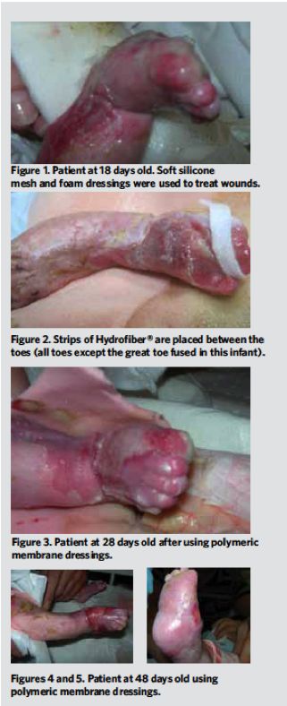
6 | INTERNATIONAL CONSENSUS

                              Key recommendations

                                                                                                                                                                                 Key recommendations
                              Key recommendations are based on the results of the literature review and the experience of the guideline development group. The
                              recommendations in this table are not arranged according to importance but rather in the order they occur in the main body of the
                              document.

                              Box 1
                              Key recommendations                                Strength of       Level of    Key references
                                                                                 recommendation    evidence
                              EB is a lifelong disorder that requires            D                 4           Badger, O'Haver et al, 2013;
                              specialist intervention and consideration to                                     Denyer 2009;
                              minimise complications and improve quality                                       Pope, Lara-Corrales et al, 2012;
                              of life. Ideally, management should take place                                   Pillay 2008, El, Zambruno et al, 2014
                              in a specialised centre by a multi-disciplinary
                              team
                              In severe EB the individual’s ability to heal can D                  4           Badger, O'Haver et al, 2013; El, Zambruno et al, 2014;
                              be compromised by malnutrition, anaemia,                                         Lara-Corrales, Arbuckle et al, 2010; Mellerio 2010;
                              pruritus and pain, and should be treated                                         Pope, Lara-Corrales et al, 2012;
                              appropriately                                                                    Schober-Flores 2003; Pope, Lara-Corrales et al, 2013
                              Careful skin and wound assessment should           D                 4           Badger, O'Haver et al, 2013; Denyer 2009; Denyer 2010;
                              be undertaken regularly. Management must                                         Elluru, Contreras et al, 2013; Pope, Lara-Corrales et al, 2012;
                              be tailored to both the type of EB and wound                                     Pope, Lara-Corrales et al, 2013; Schober-Flores 2003;
                              characteristics                                                                  Sibbald, Zuker et al, 2005; El, Zambruno et al, 2014
                              Atraumatic dressings should be used to             D                 4           Abercrombie, Mather et al, 2008; Badger, O'Haver et al,
                              prevent further blistering, skin and wound                                       2013; Denyer 2009; Denyer 2000; Denyer 2010;
                              bed damage                                                                       El, Zambruno et al, 2014; Kirkorian, Weitz et al, 2014;
                                                                                                               Lara-Corrales, Arbuckle et al, 2010; Mellerio, Weiner et al,
                                                                                                               2007; Pillay 2008; Pope, Lara-Corrales et al, 2012; Elluru,
                                                                                                               Contreras et al, 2013; Gonzalez 2013
                              People with EB and their carers are experts in     D                 4           Badger, O'Haver et al, 2013;
                              the management of their condition and their                                      Pope, Lara-Corrales et al, 2012;
                              involvement is paramount                                                         van, Lettinga et al, 2008
                              The choice of wound management strategies          D                 3,4         Kirkorian, Weitz et al, 2014;
                              should balance efficacy, patient choice and                                      Sibbald, Zuker et al, 2005; Stevens 2014
                              quality of life with cost-effectiveness
                              Staff caring for EB patients must be trained in    D                 4           Gonzalez 2013
                              specific handling techniques to avoid further
                              harm
                              Blisters are not self-limiting and intact blisters D                 4           Denyer 2009; El, Zambruno et al, 2014;
                              should be lanced and drained                                                     Elluru, Contreras et al, 2013; Lara-Corrales, Arbuckle et al,
                                                                                                               2010; Pillay 2008; Herod, Denyer et al, 2002; Schober-
                                                                                                               Flores 2003;
                                                                                                               Pope, Lara-Corrales et al, 2012
                              Management of EB wounds must address               D                 4           Badger, O'Haver et al, 2013; Denyer 2009; Denyer 2010;
                              issues such as critical colonisation, infection,                                 El, Zambruno et al, 2014; Mellerio, Weiner et al, 2007;
                              and protection from trauma                                                       Schober-Flores 2003;Sibbald, Zuker et al, 2005; Azizkhan,
                                                                                                               Denyer et al, 2007
Every effort should be made to treat the          C                    2+4          Badger, O'Haver et al, 2013; Danial, Adeduntan et al, 2015a;
                                                                                intense pruritus seen in EB and thereby                                             Danial, Adeduntan et al, 2015b; El, Zambruno et al, 2014;
                                                                                minimise scratching that leads to further skin                                      Pillay 2008; Pope, Lara-Corrales et al, 2013;
                                                                                damage                                                                              Snauwaert, Morren et al, 2011; Snauwaert, Yuen et al, 2014
                                                                                Silicone medical adhesive removers (SMARS) D                           3,4          Denyer 2009; Denyer 2010; El, Zambruno et al, 2014;
                                                                                should be used when removing adherent                                               Lara-Corrales, Arbuckle et al, 2010; Mather and Denyer
                                                                                dressings or clothing                                                               2008;
                                                                                                                                                                    Stephen-Haynes 2008
                                                                                To ensure adequate nutrition and optimise         D                    4            El, Zambruno et al, 2014; Haynes 2010; Haynes, Mellerio et
                                                                                wound healing long-term, enteral feeding                                            al, 2012;
                                                                                may be indicated in severe EB                                                       Hubbard, Haynes et al, 2011; Pope, Lara-Corrales et al, 2012

                                                                                                                                                                                                                                     w
                                                                                Optimal pain management is vital for              D                    4            Denyer 2009; Denyer 2010; El, Zambruno et al, 2014;
                                                                                patients with all forms of EB and include                                           Goldschneider and Lucky 2010;Herod, Denyer et al, 2002;
BEST PRACTICE GUIDELINES FOR SKIN AND WOUND CARE IN EPIDERMOLYSIS BULLOSA | 7

                                                                                pharmacological and non-pharmacological                                             Watterson, Howard et al, 2004; Mellerio, Weiner et al,
                                                                                interventions                                                                       2007; Goldschneider, Good et al, 2014
                                                                                When a surgical or interventional procedure       D                    4            El, Zambruno et al, 2014; Elluru, Contreras et al, 2013;
                                                                                is indicated adjustments to anaesthesia and                                         Herod, Denyer et al, 2002; Goldschneider, Lucky et al, 2010
                                                                                theatre protocols will be required to minimise
                                                                                skin damage and protect the airway
                                                                                The principles of wound bed preparation           D                    4            Lara-Corrales, Arbuckle et al, 2010; Pope, Lara-Corrales et
                                                                                (WBP) are applicable to wounds seen in                                              al, 2012;
                                                                                patients with EB, particularly wounds which                                         Mellerio, Weiner et al, 2007; Pope, Lara-Corrales et al, 2013;
                                                                                have become chronic                                                                 Sibbald, Elliott et al, 2015

                                                                                In patients with severe forms of EB there is a    D                    4            Fine, Johnson et al, 2009; Mellerio, Weiner et al, 2007;
                                                                                high risk of squamous cell carcinoma (SCC).                                         Mellerio, Robertson et al, 2016
                                                                                Regular monitoring is essential with a low
                                                                                threshold for biopsy of suspect areas.*

                                                                                * Although the evidence supplied by the US EB Registry (Fine, Johnson et al, 2009) supported by a subsequent review in 2016 (Montaudie, Chiaverini
                                                                                et al, 2016) for the high risk of SCC in severe forms of EB most notably RDEB-GS is unequivocal and are graded C -2+, the evidence for recommended
                                                                                actions are based on expert opinion.
8 | INTERNATIONAL CONSENSUS

                                                                                                                                                                                                         Evidence for wound management strategies
                              Box 2: Evidence for the use of specific wound management strategies

                              Wound management          Comments                                           Strength of      Level of   References
                              strategy                                                                     recommendation   evidence
                              Soft silicone dressings   No evaluation of products but widely used          D                4          Abercrombie, Mather et al, 2008;
                                                        and accepted in the management of EB                                           Badger, O'Haver et al, 2013; Denyer 2009; Denyer 2010;
                                                                                                                                       El, Zambruno et al, 2014; Kirkorian, Weitz et al, 2014;
                                                                                                                                       Lara-Corrales, Arbuckle et al, 2010; Mellerio, Weiner et al,
                                                                                                                                       2007; Pillay 2008; Pope, Lara-Corrales et al, 2012
                              Polymeric membrane        Limited patient numbers and case study             D                3          Stevens 2010; Pillay 2009; Denyer 2009; Clapham 2011;
                                                        evidence                                                                       Denyer, Foster & Turner 2013; Bauer, Diem & Ploder 2013;
                                                                                                                                       Carbone, Gonclaves, Grandi & Desbordes NEEDS DATE;
                                                                                                                                       Denyer, Foster & Turner NEEDS DATE
                              Honey                     Single patient case study with complete            D                3          Hon, 2005
                                                        healing of a recalcitrant wound unresponsive
                                                        to previous interventions
                              Saltwater baths to        2010 paper expert opinion. 2015 retrospective      C                2+, √      Arbuckle, 2010; Petersen, Arbuckle et al, 2015
                              reduce pain               observational study 21 patients showing
                                                        substantially reduced pain and some reduction
                                                        in other wound related symptoms
                              Lipido-colloid            Blanchet-Bardon 2005 included 20 patients          D                3          Blanchet-Bardon and Bohbot 2005; Blanchet-Bardon and
                              dressings                 with a variety of forms of EB and reported                                     Bohbot 2007; Stevens 2009
                                                        improved quality of life and healing as did
                                                        Stevens reporting on 2 patients
                              *Biological dressings     Many interventions had small patient               C,D              2+, 3      Buonocore and Ariyan 2009;
                              including cadaveric       numbers or were single case studies apart                                      Hasegawa, Mizoguchi et al, 2007; Sibbald, Zuker et al, 2005;
                              allografts, amniotic      from some artificial skin substitute studies                                   Falabella, Valencia et al, 2000; Lo, Lara-Corrales et al, 2010;
                              membrane, cord blood      that had larger numbers. All reported                                          Ng, Nguyen et al, 2014; Tadini, Pezzani et al, 2015;
                              platelet gel,             improved healing and/or reduction in wound                                     McGrath, Schofield et al, 1993; Fivenson, Scherschun et al,
                              cultured keratinocyte     related symptoms. McGrath using cultured                                       2003; Gorell, Leung et al, 2015
                              allografts, type          keratinocyte allografts reported little clinical
                              1 collagen skin           benefit
                              substitute,
                              non-biological skin
                              substitutes
Keratin gel                 Denyer variety of forms of generalised EB      D             3    Denyer, Marsh et al, 2015; Kirsner, Cassidy et al, 2012;
                                                                                                            (n=10) 6 reported faster healing, 2 gel was                       Than, Smith et al, 2013
                                                                                                            ineffective and 2 reported increased pruritus.
                                                                                                            Kirsner (n=1) improved healing. Than (n=1)
                                                                                                            improved healing. In cases where healing
                                                                                                            reported faster treated areas said to be more
                                                                                                            robust
                                                                                *Botulinum toxin in         In a case series of patients with EBS (n=6)         D        2+   Swartling, Karlqvist et al, 2010
                                                                                EBS                         5 reported some improvement in global foot
                                                                                                            related symptoms
                                                                                *Systemic G-CSF             Pilot trial in DEB (n=7) 7 showed reduction         C        2+   Fine, Manes et al, 2015
                                                                                                            in wound size and blister/erosion frequency
                                                                                *Ablative fractional        Single patient case study treatment of a            D        3    Krakowski and Ghasri 2015
BEST PRACTICE GUIDELINES FOR SKIN AND WOUND CARE IN EPIDERMOLYSIS BULLOSA | 9

                                                                                resurfacing                 chronic wound leading to almost complete
                                                                                                            healing at 8 weeks
                                                                                Systemic trimethoprim RDEB patients (n=7) with 42 wounds.            C                   2+   Lara-Corrales, Parkin et al, 2012
                                                                                                      Prospective, randomised, double-blind,
                                                                                                      placebo-controlled crossover trial. 6 of 7
                                                                                                      patients showed greater than 50% reduction
                                                                                                      in chronic wound size. In the control group 2
                                                                                                      of 7 patients showed a similar result. Limited
                                                                                                      by small sample size
                                                                                *Topical gentian violet     EBS-DM (n=5) randomised controlled pilot            D        2+   Wally, Kitzmueller et al, 2013
                                                                                in Non-H JEB                study with reported reduction in blistering.
                                                                                                            Limitation small sample size
                                                                                *Punch grafting in          Retrospective analysis of punch grafting     D               3    Yuen, Huizinga et al, 2013
                                                                                Non-H JEB                   (n=4) with 23 ulcers. Complete healing in 16
                                                                                                            lesions, 7 improved and at 3 months 2 had
                                                                                                            recurred
                                                                                *Oral epigallocatechin- RDEB (n=16). Randomised, crossover,                     C        2+   Chiaverini, Roger et al, 2016
                                                                                3-gallate               double-blind, placebo trial. Findings were
                                                                                                        improvement in healing and reduction in
                                                                                                        blisters but results did not achieve statistical
                                                                                                        significance
                                                                                Thalidomide                 RDEB-P (n=2). Two patients showed                   D        3    Ranugha, Mohanan et al, 2014
                                                                                                            reduction in pruritus and improved healing

                                                                                Piscean collagen            D: Limited case study evidence of improved                   3    Westgate S et al, 2012
                                                                                                            healing
                                                                                * Not used by guideline development group . Recommendation based on published evidence
Notes
                      Special considerations

                      Box 3: Key recommendations for management of neonates

                      Key recommendations                         Strength of       Level of   Key references
                                                                  recommendations   evidence
                      Ideally an outreach service should be       D                 4          Denyer 2009
                      offered for severely affected neonates.
                      Where this is not possible infants
                      should be transferred in appropriate
                      dressings and clothing, and in a
                      padded car seat rather than a portable
                      incubator if local policy permits
                      Advice should be sought from a              D                 4          Goldschneider and Lucky 2010
                      paediatric pain specialist to enable                                     Goldschneider, Good et al, 2014
                      adequate analgesia for procedural and                                    Herod, Denyer et al, 2002
                      chronic multi-factorial pain
                                                                                               Weiner 2004
                                                                                               Morash and Fowler 2004
                      Infant to be nursed on neonatal             D                 4          Denyer 2009
                      mattress, and staff and family trained in
                      specific handling to avoid trauma
                      Limbs and vulnerable areas should           D                 4          Denyer 2009
                      be protected with suitable dressing
                      material to reduce skin loss from baby
                      movements such as kicking
                      Secure umbilical cord with a ligature       D                 4          Denyer et all 2014
                      rather than a cord clamp
                      Use greasy emollient such as 50%            D                 4          Denyer 2009
                      liquid/50% white soft paraffin to                                        Denyer et al, 2014
                      cleanse napkin area in preference
                      to water
                      Line the napkin with a liner, a             D                 4          Denyer 2009
                      continence cloth, such as Conticloth
                      or a piece of muslin or fleece cloth,
                      to prevent friction and subsequent
                      trauma from the edges
                      Avoid bathing until inter-uterine and       D                 4          Denyer and Stevens 2010
                      birth damage have healed

10 | INTERNATIONAL CONSENSUS
BOX 4: Additional recommendations for the care of neonates with severe EB
Procedure Rationale
Remove from incubator unless prescribed for other medical condition Heat and humidity exacerbate blistering
such as prematurity
Remove cord clamp and replace with ligature To prevent trauma to umbilical area
Line nappy with soft material To prevent blistering at edges of nappy
Cleanse nappy area with 50% liquid/50% white soft paraffin in ointment or To ensure cleansing without trauma. To reduce pain
spray (Emollin) form
Delay bathing until prenatal and birth trauma have healed To avoid damage from infant being handled naked
Nurse on neonatal incubator mattress To enable infant to be lifted on mattress and avoid shearing forces
from carer’s hands
Use long soft teat such as lamb’s teat or a Haberman (specialist needs feeder) To avoid friction damage to underside of nose and oral mucosa
Apply teething gel to teat (use a preparation that is safe to use from birth) To alleviate pain from blistered mucosa
Avoid heel prick for neonatal blood screening — obtain blood via venepuncture To avoid de-gloving injury
BEST PRACTICE GUIDELINES FOR SKIN AND WOUND CARE IN EPIDERMOLYSIS BULLOSA | 11Notes
Understanding epidermolysis bullosa
EB describes a rare complex group of inherited skin fragility disorders. Ideally patients should be
QE Level 4 D managed in a specialist centre. EB is a lifelong disorder that requires specialist intervention and
considerations to minimise complications and improve quality of life.
The most recent classification for EB, agreed in 2014, names four categories of the condition defined by the
level of cleavage at the dermal/epidermal junction (Fine, Bruckner-Tuderman et al, 2014). These are:
Key recommendations ■■ EB simplex (EBS)
are highlighted in blue ■■ Junctional EB (JEB)
throughout the text and ■■ Dystrophic EB (DEB)
references can be found
in Appendix 1 ■■ Kindler syndrome.
The common factor in all types of EB is the tendency for skin and mucous membranes to blister or shear
away in response to minimal everyday friction and trauma.
The severity of EB varies from simple blistering affecting the hands and feet, particularly in warm weather, to
death in early infancy from the devastating combination of laryngeal disease and failure to thrive. Those with
DEB develop microstomia and oesophageal strictures as a result of contractures and scarring.
People with more severe forms of EB can experience recurrent blistering and skin loss. There is also a
tendency to develop chronic wounds resulting from the underlying gene defect, compromised nutrition,
chronic anaemia and repeated infection, together with constant trauma.
Non-cutaneous complications, such as anaemia due to iron deficiency and chronic disease, osteoporosis,
growth failure and pubertal delay (Haynes 2010) further compromise wellbeing. There is also a greatly increased
risk of aggressive squamous cell carcinoma in those with severe forms of EB (Mellerio, Robertson et al, 2016).
ASSESSMENT AND DIAGNOSIS
Within each of the four categories of EB there are subtypes that display individual clinical effects (see
pages 13–15). Definitive diagnosis is most commonly made from analysis of a skin biopsy using positive
immunofluorescence (IF), antigenic mapping and transmission electron microscopy. These key diagnostic
tools help confirm diagnosis and indicate the particular subtype of EB (Uitto, Richard et al, 2007).
Identification of the different causative genes responsible for EB enables the recognition of the precise
location of and type of mutation. Due to the rarity of expertise and facilities, however, diagnosis is generally
made using IF and antigen mapping. Some laboratories are moving towards molecular diagnosis from
exome sequencing of a panel of known skin fragility genes.
Pre-implantation genetic diagnosis or first trimester DNA-based prenatal diagnosis from chorionic villus
samples or amniocentesis can be offered to families in which causative mutations or informative genetic
markers have been identified. Experienced clinicians can often make a provisional diagnosis on clinical
observations, but a definitive diagnosis will always be required, particularly in neonates where a clear
diagnosis is crucial to ensure correct management.
CAUSES OF EB
EB can be inherited autosomal recessively or autosomal dominantly; in general, recessive forms tend to
be more severe. More than 1,000 recorded mutations in 14 genes contribute to the various forms of EB,
resulting in a huge variety of clinical presentations (Fine, Bruckner-Tuderman et al, 2014).
These guidelines outline each main subtype and focus on the different skin and wound management
requirements, as well as general principles for wound management for all types of EB.
EB SIMPLEX
Almost all forms of EB Simplex (EBS) are inherited autosomal dominantly, although some rare forms are
recessively inherited. EBS is a disorder of keratin proteins with the primary defects lying within proteins
encoding for keratin 5 and keratin 14. These proteins form the keratin scaffolding within the basal
epidermal cells.
Dysfunction of the keratin proteins in EBS leads to mechanical weakness of these cells; breakdown occurs
due to minor friction or rubbing resulting in blistering (Uitto, Richard et al, 2007).
12 | INTERNATIONAL CONSENSUSAll forms of EBS are most troublesome in hot and humid environments due to an increase in sweat
production, which excerbates frictional forces.
The three main types of EBS are classified as basal EBS:
■■ EBS localised (EBS-loc; previously called Weber-Cockayne) — primarily affects the hands and feet with
blisters arising from friction or spontaneously when the environment is hot or humid
■■ EBS generalised intermediate (EBS–GI; previously called Köbner), — causes more widespread blistering
at an earlier onset with blisters and skin loss frequently present at birth. Blistering persists throughout
life but becomes less troublesome, with the main problematic areas being the hands, feet and where
clothing causes friction
■■ EBS generalised severe (EBS-GS; previously called Dowling-Meara) —
a more serious type of EBS and can
be severe in the neonatal period. There is a significant risk of death in this age group resulting from sepsis
and laryngeal blistering. Blisters typically occur in clusters and often beneath and around nails and in the
mouth. Blistering tends to reduce late in childhood and hyperkeratosis develops over the palms and soles.
Additional rare basal subtypes are:
■■ EBS with mottled pigmentation (EBS-MP)
■■ EBS migratory circinate (EBS-migr)
■■ EBS autosomal recessive K14 (EBS-AR K14)
■■ EBS with muscular dystrophy (EBS-MD)
■■ EBS with pyloric atresia (EBS-PA)
■■ EBS Ogna (EBS-Og)
■■ EBS autosomal recessive BP230 deficiency (EBS-AR BP230)
■■ EBS autosomal recessive exophilin deficiency (EBS-AR exophilin 5).
EBS with muscular dystrophy is a form of autosomal recessive EBS that results from mutations in the gene
encoding plectin (PLEC1). Plectin plays an important part in both skin and muscles, helping to maintain
mechanical function. While skin involvement may be minimal, laryngeal involvement can be severe and the
patient may require a tracheostomy. Progressive muscular dystrophy commonly starts any time from the
first year onwards.
Suprabasal types of EBS are rare and include:
■■ Acral peeling skin syndrome (APSS)
■■ EBS superficialis (EBSS)
■■ Acantholytic EBS (EBS-acanth)
■■ Desmoplakin deficiency (EBS-desmoplakin) [associated cardio-myopathy]
■■ Plakoglobin deficiency (EBS-plakoglobin)
■■ Plakophilin deficiency (EBS-plakophilin).
JUNCTIONAL EB
Junctional EB (JEB) is a group of autosomal recessively inherited disorders, which are characterised by
mechanically induced blistering at the lamina lucida level of the basement membrane zone, between
the basal cells and lamina densa. All forms of JEB arise from mutations in genes that encode structural
components of the hemidesmosomes or anchoring filaments, which provide mechanical integrity across
this zone. Separation of the epithelium occurs within the lamina lucida between the lamina densa of the
basement membrane and the basal keratinocytes.
There are three main forms of junctional EB:
■■ JEB generalised severe (JEB-GS previously called Herlitz junctional EB)
■■ JEB generalised intermediate (JEB-GI previously called Non-Herlitz junctional EB)
■■ JEB with pyloric atresia (JEB-PA).
BEST PRACTICE GUIDELINES FOR SKIN AND WOUND CARE IN EPIDERMOLYSIS BULLOSA | 13Notes
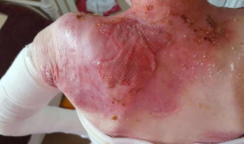
In all forms of JEB, the most problematic wounds occur on the scalp and lower legs. Open nail beds and facial
lesions occur in JEB generalised severe (JEB-GS). There is a tendency for chronic wounds to develop and a
particular feature is for wounds to over-granulate from an early age. Common features include hypoplastic
dental enamel, alopecia and genito-urinary tract involvement in longer-term patients (Fine 2010).
JEB generalised severe
In JEB-GS the protein laminin 332 is absent or greatly reduced. Laminin 332 is a major component of the
basement membrane zone, providing anchorage across the lamina lucida (Nakano, Chao et al, 2002). For
the vast majority, this type of EB carries a very poor prognosis with most not surviving beyond the first two
years of life. Death results from a combination of laryngeal blistering/respiratory distress, a profound and
uncorrectable failure to thrive, chronic wounds and sepsis (Fine, 2010). Despite the severity of the systemic
disease, good management can help reduce the severity of the wounds.
JEB generalised intermediate
The majority of JEB-GI cases result from mutations in the genes encoding type XVII collagen or laminin 332,
which are expressed in skin and other sites such as the urogenital tract. This protein has an important function
and plays a major role in the anchorage of the epidermis to the dermis. Type XVII collagen is expressed in the
skin, oral mucosa, the cornea, upper oesophagus and bladder epithelium (Van and Giudice, 2003).
JEB-GI carries a better prognosis than JEB-GS, with the majority of patients surviving to adulthood, but
there is an increased risk (up to 25%) of developing a squamous cell carcinoma (SCC) after the age of 25
(Fine, Johnson et al, 2009).
Chronic wounds may remain a lifelong problem and areas of previous wounding can become atrophied. Nail
dystrophy and scarring alopecia are common in older patients. Dental enamel defects in this subtype of EB
are characteristic and a useful diagnostic pointer.
JEB with pyloric atresia
This is associated with pyloric atresia and is a rare subtype of JEB that results from mutations in the
alpha-6-beta-4 integrin genes. This integrin is an important component of the hemidesmosomes and is
found in skin and other epithelia including the gastrointestinal and urogenital tracts. JEB-PA frequently has
a poor prognosis despite surgical correction of the atresia.
Many patients die in infancy, while milder phenotypes exhibit outcomes similar to those with JEB-GI.
However, significant morbidity from urogenital tract involvement is frequently seen in those with this
type of EB.
Other rare types of JEB
■■ JEB late onset (JEB-LO)
■■ JEB with respiratory and renal involvement (JEB-RR)
■■ JEB localised (JEB-loc)
■■ JEB inversa (JEB-I)
■■ Laryngo cutaneous syndrome (JEB-LOC syndrome; previously called Shabbir’s syndrome).
DYSTROPHIC EB
EB can be inherited either dominantly or recessively, with the more severe forms in general being inherited
recessively. In all cases there is a diminished or absent protein collagen VII, which is a crucial component
of anchoring fibrils. Anchoring fibrils act rather like Velcro® hooks attaching the epidermis to the dermis. In
DEB, separation occurs at the sub-lamina densa of the basement membrane zone.
The extent of skin fragility is extremely varied depending on whether the causative mutation predisposes to
mild or severe disease and whether the affected individual has completely absent or reduced collagen Vll.
In severe forms of DEB there are many complications, which have an impact on the individual’s ability
QE Level 4 D to heal and these should be addressed and corrected where possible. Common complications are
malnutrition, anaemia, recalcitrant pruritus, pain, infection and critical colonisation (see page 20).
14 | INTERNATIONAL CONSENSUS•
Types of dystrophic EB
Dominant dystrophic EB
Dominant dystrophic EB (DDEB) has autosomal dominant inheritance. Blistering can be localised to areas
particularly subject to trauma such as the hands, feet, knees and elbows, or more generalised. Healing is
usually accompanied by some scarring and milia are often present. Mucous membranes, particularly of the
mouth and the anal margins, can be fragile leading to difficulties with eating and constipation.
Types of DDEB
■■ DDEB generalised (DDEB-gen)
■■ DDEB acral (DDEB-ac)
■■ DDEB pretibial (DDEB-pt)
■■ DDEB pruriginosa (DDEB-pr)
■■ DDEB nails only (DDEB-na)
■■ DDEB bullous dermolysis of the newborn (DDEB-BDN).
Recessive dystrophic EB (RDEB)
■■ RDEB generalized severe (RDEB-GS)
■■ RDEB generalised intermediate (RDEB-GI)
■■ RDEB bullous dermolysis of the newborn (RDEB-BDN)
■■ RDEB inversa (RDEB-I)
■■ RDEB localised (RDEB-loc)
■■ RDEB pretibial (RDEB-pt)
■■ RDEB pruriginosa (RDEB-pr)
■■ RDEB centripetalis (RDEB-ce).
Recessive dystrophic EB — generalised severe
In RDEB-GS the skin is extremely fragile, often with extensive blistering and wounding. Patients with this form of
EB will frequently develop hard-to-heal or never-to-heal areas, or areas that do heal but can very quickly break
down. Atrophic scarring and healing leading to disabling contractures are common. Pseudosyndactyly (see pages
21–22) is often present and may require repeated surgery (Formsma, Maathuis et al, 2008; Bernardis and Box
2010). Severe pain, nutritional compromise and profoundly difficult-to-correct anaemia will all impact negatively
on wound healing. Recalcitrant pruritus can lead to destructive scratching and disruption to wound healing.
Recessive dystrophic EB — generalised intermediate
RDEB-GI has an autosomal recessive inheritance. Generally, the affected individual is able to express some
type VII collagen, with variable qualitative and quantitative abnormalities of the anchoring fibrils. The
clinical presentation will vary with a tendency for generalised blistering and consequent wounding. The
development of anaemia is common with this type of EB and there is often mucosal involvement. The hands
will be scarred with some webbing, but full pseudosyndactyly is not seen in RDEB-GI.
Dystrophic EB pruriginosa
DEB-pr can be inherited by dominant or recessive transmission. Patients will experience the skin fragility already
described above, but will also exhibit intense pruritus, which is exceptionally difficult to manage. Scratching and
skin breakdown can lead to the formation of disfiguring linear scarring in some patients, which appears almost
like keloid scarring. Other patients will have extensive blistering and skin breakdown.
Recessive dystrophic EB inversa (RDEB-I)
RDEB-I may be recessive or, less commonly, dominant. The majority of wounds develop in the flexures such as
neck, groin and axillae. Hands become scarred but do not progress to mitten deformity. Oesophageal strictures
are problematic in this group and may be particularly severe.
BEST PRACTICE GUIDELINES FOR SKIN AND WOUND CARE IN EPIDERMOLYSIS BULLOSA | 15Notes
KINDLER SYNDROME
Kindler syndrome is an autosomal recessive disorder caused by mutations in the FERMT1 gene. It is rare,
difficult to diagnose and is often confused with other subtypes of EB. Blistering, epidermal atrophy and
delayed healing result from FERMT1 gene mutations. Trauma-induced skin blisters occur in early life and are
prevalent together with skin loss and wounding during the neonatal period.
The blistering reduces in infancy but over time photosensitivity and signs of poikiloderma (a skin condition
characterised by pigmentary and atrophic changes) develop where the skin takes on a mottled appearance
(Lai-Cheong and McGrath, 2010; Lai-Cheong and McGrath, 2011).
Other clinical features include periodontitis, oesophageal strictures, malabsorption and diarrhoea in early
life, and urethral strictures. There is also an increased risk of mucocutaneous SCC in later life
(Fine, Johnson et al. 2009).
In Kindler syndrome the family fermitin homolog 1 is typically markedly reduced or absent within the
epidermis and at the dermal—epidermal junction. Unlike all other types of EB, the level of cleavage is
variable, with blister formation taking place within the epidermis, lamina lucida or beneath the lamina densa,
thus explaining the variable features demonstrated in Kindler syndrome.
16 | INTERNATIONAL CONSENSUSSkin and wound management:
general principles
All types of EB are characterised by fragile skin and a range of cutaneous involvement from blistering,
primarily on the hands and feet, through to more generalised wounding. The presence of multiple wounds of
varying duration and ability to heal makes management of EB difficult and complex.
Careful skin and wound assessment should be undertaken regularly. Management must be tailored to
suit both the type of EB and the specific characteristics of the wound.
QE Level 4 D The underlying principle of lesion management is to apply an atraumatic dressing to prevent blistering,
and skin and wound bed damage leading to pain and bleeding on removal.
People with EB and their carers are experts in the management of their condition and their involvement
in their care is paramount.
It is important to listen to the patient and/or carer as many people with EB will have a tried and tested
dressing regimen that avoids injury. For example, they may use soft padding to prevent blistering from the
edges of a dressing or apply bandaging in a certain way to reduce the risk of contractures. These dressing
techniques will have been developed over years of living with EB and many techniques will be atypical.
Careful discussion will usually reveal the reasons why patients use products in a particular way and the
clinician should be prepared to be open-minded. In countries where suitable dressings are not available,
dressings can often be modified or alternative materials used (Table 17, page 38). In addition, clinicians
need to educate patients about wound management and inform them of new products as they appear on
the market.
The choice of wound management strategies should balance efficacy, patient choice and quality of life
D
with cost effectiveness.
QE Level 4
Staff caring for EB patients must be trained in specific handling techniques to avoid causing harm.
Healthcare settings are fraught with danger for the EB patient as routine procedures, such as the use of a
PATSLIDE® to move a patient or removal of ECG electrodes, can result in extensive skin loss. Care must be
taken not to cause further injury (Gonzalez, 2013) (Table 8, page 28).
MANAGEMENT OF BLISTERS
Blisters occur in all types of EB following friction and relatively minor trauma. They can be present anywhere
on the skin and also on the mucous membranes.
QE Level 4 D Blisters are not self-limiting and will extend rapidly if left unchecked.
In contrast to recommendations for other dermatological conditions or wound management, intact blisters
should be lanced at their lowest point to limit tissue damage (Denyer 2010). A fresh hypodermic needle
should be used or, if this is not available, a sterilised sewing needle. The needle should be passed through
the blister roof, parallel to the skin, to create an entry and exit hole through which fluid can be expelled
(Figure 2, page 18).
A soft piece of material, such as gauze, can be used to gently compress the blister to encourage complete
emptying. If this compression is painful, a syringe can be attached to the needle to aspirate the fluid.
Some patients advocate using sterilised scissors or a scalpel blade to create a larger hole to prevent the
blister from refilling. The roof should be left on the blister unless personal preference is to deroof it to
prevent refilling, but deroofing can lead to additional pain and should be discouraged if possible.
The location of a particular blister may be EB-type specific. For example EBS localised will occur mainly on
the hands and feet (Figure 1, page 18) whereas mild forms of dystrophic EB will occur on the areas subject
to the most trauma, such as the bony prominences. The blisters can occur alone or in clusters depending
on the initial degree of trauma and they may be filled with serous or blood-stained fluid.
BEST PRACTICE GUIDELINES FOR SKIN AND WOUND CARE IN EPIDERMOLYSIS BULLOSA | 17Notes
Needle Blister
Skin surface
Figure 1. Blisters on toes in Figure 2. Recommended method of blister lancing.
patient with EBS Used with permission from Birmingham Children’s Hospital
SKIN AND WOUND MANAGEMENT
Dressings listed have been tried and found suitable for use on skin and wounds of children and adults
with EB. Where ‘preferred choice’ is indicated this should be selected if available. A wide range is
included to offer selection to those with a limited formulary. Dressings within tables are listed in order
of their suitability.
MANAGEMENT OF EB SIMPLEX
Dressing management in EB Simplex (EBS) focuses on preventing infection, cooling the blister sites and
protecting the skin from trauma. However, observation within the authors’ large caseloads indicates many
patients prefer to leave blisters undressed.
Dressings can lead to overheating which increases the tendency to blister as sweating increases friction.
The patient may also blister around the edge of dressings on areas subject to great pressure such as the
feet. Dressings and padding on the feet may make it impossible to wear shoes and dressings on the hands
may hamper the patient’s dexterity. For information on recommended wound dressings see Table 1. For
further information on the podiatric management of EBS see Podiatric Management of EB (Khan 2010).
The most effective management is lancing the blisters (Laimer, Lanschuetzer et al, 2010). For patients who
dislike dressings or find they exacerbate the blister sites, commercial cornflour (corn starch) may be used
to dry up the blistered areas and provide a low friction surface. Silk socks or Skinnies WEB socks can help
to reduce friction and are seam free (Grocott, Blackwell et al, 2013). Clinical experience also suggests that
silver socks may help to keep feet cool (Denyer 2010).
EBS-GS needs specific management as any dressing materials have the potential to create blisters
3 around the edges of the dressing (Table 2, page 20).
Neonates may present at birth with widespread skin loss and although dressings are required, measures
must be taken to protect the skin around the dressing. Once the wounds are healed dressings are not used
for protection in view of the resulting damage. A thin layer of equal parts of white soft and liquid paraffin
can reduce friction and soft seam–free clothing offers protection (Denyer 2009).
MANAGEMENT OF JUNCTIONAL EB
3 Wound management in Junctional EB (JEB) is focused on managing chronic wounds and excessive
granulation tissue (Table 3, page 21).
Open nail beds, and the umbilical and nappy area all pose particular challenges in infants with JEB-GS. Use
of very potent topical steroid ointments greatly reduces over-granulation and may encourage healing.
From the authors’ experience, a soft silicone mesh has a tendency to encourage over-granulation even to
the extent where the tissue grows through the mesh and forms a bridge over the dressing.
18 | INTERNATIONAL CONSENSUSSELECTING AND USING A DRESSING Removal dressings
There are number of considerations when selecting an appropriate Dressings must be removed with great care to avoid further skin
dressing, including: damage. If necessary the dressing can be soaked off in the bath,
hydrated with tepid water or saline or a silicone medical adhesive
Wear time remover (SMAR) could be used. In particular this applies to patients
Most dressings recommended in this document should be changed with RDEB or those using a bordered dressing.
every 1–3 days unless
• Patient/carer preference differs Sensitivity
• Otherwise stated by the manufacturer Any sensitivity to components of a particular dressing should
• Levels of exudate dictate more frequent change as does wound be established prior to use. Silicone sensitivity resulting from
infection or if there is obvious strikethrough. impurities in the silicone is rare but has been reported.
It is important to ensure that there are no folds or creases in the
dressing that would result in blistering and further damage to the skin.
Table 1: Recommended dressings for EBS localised and generalised ü
Preferred choice when available: blister sites — Spycra Protect (ReSkin/Bullen healthcare) or Kerralite Cool (Crawford Healthcare)
Dressing type Brand Manufacturer Indication/function Contraindication/comments
Bi-stretch soft silicone • Spycra Protect • ReSkin/Bullen • Protection
Healthcare/ • Minor blister sites and
Curea Medical non-exudating wounds
Sheet hydrogel • Kerralie Cool/ • Crawford Healthcare • Blister sites • Keep backing on to keep
Kerralite Cool Border • Cooling moist for longer
• Pain reduction • ActiFormCool — rarely pain
• ActiFormCool • Activa Healthcare reactions are reported
Bordered foam • Mepilex Border/ Border Lite • Mölnlycke Health Care • Protection
dressing • Biatain Silicone Lite • Coloplast • Blister sites
• Allevyn Gentle Border • Smith & Nephew
• Allevyn Lite
• Allevyn adhesive (non
silicone)
• UrgoTul Absorb Border • Urgo Medical
Soft silicone mesh • Mepitel • Mölnlycke Health Care • Wound contact layer
• Mepitel One
• Adaptic Touch • Acelity
• Cuticell Contact • BSN Medical
Lipido-colloid • UrgoTul • Urgo Medical • Wound contact layer • Alternative to soft
silicone mesh
Hydrogel • Intrasite Conformable • Smith & Nephew • Cooling • Do not allow to dry out
• Pain reduction
Foam • Mepilex • Mölnlycke Health Care • Protection • May cause heat-related
• Mepilex Lite blistering
• Mepilex Transfer • Soft silicone tape can be
used with for fixation
Soft silicone fixation • Siltape • Advancis Medical • Retention of non-
tape • Mepitac • Mölnlycke Health Care bordered dressings
Fixation bandage • CoFlex Haft • Aspen Medical Europe • Retention • Do not stretch bandage
• Soft-One • Snogg on application to avoid
• Acti-Wrap • Activa Healthcare tourniquet effect
Powder • Cornflour (corn starch) • Commercial • To aid drying of blister • Apply following lancing of
• Reduce friction blisters
• Do not use cornflour in
nappy area where it will
turn into a paste
BEST PRACTICE GUIDELINES FOR SKIN AND WOUND CARE IN EPIDERMOLYSIS BULLOSA | 19Notes
Table 2: Recommended dressings for patients with EBS-GS
Preferred choice when available: Spycra Protect (ReSkin/Bullen Healthcare);
Open wounds — PolyMem (Ferris Mfg Corp [Aspen Medical Europe, UK])
Dressing Brand Manufacturer Indication/ Contraindication/comments
type function
Bi-stretch • Spycra Protect • ReSkin/Bullen • Protection
protection Healthcare/ • Blister sites
Curea Medical
Polymeric • PolyMem • Ferris Mfg Corp • Wounds present • Strips of a Hydrofiber dressing
membrane (Aspen Medical at birth (see below) will need to be
Europe, UK) placed under the edges of the
dressing to protect the skin
• Change when wet in small
infants to avoid hypothermia
(See Table 7, page 27)
Lipido-colloid • UrgoTul • Urgo Medical • Wound contact • Use as a primary dressing if
layer there is risk of adhesion
Hydrofiber • Durafiber • Smith & • Protection from • Hydrate with water or saline to
Nephew the edges of remove if necessary
• Aquacel • ConvaTec dressings (see
above)
Tubular • Tubifast with • Mölnlycke • Retention • Available in a range of sizes for
bandage 2–way stretch Health Care appropriate fit
• ActiFast 2 –way • Activa • May need protection at edges to
stretch Healthcare prevent blistering
• Comfifast multi • Synergy Health
stretch
Powder • Cornflour • Commercial • To aid drying of • Do not use in the nappy area
(cornstarch) blister where it will turn into a paste
• Reduce friction • Apply following lancing of
blisters
MANAGEMENT OF DYSTROPHIC EB
QE Level 4 D Management of dystrophic EB (DEB) must address critical colonisation and infection, offer protection
from trauma, avoid contractures and reduce pruritus.
Dressings are often extensive and large sizes must be sought in order to avoid blistering where two smaller
3 dressings join (Table 4, page 23). Exudate can be copious and needs careful containment to avoid maceration
and leakage (see pages 34–35). Odour can be a feature and must be addressed to avoid embarrassment and
social compromise although eradication can be impossible (see Table 18, page 40).
Practical approaches
If the skin is lacking in moisture it has a tendency to be more itchy. However, when treating pruritus in EB
there has to be a balance between moisturising the skin without it becoming too soft and therefore more
prone to blistering. Topical emollients, including moisturisers and bath oils are helpful.
Moisturisers containing sodium lauryl sulphate should be avoided as this can exacerbate skin damage
(Kurgyis, Eros et al, 2013). Moisturisers that contain an antimicrobial agent such as benzalkonium chloride
and chlorhexidine dihydrochloride, both found in Dermol™ products (Dermal Laboratories), have been
reported to be helpful both in reducing itch and helping to reduce bacterial colonisation.
Other topical applications that may be useful include menthol in an oil-based product (such as Dermacool®,
Pern Consumer Products). Topical steroids may be helpful for particularly acute severe itch. A modified
20 | INTERNATIONAL CONSENSUSTable 3: Recommended dressings for patients with JEB
Preferred choice of dressing when available:
Infants and eroded blister sites — IntraSite Conformable
Chronic or acute wounds — PolyMem with UrgoTul as the primary dressing
Open nailbeds — Kytocel if bleeding; Mepitel One or Cuticell Contact with PolyMem as a secondary dressing if wet
Dressing type Brand Manufacturer Indication/ Contraindication/comments Wear time
function
Hydrogel • Intrasite • Smith & • Eroded blister • Small neonates at risk of • Change daily or
impregnated Conformable Nephew sites hypothermia as dressing is when dry
gauze • Neonates and cooling • May need Urgotul as
infants • May be used with topical primary contact layer
morphine only when pain is
difficult to control
Polymeric • PolyMem • Ferris Mfg Corp • Chronic and • Stimulates high levels of exudate • As determined by
membrane • PolyMem Max (Aspen Medical acute wounds — use barrier film to protect exudate level
Europe, UK) where cleansing periwound skin if required • Change frequently
is required • Distinct smell does not until exudate reduces
necessarily indicate infection
• Can be difficult to retain on
vertical surfaces
Lipido-colloid • UrgoTul • Urgo Medical • Wound contact • Can be combined with an
layer absorbant layer for moderately
to heavily exuding wounds
Soft silicone • Mepitel One • Mölnlycke • Soft silicone
mesh Health Care wound contact
• Cuticell Contact • BSN Medical layer
• Adaptic Touch • Acelity
Hydrofiber • Aquacel • ConvaTec • Very moist • Lightly exuding or dry wounds • Rehydrate with water
• Durafiber • Smith & wounds where or saline to remove, if
Nephew it is difficult to necessary
keep dressing in
place
Soft silicone • Mepilex • Mölnlycke • Protection • May adhere if placed directly on
foam • Mepilex Lite Health Care • Absorption wound bed, use an atraumatic
• Mepilex Transfer • Excessive exudate contact layer
Soft silicone • Cutimed Siltec • BSN medical • Protection • Can be cut between super-
foam with super- • Absorption absorbent crystals
absorbers • Excessive exudate
wet-wrap technique similar to that used for severe eczema may be helpful. It is important to cover the skin
with a suitable primary dressing before applying the wet-wrap to avoid adherence.
As pruritus in EB is not mediated by histamine, antihistamines tend to be of limited value. However, the
sedating effect of some antihistamines may be valuable in managing the urge to scratch, which can occur
at night when there is little else to distract the patient. Other medications that have been used for severe
recalcitrant itch include, gabapentin, amitriptyline, ondansetron, thalidomide and ciclosporin.
Management of pseudosyndactyly
Neonates with RDEB-GS are frequently born with wounds extending over their limbs, hands and feet,
QE Level 4 D caused by intrauterine movement and delivery trauma. In many cases careful dressing of these wounds,
with attention paid to separating the digits, can prevent early fusion (Denyer 2010).
De-gloving injuries are not uncommon following trauma and these also require immediate action to
separate the digits to prevent digital fusion. Despite these measures, over time and following repeated
trauma the web spaces are gradually lost and digital fusion and contractures will develop (Breitenbach,
Gruber et al, 2012).
BEST PRACTICE GUIDELINES FOR SKIN AND WOUND CARE IN EPIDERMOLYSIS BULLOSA | 21You can also read