SPINAL MUSCULAR ATROPHY: PATHOLOGY, DIAGNOSIS, CLINICAL PRESENTATION, THERAPEUTIC STRATEGIES & TREATMENTS - SMA Europe
←
→
Page content transcription
If your browser does not render page correctly, please read the page content below
SPINAL MUSCULAR ATROPHY: PATHOLOGY, DIAGNOSIS, CLINICAL PRESENTATION, THERAPEUTIC STRATEGIES & TREATMENTS
Content
1. DISCLAIMER
2. INTRODUCTION
3. SPINAL MUSCULAR ATROPHY:
   PATHOLOGY, DIAGNOSIS, CLINICAL PRESENTATION,
   THERAPEUTIC STRATEGIES & TREATMENTS
4. BIBLIOGRAPHY
5. GLOSSARY OF MEDICAL TERMS

1   SPINAL MUSCULAR ATROPHY:
    PATHOLOGY, DIAGNOSIS, CLINICAL PRESENTATION, THERAPEUTIC STRATEGIES & TREATMENTS
Disclaimer
The information in this document is provided for information purposes only. It does not constitute advice on any
medical, legal, or regulatory matters and should not be used in place of consultation with appropriate medical, legal,
or regulatory personnel. Receipt or use of this document does not create a relationship between the recipient or
user and SMA Europe, or any other third party. The information included in this document is presented as a synopsis,
may not be exhaustive and is dated November 2020. As such, it may no longer be current. Guidance from regulatory
authorities, study sponsors, and institutional review boards should be obtained before taking action based on the
information provided in this document.

This document was prepared by SMA Europe. SMA Europe cannot guarantee that it will meet requirements or be
error-free. The users and recipients of this document take on any risk when using the information contained herein.

SMA Europe is an umbrella organisation, founded in 2006, which includes spinal muscular atrophy (SMA) patient
and research organisations from across Europe.

SMA Europe campaigns to improve the quality of life of people who live with SMA, to bring effective therapies to
patients in a timely and sustainable way, and to encourage optimal patient care.

SMA Europe is a non-profit umbrella organisation that consists of 23 SMA patients and research organisations from
22 countries across Europe.

Authorship & Acknowledgements
This document was prepared by the Patient Community of SMA Europe, the principal author being Marija Krstić MD,
who is the mother of a child with SMA and a patient advocate. Marija performed this work as a volunteer, assisted by
Vanessa Christie-Brown (SMA Europe Coordinator). The design work was done by Danica Ilić, who has SMA.
It is based on a document written Cure SMA, who we would like to thank for the reference.

                         Marija Krstić, MD                           Danica Ilić, Master Designer

We would also like to thank the Cure SMA Industry Collaboration for the funding that supported the preparation
of this document. Funding was provided by members of 2019/2020 SMA Industry Collaboration, including Astellas
Pharma, Genentech/Roche, Novartis Gene Therapies, Biogen, Cytokinetics, and Scholar Rock.

2      SPINAL MUSCULAR ATROPHY:
       PATHOLOGY, DIAGNOSIS, CLINICAL PRESENTATION, THERAPEUTIC STRATEGIES & TREATMENTS
Introduction
The breadth and depth of the treatment landscape for spinal muscular atrophy (SMA) has expanded dramatically
over the past decade, with a growing number of therapeutics with multiple targets and routes of administration
being tested in clinical trials (CTs). As more of these experimental compounds move from the laboratory into clinical
trials, there is an urgent need for clinical sites that are prepared to conduct clinical trials in SMA and that can take
on patients from all over Europe.

Whilst this is very good news for the patient community, a concern is that clinical trial sites in Europe might not have
the capacity or may not be equipped to take on additional trials. SMA Europe, as part of its mission to bring effective
therapies to patients in a timely & sustainable way, has joined Cure SMA in the US and the Industry Consortium, in
taking on a series of activities to alleviate these challenges and meet the needs of trial sponsors and the SMA patient
community.

These activities include:
       1. Identifying SMA clinical trial centres in Europe:
		               a. Mapping the spread of these centres across Europe
		               b. Assessing their readiness to conduct CTs on SMA
		               c. Assessing their capacity to undertake CTs on SMA
       2. The provision of educational resources to help sites prepare for SMA Clinical trials, such as these
          information packs
       3. The provision of training opportunities such as workshops, masterclasses and conferences.

This information pack forms part of SMA Europe’s broader effort to optimise site readiness for SMA clinical trials. It
is based on a document written by Cure SMA in the US, but which has been extensively adapted to fit the European
situation and importantly, to reflect the patient’s perspective. Its goal is to provide sites with a resource for research
teams which addresses major aspects of preparing for and conducting clinical trials.

To this end, SMA Europe has written 3 booklets on the following topics:
         1. Spinal Muscular Atrophy: Pathology, diagnosis, clinical presentation, therapeutic strategies & treatments
         2. Standards of Care in spinal muscular atrophy
         3. Conducting a clinical trial

These information packs will be updated to reflect changes in the science and the clinical trial landscape for SMA.
Sites are encouraged to view it as a guide, recognising that it is one of many resources that can be helpful and that
guidance from clinical trial sponsors, institutional review boards and the regulatory authorities should always take
precedence when planning for, conducting and closing trials.

3      SPINAL MUSCULAR ATROPHY:
       PATHOLOGY, DIAGNOSIS, CLINICAL PRESENTATION, THERAPEUTIC STRATEGIES & TREATMENTS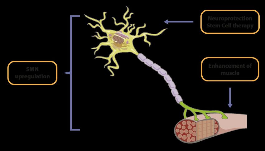
Spinal muscular atrophy
1. Background
2. Genetics
Spinal muscular atrophy modifiers
Carriers
3. Neuromuscular pathology of spinal muscular atrophy
Motor neuron changes in spinal cord in humans
Prenatal findings
Postnatal findings
Nerve pathology in humans
Neuromuscular junction (NMJ) pathology in humans
Muscle pathology in humans
SMN expression in non-SMA and SMA human tissues
Other neuronal abnormalities in SMA
Brain
Sensory pathways
Proprioception
4. SMA as a multi-organ disease
Cardiac abnormalities
Metabolic disorders
Liver defects
Pancreatic defects
Gastrointestinal defects
5. Diagnosis and differential diagnosis
Diagnosing SMA
Diagnostic genetic testing of spinal muscular atrophy
Deletion testing
Point mutation testing
SMN2 testing
Carrier testing
Newborn Screening (NBS)
Differential diagnosis
6. Clinical presentation and classification
SMA type I (severe type) or Werding-Hoffmann disease
SMA type II (intermediate type) or Dubowitz disease
SMA type III (mild type) or Kugelberg-Welander disease
SMA type IV (the mildest form)
Clinical Classification
7. Therapeutic strategies and drug treatment
Temporal requirements for SMN protein
Target cells for SMN protein restoration
Therapeutic approaches
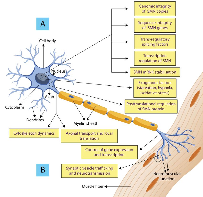
Drug treatment (SMA Drug Pipeline)
Approved drug treatments
Experimental drug treatments
4 SPINAL MUSCULAR ATROPHY:
PATHOLOGY, DIAGNOSIS, CLINICAL PRESENTATION, THERAPEUTIC STRATEGIES & TREATMENTS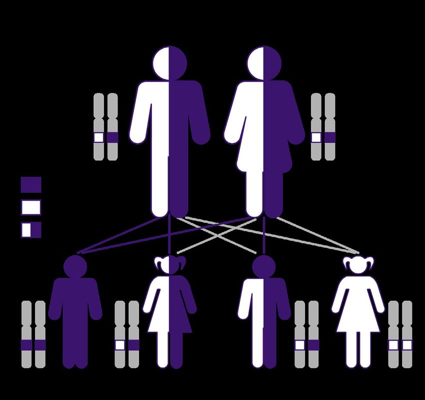
1. Background
Spinal muscular atrophy (SMA) is a rare, genetically          gene is generally associated with less severe SMA
inherited, heterogeneous group of neuromuscular               symptoms, but this finding is not correlated in every
disorders. It is characterised by progressive skeletal        case (Lefebvre, et al. 1997).
muscle weakness and subsequent atrophy, due to ab-
normalities of the motor neuron (MN) in the ventral           SMA can affect any race or gender. An estimation of
horn of the spinal cord. SMA is a debilitating disease        the incidence of all types of SMA is 1 in 10 000, while
that often takes away a person’s ability to walk, sit, eat,   the prevalence is 1-2 per 100 000 patients. SMA type 1
speak or breath and in the most severe cases, leads to        has the highest incidence (4-6 per 100,000) compared
paralysis at birth or soon after and premature death.         to other subtypes (1, 9 and 1, 7 for type II and type
In later onset SMA (childhood, adolescence or early           III respectively), but the lowest prevalence (0.004-0.28
adulthood), the affected individual loses the ability to      per 100,000) due to a short life expectancy. Prevalence
walk (Kolb & Kissel, 2015).                                   for both type II and type III is 1.5 per 100,000. Overall,
                                                              the incidence for type I is 60% and 40% for both type
SMA varies in severity, ranging from severe to mild,          II and type III. Because SMA is an autosomal recessive
with an extremely variable age of onset linked to in-         disease, both parents must be carriers for a child to
dividual severity. Different clinical presentation of the     inherit the condition. No signs of disease have been
disease may occur in the same family (Dubowitz, In-           associated with being a carrier. The carrier frequency
fantile muscular atrophy - a broad spectrum, 1967).           for the SMN1 gene has been estimated to be 1 per 50
Although primary pathology in SMA affects the neu-            (Caucasian and Asian) to 1 per 100 (African Americans)
romuscular system, increasing evidence suggests that          (Verhaart, et al. 2017).
other tissues and organs are affected (Shababi, Lorson
& Rudnik-Schoneborn, 2014; Bottai & Adami, 2013).             The first clinical presentation of SMA was described
                                                              in 1891 by Guido Werdnig, from the Department of
SMA is one of the most common autosomal recessive             Pathological Anatomy, at the University of Graz in
inherited disorders in humans after cystic fibrosis and       Austria, in two brothers with the onset of weakness
is very often called “the number one genetic killer of        at around 10 months of age. One died at age 3 and
children under the age of 2”. With a range of effective       the other at 6 years of age. Although the names of
therapies targeting the root of the disease, this defini-     Werdnig and Hoffmann have been associated with
tion tends to be historical. New therapies have signifi-      the severe subtype of SMA, they described the inter-
cantly changed the course of the disease and survival,        mediate form. In addition, Johann Hoffmann, from
especially in those who have been treated presympto-          the medical clinic in Heidelberg, described 7 patients
matically. The responsible gene for SMA is a mutated          from 3 families with an intermediate type (ability to sit
gene called survival motor neuron 1 (SMN1) and which          unaided, joint laxity and progression of scoliosis). In
is located on chromosome 5q. This gene encodes for            1899, Sylvestere presented at the paediatric society in
most of the body’s survival motor neuron (SMN) pro-           Paris, a two-month-old infant with flaccid paralysis of
tein, which is critical for the survival of lower motor       all limbs and trunk since birth and sparing of the dia-
neurons (LMNs), in contrast to other cell types (Mel-         phragm. The first description of a severe form was by
ki, et al. 1990; Lefebvre, et al. 1995; Brzustowicz, et al.   Beevor, in Brain, in 1902. The 5-week old child had an
1990; Monani, et al. 2000).                                   intercostal weakness with sparing the diaphragm and
                                                              complete paralysis of the limbs and trunk. Degenera-
Humans have another gene, SMN2, which is located in           tion of the anterior horns and posterior columns were
the same region as SMN1 and encodes a similar pro-            found at autopsy (Beevor, 1902).
tein, however, the majority of these proteins are trun-
cated and non-functional. The SMN2 gene, however,             A detailed description of the intermediate form of
produces some functional SMN protein, but it cannot           SMA was described by Dubowitz in 1964, in the UK
fully compensate for the deficit of the SMN1 gene             (Dubowitz, 1964). Through the examination of 12 pa-
(Monani, et al. 1999). The SMN2 gene is often termed          tients, he described late infantile-onset after the chil-
“the back-up” gene. Having more copies of the SMN2            dren had achieved the ability to sit, but never achieved

5      SPINAL MUSCULAR ATROPHY:
       PATHOLOGY, DIAGNOSIS, CLINICAL PRESENTATION, THERAPEUTIC STRATEGIES & TREATMENTS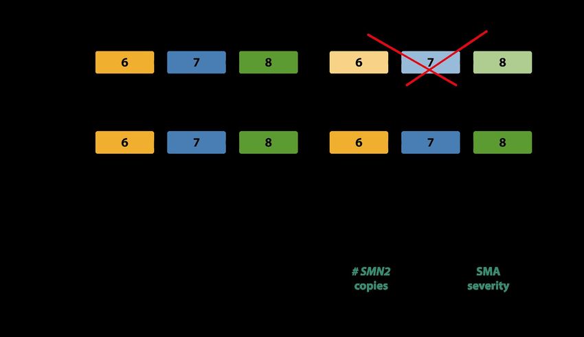
the ability to stand independently. These children had severe weakness of the legs, were static over the years, and
showed no significant deterioration.

The mild form of SMA, associated with Kugelberg and Welander, was described in 1956, through a series of 12 pa-
tients presenting with a limb-girdle muscular dystrophy with neurogenic electromyography (EMG) changes and
muscle biopsy (Kugelberg & Welander, 1956).

Over the last century, the wide clinical spectrum of SMA was recognised with detailed descriptions across severities.
Individuals with SMA often have difficulty performing the basic functions of life (breathing and swallowing). Howev-
er, SMA does not affect a person’s ability to think, learn nor their social skills. Their intellect is normal. Literature state
that the unique feature of SMA is that it is the only neuromuscular disease to spare the diaphragm although there is
intercostal weakness. Facial muscles are also spared. Some brainstem motor neuron groups, including oculomotor
and trochlear (ocular), which innervate the muscles around the eyes, are for unknown reasons spared (Nichterwitz,
et al. 2018), although, some ventilated individuals experience nearly complete paralysis of the diaphragm over time,
along with the restricted movement of the eyes and facial muscle weakness.

Identification of a gene locus, the development of animal models of SMA in the early 2000s and the recognition of
the role of SMN2 in phenotype, have all opened a path to new therapies and a better understanding of the natu-
ral history of the disease. With two approved drugs for SMA (Spinraza™ and Zolgensma™) in Europe and, the third
(Risdiplam), whose approval in the European Union is expected in the first half of 2021. with the increasing number
of clinical trials with disease-modifying therapies, there is a reason for hope. The natural history of the disease will
greatly change.

2. Genetics
The discovery of the SMN gene in 1995, as well as its              is shorter, unstable in vivo and rapidly degraded (Lefe-
localisation through studies of families with a history            bvre, et al. 1995; Monani, et al. 1999; Lorson & Andro-
of SMA, had a major impact on the non-invasive diag-               phy, 2000). Only a small amount of SMN2 transcripts
nosis of SMA, carrier testing and therapeutic options.             are correctly spliced and produce full-length SMN pro-
Localisation of the SMN gene to the 5q13.2 region was              tein. Thus, the SMN2 gene produces about 5-10% of
mapped across all SMA phenotypes.                                  full-length functional protein, since exon 7 is on oc-
                                                                   casion not spliced out of the SMN2 mRNA. In healthy
The SMA region is a large duplicated region on chro-               carriers, with one SMN1 copy and zero SMN2 copies,
mosome 5. The telomeric region contains the SMN1                   50% of functional full-length SMN protein is present,
gene, while the centromeric region contains the SMN2               which is sufficient for normal functioning and lower
gene. These genes are almost identical in the 5q13 re-             motor neurons (LMN) survival. In SMA patients with-
gion. Both SMN1 and SMN2 contain 8 exons and 99%                   out a functional SMN1 gene, the SMN2 gene serves as
homology in sequencing. They differ only by five nu-               a “back up” gene, with a range of 5-10% of functional
cleotides and produce an almost identical protein, the             protein production per SMN2 gene. In patients with
SMN protein. The differences are in exons 7 and 8, in-             SMA, the most common SMN2 copy numbers are 2-3,
trons 6 and 7. However, only one difference between                which gives an estimation of ~ 20-30% full-length
the SMN1 and SMN2 protein is functionally important:               SMN protein production. Milder phenotypes, with 4
a silent transition in exon 7 (c.840C>T), on the SMN2              copies of the SMN2 gene, have about 40% of SMN pro-
gene, which disrupts an exonic splice enhancer (ESE)               tein, which is still below the 50% level seen in healthy
and creates a new exonic splice silencer (ESS). This               carriers. Asymptomatic individuals with five copies
substitution (C to T) causes exon 7 to be excluded from            (humans can have 1 to 7 copies) of the SMN2 gene and
most of the SMN2 transcripts (which do not change                  zero copies of the SMN1 gene have been described
the amino acid sequence in the SMN protein), result-               (Mailman, et al. 2002; Lefebvre, et al. 1997) (Figure 1)
ing in the production of a truncated SMN protein that

6      SPINAL MUSCULAR ATROPHY:
       PATHOLOGY, DIAGNOSIS, CLINICAL PRESENTATION, THERAPEUTIC STRATEGIES & TREATMENTS
FIGURE 1. Diagram of SMN1 and SMN2 gene pathology in SMA

The genetics of SMA provides a unique opportunity          on the number of SMN2 genes present, which is higher
for therapeutic development. SMN2 provides attrac-         in gene conversion resulting in less severe phenotype.
tive targets and the majority of drug development
efforts in the field are focused on increasing SMN pro-    In about 5% of SMA patients, subtle (point) mutations
tein production from this gene (Iascone & Lee, 2015).      are present in the SMN1 gene localised mostly in a
                                                           region of exon 6 and 7. Those individuals are called
Individuals with SMA typically have inherited a faulty     “compound heterozygotes”, carrying a deletion/con-
(mutant) SMN1 gene from both parents. Most patients        version on one chromosome and one subtle mutation
with 5qSMA have a homozygous deletion involving            on the other. There are 23 different subtle mutations
exon 7 and 8 or only exon 7 (95-98%). While the ma-        published so far, which include nonsense, frameshift,
jority of the most severely affected SMA patients carry    missense and subtle splice site mutations. The pheno-
real homozygous deletions of SMN1, the majority of         type and severity depend both on the type of subtle
type II and III SMA patients show a homozygous ab-         mutation and the number of SMN2 copies in different
sence of SMN1 as a result of gene conversion of SMN1       combination (Wirth, et al. 1999).
into SMN2, leading to an increase of 3 - 4 copies of
SMN2. Four copies of this gene may produce enough          De novo mutations occur at a rate of about 2%. This
functional protein to result in a mild phenotype           is because the SMN region on chromosome 5 is un-
(Wirth, 2000; Campbell, et al. 1997).                      stable, which leads to the deletion and conversion of
                                                           the gene within the region. The high de novo mutation
Di Donato et al. (1997) observed a strong association      frequency is caused mainly by unequal crossing-over
between the milder forms of SMA (types II and III) and     between the repeated units during paternal meiosis
partial gene-conversion events not involving exon          (Wirth, et al. 1997).
8 from the SMN2 gene (Didonato, et al. 1997). Thus,
SMA patients with either deletion of SMN1 or a gene
conversion of SMN1 to SMN2 will produce a smaller
amount of functional SMN protein, being dependent

7      SPINAL MUSCULAR ATROPHY:
       PATHOLOGY, DIAGNOSIS, CLINICAL PRESENTATION, THERAPEUTIC STRATEGIES & TREATMENTS
Spinal muscular atrophy modifiers
The disease-modifying role of higher SMN2 gene copy number is described. It leads to later disease onset and better
prognosis. There is a statistically significant correlation between 4 SMN2 copies and SMA type IIIb or milder pheno-
types. SMA type IV patients usually have 4 - 6 SMN2 copies (Lefebvre, et al. 1997; Wirth, et al. 2006).
In populations with 2 SMN1 copies, the number of SMN2 copies varies, with 10-15% having no copies, 33% carrying
1 copy and 50% carrying 2 copies of SMN2 (Mailman, et al. 2002). As the prevalence of 1-2 SMN2 gene copies is the
most prevalent genotype in the general population, the incidence of SMA type 1 is highest. In children with SMA,
80-96% of patients with type 1 carry 1-2 copies of the SMN2 gene, while 4-20% with SMA type 2 or 3 carry 3 copies
of the SMN2 gene. About 96% of patient with SMA type 3 carry 3 or 4 SMN2 copies.
SMN2 copy number is the main disease modifier, however, it does not predict severity accurately and there is no
strict correlation. Other unknown factors must modify the SMA phenotype since affected and unaffected siblings
with homozygous deletions of SMN1 and identical haplotypes have been described in rare cases (Cobben, et al.
1995; Hahnen, et al. 1995). Intra-familiar reports describe variation in phenotype despite carrying the same number
of SMN2 copies (Mailman, et al. 2002). Sometimes, less than five SMN2 copies are found in asymptomatic carriers of
a homozygous deletion of the SMN1 gene and in some cases, six SMN2 copies cannot rescue from SMA symptoms.
Some SMA type I patients carry three SMN2 copies and SMA type III patients harbour two copies of the SMN2 gene
(Cusco, et al. 2006). This shows that other modulators of SMN2 gene splicing and other modifier genes exist, that al-
ter the amount of full-length SMN protein it produces, influencing disease manifestation and high clinical heteroge-
neity in SMA as a monogenic disease. Study of these factors reveals other underlying SMA pathological mechanisms
than in MNs and that can have an important clinical and therapeutic implication.
An inverse correlation has been observed between SMN2 copy number and duration of survival and age of onset in
adults (patients with 3 copies had significantly earlier onset compared to those with 4 copies). An inverse correlation
was observed between SMN2 copy numbers and brainstem involvement (facial weakness, chewing and swallowing
problems, tongue fasciculation and dysphonic voice). SMN2 copy number was associated with higher scores on the
Hammersmith Functional Motor Scale (HFMS) (Elsheikh, et al. 2009). An influence of SMN2 copy number on denerva-
tion was observed in SMA patients: the less SMN2 copies, the less Motor Unit Number Estimation (MUNE) (Swoboda,
et al. 2005) and maximum compound Motor Action Potential amplitude (CMAP). Heart defects were seen in those
patients who carry one SMN2 copy, but not in patients with increased SMN2 copies (Rudnik-Sconeborn, et al. 2008).
The first candidate phenotypic modifiers for SMA besides the SMN2 gene were 3 other genes localised in the SMA
region (neuronal apoptosis inhibitory protein gene (NAIP); basal transcription factor subunit p44 (p44c) and H4F5
(also known as SERF1) genes. Deletion of those genes was observed more frequently in SMA type 1 patients than
in milder forms (type 2 and 3). There is still no proof of these genes’ involvement in the modification of SMA pheno-
types since deletions of these genes in type 1 patients possibly reflect large deletions in SMN1 and SMN2 regions
(Campbell, et al. 1997).
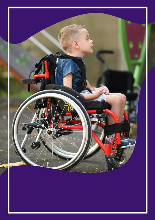
At the moment, SMN2 gene copy number is the only accepted modifier of SMA phenotype in patients. A summary
of other modifying factors and pathways connected with SMA pathology is given in Figure 2.
Modifying factors can be:
• SMN - dependent
• SMN - independent
SMN - dependent modifiers:
Genomic integrity (refers to SMN1/SMN2 copy numbers)
Sequence integrity (refers to gene conversion and point mutation)
Splicing factors (factors which alter SMN2 pre-mRNA splicing). Upregulation of exon 7 inclusion in SMN2 mRNA
8 SPINAL MUSCULAR ATROPHY:
PATHOLOGY, DIAGNOSIS, CLINICAL PRESENTATION, THERAPEUTIC STRATEGIES & TREATMENTS
can promote the amount of functional SMN protein produced). Splicing factors, namely TRA2-β1 (SFRS10), SF2/ASF
(SRSF1), SAM68(KHDRBS1), SRp30c (SRSF9), SRSF2, TIA1 (SRFS10), and hnRNP proteins (hnRNP-A1, hnRNP-G, hn-
RNP-M,hnRNP-U, hnRNP-Q) bind either directly or indirectly to cis-regulatory elements and facilitate the integration
or skipping of exon 7. Besides the above-listed elements, introns adjacent to SMN2 exon 7 comprise other essential
cis-elements, such asISS-N1, A+100G, Element 1, that bind splicing repressors and represent compelling targets for
SMA treatment. The principle of splicing correction is the basis of major approaches to SMA therapy with antisense
oligonucleotides (ASO). Importantly, the first approved treatment for SMA, SpinrazaTM, is based on this principle:
targeting the ISS-N1 negative element. Thus, factors altering SMN2 pre-mRNA splicing may be considered as SMA
severity modifiers through changing the amount of functional SMN protein produced.

Transcription regulation of SMN2 gene - various signalling cascades induced by hormones (prolactin) or G proteins,
including DNA methylation

Stabilisation of SMN mRNA

Posttranslational regulation and degradation of SMN protein (Survival Motor Neuron Protein phosphorylation,
ubiquitination, sumoylation)

Exogenous factors: starvation, hypoxia, and oxidative stress. Starvation and hypoxia strongly reduce full-length
SMN2 levels.

Identification of key SMN-independent pathways is essential to understand the contribution of SMN to neuromus-
cular and systemic pathology. SMN independent factors are: cytoskeleton dynamics, synaptic vesicle (SV) trafficking
and neurotransmission, axonal transport and local translation, and control of gene expression. Motor neurons are
highly specialised cells whose cytoskeleton has a crucial role in numerous cellular functions, ranging from axonal
growth and morphology maintenance to more specific functions such as axonal transport, SV trafficking and neu-
rotransmission. These factors, to a certain extent, have been shown to affect and modify these processes in SMA.

For a more detailed description of modifying factor in SMA pathology, see the following references: (Maretina, et al.
2018; Oprea, et al. 2008; Wirth, Garbes & Riessland, 2013; Janzen, et al. 2018).

The apoptotic pathway plays a crucial role in motor neuron loss and an important one in the pathogenesis of other
neurodegenerative diseases. Some apoptosis proteins are discussed as possible therapeutic targets. Some studies
have reported an increase in autophagic features in SMA motor neurons, suggesting that autophagy dysregulation
can represent a new pathogenetic hypothesis in SMA. Its pharmacological manipulation could influence the pro-
gression of the disease. Pharmacological and genetic inhibition of autophagy increases SMN levels, while induction
of autophagy decreases these levels (Rodriguez-Muela, et al. 2018; Piras & Boido, 2018).

Identification of modifying factors and pathways, as well as molecules or drugs that upregulate full-length SMN2
gene product or stabilise the SMN protein, are the most promising strategies for SMA therapy. In addition to SMN-de-
pendent strategies, identification of SMN-independent modifiers unveils combined therapy approaches in a broad
neurodegenerative context.

9      SPINAL MUSCULAR ATROPHY:
       PATHOLOGY, DIAGNOSIS, CLINICAL PRESENTATION, THERAPEUTIC STRATEGIES & TREATMENTS
FIGURE 2. Overview of select SMA modifiers.
(A) depicts the SMN-dependent modifiers (B) depicts SMN-independent modifiers.
Carriers
A carrier is a healthy individual who is not at risk of developing the disease but has a risk of passing the gene muta-
tion to his or her offspring. Carriers are unaffected heterozygous individuals, which means that they have one faulty
and one functioning copy of the SMN1 gene. Carriers of autosomal recessive disorders typically find out that they
are both carriers by having an affected child. A child will only have SMA if both parents pass on the faulty copy of the
SMN1 gene, which occurs in 25% of cases (Figure 3). If only one parent is a carrier, the child is usually not at risk of
SMA but does have a 50% risk of being a carrier.
Carriers fall into four main genotypic groups (Figure 4). The most common is the “1 + 0” genotype (one normal
functional allele and an SMN1-deleted, disease allele). A much less common category is the “2 + 0” genotype with
10 SPINAL MUSCULAR ATROPHY:
PATHOLOGY, DIAGNOSIS, CLINICAL PRESENTATION, THERAPEUTIC STRATEGIES & TREATMENTStwo functional genes on one chromosome and none on the other. Furthermore, there are also “1 + 1D” and “2 + 1D”
genotypes, which have one or two functional genes on one chromosome and a non-functional gene due to either
a point mutation or a microdeletion on the other. These last two genotypes are very rare and are called compound
heterozygotes. Four or even more copies of the SMN1 gene have also been found, indicating a “2 + 2” or possibly a
“3 + 1” genotype. This suggests “3 + 0” or “3 + 1D” carrier genotypes might also be possible however these are even
rarer (Verhaart, et al. 2017).
A higher percentage of hidden carriers (“2 + 0”) with 2, even more copies of the SMN1 gene is 3-8 times more prev-
alent in African American when compared to other ethnic groups, thereby decreasing the sensitivity of most carrier
tests used (Verhaart, et al. 2017).
FIGURE 3. SMA is an autosomal recessive disease. Parents of an affected individual are typically carriers of one faulty copy. In
families where both parents are carriers, there is a 25% chance that each of their children will have two faulty copies and have
SMA, a 50% chance each that their children will be carriers and not have the disease, and a 25% chance that each of their chil-
dren will have two normal SMN1 genes and not have SMA, nor be carriers.
FIGURE 4. Most common SMN genotypes among carriers and non-carriers. This image was published in Verhaat et al. Preva-
lence, incidence and carrier frequency of 5q-linked spinal muscular atrophy – a literature review. Orphanet J Rare Dis. 2017;12(1):124.
11 SPINAL MUSCULAR ATROPHY:
PATHOLOGY, DIAGNOSIS, CLINICAL PRESENTATION, THERAPEUTIC STRATEGIES & TREATMENTS3. Neuromuscular pathology of spinal
muscular atrophy
The main pathological feature of SMA, present in all (scAAV9-SMN) in a mouse model of SMA (Foust KD, et
human patients, is a specific loss of lower motor neu- al. 2010). AAV9 gene vector injected in neonatal mice
rons (MNs) (“spinal”) and small myofibers (“muscular at postnatal day 1 rescued neuromuscular physiology,
atrophy”). The name SMA was first used by Johann motor function and life span.
Hoffmann. It is now recognised that SMA is not just a
disease of MNs but extends far beyond them. Loss of This study implies that SMN protein delivered selec-
motor neuron cells from the spinal cord occurred sub- tively to SMA motor neurons provide therapeutic ben-
sequently to the development of other components efit, but the mitigating effects on the overall disease
of the neuromuscular system (axons and muscles) and survival are modest. Restoring SMN protein in
and specifically defects at the neuromuscular junction other cell types appears important, especially in se-
(NMJ). Motor neurons are particularly sensitive to SMN vere SMA.
protein paucity, but it is still uncertain if this is a se-
lective vulnerability of motor neurons to depletion of SMN is ubiquitously expressed and developmentally
SMN protein. These assumptions have been explored regulated. High levels of SMN expression occurs in
in numerous studies of animal models and, the results most tissues during embryogenesis (skeletal muscles,
strongly indicate that there is a unique requirement heart, kidney, thymus, pancreas, brain and other neu-
for adequate levels of SMN protein in other cells ex- ronal cells, but not in lungs), followed by a significant
cept those in the central nervous system. Tissue-spe- reduction after birth in all tissues except in the brain
cific requirements for SMN protein was investigated in and spinal cord, which remains relatively high until 2
animal transgenic mice. To investigate the role of spi- weeks post-birth. These findings suggest that higher
nal neurons in the disease process and as therapeutic levels of SMN protein are required prenatally in com-
targets, researchers have restored SMN protein selec- parison to the postnatal period and that most tissues
tively to all CNS neurons in severe SMA model mice, require SMN protein for normal development (Burlet,
using Nes-Cre recombinase transgene to activate the et al. 2018). The contribution of SMN2 to the level of
expression of an inducible SMN rescue allele (Lee A full-length transcript in non-SMA foetuses in other
J-H, et al. 2012). Restoring the SMN proteins to the tissues except the spinal cord was greater and virtu-
spinal neurons arrested the loses of motor neurons in ally absent in the spinal cord. This suggests SMN2 is a
transgenic mice. There was no significant difference in potentially important contributor to both the disease
the number of motor neuron cells in transgenic mice mechanism in SMA and in compensating for the pa-
compared to control at postnatal day 7. Restoring SMN thology seen in tissues clinically unaffected by the dis-
protein to the spinal neurons of the mutant mice pre- ease. But, SMN2 gene full-length transcripts become
vented the loss of proprioceptive synapses on the mo- critical in the spinal cord upon homozygous loss of
tor neuron cell bodies. SMA mice expressing neuronal the SMN1 gene (Soler-Botija, et al. 2005).
SMN exhibited significantly fewer distal defects of the
neuromuscular synapses. Accumulation of neurofila-
ments at pre-synapses was significantly reduced. The
size and complexity of post-synapses were increased.
Motor performances of transgenic mice improved as
a four-fold increase in median survival. Nevertheless,
mutants expressing neuronal SMN did not live beyond
3 weeks of birth. These effects were modest relative
to the level of phenotypic rescue previously achieved
following ubiquitous SMN expression with system-
ic delivery of self-complementary AAV9-mediated
gene vector expressing SMN1 cDNA to replace SMN
12 SPINAL MUSCULAR ATROPHY:
PATHOLOGY, DIAGNOSIS, CLINICAL PRESENTATION, THERAPEUTIC STRATEGIES & TREATMENTSMotor neuron changes in spinal cord in humans
Prenatal findings
During embryological development, excess motoneurons undergo a phase of degeneration and death via a process
known as programmed cell death (PCD) or apoptosis, allowing the final organisation of the nervous system. This
process is very rapid and coincides with a stage of synaptogenesis. Prenatally, the normal apoptotic process is more
pronounced in SMA, resulting in a prenatal deficiency in MN numbers in the spinal cord of SMA foetuses and the
lumbar part of the spinal cord. Motor neurons are reduced by 15-35% compared to normal foetuses and by 50% in
the early neonatal period (Simic, et al. 2000). The remaining SMA foetal MNs express normal levels of choline acetyl-
transferase (ChAT), the enzyme that catalyses the biosynthesis of acetylcholine (Ach), whose reduction is attributed
to the loss of MNs, suggesting that the function of surviving MNs in this period may be adequate (Soler-Botija, et al.
2005).
Postnatal findings
The classical pathological features evident in SMA spi- ogy or quantity, based on relative susceptibility within
nal cords are referred to as the neuropathological tet- different MNs subtypes within a spinal cord or caudal-
rad and include the loss of MNs, empty cell beds, glial rostral axis. Not all motor neurons and their NMJs are
bundles in ventral spinal roots (VRs) and heterotopic similarly susceptible to SMN protein deficiency. Pa-
MNs. tients with SMA have more weakness proximally than
distally. They retain normal eye movements, external
Increased disease duration (increased patients age) sphincter continence and diaphragm function (Mar-
is associated with higher MN losses. In severe cases, tinez-Hernandez, Bernal, Alias & Tizzano, 2014). The
about 73% of MNs were lost in individuals aged 5-22 precise origin of the selective vulnerability of some
months (Simic, et al. 2000). Empty cell beds and glial motor neurons and their NMJs remains unclear but it
bundles are frequently observed in the ventral horns clearly demonstrates that not all cells respond to low
(VHs) of spinal cords, representing a space where MNs levels of SMN in the same way. On the other hand, a
previously resided. There are few post-mortem stud- study from 2012 investigated a range of morpholog-
ies involving patients with intermediate or mild forms ical neuromuscular parameters and none conferred
of SMA. MN loss was less severe in teenage SMA type II the risk to degeneration of different subtypes, sug-
patients than in SMA type 1 patients of different ages gesting that more subtle molecular differences are
(Ito, Shibata, Saito, Kobayashi & Osawa, 2011). In a lim- likely to underlie the relative susceptibility of some
ited number of patients, a severe reduction in MNs, as motor neuron pools (Thomson, et al. 2012)
well as the hypoglossal and facial nucleus in the brain-
stem, were also reported in SMA type II (Araki, et al.
2003). MNs of the phrenic nerve are preserved. Many
of the MNs remaining in SMA spinal cords are chrom-
atolytic, have an accumulation of neurofilaments (Nf )
and breakdown of the plasma membrane consistent
with necrotic cell death. Neurofilaments are elevated
in the cerebrospinal fluid (CSF) in infantile-onset SMA
and decrease under Nusinersen (SpinrazaTM), the first
approved treatment for SMA. This is used as an addi-
tional marker to monitor therapy efficacy in clinical
trials (Winter, et al. 2019).
Heterotopic MNs are aberrantly localised in the white
matter. They are presumed to be undifferentiated in
the very early stage of development, without axons,
dendrites and synaptic connections (Simic, et al. 2000).
No human studies to date have examined MNs pathol-
13 SPINAL MUSCULAR ATROPHY:
PATHOLOGY, DIAGNOSIS, CLINICAL PRESENTATION, THERAPEUTIC STRATEGIES & TREATMENTSNerve pathology in humans
Reduced number of long myelinated axons with rare demyelination or degeneration are frequently observed in
proximal VRs. An alternative hypothesis to explain the increased vulnerability of motor neurons to low SMN levels
may be the consequence of disrupted axonogenesis. At post-symptomatic stages in severe SMA mice, only 20–35%
of motor neurons are lost from the spinal cord, despite severe neuromuscular degeneration, indicating that motor
neuron function is compromised prior to cell death (Monani, et al. 2000). This indicates that axonal pathology may
precede MNs somal death.
There is also a degeneration of small unmyelinated axons in VRs and distal nerve terminals, as well as a reduced
number of myelinated peripheral nerves and intramuscular nerves.
One of the hallmark pathological features in SMA are glial bundles in the VRs, which are present in all SMA types.
They were first described in the lumbar segment of the spinal cord. They taper off with increasing distance from the
spinal cord. Glial bundles are considered to be an astrocytic response to early-onset MN degeneration (Winter, et
al. 2019). Astrocytes are an important source of trophic factors that provide essential signals for neuronal survival.
In diseased and/or injured environments, astrocytes become reactive and undergo distinct morphological restruc-
turing including the upregulation of glial fibrillary acidic protein (GFAP) and other neurofilament proteins. Reactive
astrocytes secrete proinflammatory cytokines and, alter growth factor production, all of which promote neuron loss.
Microglia, which are another important source of neurotrophic molecules, undergo similar morphological and func-
tional changes when activated and serve as inflammatory cells in the CNS. In severe SMA mice models, astrocyte
dysfunction and/or activation preceded motor neuron loss (Rindt, et al. 2015). Astrocytes switch away from their
protective functions toward reactive and inflammatory cells and create a neural environment that exacerbates mo-
tor neuron malfunction. The phenotypic changes in SMA astrocytes, such as altered morphology, altered signalling
and decreased growth factor production, could negatively impact motor neuron health (Sison, et al. 2017).
Neuromuscular Junction (NMJ) Pathology in humans
Prenatal findings point to early alteration of the motor unit in patients with severe SMA. NMJ pathology seems to
start during the foetal period. As a result, initial muscle innervation cannot be maintained.
Disaggregated AcH receptors (AcHRs) were found around the myotubes in SMA type 1 foetuses, but not in SMA type
II. Accumulation of presynaptic vesicles, which were not in contact with muscle cells was observed both in humans
(Martinez-Hernandez, et al. 2013) and mouse models (Kong, et al. 2009). Involvement of NMJ pathology in mice
could be translated to human patients.
In postnatal findings, NMJ immaturity was documented in a form of simple motor terminal arborisations with no
synaptic vesicles and persistent expression of immature forms of γ AcHRs (Kong, et al. 2009). In mice models, neu-
rofilament aggregation in axons ends, abnormally small and not developed motor endplates, loss of Schwann cells,
delayed AcHRs and axon maturation, were also documented (Kariya, et al. 2008). Potential cellular and molecular
mechanisms underlying NMJ pathology are: Nf accumulation in motor nerve terminals, axonal transport and cy-
toskeletal abnormalities, altered calcium channels, mitochondrial dysfunction with decreased ATP levels and ubiq-
uitin pathways.
Muscle pathology in humans
Motor neuron survival and skeletal muscle development are closely linked and depend upon continued cellular
contact between these two cell types. Neuronal dysfunction can contribute significantly to muscle atrophy, but
there is evidence to suggest that intrinsic abnormalities in SMA skeletal muscle, due to lack of SMN protein, could
contribute directly to disease pathogenesis.
14 SPINAL MUSCULAR ATROPHY:
PATHOLOGY, DIAGNOSIS, CLINICAL PRESENTATION, THERAPEUTIC STRATEGIES & TREATMENTSPresence of smaller and thinner myotubes in SMA patients was seen prenatally, with impairment in myotubes width,
indicating a delay in muscle growth and maturation. These findings are consistent in the postnatal period (Martin-
ez-Hernandez, et al. 2009). Expression of proteins required for myofiber development is altered, with higher expres-
sion of immature isoforms of myosin heavy chains, which persist postnatally in atrophic areas of more affected mus-
cles. There is a correlation between myofiber maturity and innervational status (persistent expression of immature
myofiber cell surface marker has been observed on denervated myofibers in contrast to large preserved innervated
myofibers (Walsh & Moore, 1986).
Clinical diagnosis of SMA before genetic testing relied on muscle pathology from biopsy tissue acquired from
quadriceps muscle. Large areas of small rounded myofibers of all types (hypotrophic) often involving all fascicles,
interspersed by hypertrophic fibres were regularly seen. Hypotrophic fibres are evident in all SMA types, with a less-
er percentage in less severe cases. There is a continuous growth of hypertrophic fibres type I (probably fibres which
retained innervation or have been reinnervated from regenerative sprouts of surviving MNs) and arrest of growth of
small fibres, due to early denervation which begins at ages 2 and above. With SMA disease progression, there is an
increased replacement of muscles by fibrosis and fat.
Pathological changes in muscle are dependent on innervating MNs, but evidence showed that changes in mus-
cles could occur independently of nerve degeneration, confirming that SMN has cell-autonomous roles in muscle
(Mutsaers, et al. 2011; Arnold, et al. 2004). These findings suggest that treatments for SMA that target muscle, as well
as motor neurons, are likely to be beneficial.
An early study that increased SMN in muscle did not find significant improvements in motor phenotype and re-
storing SMN in skeletal muscle alone is insufficient to correct disease pathology in SMA mice (Gavrilina, et al. 2008).
On the other hand, a growing body of literature suggests that selective restoration of SMN levels by 50% in muscle
satellite cells (regenerative cells) improved phenotype in SMA mice (Nicole, et al. 2003).
It is possible that postnatal muscle loss could secondarily trigger MN cell death as well as NMJ maturation defect
and abnormal synapses.
SMN expression in non-SMA and SMA human tissues
There is a small number of data about baseline nor- sample as spinal cord tissue examined. The highest
mal or disease-associated SMN levels in disease-rele- SMN protein level of all SMA affected tissues was in
vant human tissues. There is even less understanding this sample in 18 gestational week, but this level was
of SMN induction in the CNS tissues with treatment approximately 5 fold reduced compared with medi-
because SMN levels cannot be measured in the CNS an SMN levels of prenatal non SMA control samples.
tissues of living patients. To understand the dynam- Compared to levels of SMA protein before 3 months
ics of SMN expression in the human CNS and non-CNS of age and after 3 months of age there were no statis-
tissues of unaffected and SMA patients (with 2 SMN2 tically significant differences between SMA and non-
copies), researchers have recently quantified SMN- SMA samples. SMN protein become restricted at low
mRNA and SMN protein levels in human tissue (spinal levels postnatally, particularly after 3 months of age
cord, brain and muscle) during expedited autopsies and remain low in cases 3 month-14 years. This study
(Ramos, et al. 2019) SMN protein levels showed a broad showed that the decrease in SMA protein levels in the
range in the spinal cord, cortex and muscle tissues iso- spinal cord was most evident in samples spanning
lated during fetal development in non-SMA controls 3 months before and 3 months after birth (perinatal
and decreased during development. Postnatal con- development). A similar result was observed in fron-
trol group less than 3 months of age had 2,3 fold de- tal cortex tissues and skeletal muscles. There was a 3
crease in SMA protein levels in spinal cord compared fold decrease in the early postnatal period compared
to prenatal levels. SMA affected spinal cords samples to the prenatal period in non-SMA iliopsoas muscle,
had a 6 fold decrease in the same age group (less than while approximately 2 fold decrease was found in
3 months postnatally) compared to non-SMA spinal postnatal samples of iliopsoas muscle and diaphragm
cord samples. There was only one fetal SMA affected in SMA compared to the same muscles in postnatal
15 SPINAL MUSCULAR ATROPHY:
PATHOLOGY, DIAGNOSIS, CLINICAL PRESENTATION, THERAPEUTIC STRATEGIES & TREATMENTSnon SMA controls. These findings imply a particular SMN2 copies in spinal cord or cortex. SMN2 transcript
need for SMN protein in the CNS and muscles during expression is unaltered by the loss of SMN1 in the CNS.
gestational and early neonatal developmental stag- When controlled for SMN2 copy number, postnatal
es. SMN protein may decrease rapidly in the last tri- control cases had median 2.1-fold more SMN protein
mester and the first 3 months after birth, which high- than postnatal SMA cases.
lights a possible optimal therapeutic window. Spinal Five patients who were treated with Nusinersen in this
cord SMN mRNA expression correlated moderately research showed ASO drug uptake in CNS samples
with SMN protein expression. The range of SMN tran- (spinal cord and brain). ASO drug concentrations were
script levels was restricted compared with that of pro- 2 fold lower in the cervical spinal cord compared to the
tein. The decreases in median SMN1-FL or SMN2-FL lumbar and thoracic spinal cord. Variable drug levels
mRNA levels between prenatal and postnatal samples were seen in brain tissue. This pattern of drug concen-
(SMN1-FL, 25%; SMN2-FL, 29%) were modest com- tration was associated with a 3-fold increase in SMN2-
pared with those of protein (79%). In non-SMA con- FL mRNA levels in cervical, thoracic, and lumbar/sacral
trol samples, a significant drop of median SMN2-FL spinal cord samples isolated from Nusinersen-treated
transcript occurred only between prenatal and early cases. There was no change in SMN2-FL expression in
postnatal control samples, whereas median SMN1-FL brain tissues. Muscle and liver tissues also showed no
transcript decreased significantly only between early increase in SMN-FL transcripts. Those with the highest
and late postnatal samples. This may indicate an earli- level of SMN2 mRNA expression received Nusinersen
er developmental decrease in SMN2mRNA expression prior to death or received multiple doses. ASO uptake
relative to that of SMN1mRNA in non-SMA spinal cord and SMN protein expression were also assessed by im-
and brain samples. This earlier decrease of SMN2 tran- munostaining. ASO uptake were particularly evident
scriptional activity could further contribute to earlier in ventral horns and the staining was highest in the
developmental reductions in SMN protein in SMA pa- lumbo-sacral region and thoracic region. Less was ob-
tients compared with controls. SMN protein expres- served in the cervical spinal cord and upper brain re-
sion only modestly correlated with a sum of SMN1 and gions indicating caudal-to-rostral gradient in the CNS.
SMN2 mRNA transcripts in prenatal control samples. In The caudal-rostral gradient paralleled with SMN2-FL
SMA postnatal samples SMN protein levels modestly transcript expression. Immunostaining of SMN protein
correlated with SMN2-FL transcripts. The highest SMN was evident in the ventral horn, macroglia and epend-
protein expression and SMN2-FL mRNA expression ymal cells at all spinal levels and, the percentage of
was observed only in one fetal SMA sample. This may SMN positive cells in treated patients were either not
indicate that SMN protein independently decreased significantly different or higher relative to those of un-
with age between prenatal and late postnatal sam- affected control samples.
ples and that additional, posttranscriptional mecha-
nisms contribute to the decrease in SMN protein levels
during perinatal development. This implies that other
mechanisms, except SMN1 and SMN2 mRNA expres-
sion, could also account for variations of therapeutic
response in patients with SMA. When controlled for
copy number, SMN2-FL, SMN2-Δ7, and SMN2-FL/Δ7
transcript expression did not significantly differ be-
tween postnatal control and postnatal SMA with 2
Other neuronal abnormalities in SMA
Brain
A correlation between neuronal pathology in other parts of the brain in relation to disease severity and SMN2 copy
numbers was reported. Morphologic analyses of post-mortem central nervous system in most severely affected
SMA type I patients with one copy of SMN2 gene, showed widespread neuronal degeneration in the motor cortex,
nuclei pontis, reticular formation, substantia nigra, cerebellar structures and thalamus (Harding, et al. 2015). This
indicates a potentially important role for SMN in regulating brain development.
16 SPINAL MUSCULAR ATROPHY:
PATHOLOGY, DIAGNOSIS, CLINICAL PRESENTATION, THERAPEUTIC STRATEGIES & TREATMENTSSensory pathways
Several cases of defective sensory neurons and thalamic lesions have been detected in genetically confirmed SMA.
SMN levels are high in dorsal root ganglia and posterior horn of the spinal cord in the human foetus. Restoration
of SMN in motor neurons of SMA mice repairs the NMJ defects and also restores synapses in the sensory neurons
(Gogliotti, et al. 2012), suggesting that the sensory-motor circuit function is dependent on SMN levels in motor neu-
rons.
Within spinal motor circuits, motor neurons (MNs) bridge the central and peripheral nervous systems by conveying
central commands to the skeletal muscles. MNs receive synapses from sensory neurons, spinal interneurons and
supraspinal pathways.
Whether synaptic dysfunction is responsible for the MN death or synaptic loss occurs in response to MN dysfunction
is unresolved. The key role of excitatory synaptic drive in shaping the function of motor neurons during develop-
ment was investigated by researchers from Columbia University in the United States (Fletcher, et al. 2017). SMN
depleted motoneurons exhibited reduced monosynaptically - induced excitatory postsynaptic potential (EPSP). The
modest overall reduction in a number of proprioceptive synapses on hyperexcitable motor neurons soma and den-
drites were also found, indicating that synaptic dysfunction precedes synaptic loss and correlates with an increase
in motor neurons sensory input resistance early in disease before motor neuron death.
Dysfunction of proprioceptive synapses caused increased input resistance of motor neurons and reduction in motor
neuron firing in SMA. SMA MNs fire at significantly reduced frequencies after the blockade of proprioceptive trans-
mission through expression of the tetanus toxin light chain subunit (TeNT), which inhibits neurotransmitter release
(glutamate). Increasing neuronal activity pharmacologically by chronic exposure with kainate, a glutamate receptor
agonist. in vivo, increased neuronal activity and improved righting time and led to the improvement in motor func-
tion.
Selective restoration of SMN protein in proprioceptive neurons rescued synaptic loos. The rescue of proprioceptive
synapses or enhanced presynaptic function of the remaining synapses was demonstrated by improved EPSP. Selec-
tive restoration of SMN protein in motor neurons had no effect on the synaptic rescue.
SMN upregulation in proprioceptive neurons in SMA mice improved NMJ function and motor behaviour. Combi-
nation of SMN upregulation in both motorneurons and proprioceptive neurons significantly contributed to the im-
provement in motor deficit by improving righting time and increasing CMAP amplitude.
This study showed that synaptic mechanisms are also responsible for motor neuron dysfunction in spinal muscular
atrophy.
Proprioception
In adults, proprioceptive afferent input contributes a significant amount of synaptic drive that continually modulates
motor output, muscle stiffness and tone, which are critical for postural control. During development, motor neurons
and muscles are dependent on each other, and their connection is essential to each component’s growth and sur-
vival. This is evident in the clinical manifestations of SMA, which include hyporeflexia, whereby patients’ reflexes are
below normal, or absent and selective muscle weakness of proximal, axial, and intercostal muscle groups. Severely
affected babies cannot sit without support. The stretch reflex is primary for movements. The circuitry involved in the
stretch reflex comprises three different cell types: alpha motor neurons, muscle fibres, and proprioceptive sensory
neurons. Upon muscle stretch, specialised sensory endings embedded in the muscle, called muscle spindles, length-
en and increase the firing frequency of primary proprioceptive sensory afferents. In SMA patients, muscle spindles
are much thicker and cellular, capsules are thickened and denervated from sensory afferents. These observations
suggest that spindle degeneration is a progressive process and that the loss of proprioceptive information from
afferents may result from the inhibited contractile shortening of the spindle or complete sensory denervation. On
electromyography (EMG) H (Hoffmann’s)–reflex was absent in 86% of cases, while the M-wave was absent in 30%
of cases, suggesting impaired sensory-motor function; H-reflexes were absent in 40% of SMA III patients, whereas
milder abnormalities in M-wave responses were observed (Chiriboga, et al. 2015). These observations suggest that
H-reflex responses may be a more sensitive method for detecting the initial stages of the disease.
17 SPINAL MUSCULAR ATROPHY:
PATHOLOGY, DIAGNOSIS, CLINICAL PRESENTATION, THERAPEUTIC STRATEGIES & TREATMENTS4. SMA as a multi-organ disease
Although the primary pathology in SMA affects the neuromuscular system, evidence from clinical reports and ani-
mal studies have shown that other tissues and organs are involved. These patients have various complications and
die prematurely, before damage to other organs occurs. Multi-organ dysfunction, including cardiac and vascular
defects, is not a general feature of human SMA.
Cardiac abnormalities
Most of the cardiac abnormalities have been reported in SMA type I patients with one SMN2 copy, while they are
also present in patients with 2 SMN2 copies. Congenital defects were most present in SMA type I patients. About
50% of SMA type I patients with structural cardiac defects had a single structural abnormality, atrial septal defect
(ASD) as the most prevalent, followed by a ventricular septal defect (VSD). The other 50% had multiple structural
abnormalities, ASD combined with VSD or hypoplastic left heart syndrome as the most frequent combination. Oth-
er congenital abnormalities were also reported: valvular aortic stenosis, hypoplastic aortic arch, coarctation of the
aorta, tricuspid athrepsia and hypoplastic left heart syndrome. Patients with 1 SMN2 copy had haemodynamically
relevant congenital defects, which points to the role of SMN protein in cardiogenesis in SMA patients (Wijngaarde, et
al., 2017). Ultrasonographic assessments of the heart in SMA foetuses with one SMN2 copy revealed a high propor-
tion of congenital abnormalities, associated with increased nuchal translucency (NT) (Parra, et al. 2012).
Abnormal electrocardiographic findings were artefacts due to baseline tremor from peripheral muscular fascicula-
tions, which did not alter cardiac rhythm and function. Bradycardia (less than 40 beats per minute) was reported in
severe cases, who survived many years due to assisted ventilation with signs of right ventricular overload. Bradycar-
dia, fluctuation in blood pressure, irregular skin reaction to temperature changes and vascular abnormalities were
interpreted as an autonomic nervous system dysfunction (nije nerve nego nervous)(ANS). Vascular abnormalities,
like distal digital necrosis, were associated with cardiac abnormalities but also as an isolated feature. And dysfunc-
tion may lead to impaired regulation of vascular tone (Bottai & Adami, 2013; Parra, et al. 2012).
Interpretation of cardiac dysfunction in SMA type II and III patients should be taken with caution, as the normal pop-
ulation is also at risk to develop cardiac deficiency. Patients with long-standing respiratory dysfunction are prone to
right heart overloading and pulmonary hypertension, with ECG pathological findings. Patients with atrioventricular
disturbances (AV) have to be considered as having other types of dystrophy such as Emery - Dreifuss or myotonic,
or autosomal dominant proximal SMA. Baseline tremor on ECG was reported in all SMA type II patients and in 50%
of patients with SMA type III. A small number of patients had other rhythm abnormalities. Cardiac abnormalities are
rare and, usually involve mitral valve prolapse. Complex cardiac malformations were reported only in 2 patients with
later onset disease (Parra, et al. 2012).
Metabolic disorders
Metabolic defects on the level of lipid metabolism and glucose metabolism have been reported, implicating the
liver and pancreas respectively.
Liver defects
Abnormal fatty acid metabolism is the most common metabolic defect exclusive to SMA type I patients with a few
exceptions seen in milder patients. Mild to moderate ketonuria with elevated fatty acid levels in blood upon fast-
ing was found, due to mitochondrial β oxidation abnormalities as well as elevated esterified carnitine (Crawford,
Sladky, Hurko, Besner-Johnston & Kelley, 1999). Extreme muscle wasting in severe SMA patients could be explained
by defects in fatty acid transport/oxidation as a major source of energy production during prolonged fasting (in
18 SPINAL MUSCULAR ATROPHY:
PATHOLOGY, DIAGNOSIS, CLINICAL PRESENTATION, THERAPEUTIC STRATEGIES & TREATMENTSYou can also read