Strengths and Weaknesses of the Current Strategies to Map and Characterize R-Loops - MDPI
←
→
Page content transcription
If your browser does not render page correctly, please read the page content below
non-coding
          RNA
Review
Strengths and Weaknesses of the Current Strategies
to Map and Characterize R-Loops
Vincent Vanoosthuyse        ID

 UMR5239 CNRS, Université de Lyon, ENS-Lyon, 46 allée d’Italie, 69007 Lyon, France;
 vincent.vanoosthuyse@ens-lyon.fr

 Received: 1 March 2018; Accepted: 23 March 2018; Published: 27 March 2018

 Abstract: R-loops are evolutionarily conserved three-stranded structures that result from the
 formation of stable DNA:RNA hybrids in the genome. R-loops have attracted increasing interest in
 recent years as potent regulators of gene expression and genome stability. In particular, their strong
 association with severe replication stress makes them potential oncogenic structures. Despite their
 importance, the rules that govern their formation and their dynamics are still controversial and
 an in-depth description of their direct impact on chromatin organization and DNA transactions is still
 lacking. To better understand the diversity of R-loop functions, reliable, accurate, and quantitative
 mapping techniques, as well as functional assays are required. Here, I review the different approaches
 that are currently used to do so and to highlight their individual strengths and weaknesses.
 In particular, I review the advantages and disadvantages of using the S9.6 antibody to map R-loops
 in vivo in an attempt to propose guidelines for best practices.

 Keywords: R-loops; S9.6; RNase H1; DRIP; R-ChIP

1. Introduction
     R-loops form when a complementary RNA molecule invades the DNA double helix and
hybridizes with one strand through Watson–Crick pairing, leaving the second DNA strand
single-stranded. It is believed that most R-loops form in a co-transcriptional manner, when the nascent
RNA invades its template in the wake of the RNA polymerase (reviewed in [1–3]). In eukaryotes,
R-loops can form both at protein-coding and non-coding transcription units, whatever the RNA
Polymerase involved and it is estimated that 5% of the human genome has the potential to form
detectable R-loops [4]. R-loop biology is the focus of growing scrutiny, as increasing evidence suggest
that the defective control of R-loop formation is associated with a number of human diseases [5].
Many questions remain however regarding the direct consequences of R-loop formation on the
surrounding chromatin.
     Current data demonstrate that specific features facilitate R-loop formation. These include negative
topological stress in the DNA, a strong bias for Guanine residues (G-skew) or the presence of
homopolymeric Adenine tracts in the RNA sequence, and high rates of transcription (reviewed
in [2]). Some of these features are inter-dependent, as high rates of transcription will lead to greater
negative topological stress, and, incidentally, will also increase the likelihood of forming and hence
detecting R-loops. Nevertheless, a consensus is emerging that the beginning of G-skewed genes
represents strong R-loop forming regions [4,6–8], although it could be that those particular R-loops
are thermodynamically more stable because of the G-skew, and hence easier to detect. Using some
mapping methods at least, R-loops have also been shown to form in gene bodies and in terminator
regions [4], although the presence of R-loops in gene terminators was not significantly detected in
Arabidopsis thaliana [8]. Recent observations have suggested that promoter R-loops tend to form over
DNA sequences where RNA Polymerase II is frequently in pause [6,9,10] and that have the potential to

Non-coding RNA 2018, 4, 9; doi:10.3390/ncrna4020009                            www.mdpi.com/journal/ncrna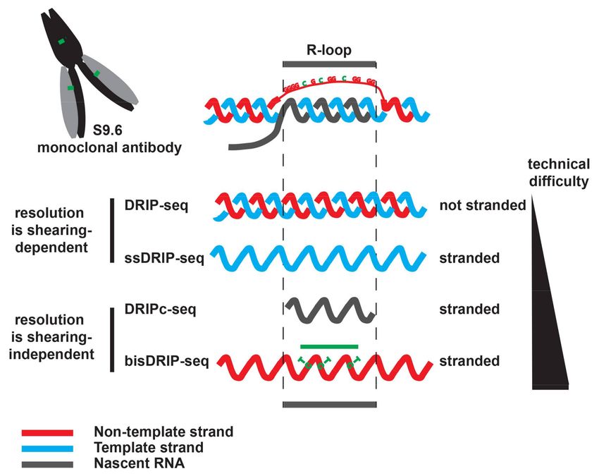
Non-coding RNA 2018, 4, 9                                                                             2 of 15

form G-quadruplex structures [6]. It is possible that R-loops, by displacing G-rich single-stranded DNA
(ssDNA), facilitate the formation of intra-molecular G-quadruplexes, which in turn, could stabilize
R-loops. Taken together, these observations clearly show that there are strong sequence determinants
to R-loop formation.
      In eukaryotes, chromatin structure (i.e., the position and dynamic of nucleosomes) also modulates
R-loop formation [11–13], presumably because the presence of nucleosomes counter-acts the propensity
of the double-helix to open and be invaded by the nascent RNA. The folding of the nascent RNA and
its coating by RNA-binding proteins such as the spliceosome is also believed to strongly antagonize
R-loop formation, most likely by physically preventing the nascent RNA from threading back into
its DNA template [14,15]. Consistent with this, the presence of introns was shown to limit R-loop
formation [15] and conversely, R-loop formation was shown to increase significantly in many mutants
of the RNA processing machinery [16,17]. Note however that, somewhat in apparent contradiction
with this simple model, there is evidence that the homologous recombination (HR) machinery is
essential for R-loop formation in many RNA processing mutants in budding yeast, suggesting that
R-loop formation is an active process that requires the Rad51 recombinase [18]. The significance of this
observation is not yet fully understood. More importantly, recent data suggest that DNA replication
has a strong influence on R-loop formation [19,20]: head-on conflicts between the transcription and
replication would facilitate R-loop formation, possibly because topological constraints associated with
such conflicts induce more negative topological stress over R-loop forming genes, whilst co-directional
conflicts would reduce R-loop formation, possibly because replisome-associated helicases could
disassemble R-loops. In addition, recent data suggest that in human cells at least, R-loop formation in
mitosis is largely restricted to centromeres [21]. Taken together, these observations strongly suggest
that the cell cycle stage greatly influences the abundance and localization of R-loops. To sum up,
although our understanding of the features that modulate R-loop formation has increased considerably
in recent years, in-depth and consensual mechanistic details of R-loop formation at the molecular level
are still missing.
      R-loops have been previously proposed to act as double-edged swords in the genome [1],
because they can be both physiological intermediates in some key processes and pathological structures.
For example, R-loops have been shown to participate in class-switch recombination at immunoglobulin
genes [22,23], in the initiation of mitochondrial replication [24], in the hypo-methylation of
promoters [25,26] or in transcription termination [27–29]. Recently, R-loops have also been proposed to
form on either side of double-strand breaks [30]. Conversely, R-loops are mutagenic [31], they stimulate
transcription-associated recombination (TAR) [32], and they are associated with DNA damage in
a replication-dependent manner [33]. In particular, R-loops are hugely detrimental in head-on
collisions between transcription and replication, where they lead to severe genome instability [19,20].
What features determine when R-loops become pathological structures remains poorly understood,
but there is general agreement that R-loop stability and the chromatin context in which they form are
likely to be key contributing factors. It is also possible that there are in fact different types of R-loops
in the cell, depending, for example, on their size or their sequence, and that those different R-loops
have different levels of toxicity depending on their architecture or the set of proteins that recognize
them. Consistent with the idea that there could be different types of R-loops, R-loops that form in the
absence of the THO complex in Saccharomyces cerevisiae display much higher levels of TAR than those
that form when both ribonuclease (RNase) H1 and H2 are missing [32].
      To address further the possible functions of R-loops, reproducible, accurate, and quantitative
R-loop mapping techniques are needed. The most widely used of these mapping techniques
is DNA:RNA immunoprecipitation (DRIP) [25] that relies on the use of the S9.6 antibody,
which recognizes DNA:RNA hybrids with great affinity [34]. Recently, several studies have questioned
the robustness of this approach [6,7,35] and an alternative strategy to map R-loops has been
developed [6]. In addition, the understanding of R-loop functions necessitates robust and specific
approaches to modulate R-loop formation. For this, the available toolkit remains very limited and
Non-coding RNA 2018, 4, 9 3 of 15
most studies rely on the nuclear over-expression of RNase H, an enzyme that destroys R-loops without
specificity, whether they are physiological or pathological.
Here, I aim to conduct an impartial assessment of the strengths and weaknesses of the different
methods to map and evaluate the functions of R-loops. Based on these observations, I propose
guidelines for best practices when working with R-loops.
2. Non-Denaturing Bisulfite Footprinting to Map R-Loops
Exposure of single-stranded DNA (ssDNA) to sodium bisulfite converts unmethylated cytosine
residues into uracil and this property has been used to map the ssDNA that is associated with
R-loops [7,22]. In a non-denatured R-loop, only the unmethylated cytosine residues present in the
non-template strand should be converted, as those in the template strand are protected from conversion
by the hybridized RNA. The presence of an R-loop is confirmed by the fact that cytosine conversion is
abolished after exogenous RNase H treatment. The great advantage of this approach is that it allows
for the footprinting of R-loops at near nucleotide resolution and at the single molecule resolution.
For a given locus, this technique gives access to the distribution of R-loop sizes and positions. Its main
limitation, however, at least in its current state, is that it is very low-throughput and that its resolution is
strongly constrained by the number and position of unmethylated cytosine residues in the non-template
strand. For instance, this approach would be inappropriate to map R-loops forming within polyA
tracts [36]. Nevertheless, it is tempting to speculate that the development of novel long-read and
single-molecule sequencing technologies, such as PacBio single-molecule real time (SMRT) sequencing
could soon make the genome-wide bisulfite mapping of R-loops a reality. Alternatively, to map
R-loop associated ssDNA, the use of the ssDNA-binding protein replication protein A (RPA) could be
envisaged. However, to be precise and specific for R-loops, stranded and quantitative RPA chromatin
immunoprecipitation (RPA-ChIP) would have to be set up to precisely identify RNase H-sensitive
RPA-ChIP signals.
3. The S9.6-Based Methods to Map and Quantify R-Loops
To date, most of the methods to quantify and map R-loops both in vitro and in vivo rely on
the use of the S9.6 monoclonal antibody [34]. S9.6 recognizes predominantly DNA:RNA hybrids,
although several studies have shown that it has some weaker, but nevertheless significant affinity for
RNA:RNA hybrids as well [37–39]. In addition, it was proposed that the affinity of S9.6 for DNA:RNA
hybrids is influenced by their nucleotide composition and that this might introduce bias in S9.6-based
maps [35]. Nevertheless, S9.6 is classically used for dot blot analysis, immunofluorescence studies and
immunoprecipitation approaches.
It is important to note that, besides the technical challenges and the potential limitations that are
associated with using S9.6 (see below), S9.6 should recognize any DNA:RNA hybrids greater than
six nucleotides in length [37] in the genome, whether they correspond to R-loops or to DNA:RNA
hybrids produced during normal DNA transactions, such as DNA damage repair or Okazaki fragments
synthesis. Theoretically at least, both genuine R-loops and other DNA:RNA hybrids should yield
RNase H-sensitive S9.6 signals. In practice, this could be a problem for example when mapping
DNA:RNA hybrids at stalled replication forks, where it is conceivable that the RNA primers required
to synthesize Okazaki fragments could be stabilized and might give significant S9.6 signals. Therefore,
unless one works with synchronized populations of cells, it is possible that DNA:RNA hybrids other
than R-loops will contribute to the signals obtained using S9.6-based methods. At this point, one should
stress that this is still only a theoretical limitation of using S9.6 to characterize R-loops, but one that
seems important to keep in mind and that should be carefully investigated in the future.
3.1. Quantifying R-Loop Formation Using Dot Blots
When properly controlled using RNase H treatment and accurate DNA quantification, dot blot
analysis is a reliable and straightforward way of quantifying DNA:RNA hybrids [33,40,41].Non-coding RNA 2018, 4, 9 4 of 15
Note however, that, unless nucleic acids have been extracted from purified nuclei, the material
that is probed tends to be mostly a mixture of genomic, mitochondrial, and in some cell-types, cytosolic
DNA. As a result, dot blot analysis is probably only sensitive enough to detect major changes in R-loop
accumulation but not to detect small, localized changes in R-loop formation in genomic DNA.
3.2. Visualizing R-Loop Formation Using Immunofluorescence Approaches
S9.6 has been used extensively in immunofluorescence studies, both on whole cells and on
chromosome spreads. On whole cells especially, the available results are somewhat confusing
because the pattern obtained with S9.6 varies significantly from one study to another (see below).
There might be cell-type specificities to explain this varying pattern, but it is more likely, as shown
by Skourti-Stathaki et al., that the conditions used to fix the cells have a strong influence on the S9.6
pattern [28]. Note however, that to date, no objective criteria have been established to help decide
which fixating condition best preserves genuine R-loops.
Most studies detect strong cytoplasmic and nucleolar signals with the S9.6 antibody, whilst the
4’,6-diamino-2-phenylindole (DAPI)-stained nuclear signal is surprisingly weak in comparison.
Although the nucleolar signal could be explained at least in part by the well-described and
evolutionarily conserved formation of R-loops at the ribosomal DNA (rDNA) [42–44], a very strong
cytoplasmic signal is unexpected and somewhat confusing. In some studies, this cytoplasmic signal is
restricted to a region around the nucleus that is known to be rich in mitochondria. In the majority of
studies, however, the cytoplasmic signal is much more widespread.
The cytoplasmic signal is often attributed to R-loops produced in the mitochondria [25], but others
have disputed that fact by showing that there is no significant co-localization between the S9.6 signal
and mitochondria [45]. Koo et al. proposed instead that the cytoplasmic signal that is detected by S9.6
comes for a large part from cytosolic DNA:RNA hybrids whose RNA moiety are produced by RNA
polymerase III (RNAPIII) [45]. Note however that the origin of the DNA in these putative cytoplasmic
RNAPIII-dependent DNA:RNA hybrids remains mysterious. Intriguingly, a strong cytoplasmic signal
was also obtained when cells were stained with the J2 antibody that recognizes RNA:RNA hybrids [28].
As S9.6 has a significant affinity for RNA:RNA hybrids [37–39], it is possible that at least part of the
cytoplasmic signal detected with S9.6 can be attributed to highly structured RNAs or even RNA:RNA
hybrids. The same can be said for the nucleolar signal because the nucleolus also contains a lot of
hairpin-forming RNAs. These observations suggest that the S9.6 pattern after immunofluorescence on
whole cells might result from both R-loop and non R-loop structures.
Because the cytoplasmic signal obtained with S9.6 is not fully understood, it is often ignored,
even if it is much stronger than the nuclear signal. Surprisingly, however, there are many mutant
conditions where the S9.6 signal increases both in the cytoplasm and in the nucleus, even when proteins
that are thought to be exclusively nuclear are mutated: for example, the deficiencies of BRCA2 [10,46],
Senataxin [47], U2 snRNP [48], SRPK2, and DDX23 [49] all lead to a significant increase in cytoplasmic
S9.6 staining. This counter-intuitive observation remains unexplained. One possible explanation is
that S9.6 detects a stress-associated structure that is found in the cytoplasm and perhaps also in the
nucleus and the nucleolus. If this is true, quantifying the nuclear S9.6 signal by immunofluorescence
as a measure of R-loop formation is probably inaccurate. Finally, these observations suggest that
S9.6 probably recognizes other structures than DNA:RNA hybrids in situ and that its specificity for
DNA:RNA hybrids is very sensitive to the way the DNA/chromatin is fixed or extracted.
What steps could be taken to improve this otherwise convenient approach to monitor R-loop
formation in situ? First, it seems imperative that the wider community should quickly agree on
a fixation protocol to best preserve R-loops and their sensitivity to RNase H. Then, the quantification of
the cytoplasmic, nucleolar and nuclear S9.6 signals and their respective sensitivity to exogenous RNase
H treatment should be systematically presented in a statistically significant number of cells. In addition,
a small improvement to a previously developed non S9.6-based strategy could be implemented in
parallel for corroboration: the Aguilera lab used the 52-residue DNA:RNA hybrid-binding domain ofNon-coding RNA 2018, 4, 9 5 of 15
RNase H1 (HB-GFP) to quantify R-loops in vivo after cell permeabilization [46]. An additional control
for this approach could be to express a mutated version of HB-GFP that cannot recognize DNA:RNA
hybrids anymore (see below). Note, that as the over-expression of HB-GFP might interfere with the
binding of the endogenous RNase H1 to R-loops, it is possible that HB-GFP over-expression might
artificially stabilize R-loops. When implementing this strategy, it is therefore essential to show that the
expression levels of HB-GFP are comparable in the different backgrounds/conditions where R-loops
are to be quantified.
3.3. DNA:RNA Immunoprecipitation (DRIP) and DRIP-Like Methods
The use of the S9.6 antibody to map DNA:RNA hybrids at specific loci was first developed by the
Tollervey and the Proudfoot labs in yeast [44] and in human cells [27], respectively. A similar strategy
was then implemented by the Chédin lab to obtain genome-wide maps of RNase H-sensitive R-loops in
human cells [25]. The Chédin lab coined the now widely-used name DNA:RNA immunoprecipitation
(DRIP) to describe this approach. The principle of DRIP is very simple: nucleic acids are extracted and
sheared, and the DNA:RNA hybrids are immuno-precipitated using the S9.6 antibody. Pre-treatment of
half the sample with exogenous RNase H validates the specificity of the immunoprecipitation, although
this crucial control is not always shown (see below). When studying a small set of loci, the enrichment of
DNA:RNA hybrids is classically estimated using quantitative polymerase chain reaction (DRIP-qPCR).
To better demonstrate the presence of R-loops, a few studies have used reverse transcription qPCR
(RT-qPCR) on DRIP material (DRIP-RT-qPCR) to definitely demonstrate that the immuno-precipitated
material did indeed contain RNase H-sensitive RNA molecules [4,8]. At the genome-wide scale,
various sequencing techniques have been implemented (Figure 1): DRIP-seq sequences the DNA
present in the immuno-precipitated material [25]; for increased resolution, DRIPc-seq sequences that
the RNA molecules present in the immuno-precipitated fraction [4,38]; ssDRIP-seq sequences the
template strand hybridized to the R-loop RNA [8]; bisDRIP-seq combines S9.6 immunoprecipitation
with bisulfite footprinting to map the R-loop-associated ssDNA [7]. DRIPc-seq and bisDRIP-seq map
R-loops in a strand-specific manner at near nucleotide resolution. Although DRIP has considerably
improved our understanding of R-loops in recent years, it suffers from an apparent lack of robustness
between studies, which begs some questions.
Figure 1. Genome-wide mapping of DNA:RNA hybrids using DNA:RNA immunoprecipitation
(DRIP)-like approaches.Non-coding RNA 2018, 4, 9 6 of 15
After DNA extraction and shearing, the S9.6 monoclonal antibody is used to precipitate
DNA:RNA hybrids. After purification, the precipitated DNA:RNA hybrids are sequenced. DRIP-seq
([25], not stranded) and ssDRIP-seq ([8], stranded) sequences the associated DNA. With these strategies,
DNA:RNA hybrids are mapped with a resolution that is dependent on the size of the starting DNA
fragments, which could be bigger than the R-loop. DRIPc-seq sequences the RNA moiety in the
DNA:RNA hybrid in a strand-specific manner ([4,38]). Paired-end sequencing of the RNA ensures that
the exact borders of the sequenced RNA are known. As a result, DRIPc-seq maps DNA:RNA hybrids
in a strand-specific manner at near nucleotide resolution. Because it requires working with RNA,
its implementation and the building of the sequencing libraries is technically more challenging than
DRIP-seq. bisDRIP-seq [7] maps R-loops by combining the precipitation of the DNA:RNA hybrids
with S9.6 with the bisulfite footprinting of the R-loop associated ssDNA. It is stranded (see text),
but its resolution depends on the number of unmethylated cytosine residues that are present in the
non-template strand and as a result it might under-estimate the size of R-loops (green line). However,
with bisDRIP-seq, the R-loop associated ssDNA is modified before DNA extraction, which could limit
the loss of unstable R-loops that might happen during DNA extraction. Of those four approaches,
bisDRIP-seq is probably the hardest to implement and to analyze.
3.3.1. Apparent Lack of Robustness of the DRIP Method
DRIP in different studies sometimes gives inconsistent results. For example, in S. cerevisiae, R-loops
were shown by some [50] but not others [51] to accumulate at PMA1 when the DNA&RNA helicase
Sen1 is deficient. The pattern of R-loop formation at the commonly-used gene model Actß varies
also unexpectedly between studies: R-loops would be as likely to form within the first intron and the
terminator of the gene for some [28,52], much more likely to form within the terminator for others [26],
and finally far less likely for a fourth study [10]. Similarly, R-loops were shown by some to accumulate
at terminator but not promoter regions when BRCA1 is deficient [10,52], whilst others detected R-loop
accumulation particularly at promoter regions in those conditions [9]. In addition, DRIP enrichment
for a given locus sometimes varies significantly from one study to the next or even within one study
from one figure to another. At the genome-wide scale, there is also apparent variability in the number
of R-loop forming regions that were identified using DRIP: for example, after DRIPc-seq, one study
reported 8112 promoters forming R-loops forming regions in human embryonic carcinoma Ntera2
cells [25], whilst another DRIP study only identified 3257 R-loop forming promoters in human primary
fibroblasts [26]. It is conceivable that at least some of these apparent discrepancies could be explained
by the fact that cell types and hence transcriptional patterns often differ between studies, although there
is evidence that patterns of R-loop formation are conserved between cell-types [4]. Moreover, different
sequencing depths or peak calling algorithms could conceivably account for the inconsistencies in
genome-wide DRIP studies. However, until it is firmly established that such parameters do indeed
explain those differences, one is left feeling that, despite its very simple principle, DRIP is either not
that straightforward to implement or not robust enough for its purpose. What could explain this
apparent lack of robustness?
Many different DRIP protocols have been set up. Some of those protocols contain steps that are
considered as very detrimental by others: for example some protocols include fixation and sonication
steps [40], whilst both of these steps are to be absolutely avoided according to other protocols [36].
Although the aim of this review is not to discuss the different DRIP protocols, but rather to highlight
the intrinsic strengths and weaknesses of the DRIP approach as compared to other R-loop mapping
methods, it is likely that the different protocols and in particular the way the DNA is sheared and
extracted could explain part of the variability that is observed. As explained below, other parameters
could also contribute to this variability.Non-coding RNA 2018, 4, 9 7 of 15
3.3.2. Factors Contributing to the Lack of Robustness of DRIP
As discussed above, it is highly likely that the way the DNA is extracted has a very significant
impact on the DRIP signal. In addition, the percentage of cells in S-phase in the starting material and
the specificity of the antibody are also key factors to take into account to explain those discrepancies.
The cell-cycle profile. The recent observation that DNA replication is a modulator of R-loop
accumulation [19,20] demonstrates that the cell-cycle profile of the starting material, and in particular,
the proportion of cells in S-phase and in mitosis, will greatly influence the DRIP results. One should
therefore either synchronize the samples of interest or endeavor to compare cultures with similar
proportions of cells in S-phase and mitosis.
The specificity of the antibody. As mentioned above, although the greater affinity of the S9.6
antibody for DNA:RNA hybrids is very clear on in vitro substrates, several observations indicate
that it can also recognize other structures when used on more complex material. For example, we
recently showed that the known affinity of S9.6 for RNA:RNA hybrids, albeit weaker than its affinity
of DNA:RNA hybrids, remained a confounding parameter for the accurate mapping of R-loops in
fission yeast using DRIPc-seq [38]. In addition, the use of bisDRIP-seq has shown that S9.6 could
efficiently immuno-precipitate single-stranded promoter regions that did not contain R-loops [7].
These observations strongly suggest that S9.6 is able to recognize a variety of nucleic acids that do
not adopt the conformation of the ordinary B form of DNA. It is therefore conceivable that the way
the genomic DNA is extracted will greatly influence the extent to which these other non-B forms of
DNA are preserved, and hence the level of RNase H-resistant signal in DRIP experiments (Figure 2).
This could at least partly explain the discrepancies between studies. DRIP enrichments might result
from a combination of features recognized by S9.6 and might not necessarily only reflect the presence
of R-loops. For example, the strong DRIP signal at the termination site (TES) of the TEFM gene in
human fibroblasts is fully resistant to RNase H treatment [10]. This is why the RNase H-treated sample
is such an important control, which cannot be replaced by complicated normalization methods as it
is sometimes done. These observations strengthen the argument that the best way to demonstrate
the presence of R-loops at a particular locus is to show the presence of strand-specific and RNase
H-sensitive RNA using RNA-based methods, such as DRIP-RTqPCR or DRIPc-seq [4,8,38].
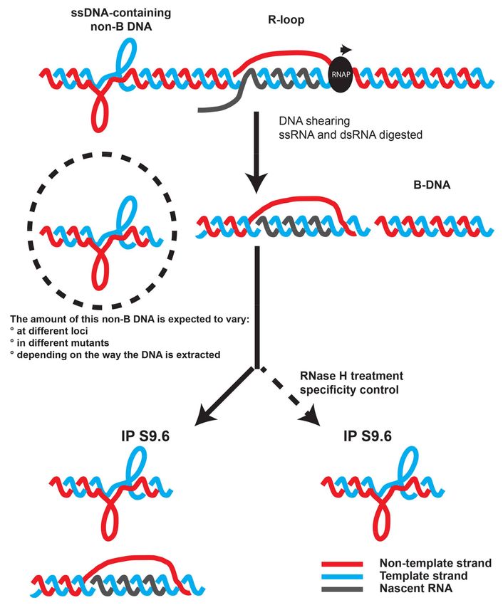
Scheme illustrating the fact that DRIP signals could result from R-loop and non R-loop structures
and the importance of the RNase H treatment to accurately estimate DNA:RNA hybrid formation at
a locus of interest.Non-coding RNA 2018, 4, 9 8 of 15
Figure 2. The S9.6 antibody is likely to recognize also ssDNA-containing non-B DNA forms that do not
correspond to R-loops [7].
4. RNase H1-Based Methods to Map R-Loops
RNase H1 is a highly conserved enzyme that recognizes DNA:RNA hybrids and cleaves their
RNA moiety. In human cells, the low abundant RNase H1 is particularly enriched in mitochondria
and in the nucleolus in a transcription-dependent manner [42]. Its role in the nucleus is not yet
very well characterized however. As it is an endogenous activity that evolved to recognize and to
disassemble DNA:RNA hybrids, RNase H1 has been used as a tool to map R-loops. In particular,
several studies have used a mutant of RNase H1 that could recognize but not process DNA:RNA
hybrids [6,25,43,53]. The earliest strategy was to express this catalytically inactive human RNase H1
(hRNase H1-D145N) in bacteria and to couple it to amylose beads. R-loops were then purified by
affinity from sheared genomic DNA. This strategy, called DNA:RNA in vitro enrichment (DRIVE-seq),
identified fewer R-loops than DRIP-seq [25,53]. To improve the sensitivity of this strategy, we made
the same mutation in the endogenous RNase H1 enzyme in fission yeast (Rnh1-D129N), and we used
chromatin immunoprecipitation (ChIP) of Rnh1-D129N to demonstrate the formation of unstable
R-loops at tRNA genes [43]. The enrichment of Rnh1D129N at tRNA genes was fully sensitive to
the strong in vivo expression of catalytically-active RNase H1 from Escherichia coli (RnhA), validating
the use of Rnh1D129N as an R-loop reporter at these sites [43]. A similar approach was recently
implemented in human cells using the nuclear over-expression of the catalytically inactive RNase
H1-D210N and was renamed R-ChIP [6]. Importantly, the sequencing of R-ChIP reactions in this
latest study was strand-specific and was controlled using a mutant of RNase H1 that cannot recognize
DNA:RNA hybrids [6].
As with DRIVE-seq, R-ChIP identified fewer R-loop forming regions than the S9.6-based
DRIP-seq or DRIPc-seq approaches [6]. Importantly, R-ChIP identified R-loops mostly at promoters,Non-coding RNA 2018, 4, 9 9 of 15
but not at terminator regions [6]. In addition, R-ChIP identified smaller R-loop forming regions
than DRIPc-seq [6]. The size of R-loops identified by R-ChIP was very similar to the archetypical
size of R-loops mapped in vitro at nucleotide resolution using non-denaturing bisulfite footprinting.
Moreover, the R-loop forming regions identified by R-ChIP contained sequence motifs, such as clusters
of Gs in the non-template strand that were previously identified in vitro as potential triggers for
R-loop formation [54]. Finally, in both fission yeast and human cells, R-ChIP identified tRNA genes
as hotspots of R-loop formation in otherwise wild-type cells [6,43], whilst tRNAs were identified
as R-loop forming regions by DRIP in A. thaliana [8], but not in human cells [4,25]. Taken together,
these observations led Chen et al. [6] to conclude that R-ChIP is better at identifying genuine R-loops
than S9.6-based methods.
There are however significant down sides to R-ChIP. Although it is easy to implement in yeast [43],
it is harder to implement in vertebrate cells, because it requires the stable expression of a mutant
enzyme. As shown recently [21], the over-expression of a catalytically-inactive RNase H1 enzyme
presents the risk of interfering with the dynamics of R-loops in vivo, when there is good evidence that
R-loop dynamics is likely to be critical for gene expression and genome stability. To implement R-ChIP
in mammalian cells, it is therefore important that the catalytically-inactive RNase H1 is expressed
at the right level: too much expression and there is a risk of dominant-negative effects; too little
expression and there is a risk that the endogenous, catalytically-active enzyme could interfere with
the binding of the catalytically-inactive mutant and the efficiency of R-ChIP. More importantly even,
there is a significant risk that R-ChIP is only going to map the R-loops that are recognized by RNase
H1. As an increasing number of proteins have been postulated to recognize and disassemble R-loops
in vivo (see for example Senataxin [55], FANCM [56], BLM [57], DDX19 [58], MTR4 [31] among others),
it is conceivable that there might be different types of R-loops that could be recognized by different
types of proteins. This might be why R-ChIP did not identify terminator regions as R-loop hotspots
in human cells [6], where R-loops might be recognized and disassembled by DNA&RNA helicases,
such as Senataxin and not by RNase H1 [27]. This could also explain why R-ChIP and DRIVE-seq
identified fewer R-loop forming regions than DRIP-seq or DRIPc-seq.
We argued previously that R-ChIP and DRIP are complementary approaches because their use in
parallel could give information about the stability of R-loops at specific loci [43]. For example in fission
yeast, RNase H1 is most abundant at RNAPIII-transcribed genes, suggesting that R-loops are constantly
formed and detected there [43]. The R-ChIP signal is therefore very strong at RNAPIII-transcribed
genes and this was also true in human cells [6]. On the contrary, DRIP-qPCR and DRIPc-seq only
gave significant signals at RNAPIII-transcribed genes in fission yeast in the absence of RNase H1 and
RNase H2 [38,43], thus confirming that RNAPIII-transcribed genes produce R-loops that are efficiently
degraded by RNase H enzymes. To summarize these observations, it is conceivable that DRIP is better
suited at detecting long-lived R-loops, whilst R-ChIP could be better at detecting highly dynamic
R-loops that are processed by RNase H1.
5. RNase H Over-Expression as a Tool to Probe R-Loop Functions
As discussed above, the direct consequences of R-loop formation on the surrounding chromatin
are still largely unclear. An in-depth understanding of R-loop contribution to gene expression and
genome stability necessitates functional assays where R-loop formation could be specifically modulated.
Importantly, to secure an unequivocal interpretation of the data, R-loop formation should be modulated
without interfering with transcript synthesis or integrity.
The most common functional assay to probe R-loop functions relies on the long-term
modulation of R-loop levels by affecting RNase H activity in vivo: RNase H-sensitive R-loops are
classically stabilized by deleting or down-regulating RNase H enzymes and in most model systems,
RNase H-sensitive R-loops can be de-stabilized by artificially increasing the concentration of RNase
H enzymes in the nucleus. When increasing (or decreasing) RNase H activity in the nucleus alters
a phenotype of interest, it is concluded that R-loops contribute to this phenotype. Note however thatNon-coding RNA 2018, 4, 9 10 of 15
this approach does not demonstrate that the contribution of R-loops to the phenotype of interest is
direct or indirect.
The most widely used strategy is to over-express RNase H. Although being widely used,
this strategy presents significant disadvantages. Its biggest limitation is that the amount of R-loops is
reduced genome-wide and without specificity, meaning that both the physiological and the pathological
RNase H-sensitive R-loops are affected. In addition, although this has never been shown, there is
at least a theoretical risk that a strong concentration of RNase H in the nucleus might also interfere
with the steady-state of other DNA:RNA hybrids, such as the primers of Okazaki fragments or
the DNA:RNA hybrids that were recently detected at double-strand breaks during repair [30,59].
This could be why RNase H1 over-expression in human cells was associated with persistent DNA
damage [42]. Finally, as discussed above, the possibility that there are RNase H-resistant R-loops
in vivo that would resist such treatment has not yet been excluded.
Importantly, the fact that RNase H over-expression has an indiscriminate effect on DNA:RNA
hybrids opens the possibility that it will have a significant impact on the transcriptome and the
proteome, and that those changes could indirectly contribute to alter the phenotype of interest.
If true, this would seriously complicate the interpretation of such experiments. For example,
it was shown that the over-expression of RNase H1 in human cells significantly affects the protein
levels of Top1 and other DNA repair proteins [42]. In addition, we recently demonstrated that the
strong expression of E. coli RnhA in fission yeast had a significant impact on the transcriptome and
affected the steady-state levels of many RNAs that did not form R-loops, according to our DRIPc-seq
maps [38]. These observations confirmed that long-term manipulation of RNase H activity imparts
significant and indirect changes to the transcriptome and proteome. In addition, the transcriptome
and proteome alterations that are associated with RNase H over-expression are likely to differ in
different genetic backgrounds. In particular, the modifications to the transcriptome imparted by RNase
H over-expression could combine with the existing alterations to the transcriptome in some mutant
backgrounds. Consistent with this, our unpublished results indicate that the strong expression of RnhA
in fission yeast differently alters the transcriptome in different mutant backgrounds. Therefore, the fact
that a phenotype of interest in a mutant background is sensitive to RNase H over-expression does
not necessarily mean that R-loops contribute to this phenotype directly. Nevertheless, if this strategy
were to be implemented to probe R-loop function because of its relative ease, it should probably
be backed up by the demonstration that the over-expression of another R-loop removing enzyme
also impacts the phenotype of interest in a similar way. Alternatively, ectopic expression of AID,
a cytidine deaminase that targets cytosine residues in ssDNA, was shown previously to enhance
R-loop dependent recombination and mutagenesis in yeast [13,60], and could be used to probe R-loop
functions at the genome-wide level. Note however that AID could also target ssDNA present in other
forms of non-B DNA [61], which could complicate the interpretation of such experiments.
However, if the consequences of R-loop formation on gene expression and genome stability are
highly dependent on the chromatin context in which they form, locus-specific ways of manipulating
R-loops must be developed. A corollary to what was discussed above is that locus-specific assays
should be preferred to genome-wide approaches anyway because they are probably at lower risk of
imparting indirect effects. To modulate R-loop formation locally, promoter-inactivating mutations [62]
or ribozyme-induced transcript cleavage [32] have been used previously. The down side of these
approaches is that they affect R-loop formation by interfering with the synthesis or the integrity of
the transcript itself. Unless one provides strong arguments establishing that the sole purpose of
transcription at the locus of interest is to form R-loops, the interpretation of such experiments can
be complicated because they remove both the R-loop and the transcript. Ideally, one should aim
to modulate the R-loop formation without affecting the synthesis of the transcript. To increase the
concentration of RNase H (or other R-loop removing enzymes) locally using chromosome-targeting
approaches could be an alternative strategy. A down side to this approach is that it is possible that
the chromosome-targeting mechanism would interfere with the activity, the dynamics or efficiency ofNon-coding RNA 2018, 4, 9 11 of 15
RNase H, which would result in only an incomplete reduction of R-loop levels. Another possibility
could be to use genome editing to alter the sequence determinants leading to R-loop formation in
a sequence of interest, without interfering with the synthesis of the transcript. Although this should be
in theory the best approach, it also presents down sides: it is cumbersome to implement because the
sequence determinants would have to be identified and validated in vitro before being mutated in vivo;
in addition, it would be difficult to alter such sequence determinants in protein-coding sequences
without altering the protein sequence itself, because some amino-acids, such as Glycine, are exclusively
encoded by G-rich codons. Depending on the phenotype that is investigated, this could complicate the
interpretation of the results. This approach might therefore be better implemented to understand the
role of R-loop formation within non-coding transcription units.
To conclude, to go further forward in the in-depth characterization of R-loop contributions
to gene expression and genome stability, there are some serious technical hurdles: RNase H
over-expression, which is the strategy that is easiest to implement, is subject to potential indirect effects,
whilst locus-specific strategies that would be better adapted at evaluating the direct consequences of
R-loop formation on the surrounding chromatin are both risky and difficult to implement. Consensual
proof-of-concept experiments are required to move the field forward.
6. Conclusions
This short survey of the strengths and weaknesses of the different methods used to characterize
R-loop distribution and functions highlighted several important conclusions:
1. Current immunofluorescence approaches are unlikely to be a reliable way of quantifying R-loops.
2. The affinity of the S9.6 antibody for RNA:RNA hybrids and other non-B forms of DNA is
potentially a confounding parameter for the quantification and the mapping of genuine R-loops
using DRIP-like approaches.
3. The possible influence of the cell-cycle on the formation and/or stability of R-loops and the fact
that the S9.6 antibody could theoretically recognize DNA:RNA hybrids associated with Okazaki
fragments suggest that the cell-cycle profile of the starting material is a parameter to consider
when performing DRIP.
4. More evidence is needed to demonstrate that R-ChIP is able to map all types of R-loops rather
than a subset of abundant, G-skewed, promoter-associated R-loops.
5. Possible indirect perturbations to the transcriptome and proteome associated with prolonged
manipulation of RNase H levels must be taken into consideration when using this strategy to
characterize R-loop functions.
Several recommendations can be drawn from this. Because DNA replication has recently emerged
as a significant modulator of DNA:RNA hybrids formation, it is important to know the proportion of
cells undergoing DNA replication when quantifying R-loop formation in different mutant backgrounds
(if one mutant spends more time in S-phase than the other, the abundance of DNA:RNA hybrids
might be artificially skewed). Otherwise, and unless one is specifically interested in the role of
R-loops during DNA replication, R-loops might be best quantified and mapped on populations of cells
synchronized in G1 or G2. In any case, the genomic DNA should be pre-treated with RNases to remove
both RNA:RNA hybrids and remnants of chromatin-associated RNAs [38]. Importantly, the wider
community should quickly agree on a set of guidelines on how best to extract DNA to preserve R-loops.
To improve resolution and demonstrate without doubt that the mapped R-loops do indeed contain
hybridized RNA molecules, RNA-based methods such as DRIP-RTqPCR and DRIPc-seq are probably
better. As an improvement to the current DRIPc-seq data and to reduce potential priming bias during
retro-transcription of the hybridized RNA, adapter-based primers rather than random hexamers could
be used, as is commonly done to sequence small RNAs. In addition, because R-loop formation is
mainly a co-transcriptional event, methods should be developed to normalize DRIP or R-ChIP signals
to nascent RNA transcription. When possible, R-ChIP and DRIP-like approaches could be used inNon-coding RNA 2018, 4, 9 12 of 15
parallel for corroboration as was done previously [43]. Finally, new locus-specific approaches should
be developed to evaluate the direct consequences of R-loop formation on the surrounding chromatin.
Acknowledgments: I thank Frédéric Chédin for helpful discussions and the four anonymous referees for
their constructive criticisms. I apologize to the many colleagues whose work I did not refer to in this short
review. My work was funded by a “Chaire d’Excellence” awarded by the Agence Nationale pour la Recherche
(ANR, Project TRACC, CHX11), by a “Projet Pluri-Equipe” (PPE 2016–2018) awarded by la Ligue contre le Cancer,
comité du Rhône and by a “Projet Emergent” de l’ENS-LYON (2016–2018).
Conflicts of Interest: The author declares no conflict of interest.
References
1. Skourti-Stathaki, K.; Proudfoot, N.J. A double-edged sword: R loops as threats to genome integrity and
powerful regulators of gene expression. Genes Dev. 2014, 28, 1384–1396. [CrossRef] [PubMed]
2. Chedin, F. Nascent connections: R-loops and chromatin patterning. Trends Genet. 2016, 32, 828–838.
[CrossRef] [PubMed]
3. Santos-Pereira, J.M.; Aguilera, A. R loops: New modulators of genome dynamics and function.
Nat. Rev. Genet. 2015, 16, 583–597. [CrossRef] [PubMed]
4. Sanz, L.A.; Hartono, S.R.; Lim, Y.W.; Steyaert, S.; Rajpurkar, A.; Ginno, P.A.; Xu, X.; Chedin, F. Prevalent,
dynamic, and conserved R-loop structures associate with specific epigenomic signatures in mammals.
Mol. Cell 2016, 63, 167–178. [CrossRef] [PubMed]
5. Richard, P.; Manley, J.L. R loops and links to human disease. J. Mol. Biol. 2016, 429, 3168–3180. [CrossRef]
[PubMed]
6. Chen, L.; Chen, J.Y.; Zhang, X.; Gu, Y.; Xiao, R.; Shao, C.; Tang, P.; Qian, H.; Luo, D.; Li, H.; et al. R-ChIP
using inactive RNase H reveals dynamic coupling of R-loops with transcriptional pausing at gene promoters.
Mol. Cell 2017, 68, 745–757.e5. [CrossRef] [PubMed]
7. Dumelie, J.G.; Jaffrey, S.R. Defining the location of promoter-associated R-loops at near-nucleotide resolution
using bisDRIP-seq. eLife 2017, 6, e28306. [CrossRef] [PubMed]
8. Xu, W.; Xu, H.; Li, K.; Fan, Y.; Liu, Y.; Yang, X.; Sun, Q. The R-loop is a common chromatin feature of the
Arabidopsis genome. Nat. Plants 2017, 3, 704–714. [CrossRef] [PubMed]
9. Zhang, X.; Chiang, H.C.; Wang, Y.; Zhang, C.; Smith, S.; Zhao, X.; Nair, S.J.; Michalek, J.; Jatoi, I.;
Lautner, M.; et al. Attenuation of RNA polymerase II pausing mitigates BRCA1-associated R-loop
accumulation and tumorigenesis. Nat. Commun. 2017, 8, 15908. [CrossRef] [PubMed]
10. Shivji, M.K.K.; Renaudin, X.; Williams, C.H.; Venkitaraman, A.R. BRCA2 regulates transcription elongation
by RNA polymerase ii to prevent R-loop accumulation. Cell Rep. 2018, 22, 1031–1039. [CrossRef] [PubMed]
11. Taneja, N.; Zofall, M.; Balachandran, V.; Thillainadesan, G.; Sugiyama, T.; Wheeler, D.; Zhou, M.; Grewal, S.I.
SNF2 family protein FFT3 suppresses nucleosome turnover to promote epigenetic inheritance and proper
replication. Mol. Cell 2017, 66, 50–62.e6. [CrossRef] [PubMed]
12. Herrera-Moyano, E.; Mergui, X.; Garcia-Rubio, M.L.; Barroso, S.; Aguilera, A. The yeast and human fact
chromatin-reorganizing complexes solve R-loop-mediated transcription-replication conflicts. Genes Dev.
2014, 28, 735–748. [CrossRef] [PubMed]
13. Garcia-Pichardo, D.; Canas, J.C.; Garcia-Rubio, M.L.; Gomez-Gonzalez, B.; Rondon, A.G.; Aguilera, A.
Histone mutants separate R loop formation from genome instability induction. Mol. Cell 2017, 66,
597–609.e595. [CrossRef] [PubMed]
14. Chen, X.; Yang, J.R.; Zhang, J. Nascent RNA folding mitigates transcription-associated mutagenesis.
Genome Res. 2016, 26, 50–59. [CrossRef] [PubMed]
15. Bonnet, A.; Grosso, A.R.; Elkaoutari, A.; Coleno, E.; Presle, A.; Sridhara, S.C.; Janbon, G.; Geli, V.;
de Almeida, S.F.; Palancade, B. Introns protect eukaryotic genomes from transcription-associated genetic
instability. Mol. Cell 2017, 67, 608–621.e606. [CrossRef] [PubMed]
16. Stirling, P.C.; Chan, Y.A.; Minaker, S.W.; Aristizabal, M.J.; Barrett, I.; Sipahimalani, P.; Kobor, M.S.; Hieter, P.
R-loop-mediated genome instability in mRNA cleavage and polyadenylation mutants. Genes Dev. 2012, 26,
163–175. [CrossRef] [PubMed]Non-coding RNA 2018, 4, 9 13 of 15
17. Wahba, L.; Amon, J.D.; Koshland, D.; Vuica-Ross, M. RNase H and multiple RNA biogenesis factors cooperate
to prevent RNA:DNA hybrids from generating genome instability. Mol. Cell 2011, 44, 978–988. [CrossRef]
[PubMed]
18. Wahba, L.; Gore, S.K.; Koshland, D. The homologous recombination machinery modulates the formation of
RNA–DNA hybrids and associated chromosome instability. eLife 2013, 2, e00505. [CrossRef] [PubMed]
19. Hamperl, S.; Bocek, M.J.; Saldivar, J.C.; Swigut, T.; Cimprich, K.A. Transcription-replication conflict
orientation modulates R-loop levels and activates distinct DNA damage responses. Cell 2017, 170,
774–786.e719. [CrossRef] [PubMed]
20. Lang, K.S.; Hall, A.N.; Merrikh, C.N.; Ragheb, M.; Tabakh, H.; Pollock, A.J.; Woodward, J.J.; Dreifus, J.E.;
Merrikh, H. Replication-transcription conflicts generate R-loops that orchestrate bacterial stress survival and
pathogenesis. Cell 2017, 170, 787–799.e718. [CrossRef] [PubMed]
21. Kabeche, L.; Nguyen, H.D.; Buisson, R.; Zou, L. A mitosis-specific and R loop-driven ATR pathway promotes
faithful chromosome segregation. Science 2018, 359, 108–114. [CrossRef] [PubMed]
22. Yu, K.; Chedin, F.; Hsieh, C.L.; Wilson, T.E.; Lieber, M.R. R-loops at immunoglobulin class switch regions in
the chromosomes of stimulated B cells. Nat. Immunol. 2003, 4, 442–451. [CrossRef] [PubMed]
23. Zhang, Z.Z.; Pannunzio, N.R.; Han, L.; Hsieh, C.L.; Yu, K.; Lieber, M.R. The strength of an ig switch region is
determined by its ability to drive R loop formation and its number of WGCW sites. Cell Rep. 2014, 8, 557–569.
[CrossRef] [PubMed]
24. Lee, D.Y.; Clayton, D.A. Initiation of mitochondrial DNA replication by transcription and R-loop processing.
J. Biol. Chem. 1998, 273, 30614–30621. [CrossRef] [PubMed]
25. Ginno, P.A.; Lott, P.L.; Christensen, H.C.; Korf, I.; Chedin, F. R-loop formation is a distinctive characteristic
of unmethylated human CpG island promoters. Mol. Cell 2012, 45, 814–825. [CrossRef] [PubMed]
26. Grunseich, C.; Wang, I.X.; Watts, J.A.; Burdick, J.T.; Guber, R.D.; Zhu, Z.; Bruzel, A.; Lanman, T.; Chen, K.;
Schindler, A.B.; et al. Senataxin mutation reveals how R-loops promote transcription by blocking DNA
methylation at gene promoters. Mol. Cell 2018, 69, 426–437.e427. [CrossRef] [PubMed]
27. Skourti-Stathaki, K.; Proudfoot, N.J.; Gromak, N. Human senataxin resolves RNA/DNA hybrids formed at
transcriptional pause sites to promote Xrn2-dependent termination. Mol. Cell 2011, 42, 794–805. [CrossRef]
[PubMed]
28. Skourti-Stathaki, K.; Kamieniarz-Gdula, K.; Proudfoot, N.J. R-loops induce repressive chromatin marks over
mammalian gene terminators. Nature 2014, 516, 436–439. [CrossRef] [PubMed]
29. Ginno, P.A.; Lim, Y.W.; Lott, P.L.; Korf, I.; Chedin, F. GC skew at the 50 and 30 ends of human genes links
R-loop formation to epigenetic regulation and transcription termination. Genome Res. 2013, 23, 1590–1600.
[CrossRef] [PubMed]
30. Cohen, S.; Puget, N.; Lin, Y.L.; Clouaire, T.; Aguirrebengoa, M.; Rocher, V.; Pasero, P.; Canitrot, Y.; Legube, G.
Senataxin resolves RNA:DNA hybrids forming at DNA double-strand breaks to prevent translocations.
Nat. Commun. 2018, 9, 533. [CrossRef] [PubMed]
31. Lim, J.; Giri, P.K.; Kazadi, D.; Laffleur, B.; Zhang, W.; Grinstein, V.; Pefanis, E.; Brown, L.M.; Ladewig, E.;
Martin, O.; et al. Nuclear proximity of Mtr4 to RNA exosome restricts DNA mutational asymmetry. Cell
2017, 169, 523–537.e515. [CrossRef] [PubMed]
32. Huertas, P.; Aguilera, A. Cotranscriptionally formed DNA:RNA hybrids mediate transcription elongation
impairment and transcription-associated recombination. Mol. Cell 2003, 12, 711–721. [CrossRef] [PubMed]
33. Stork, C.T.; Bocek, M.; Crossley, M.P.; Sollier, J.; Sanz, L.A.; Chedin, F.; Swigut, T.; Cimprich, K.A.
Co-transcriptional R-loops are the main cause of estrogen-induced DNA damage. eLife 2016, 5, e17548.
[CrossRef] [PubMed]
34. Boguslawski, S.J.; Smith, D.E.; Michalak, M.A.; Mickelson, K.E.; Yehle, C.O.; Patterson, W.L.; Carrico, R.J.
Characterization of monoclonal antibody to DNA·RNA and its application to immunodetection of hybrids.
J. Immunol. Methods 1986, 89, 123–130. [CrossRef]
35. Konig, F.; Schubert, T.; Langst, G. The monoclonal S9.6 antibody exhibits highly variable binding affinities
towards different R-loop sequences. PLoS ONE 2017, 12, e0178875. [CrossRef] [PubMed]
36. Wahba, L.; Costantino, L.; Tan, F.J.; Zimmer, A.; Koshland, D. S1-DRIP-seq identifies high expression and
polyA tracts as major contributors to R-loop formation. Genes Dev. 2016, 30, 1327–1338. [CrossRef] [PubMed]Non-coding RNA 2018, 4, 9 14 of 15
37. Phillips, D.D.; Garboczi, D.N.; Singh, K.; Hu, Z.; Leppla, S.H.; Leysath, C.E. The sub-nanomolar binding of
DNA-RNA hybrids by the single-chain Fv fragment of antibody S9.6. J. Mol. Recognit. 2013, 26, 376–381.
[CrossRef] [PubMed]
38. Hartono, S.R.; Malapert, A.; Legros, P.; Bernard, P.; Chedin, F.; Vanoosthuyse, V. The affinity of the S9.6
antibody for double-stranded RNAs impacts the accurate mapping of R-loops in fission yeast. J. Mol. Biol.
2018, 430, 272–284. [CrossRef] [PubMed]
39. Kinney, J.S.; Viscidi, R.P.; Vonderfecht, S.L.; Eiden, J.J.; Yolken, R.H. Monoclonal antibody assay for
detection of double-stranded RNA and application for detection of group a and non-group a rotaviruses.
J. Clin. Microbiol. 1989, 27, 6–12. [PubMed]
40. Halasz, L.; Karanyi, Z.; Boros-Olah, B.; Kuik-Rozsa, T.; Sipos, E.; Nagy, E.; Mosolygo, L.A.; Mazlo, A.;
Rajnavolgyi, E.; Halmos, G.; et al. RNA–DNA hybrid (R-loop) immunoprecipitation mapping: An analytical
workflow to evaluate inherent biases. Genome Res. 2017, 27, 1063–1073. [CrossRef] [PubMed]
41. Yang, Z.; Hou, Q.; Cheng, L.; Xu, W.; Hong, Y.; Li, S.; Sun, Q. RNase H1 cooperates with DNA gyrases to
restrict R-loops and maintain genome integrity in arabidopsis chloroplasts. Plant Cell 2017, 29, 2478–2497.
[CrossRef] [PubMed]
42. Shen, W.; Sun, H.; De Hoyos, C.L.; Bailey, J.K.; Liang, X.H.; Crooke, S.T. Dynamic nucleoplasmic and nucleolar
localization of mammalian RNase H1 in response to RNap I transcriptional R-loops. Nucleic Acids Res. 2017,
45, 10672–10692. [CrossRef] [PubMed]
43. Legros, P.; Malapert, A.; Niinuma, S.; Bernard, P.; Vanoosthuyse, V. RNA processing factors Swd2.2 and
Sen1 antagonize RNA Pol III-dependent transcription and the localization of condensin at Pol III genes.
PLoS Genet. 2014, 10, e1004794. [CrossRef] [PubMed]
44. El Hage, A.; French, S.L.; Beyer, A.L.; Tollervey, D. Loss of topoisomerase I leads to R-loop-mediated
transcriptional blocks during ribosomal RNA synthesis. Genes Dev. 2010, 24, 1546–1558. [CrossRef] [PubMed]
45. Koo, C.X.; Kobiyama, K.; Shen, Y.J.; LeBert, N.; Ahmad, S.; Khatoo, M.; Aoshi, T.; Gasser, S.; Ishii, K.J. RNA
polymerase III regulates cytosolic RNA:DNA hybrids and intracellular microRNA expression. J. Biol. Chem.
2015, 290, 7463–7473. [CrossRef] [PubMed]
46. Bhatia, V.; Barroso, S.I.; Garcia-Rubio, M.L.; Tumini, E.; Herrera-Moyano, E.; Aguilera, A. BRCA2 prevents
R-loop accumulation and associates with TREX-2 mRNA export factor PCID2. Nature 2014, 511, 362–365.
[CrossRef] [PubMed]
47. Becherel, O.J.; Sun, J.; Yeo, A.J.; Nayler, S.; Fogel, B.L.; Gao, F.; Coppola, G.; Criscuolo, C.; De Michele, G.;
Wolvetang, E.; et al. A new model to study neurodegeneration in ataxia oculomotor apraxia type 2.
Hum. Mol. Genet. 2015, 24, 5759–5774. [CrossRef] [PubMed]
48. Tanikawa, M.; Sanjiv, K.; Helleday, T.; Herr, P.; Mortusewicz, O. The spliceosome U2 snRNP factors
promote genome stability through distinct mechanisms; transcription of repair factors and R-loop processing.
Oncogenesis 2016, 5, e280. [CrossRef] [PubMed]
49. Sridhara, S.C.; Carvalho, S.; Grosso, A.R.; Gallego-Paez, L.M.; Carmo-Fonseca, M.; de Almeida, S.F.
Transcription dynamics prevent RNA-mediated genomic instability through SRPK2-dependent DDX23
phosphorylation. Cell Rep. 2017, 18, 334–343. [CrossRef] [PubMed]
50. Mischo, H.E.; Gomez-Gonzalez, B.; Grzechnik, P.; Rondon, A.G.; Wei, W.; Steinmetz, L.; Aguilera, A.;
Proudfoot, N.J. Yeast Sen1 helicase protects the genome from transcription-associated instability. Mol. Cell
2011, 41, 21–32. [CrossRef] [PubMed]
51. Grzechnik, P.; Gdula, M.R.; Proudfoot, N.J. Pcf11 orchestrates transcription termination pathways in yeast.
Genes Dev. 2015, 29, 849–861. [CrossRef] [PubMed]
52. Hatchi, E.; Skourti-Stathaki, K.; Ventz, S.; Pinello, L.; Yen, A.; Kamieniarz-Gdula, K.; Dimitrov, S.; Pathania, S.;
McKinney, K.M.; Eaton, M.L.; et al. BRCA1 recruitment to transcriptional pause sites is required for
R-loop-driven DNA damage repair. Mol. Cell 2015, 57, 636–647. [CrossRef] [PubMed]
53. Marinello, J.; Bertoncini, S.; Aloisi, I.; Cristini, A.; Malagoli Tagliazucchi, G.; Forcato, M.; Sordet, O.;
Capranico, G. Dynamic effects of topoisomerase I inhibition on R-loops and short transcripts at active
promoters. PLoS ONE 2016, 11, e0147053. [CrossRef] [PubMed]
54. Roy, D.; Lieber, M.R. G clustering is important for the initiation of transcription-induced R-loops in vitro,
whereas high G density without clustering is sufficient thereafter. Mol. Cell. Biol. 2009, 29, 3124–3133.
[CrossRef] [PubMed]Non-coding RNA 2018, 4, 9 15 of 15
55. Kim, H.D.; Choe, J.; Seo, Y.S. The sen1+ gene of Schizosaccharomyces pombe, a homologue of budding yeast
SEN1, encodes an RNA and DNA helicase. Biochemistry 1999, 38, 14697–14710. [CrossRef] [PubMed]
56. Schwab, R.A.; Nieminuszczy, J.; Shah, F.; Langton, J.; Lopez Martinez, D.; Liang, C.C.; Cohn, M.A.;
Gibbons, R.J.; Deans, A.J.; Niedzwiedz, W. The fanconi anemia pathway maintains genome stability by
coordinating replication and transcription. Mol. Cell 2015, 60, 351–361. [CrossRef] [PubMed]
57. Chang, E.Y.; Novoa, C.A.; Aristizabal, M.J.; Coulombe, Y.; Segovia, R.; Chaturvedi, R.; Shen, Y.; Keong, C.;
Tam, A.S.; Jones, S.J.M.; et al. RECQ-like helicases Sgs1 and BLM regulate R-loop-associated genome
instability. J. Cell Biol. 2017, 216, 3991–4005. [CrossRef] [PubMed]
58. Hodroj, D.; Recolin, B.; Serhal, K.; Martinez, S.; Tsanov, N.; Abou Merhi, R.; Maiorano, D. An ATR-dependent
function for the ddx19 RNA helicase in nuclear R-loop metabolism. EMBO J. 2017, 36, 1182–1198. [CrossRef]
[PubMed]
59. Ohle, C.; Tesorero, R.; Schermann, G.; Dobrev, N.; Sinning, I.; Fischer, T. Transient RNA–DNA hybrids are
required for efficient double-strand break repair. Cell 2016, 167, 1001–1013.e1007. [CrossRef] [PubMed]
60. Gomez-Gonzalez, B.; Aguilera, A. Activation-induced cytidine deaminase action is strongly stimulated by
mutations of the THO complex. Proc. Natl. Acad. Sci. USA 2007, 104, 8409–8414. [CrossRef] [PubMed]
61. Pannunzio, N.R.; Lieber, M.R. AID and reactive oxygen species can induce DNA breaks within human
chromosomal translocation fragile zones. Mol. Cell 2017, 68, 901–912.e903. [CrossRef] [PubMed]
62. Cloutier, S.C.; Wang, S.; Ma, W.K.; Al Husini, N.; Dhoondia, Z.; Ansari, A.; Pascuzzi, P.E.; Tran, E.J. Regulated
formation of lncRNA–DNA hybrids enables faster transcriptional induction and environmental adaptation.
Mol. Cell 2016, 61, 393–404. [CrossRef] [PubMed]
© 2018 by the author. Licensee MDPI, Basel, Switzerland. This article is an open access
article distributed under the terms and conditions of the Creative Commons Attribution
(CC BY) license (http://creativecommons.org/licenses/by/4.0/).You can also read


