Stress response in periodontal ligament stem cells may contribute to bisphosphonate associated osteonecrosis of the jaw: A gene expression array ...
←
→
Page content transcription
If your browser does not render page correctly, please read the page content below
Molecular Medicine REPORTS 22: 2043-2051, 2020

                  Stress response in periodontal ligament stem cells may
                  contribute to bisphosphonate‑associated osteonecrosis
                       of the jaw: A gene expression array analysis
                             YUEQI SHI1,2*, MENGYU LI1,2*, YEJIA YU1,2, YUQIONG ZHOU1,2,
                               WENJIE ZHANG2,3, HONGFEI HUA1,2 and SHAOYI WANG1,2

              1
              Department of Oral Surgery, Ninth People's Hospital, Shanghai Jiao Tong University School of Medicine;
                     2
                       Shanghai Key Laboratory of Stomatology and Shanghai Research Institute of Stomatology;
                          National Clinical Research Center of Stomatology; 3Department of Prosthodontics,
              Ninth People's Hospital, Shanghai Jiao Tong University School of Medicine, Shanghai 200011, P.R. China

                                         Received October 26, 2019; Accepted June 3, 2020

                                                    DOI: 10.3892/mmr.2020.11276

Abstract. Gene expression alterations in periodontal liga-            with both stress‑ (e.g. caspase‑1) and ossification‑associated
ment stem cells (PDLSCs) during bisphosphonate (BP) usage             genes [e.g. secreted phosphoprotein 1 and collagen type I α1
and the transcriptomic mechanism underlying BP‑related                chain (COL1A1)]. BP treatment induced stress response‑like
osteonecrosis of the jaw have not been fully elucidated. In           transcriptional alterations in PDLSCs, followed by inhibi-
the present study, human PDLSCs were isolated from adults             tion of proliferation and ossification. These alterations may
with no history of periodontal disease, and subsequently              contribute to the onset of jaw osteonecrosis. PARP1 and CYLD
incubated and treated with zoledronate on days 3 and 5.               may be two key genes involved in this pathological procedure.
Subsequently, PDLSCs from all timepoints were screened
using an Affymetrix Gene Expression Array. Limma differen-            Introduction
tial expression analysis was performed on a normalized gene
expression matrix, followed by cluster analysis, pathway and          Bisphosphonate (BP) is an effective treatment for osteopo-
network analyses. Overall, 906 genes (352 upregulated and             rosis, multiple myeloma, Paget's disease and bone‑metastatic
554 downregulated) exhibited differential expression levels           cancer (1). BP decreases the rate of complications of bone metas-
between days 0 and 5, and these were termed slow‑response             tasis and minimizes skeletal‑associated events in malignancies.
genes. These slow‑response genes were enriched in cellular            However, unwanted side effects, such as osteonecrosis of the
stress response signaling pathways (upregulated genes), as            jaw and atypical femur fractures, still occur (2). Lo et al (3)
well as proliferation‑ and ossification‑associated signaling          used thorough screening to show an increased number of cases
pathways (downregulated genes). Furthermore, 168 (day 3               of BP‑related osteonecrosis of the jaw (BRONJ; prevalence in
vs. 0) and 105 (day 5 vs. 3) genes were differentially expressed      America in 2006, 0.10%; 95% CI, 0.05‑0.20) due to chronic
between adjacent timepoints. These genes were also enriched           oral BP usage. The highest reported incidence rate of BRONJ
in stress response‑ and proliferation‑associated signaling path-      is 5‑10%, occurring in patients using high doses of BP, which
ways, but not in ossification‑associated signaling pathways.          leads to decreased drug prescription and increased occurrence
Poly(ADP‑ribose) polymerase 1 (PARP1) and CYLD lysine 63              of osteoporosis‑associated complications (4,5).
deubiquitinase (CYLD) had the most protein‑protein interaction            The majority of BRONJ cases occur following dental
partners among the slow‑response genes and were connected             extraction; incidence rates depend on the severity of dental
                                                                      disease or adjacent active periodontal disease. Marx (6)
                                                                      reported that 85/152 BRONJ cases were associated with dental
                                                                      extraction, and suggested prevention strategies, such as inter-
                                                                      rupting BP treatment before such procedures. As periodontal
Correspondence to: Dr Shaoyi Wang, Department of Oral Surgery,        disease is present in the majority of patients with osteonecrosis,
Ninth People's Hospital, Shanghai Jiao Tong University School of
                                                                      oral infections and surgical dental treatments are considered
Medicine, 639 Zhizaoju Road, Shanghai 200011, P.R. China
E‑mail: wangshaoyi163@aliyun.com
                                                                      to be a triggering event for BRONJ (7). Previous case reports
                                                                      and animal studies have identified periodontal disease as a key
*
    Contributed equally                                               risk factor for the development of BRONJ (8,9). Clinically, a
                                                                      number of reports have noted that BRONJ occurs following
Key words: bisphosphonate, osteonecrosis of the jaw, transcriptome,   tooth extraction due to the severity of dental disease or the pres-
inflammation, periodontal ligament stem cells, stress response        ence of active periodontal or periapical disease in surrounding
                                                                      tissues (10,11). In 2014, Oteri et al (8) conducted a case‑control
                                                                      study and revealed that patients with BRONJ have fewer teeth,
2044                 SHI et al: STRESS RESPONSE IN BISPHOSPHONATE-RELATED OSTEONECROSIS OF THE JAW

greater clinical attachment level and less alveolar bone support    plates and incubated in growth medium (DMEM, HyClone;
than control subjects. Our previous study demonstrated that         Cytiva) with 10% FBS (Gibco; Thermo Fisher Scientific, Inc.),
patients with BRONJ had deep periodontal pockets and severe         100 U/ml penicillin and 100 µg/ml streptomycin) containing
periodontal bone defects adjacent to the exposed necrotic           10 nM dexamethasone (Sigma‑Aldrich; Merck KGaA), 5 mM
bone (12). However, at present, the exact molecular signaling       β ‑glycerophosphate (Sigma‑Aldrich; Merck KGaA) and
pathway of BRONJ pathogenesis, as well as its association           500 µM ascorbic acid (Sigma‑Aldrich; Merck KGaA) for
with infections or other stress, remains poorly understood.         14 days to induce mineralization. The cells were then stained
    In order to explore the underlying mechanism of BRONJ,          with 1% Alizarin Red (Sigma‑Aldrich; Merck KGaA) for 1 h
the effect of the most widely used BP (zoledronic acid) on          at 37˚C. In order to investigate adipogenic ability, PDLSCs
periodontal ligament stem cells (PDLSCs) was analyzed along         at passage 3 were seeded at a density of 5x10⁴ cells/well in
with intracellular molecular changes. Human PDLSCs isolated         12‑well plates and incubated in basal medium (Gibco; Thermo
from human periodontal ligament (PDL) tissue exhibited a            Fisher Scientific, Inc.) (DMEM with 10% FBS, 100 U/ml
capacity for self‑renewal and multipotency, thereby serving a       penicillin and 100 µg/ml streptomycin) at 37˚C supplemented
key role in regeneration of periodontal tissue. The aim of the      with 0.5 mM methylisobutylxantine, 0.5 mM hydrocortisone,
present study was to analyze the impact of BP treatment on the      200 µM indomethacin and 10 µg/ml insulin for 4 weeks and
transcriptome of PDLSCs and to identify key processes and           stained with Oil Red O (5 mg/ml; Sigma‑Aldrich; Merck
genes involved in BRONJ that may serve as potential thera-          KGaA) for 1 h at 37˚C. All images of the cells were captured
peutic targets. A schematic outlining the current experiments       using an inverted contrast‑phase light microscope (Nikon
is presented in Fig. 1.                                             Corporation).

Materials and methods                                               Zoledronate treatments. Zoledronate was purchased from
                                                                    Novartis Pharmaceuticals UK Ltd. and diluted in 0.9% NaCl
Cell culture. Human PDLSCs were isolated from healthy               infusion solution. PDLSCs were treated with an oncologic dose
premolars or third molars extracted from eight young adults         of zoledronate (50 µM) at days 3 and 5 at 37˚C. Control cells were
(five males and three females, aged 15‑23 years) under orth-        incubated with the same procedure, except that they were treated
odontic treatment with no history of periodontal disease.           with vehicle only (0.9% NaCl). The oncologic dose of zoledro-
Sample collection was conducted at Shanghai Ninth People's          nate was selected based on published pharmacokinetic data (13).
Hospital between January, 2018 and April, 2018. The experi-         Calculations of the concentrations of zoledronate exposure
mental protocols were approved by the Ethics Committee of           equivalents on cells were performed as previously described (14).
the Ninth People's Hospital Affiliated with Shanghai Jiao Tong
University, School of Medicine (Shanghai, China). All donors        RNA extraction, purification and quality control. Total
provided written informed consent prior to participating            RNA from each cell sample was extracted using TRIzol®
in the present study. PDLSCs were isolated and cultured as          (cat. no. 15596‑018; Thermo Fisher Scientific, Inc.) according
previously reported, with a slight modification (12). Following     to the manufacturer's protocol. RNA integrity was evaluated
extraction, teeth were immediately placed into a solution of        using an Agilent Bioanalyzer 2100 (Agilent Technologies, Inc.).
PBS containing 100 U/ml penicillin and 100 U/ml strepto-            Qualified total RNA was further purified using an RNeasy Micro
mycin. After washing thoroughly in PBS (3‑5 times) to remove        kit (cat. no. 74004; Qiagen GmbH) and RNase‑Free DNase Set
blood components, the PDL tissues were gently scraped from          (cat. no. 79254; Qiagen GmbH). Extracted RNA was analyzed
the surface of the middle of the root, minced into 1‑3 cubes        using a NanoDrop ND‑2000 spectrophotometer (NanoDrop
(2 mm3), and placed into 6‑well culture dishes. Subsequently,       Technologies; Thermo Fisher Scientific, Inc.) and Agilent
glass cover slips were placed over the tissues to prevent           Bioanalyzer 2100 (Agilent Technologies, Inc.). Quality thresholds
floating, and PDL tissues were incubated in culture medium          were set as RNA integrity number >6 and S28/S18 >0.7.
(DMEM, HyClone; Cytiva) with 10% FBS (Gibco; Thermo
Fisher Scientific, Inc.), 100 U/ml penicillin and 100 µg/ml         Array hybridization. Prior to hybridization, total RNA was
streptomycin) in a humidified atmosphere of 95% air and             first amplified, labeled and purified using a GeneChip 3'IVT
5% CO2 at 37˚C. The tissues were subsequently maintained by         PLUS Reagent kit (cat. no. 902416; Affymetrix; Thermo Fisher
replacing the medium every 3‑4 days until cell density reached      Scientific, Inc.) according to the manufacturer's protocol to
90% confluence. Subsequently, the cells were detached               obtain biotin‑labeled cRNA. Array hybridization and washing
using 0.25% trypsin/EDTA and sub‑cultured at a density of           were performed using a GeneChip® Hybridization, Wash and
1x10⁵ cells/cm2 in 100‑mm dishes. Cells at passage 3‑5 were         Stain kit (cat. no. 900720; Affymetrix; Thermo Fisher Scientific,
used for subsequent experiments.                                    Inc.) in a Hybridization Oven 645 (cat. no. 00‑0331‑220V;
                                                                    Affymetrix; Thermo Fisher Scientific, Inc.) and Fluidics Station
Self‑renewal and multiple differentiation of PDLSCs in vitro.       450 (cat. no. 00‑0079; Affymetrix; Thermo Fisher Scientific,
In order to assess the self‑renewal capacity of PDLSCs, cells       Inc.) according to the manufacturer's protocols.
(200/well) were plated in a 6‑well plate. The 14‑day cultures
were fixed with 4% formalin for 15 min at 37˚C and stained          Data acquisition and pre‑processing. Slides were scanned
with 0.1% crystal violet (Beyotime Institute of Biotechnology)      using a GeneChip ® Scanner 3000 (Affymetrix; Thermo
for 15 min at 37˚C. Aggregates of ≥50 cells were scored as colo-    Fisher Scientific, Inc.) and Command Console software
nies. In order to investigate the osteogenic ability, PDLSCs at     (version 4.0; Affymetrix; Thermo Fisher Scientific, Inc.)
passage 3 were seeded at a density of 5x10⁴ cells/well in 12‑well   using the default settings. Raw data for each sample were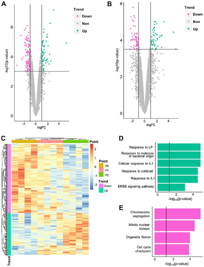
Molecular Medicine REPORTS 22: 2043-2051, 2020                                                          2045

Figure 1. Flowchart of the present study. Top left: Cell culture procedures. Top Right: Expression quantification. Bottom right: Bioinformatic analysis. Bottom
left: Conclusion.

filtered using the following thresholds: i) Average background                   Cluster analysis. Hierarchical cluster analysis based on
value
2046                     SHI et al: STRESS RESPONSE IN BISPHOSPHONATE-RELATED OSTEONECROSIS OF THE JAW

Figure 2. In vitro characteristics of cultured PDLSCs. Scale bars=200 µm. (A) PDLSCs grew around periodontal ligament tissues five days after incubation.
(B) PDLSCs reached confluence seven days after incubation. (C) Entire plate view of colony‑forming PDLSCs, which were plated at a low density and
cultured for 7 days before staining with crystal violet. (D) Cell clusters derived from PDLSCs with typical fibroblast‑like morphology seven days after
incubation. (E) Alizarin Red‑S staining 14 days after incubation. (F) Oil Red O staining four weeks after incubation. PDLSC, periodontal ligament stem cell.

physiology of PDL tissues. Cells were found growing around                      P1 and P0 samples (Fig. 3B‑D). It was hypothesized that these
the PDL tissues 5‑10 days after initial incubation (Fig. 2A) and                genes required a long period of BP exposure to induce signifi-
proliferated to reach 90% confluence in 7‑10 days (Fig. 2B).                    cant alteration. The DEGs between P2 and P0 were referred
Similar to other mesenchymal stem cells, PDLSCs formed                          to as slow‑response DEGs. One of these DEGs, COL1A1
adherent clonogenic cell clusters of fibroblast‑like cells                      (logFC, ‑2.89; P=0.007), was highlighted in a previous genetic
(Fig. 2C and D). The multipotent capacity of PDLSCs was                         association study, which reported that COL1A1 polymorphism
verified by induction in the osteogenic and adipogenic media                    increased the risk of BRONJ (22).
in vitro. Alizarin Red staining (Fig. 2E) revealed a number                         Similarly, expression level data was compared between
of calcified nodules in the cultures after 14 days of induction.                P2 and P1, and P1 and P0. It was hypothesized that such
After 4 weeks of culture in adipogenic medium, intracellular                    comparisons would identify genes that were altered rapidly
lipid vacuoles appeared in PDLSCs, the presence of which                        following BP exposure; hence, these DEGs between adjacent
was confirmed by Oil Red O staining (Fig. 2F).                                  time points were referred to as fast‑response DEGs. A total of
                                                                                264 fast‑response DEGs, including 168 DEGs for P1 vs. P0
Quantification of the PDLSC transcriptome via gene expression                   (Fig. 4A; Table SIII) and 105 DEGs for P2 vs. P1 (9 overlap-
level array. In order to evaluate the impact of BP on PDLSCs,                   ping genes; Fig. 4B; Table SIV), were identified. The majority
genome‑wide transcription levels of all samples were measured                   (229/264) were also slow‑response DEGs (i.e. differentially
at three time points (days 0, 3 and 5) using the Affymetrix                     expressed between P0/1 and 2). Among all fast‑response
PrimeView Human Gene Expression Array. A total of 49,282                        DEGs, 163 genes were downregulated and 101 were upregu-
probes were detected and mapped to 20,608 genes. Following                      lated over time (Fig. 4C). No DEGs were identified in adjacent
normalization and filtration, the expression levels of 18,384 genes             comparisons of control cells (Table SII), which indicates that
for all samples were obtained. These expression level data                      the fast‑response DEGs were not driven by experimental
enabled further analysis of the biological effects of BP usage.                 procedures. Fast‑response DEGs successfully separated three
                                                                                time points in hierarchical clustering analysis (Fig. 4C).
Genes exhibit different patterns of expression in response to
BP. Having quantified the gene expression levels of PDLSCs                      BP exposure influences stress response and cartilage
at different time points (P0, 1 and 2), gene expression levels                  generation. Having identified genes influenced by BP
which were significantly altered following BP exposure were                     treatment, the biological consequences of BP usage were inves-
identified. First, gene expression levels were compared at                      tigated. GO‑BP analysis was applied separately to upregulated
P2 and P0, which represented the longest exposure to BP. A                      and downregulated DEG lists.
total of 906 genes (352 upregulated and 554 downregulated)                          For slow‑response DEGs, a notable cellular stress reaction
were demonstrated to be significantly differentially expressed                  was observed (Fig. 3C; Table SV). Upregulated slow‑response
between P2 and P0 (Fig. 3A; Table SI). Among these                              DEGs were significantly enriched in response to inflammatory
genes, two were also differentially expressed in control cell                   factors, such as lipopolysaccharide (P‑adjusted=2.27x10 ‑7),
lines (Table SII) and were, therefore, excluded from down-                      ba c t e r i a l m ole cu le s ( P‑ a dj u st e d =2. 69 x10 ‑7 ) a n d
stream analysis. The remaining DEGs could distinguish P2                        interleukin‑1 (P‑adjusted=3.93x10 ‑4). Another inflamma-
from P1 and P0 samples but could not distinguish between                        tion‑associated signaling pathway, ‘ERBB signaling pathway’
Molecular Medicine REPORTS 22: 2043-2051, 2020 2047
Figure 3. Biological significance of slow‑response DEGs. (A) Volcano plot for differential expression analysis at day 5 vs. 0. (B) Hierarchical clustering analysis
for all 12 samples, using all DEGs identified. Gene Ontology‑Biological Process results for (C) upregulated and (D) downregulated DEGs. DEG, differentially
expressed gene; FC, fold change; IL, interleukin; LP, lipopolysaccharide.
(P‑adjusted=7.40x10 ‑4), was also moderately enriched. CYLD lysine 63 deubiquitinase (CYLD) and poly(ADP‑ribose)
Downregulated genes (Fig. 3D; Table SVI) inhibited cartilage polymerase 1 (PARP1) function as hub genes in BP pathology.
generation and were notably enriched in ‘chondrocyte differen- Having identified the key biological processes in BP pathology,
tiation’ (P‑adjusted=3.92x10‑9) and ‘extracellular organization’ it was investigated whether any key genes (hubs) served a
(P‑adjusted=4.38x10 ‑9). ‘Ossification’ (P‑adjusted=1.36x10 ‑5) central role in these processes. Such hubs may serve as poten-
was also significantly enriched by downregulated genes, tial interventional targets to prevent osteonecrosis.
which suggested that it was inhibited during BP treatment. In order to investigate the potential hub genes, a PPI
Cell proliferation was also inhibited, demonstrated by enrich- network of all DEGs was constructed (Fig. 5). It was hypoth-
ment of ‘mitotic nuclear division’ (P‑adjusted=9.98x10 ‑6) and esized that hub genes might act as ‘bridges’ between fast‑ and
‘mesenchymal cell proliferation’ (P‑adjusted=1.12x10‑4). slow‑response DEGs and connect more slow‑response DEGs.
For fast‑response DEGs, different enrichment patterns If such hub genes exist, they may be potential drug targets.
were observed. All enriched pathways were associated with Thus, the number of slow‑response DEGs connected by
proliferation and cell cycle regulation (e.g. ‘chromosome each gene was calculated (Table SVIII). The top gene,
segregation’; P‑adjusted=7.62x10 ‑6; Table VII). For upregu- PARP1, which connected 14 slow‑response DEGs, was an
lated fast‑response DEGs, the induction of cellular stress upregulated slow‑response DEG. PARP1 was connected and
reaction was similar to that of slow‑response DEGs. These positively correlated with caspase‑1 (CASP1), an upregulated
results indicated that stress reaction was induced more rapidly fast‑response DEG that serves a vital role in response to
than inhibition of cartilage generation and ossification. inflammation and stress (23).2048 SHI et al: STRESS RESPONSE IN BISPHOSPHONATE-RELATED OSTEONECROSIS OF THE JAW
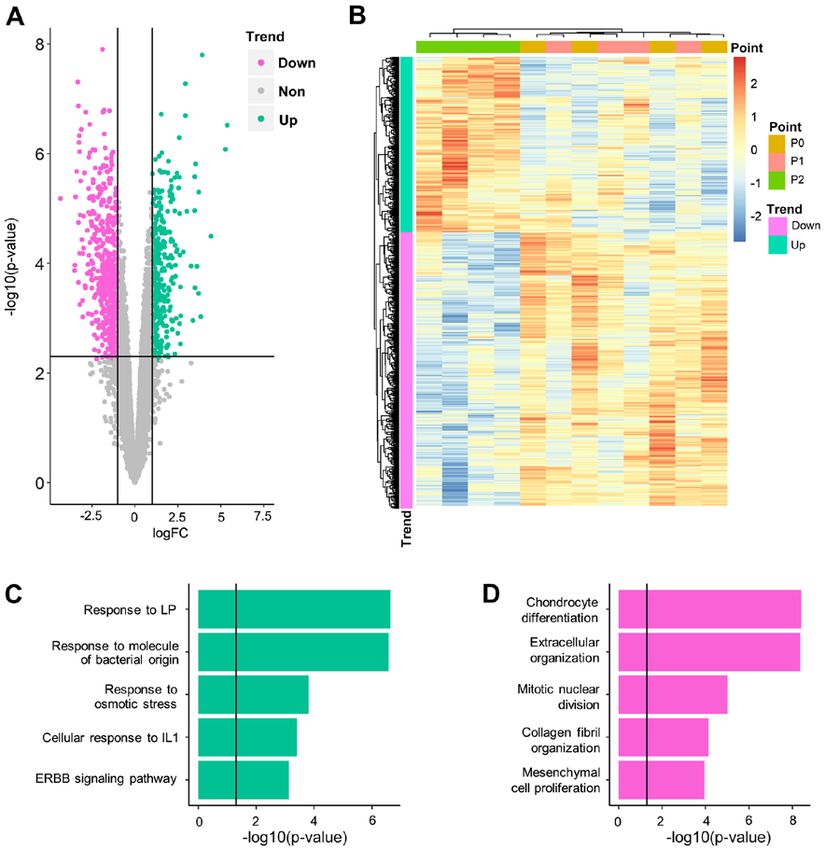
Figure 4. Biological significance of fast‑response differentially expressed genes. Volcano plot for differential expression analysis of (A) day 3 vs. 0 and
(B) day 5 vs. 3. (C) Hierarchical clustering analysis for all 12 samples, using all fast‑response DEG. Gene Ontology‑Biological Process results for (D) upregu-
lated and (E) downregulated DEGs. PDLSC, periodontal ligament stem cell; IL, interleukin; LP, lipopolysaccharide.
PARP1 was connected and negatively correlated with genes and may inhibit downstream extracellular matrix
secreted phosphoprotein 1 (SPP1), a gene involved in the generation of bone.
attachment of osteoclasts to the mineralized bone matrix (24). PARP1 and CYLD were slow‑response DEGs. It was further
The CYLD gene, which connected 13 slow‑response DEGs, determined whether any fast‑response DEGs could serve as
had three PPI partners that are involved in collagen biosyn- hub genes. KRAS and CDK1, which connect 11 slow‑response
thesis [serpin family H member 1 (SERPINH1) (24), filamin A DEGs, were the top genes among fast‑response DEGs. They
(FLNA) (25) and COL1A1]. A negative correlation was both connected a number of genes involved in ‘cell prolif-
observed between CYLD and these collagen‑associated genes, eration’ and ‘cell cycle’ (Table SVI). KRAS was negatively
which indicated that CYLD is upregulated by fast‑response corelated with PPI partners WW and C2 domain containing 1Molecular Medicine REPORTS 22: 2043-2051, 2020 2049
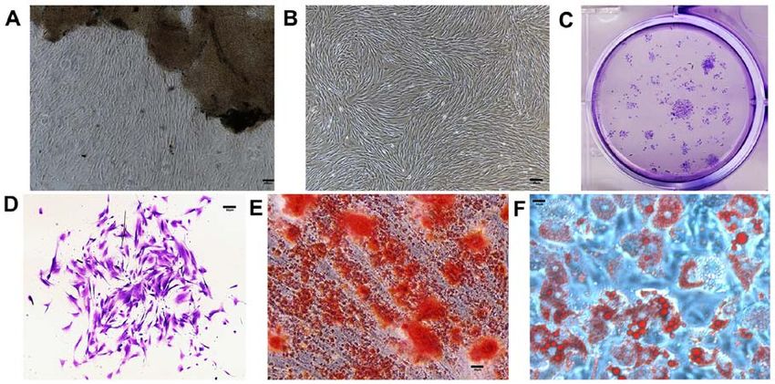
Figure 5. Network analysis of all DEGs. Green represents fast‑response DEGs; purple represents slow‑response DEGs. Each line represents a protein‑protein
interaction connection recorded in Biogrid. Line color represents Pearson correlation coefficients between genes. Key genes (points) and key interactions
(lines) are enlarged. DEG, differentially expressed gene; FLNA, filamin A; CYLD, CYLD lysine 63 deubiquitinase; COL1A1, collagen type I α1 chain;
SERPINH1, serpin family H member 1; PARP1, poly(ADP‑ribose) polymerase 1; SPP1, secreted phosphoprotein 1; CASP1, caspase 1.
(PC, ‑0.70503), kinetochore scaffold 1 (PC, ‑0.56551) and ical research of BRONJ, these hypotheses are treated as being
cell division cycle associated 8 (PC, ‑0.7047), whereas CDK1 exclusive and require validation (31). The present results indi-
exhibited a notable positive correlation with a number of cell cated that these signaling pathways may be different phases of
cycle genes, such as cyclin B1 (PC, 0.922838) and non‑SMC a single uniform pathological procedure and therefore should
condensin I complex subunit G (PC, 0.975156; Table SIX). not be considered separately.
Another debate regarding BRONJ pathology is the
Discussion mutual association between inflammatory damage and
infection. Infection can directly damage the bone structure
Osteonecrosis of the jaw is one of the most severe complica- and exposed bones are further susceptible to bacterial
tions in patients using BP (26). The present study reports the colonization (32). To date, the causative factors from this
results of a genome‑wide landscape of transcriptomic altera- mutual association have not been fully elucidated. Based
tion in PDLSCs during BP usage, which may increase the on the present sterile in vitro experiments, it was observed
understanding of the mechanism of BRONJ and putative drug that inflammation and stress responses can arise in the
targets for BRONJ. absence of infection. Despite being a sterile environment,
With the advantage of time series designation, the present PDLSCs exhibited signs of fighting against infection (e.g.,
analysis demonstrated that genes respond to BP exposure at enrichment in ‘response to molecules of bacterial origin’).
different rates. The majority of BP‑associated genes (676/941) It can be inferred that BP can induce inflammatory reac-
exhibited significant differential expression levels only at tions in PDLSCs in the absence of other causative factors.
day 5 vs. 0, indicating that they responded to BP in a slow and Therefore, it was hypothesized that inflammation and stress
gradual manner. The distinct expression level patterns of fast‑ response are causative factors in the pathology of BRONJ,
and slow‑response genes built a ‘two‑wave’ alteration, which is and prevention of BP‑induced inflammation is a potential
in accordance with the clinical observation that osteonecrosis target for the management of BRONJ.
of the jaw follows a chronic course following BP exposure (27). As the stress response occurs prior to inhibition of ossi-
The two‑wave expression level alteration affected different fication, it was hypothesized that the former process gives
biological functions. As indicated by the GO‑BP analysis, rise to the latter via key intermediate molecules, namely
fast‑response DEGs were notably enriched in inflammatory hubs. PPI network analysis identified two potential hub genes:
and stress responses, as well as in cell proliferation signaling PARP1 and CYLD. PARP1 encodes a chromatin‑associated
pathways. However, slow‑response DEGs exhibited enrichment enzyme, poly(ADP‑ribosyl)transferase (33), and acts as a
in ossification and cartilage‑associated signaling pathways. As bridge between fast‑response inflammation and slow‑response
reported by Aghaloo et al (27) and Lorenzo‑Pouso et al (28), ossification inhibition by interacting with CASP1 and SPP1,
inflammation and inhibition of bone remodeling have been respectively (23,24). This bridge‑like structure suggests that
suggested as putative mechanisms of BRONJ. While isolated PARP1‑based intervention may have a decoupling effect on
evidence of resorption‑generation imbalance (29), periodontal the inflammation‑ossification inhibition cascade. Another
disease (7) and infection (30) has been discovered in patholog- potential hub, CYLD, encodes a cytoplasmic protein with2050 SHI et al: STRESS RESPONSE IN BISPHOSPHONATE-RELATED OSTEONECROSIS OF THE JAW
three cytoskeletal‑associated protein‑glycine‑conserved Ethics approval and consent to participate
domains that functions as a deubiquitinating enzyme (34).
Previous studies have linked CYLD with extracellular matrix The experimental protocols were approved by the ethics committee
generation (35) and bone formation/resorption balance (36). In of Ninth Peoples Hospital affiliated to Shanghai Jiao Tong
PDLSCs, CYLD was observed to interact with slow‑response University, School of Medicine (approval no. SH9H‑2020‑T37‑2).
DEGs (SERPINH1, FLNA and COL1A1) that are involved in Written informed consent was obtained from the participants
collagen biogenesis, a key process in extracellular genera- prior to participation in the study. Informed consent was obtained
tion (37) and bone formation (38). Although no evidence was from the parents of the one patient who was a minor.
found to support the association between CYLD and inflam-
mation in PDLSCs, the strong connection between CYLD and Patient consent for publication
collagen function make it a putative hub gene in the network of
BP pathology. As for the fast‑response gene, the upregulation Not applicable.
of KRAS and downregulation of CDK1 supported the hypoth-
esis that cell proliferation was inhibited by BP usage. KRAS Competing interests
and CDK1 may be key drivers of such inhibition.
The present study has certain limitations. The duration The authors declare that they have no competing interests.
of BP treatment was relatively short, and all PDLSC samples
were collected from patients agedMolecular Medicine REPORTS 22: 2043-2051, 2020 2051
15. Ritchie ME, Phipson B, Wu D, Hu Y, Law CW, Shi W and 27. Aghaloo T, Hazboun R and Tetradis S: Pathophysiology of osteo-
Smyth GK: limma powers differential expression analyses for necrosis of the jaws. Oral Maxillofac Surg Clin North Am 27:
RNA‑sequencing and microarray studies. Nucleic Acids Res 43: 489‑496, 2015.
e47, 2015. 28. Lorenzo‑Pouso AI, Pérez‑Sayáns M, González‑Palanca S,
16. R Core Team: R: A language and environment for statistical Chamorro‑Petronacci C, Bagán J and García‑García A:
computing. R Foundation for Statistical Computing, Vienna, Biomarkers to predict the onset of biphosphonate‑related osteo-
Austria, 2019. necrosis of the jaw: A systematic review. Med Oral Patol Oral Cir
17. Raivo Kolde: Pheatmap: Pretty Heatmaps. R package Bucal 24: e26‑e36, 2019.
version 1.0.8. https://CRAN.R-project.org/package=pheatmap. 29. Coleman RE, Major P, Lipton A, Brown JE, Lee KA, Smith M,
2018. Saad F, Zheng M, Hei YJ, Seaman J and Cook R: Predictive value
18. Ashburner M, Ball CA, Blake JA, Botstein D, Butler H, of bone resorption and formation markers in cancer patients with
Cherry JM, Davis AP, Dolinski K, Dwight SS, Eppig JT, et al: bone metastases receiving the bisphosphonate zoledronic acid.
Gene ontology: Tool for the unification of biology. Nat Genet 25: J Clin Oncol 23: 4925‑4935, 2005.
25‑29, 2000. 30. Ruggiero SL, Dodson TB, Fantasia J, Goodday R, Aghaloo T,
19. Yu G, Wang LG, Han Y and He QY: clusterProfiler: An R Mehrotra B and O'Ryan F; American Association of Oral and
package for comparing biological themes among gene clusters. Maxillofacial Surgeons: American association of oral and
OMICS 16: 284‑287, 2012. maxillofacial surgeons position paper on medication‑related
20. Chatr‑aryamontri A, Oughtred R, Boucher L, Rust J, Chang C, osteonecrosis of the jaw‑2014 update. J Oral Maxillofac Surg 72:
Kolas NK, O'Donnell L, Oster S, Theesfeld C, Sellam A, et al: 1938‑1956, 2014.
The BioGRID interaction database: 2017 update. Nucleic Acids 31. Allen MR and Burr DB: The pathogenesis of bisphos-
Res 45: D369‑D379, 2017. phonate‑related osteonecrosis of the jaw: So many hypotheses,
21. Shannon P, Markiel A, Ozier O, Baliga NS, Wang JT, Ramage D, so few data. J Oral Maxillofac Surg 67 (Suppl 5): S61‑S70, 2009.
Amin N, Schwikowski B and Ideker T: Cytoscape: A software 32. Reid IR and Cornish J: Epidemiology and pathogenesis of osteo-
environment for integrated models of biomolecular interaction necrosis of the jaw. Nat Rev Rheumatol 8: 90‑96, 2012.
networks. Genome Res 13: 2498‑2504, 2003. 33. Zhu X, Ma X and Hu Y: PARP1: A promising target for the devel-
22. Katz J, Gong Y, Salmasinia D, Hou W, Burkley B, Ferreira P, opment of PARP1‑based candidates for anticancer intervention.
Casanova O, Langaee TY and Moreb JS: Genetic polymorphisms Curr Med Chem 23: 1756‑1774, 2016.
and other risk factors associated with bisphosphonate induced osteo- 34. Mathis BJ, Lai Y, Qu C, Janicki JS and Cui T: CYLD‑mediated
necrosis of the jaw. Int J Oral Maxillofac Surg 40: 605‑611, 2011. signaling and diseases. Curr Drug Targets 16: 284‑294, 2015.
23. Franchi L, Eigenbrod T, Muñoz‑Planillo R and Nuñez G: The 35. Wang H, Lai Y, Mathis BJ, Wang W, Li S, Qu C, Li B, Shao L,
inflammasome: A caspase‑1‑activation platform that regulates Song H, Janicki JS, et al: Deubiquitinating enzyme CYLD
immune responses and disease pathogenesis. Nat Immunol 10: mediates pressure overload‑induced cardiac maladaptive
241‑247, 2009. remodeling and dysfunction via downregulating Nrf2. J Mol Cell
24. Chellaiah MA, Kizer N, Biswas R, Alvarez U, Strauss‑Schoenberger J, Cardiol 84: 143‑153, 2015.
Rifas L, Rittling SR, Denhardt DT and Hruska KA: Osteopontin 36. Vriend J and Reiter RJ: Melatonin, bone regulation and the ubiq-
deficiency produces osteoclast dysfunction due to reduced CD44 uitin‑proteasome connection: A review. Life Sci 145: 152‑160, 2016.
surface expression. Mol Biol Cell 14: 173‑189, 2003. 37. Rodriguez‑Pascual F and Slatter DA: Collagen cross‑linking:
25. Falet H, Pollitt AY, Begonja AJ, Weber SE, Duerschmied D, Insights on the evolution of metazoan extracellular matrix. Sci
Wagner DD, Watson SP and Hartwig JH: A novel interaction Rep 6: 37374, 2016.
between FlnA and Syk regulates platelet ITAM‑mediated 38. Saito M and Marumo K: Effects of collagen crosslinking on bone
receptor signaling and function. J Exp Med 207: 1967‑1979, 2010. material properties in health and disease. Calcif Tissue Int 97:
26. Khosla S, Burr D, Cauley J, Dempster DW, Ebeling PR, 242‑261, 2015.
Felsenberg D, Gagel RF, Gilsanz V, Guise T, Koka S, et al:
Bisphosphonate‑associated osteonecrosis of the jaw: Report This work is licensed under a Creative Commons
of a task force of the American Society for bone and mineral Attribution-NonCommercial-NoDerivatives 4.0
research. J Bone Miner Res 22: 1479‑1491, 2007. International (CC BY-NC-ND 4.0) License.You can also read