Structural Stabilization of Human Transthyretin by Centella asiatica (L.) Urban Extract: Implications for TTR Amyloidosis - MDPI
←
→
Page content transcription
If your browser does not render page correctly, please read the page content below
biomolecules

Article
Structural Stabilization of Human Transthyretin by
Centella asiatica (L.) Urban Extract: Implications for
TTR Amyloidosis
Fredrick Nwude Eze , Ladda Leelawatwattana and Porntip Prapunpoj *
 Department of Biochemistry, Faculty of Science, Prince of Songkla University, Hat Yai, Songkhla 90112, Thailand;
 fredrickeze@rocketmail.com (F.N.E.); ladda.l@psu.ac.th (L.L.)
 * Correspondence: porntip.p@psu.ac.th; Tel.: +66-74-288275

 Received: 4 February 2019; Accepted: 21 March 2019; Published: 29 March 2019

 Abstract: Transthyretin is responsible for a series of highly progressive, degenerative, debilitating, and
 incurable protein misfolding disorders known as transthyretin (TTR) amyloidosis. Since dissociation
 of the homotetrameric protein to its monomers is crucial in its amyloidogenesis, stabilizing the native
 tetramer from dissociating using small-molecule ligands has proven a viable therapeutic strategy.
 The objective of this study was to determine the potential role of the medicinal herb Centella asiatica
 on human transthyretin (huTTR) amyloidogenesis. Thus, we investigated the stability of huTTR with
 or without a hydrophilic fraction of C. asiatica (CAB) against acid/urea-mediated denaturation. We
 also determined the influence of CAB on huTTR fibrillation using transmission electron microscopy.
 The potential binding interactions between CAB and huTTR was ascertained by nitroblue tetrazolium
 redox-cycling and 8-anilino-1-naphthalene sulfonic acid displacement assays. Additionally, the
 chemical profile of CAB was determined by liquid chromatography quadruple time-of-flight mass
 spectrometry (HPLC-QTOF-MS). Our results strongly suggest that CAB bound to and preserved
 the quaternary structure of huTTR in vitro. CAB also prevented transthyretin fibrillation, although
 aggregate formation was unmitigated. These effects could be attributable to the presence of phenolics
 and terpenoids in CAB. Our findings suggest that C. asiatica contains pharmaceutically relevant
 bioactive compounds which could be exploited for therapeutic development against TTR amyloidosis.

 Keywords: transthyretin; TTR amyloidosis; protein misfolding; neuroprotection; Centella asiatica;
 antioxidants; triterpenoids; phenolics; HPLC-MS

1. Introduction
      Misfolding and/or aggregation of proteins is at the heart of the etiopathogenesis of a group of
debilitating disorders referred to as amyloidosis [1]. Many of these diseases such as Parkinson’s
disease, Huntington’s, Alzheimer’s, and transthyretin (TTR) amyloidosis are still incurable. As a result,
a lot of investigative effort is focused on developing effective, safe, and reliable therapeutic agents.
      In humans, TTR amyloidosis is a group of proteinopathies triggered by partial unfolding,
misfolding, aggregation, and accumulation of TTR into a spectrum of cytotoxic aggregates [2].
Under normal physiological circumstances, TTR is a homotetrameric protein that transports thyroid
hormones and retinol A via its interaction with a holo-retinol-binding protein [3]. In mature adults, a
late-onset acquired TTR amyloidosis also known as senile systemic amyloidosis (SSA) is caused by the
accumulation of wild-type in organs. SSA affects at least 25% of octogenarians, accounts for more than
10% of heart failure with preserved fraction [4], and is associated with the development of congestive
heart failure [5,6]. Accumulation of variant TTR precipitates familial TTR amyloidosis, such as familial
amyloidosis cardiomyopathy (TTR-FAC) [5], familial amyloidotic polyneuropathy (TTR-FAP), and

Biomolecules 2019, 9, 128; doi:10.3390/biom9040128                            www.mdpi.com/journal/biomolecules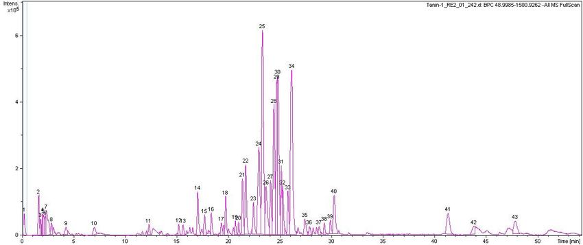
Biomolecules 2019, 9, 128                                                                             2 of 18

central nervous system selective amyloidosis (CNSA) [7]. Onset for familial TTR amyloidosis could
occur within the second decade of life depending on the TTR mutant involved. TTR amyloidosis
can be very progressive and debilitating with mortality within the first 10 to 15 years of onset [8].
Orthotopic liver transplantation, with several limitations including highly invasive, unsuitability of
patients, lack of donors, etc., is still the primary form of disease-modifying therapy for most TTR-FAP
patients. Recently, the disease-modifying drug, Tafamidis, was approved for treating TTR-FAP [9].
However, in a just-concluded long-term study, it was observed that the slow progression of neuropathy
in TTR-FAP patients could not be prevented by Tafamidis [10]. Thus, it is pertinent to search for safe,
more effective, and less invasive therapeutic alternatives.
      Although the current understanding of the molecular pathophysiology underlying TTR
amyloidosis is still incomplete, it is widely regarded that tetramer dissociation into monomers is the
initial and consequential step in its amyloidogenesis [11,12]. Thus, preventing tetramer dissociation by
enhancing its kinetic stability has been proposed as an effective therapeutic strategy [8]. The kinetic
stabilization strategy involves the use of small-molecule ligands to prevent conformational excursions
of the native tetramer by binding to its thyroxine (T4)-binding sites [8]. Kinetic stabilization of TTR
tetramers formed the rational basis for the development of the TTR-FAP drug, Tafamidis [13].
      Native wild-type TTR, while significantly stable under normal physiological conditions [14], is
susceptible to oxidative modifications, which could alter its conformation and stability [15]. The role
of protein oxidation was noted in the increased amyloidogenicity of nitric oxide-modified TTR but not
in unmodified TTR [15]. Also, age-associated oxidation of TTR reportedly impacts the onset of senile
systemic amyloidosis [16]. Furthermore, while prefibrillar and mature TTR amyloid fibrils were not
or less cytotoxic, all forms of oxidized native TTR were shown to be cytotoxic [2,16]. This backdrop
highlights the role of oxidative damage in TTR amyloidogenesis and prompted the notion that, in
addition to kinetic stabilizers, antioxidant treatment could be considered as an alternative/supplement
therapy. Interestingly, most of the natural bioactives that have been reported to stabilize TTR such as
quercetin, epigallocatechin gallate (EGCG), gallic acid, curcumin, and propolis extract are also potent
antioxidants [17,18].
      In addition, it has been noted that the protective effect exerted by antioxidants in chronic and
neurodegenerative diseases in vivo is derived mainly from the additive, synergistic, and complementary
effects of the several bioactive constituents present in the phytocomplex, such as plant extracts, fruits,
and vegetables, rather than individual phytocomponents [19]. The antioxidant activity of medicinal
plant extracts was also found to strongly correlate with their ability to limit the assembly of beta-amyloid,
which is central to Alzheimer’s disease pathology [20]. For a multifactorial disease with a complex
pathology such as TTR amyloidosis, plant extracts or products could provide potential multi-target
therapeutic agents.
      Centella asiatica is a small, succulent, and herbaceous plant popular in several parts of the world for
its medicinal and culinary value. It is indigenous to the tropical and subtropical regions of Asia, Africa,
and the southern parts of the USA. In Thailand, it is known locally as Bua-bok and is used for making a
very popular herbal drink, Nam Bai Bua Bok. The fresh aerial parts are eaten with rice and are part of
many local food recipes. In folk medicine, C. asiatica is a popular nervine and adaptogen. It is used for
wound healing, treatment of neurological disorders, and promoting general wellbeing [21]. The major
bioactive components of C. asiatica are a group of pentacyclic triterpenoids known as centellosides. In
addition, C. asiatica is richly endowed with phenolics and flavonoids [22]. C. asiatica reportedly has
many pharmacological and biological effects including antioxidant, anti-inflammatory, inhibition of
β amyloid peptide aggregation and toxicity, and anti-α-synuclein aggregation [23,24]. Despite these
reports, currently, there is no data on its potential pharmacological effect on TTR amyloidogenesis.
Together with its rich phytochemical and good safety profile, we decided to investigate the potential
role of C. asiatica in the modulation huTTR amyloidogenesis.
      Thus, we examined the impact of a hydrophilic fraction of C. asiatica (CAB) on native huTTR
structural stability and fibril formation using urea/acid-mediated denaturation assays, and transmission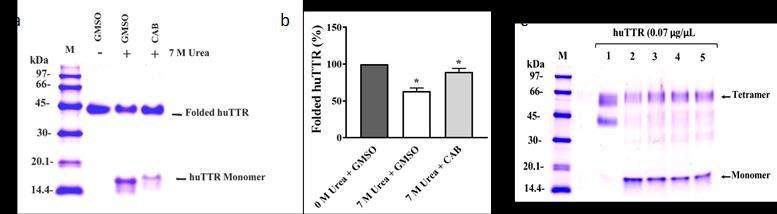
Biomolecules 2019, 9, 128                                                                            3 of 18

electron microscopy, respectively. We also determined the plausible binding interactions of CAB-huTTR
complex and the chemical properties CAB. The present results provide relevant insight into the
neuroprotective potential of C. asiatica, specifically, with regards to TTR amyloidosis.

2. Materials and Methods

2.1. Purification of huTTR from Plasma
     Human TTR from plasma was isolated in a two-step protocol which consisted of a decrease
in albumin burden and followed by preparative discontinuous native-PAGE as previously
described [25,26]. The eluting fractions were monitored for the presence of huTTR by 10% native-PAGE.
Protein bands were detected by silver staining. Fractions containing only huTTR were pooled,
concentrated, and checked for purity by SDS-PAGE with Coomassie blue R-250 staining. The
concentration of purified huTTR was determined by the Bradford assay [27] and stored at −20 ◦ C.

2.2. Preparation of CAB
      C. asiatica was obtained locally in Hat Yai city, Southern Thailand. The identity of the whole
plant specimen was authenticated by Associate Professor Dr. Kitichate Sridith, Curator-in-Chief of the
National Herbarium at Prince of Songkla University, Hat Yai, Thailand. A specimen was deposited in
the herbarium with voucher number F.N.1 (PSU). Plant aerial parts were repeatedly washed with tap
water followed by reverse osmosis water. The plant sample was air-dried for 12 h to reduce moisture
content and then oven-dried at 60 ◦ C for another 12 h. Dried C. asiatica was ground into a fine powder
and stored in an opaque container at −20 ◦ C for extraction within 24 h.
      C. asiatica powder (450 g) was extracted with 2 L of cold acetone/methanol/water (2:2:1 v/v/v)
containing 0.5% glacial acetic acid for 2 h as previously reported [28]. The plant mixture was filtered
through a Buchner funnel overlaid with filter paper (Whatman No. 1) using a vacuum pump. The
filtrate was concentrated to one-third of its original volume by a rotary evaporator at a temperature of
40 ◦ C. Thereafter, the filtrate was defatted by partitioning in an equal volume of n-hexane twice, using
a separatory funnel. The lower phase was further partitioned in dichloromethane twice to remove
chlorophyll, waxes, and other less polar components. The upper hydrophilic phase in the separatory
funnel was collected and residual organic solvents were removed under vacuum by speed-vac to
remove residual organic solvents, and then lyophilized to a hygroscopic powder. This powder obtained
from the hydrophilic fraction of C. asiatica was hereafter referred to as CAB (C. asiatica bioactives). CAB
was aliquoted into opaque vials and stored at −20 ◦ C. The scheme for CAB preparation is shown in
Figure S2.

2.3. Nitroblue Tetrazolium (NBT) Redox-Cycling Assay
      The binding of CAB to huTTR was determined by NBT staining which distinguishes
quinone-modified from unmodified proteins [29]. Human TTR (2.1 µg/µL) in 50 mM Tris-HCl
pH 7.5 was incubated in the presence of CAB or gallic acid (GA), or DMSO (vehicle), at 10× the molar
equivalent of human TTR concentration. The samples were mixed with sample buffer containing 4%
SDS and immediately boiled for 10 min prior to separation by SDS-PAGE (15% resolving gel). The
gel was electrotransferred onto a nitrocellulose membrane. After transfer, the membrane was stained
with Ponceau S dye (0.1% Ponceau S in 5% acetic acid) for 1 h to confirm blotting. Subsequently, the
membrane was washed with distilled water, Tris-buffered saline with Tween 20 (TBS-T) and rinsed
with Milli-Q water to remove the Ponceau S stain. Then, it was re-stained with glycinate/NBT solution
(10 mg NBT tablet in 14 mL of 2 M potassium glycinate buffer, pH 10) for 45 min to identify proteins
that interacted with phenolics or related compounds.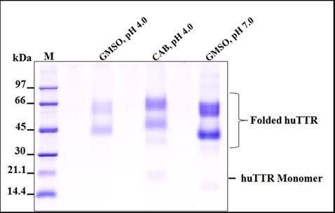
Biomolecules 2019, 9, 128                                                                        4 of 18

2.4. Determination of the Stability of huTTR in the Presence of CAB
     TTR tetramer dissociation into monomeric subunits is quite slow under normal physiological
pH. However, dissociation of TTR tetramer and subsequent misfolding of monomers is significantly
increased in vitro under conditions of high urea concentration or mild acidity [14]. Resistance to
urea-induced or acid-induced dissociation of TTR tetramer to monomers provides insight into the
stability of native TTR against the partial denaturation of monomers required for amyloidogenesis. This
is because tetramer dissociation precedes monomer unfolding and misfolding [30]. Purified huTTR
in 50 mM Tris-HCl buffer pH 7.5 was dialyzed against 10 mM sodium phosphate buffer containing
100 mM KCl and 1 mM ethylenediamine tetraacetic acid (EDTA), pH 7.4 (GF buffer) for 24 h with 3
buffer changes prior to stability assays as follows.

2.4.1. Against Urea-Mediated Denaturation
     The influence of CAB on huTTR stability against urea-mediated dissociation was evaluated as
previously described [31]. In brief, huTTR (1 µg) in a GF buffer, pH 7.4 was supplemented with
CAB (100× molar equivalents with respect to huTTR) dissolved in GF buffer containing DMSO, 50:50
v/v (GMSO) or GMSO only, and incubated for 2 h at 37 ◦ C. Denaturation was triggered by adding
urea (in GF buffer containing 1 mM 1,4-Dithiothreitol, pH 7.4) into the protein complex to a final
concentration of 7 M and subsequently incubating for 96 h at ~6 ◦ C in the dark. Folded huTTR (i.e.,
tetramers, trimers, and dimers) that remained after the denaturation was determined by 14% Tricine
SDS-PAGE. The running buffer contained 0.025% SDS and the sample loading buffer 0.2% SDS. It has
been suggested that these detergent concentrations are enough to prevent monomer reassociation,
but too low to trigger tetramer dissociation [32]. Resolved proteins were identified by staining with
Coomassie brilliant blue R-250 dye (0.1%) solution. Bands representing folded huTTR on the gel were
quantified by densitometry using gel documentation on LabWorks 4.0 software (UVP Ltd., Cambridge,
UK). The relative percentage of folded huTTR that remained after denaturation was calculated as an
indication of native huTTR stability.

2.4.2. Against Acid-Mediated Denaturation
      The effect of CAB on the stability of huTTR under mildly acidic conditions was performed as
described previously [33]. Briefly, huTTR (0.5 µg/uL) in a GF buffer, pH 7.4 was supplemented with or
without 100× molar excess of CAB followed by incubation for 2 h at 37 ◦ C. Tetramer dissociation was
initiated by reducing the pH of the reaction mixture to 4.0 by adding an equal volume of acetate/acetic
acid buffer containing 100 mM KCl, 1 mM EDTA, and 2 mM dithiothreitol (DTT), pH 4.0. Thereafter,
the protein complex was incubated at 37 ◦ C for 14 days in the dark and under aseptic condition.
Glutaraldehyde cross-linking assay was performed to determine the amount and quaternary structure
of huTTR that remained after 14 days of denaturation stress. Briefly, glutaraldehyde was added to the
protein complex to a final concentration of 2.5%, and the assay mixture was incubated for 4 min at room
temperature. Cross-linking was terminated by the addition of NaBH4 (7% in 0.1 M NaOH) at an equal
volume to glutaraldehyde. Then, the mixture was mixed with 4x loading buffer containing 8% SDS
and 5% beta-mercaptoethanol and boiled for 10 min prior to analysis by 10% Tricine SDS-PAGE. The
separated protein bands were visualized by Coomassie blue staining. The intensity of the tetrameric
form of huTTR was quantified by densitometry using gel documentation. The stability of huTTR was
inferred from the relative percentage of tetramer that remained after denaturation.

2.5. Determination of 8,1-ANS Binding Displacement from huTTR by CAB
     Fluorophore, 8-anilino-1-naphthalene sulfonic acid (8,1-ANS), reportedly binds at the T4-binding
site of native huTTR in solution [34]. Thus, its displacement by small-molecule ligands is often
used to probe for ligand binding to the T4-binding site of TTR tetramer [33]. To perform 8,1-ANS
binding-displacement assay, huTTR (0.055 µg/µL) was incubated in the presence or absence of 8,1-ANS,
Biomolecules 2019, 9, 128 5 of 18
(10 µM) for 10 min, followed by the addition of CAB at 25× and 50× molar equivalents with respect
to human TTR. To measure fluorescence, protein samples were excited at 385/40 and emission was
collected at 480/20 using a Synergy HT (Bio-Tek Instruments, Winooski, VT, USA) microplate reader.
2.6. Determination of the Influence of CAB on huTTR Fibrillation by Transmission Electron Microscopy (TEM)
TEM was used to study the impact of CAB on huTTR fibril formation as previously described [35].
Human TTR (1 µg/µL) in a GF buffer was supplemented with or without CAB (20× molar equivalents
with respect to huTTR). The mixture was subsequently incubated for 2 h at 37 ◦ C to facilitate binding.
Fibril formation was initiated by reducing pH to 4.0, via the addition of an equal volume of acetate
buffer (pH, 4.0) to the mixture, and followed by incubation at 37 ◦ C for 7 days. The extent of aggregate
and fibril formation was ascertained by TEM of the TTR mixtures. In brief, aliquots of the TTR mixtures
were diluted with Milli-Q water (1:20, v/v). A drop (~10 µL) of the solution was applied onto a 400-mesh
formvar-coated copper grid for 1 min. Excess sample was removed with a wedge of filter paper. The
grid was immediately rinsed with a drop of Milli-Q water, dried, and negatively stained with 2%
uranyl acetate in 70% methanol for 2 min. Excess stain was blotted out and the grid was thoroughly
air-dried. TEM images were obtained with a JEM-2010 (JEOL Ltd., Akishima, Tokyo, Japan) electron
microscope, running at 160 KV.
2.7. Determination of CAB Antioxidant Properties
2.7.1. DPPH (1,1-Diphenyl-2-picrylhydrazyl) Radical Scavenging Activity
CAB was tested for its scavenging effect on DPPH radical following the method as described [36].
CAB was prepared in 50% aqueous methanol while Trolox (purchased from Aldrich Chemical Company,
Steinheim, Germany) was prepared in distilled water and served as an antioxidant standard. CAB or
Trolox solution (10 µL) was diluted with methanol (140 µL), and then DPPH solution in methanol (150
µL, 0.1 mM) was added. The reaction mixture in which CAB was replaced with aqueous methanol and
Trolox was replaced with distilled water were included as controls. Blank probes contained methanol
(290 µL) and 10 µL of CAB or Trolox. Blank probes for controls contained only methanol (300 µL).
Sample and control reactions were incubated for 30 min before reading absorbance at 515 nm. After
blank correction, DPPH radical scavenging activity (%) was obtained from the equation:
inhibition (%) = (absorbance of control − absorbance of sample)/absorbance of sample × 100 (1)
A curve was plotted for inhibition versus concentration. The antioxidant activity of CAB and
Trolox was obtained from the curve and expressed as the concentration that scavenged the DPPH
radical by 50% (IC50 ) in a mean ± standard deviation of triplicates determination.
2.7.2. Ferric Reducing Antioxidant Power (FRAP)
The reducing power of CAB was determined by FRAP assay as described by [37]. FRAP solution
containing 300 mM acetate buffer pH 3.6, 10 mM TPTZ 2,4,6-Tri(2-pyridyl)-s-triazine (TPTZ) solution,
and 20 mM FeCl3 .6H2 O solution (10:1:1, v/v/v) was prepared and incubated for 30 min at 37 ◦ C. CAB
solution was prepared by diluting the powder with 50% aqueous methanol. Trolox (up to 600 µmol/L)
was included as the antioxidant standard for calibration. The assay was performed in a 96-well plate.
Aliquots (10 µL) of CAB solution or Trolox solution were mixed with FRAP solution (200 µL) and
incubated for 30 min at 37 ◦ C. Then, absorbance was monitored at 650 nm. CAB antioxidant activity
was expressed as a micromole of Trolox equivalents per gram of CAB. The higher the FRAP value, the
stronger the reducing potential.Biomolecules 2019, 9, 128 6 of 18
2.8. Determination of the Chemical Composition of CAB
2.8.1. Total Phenolic Content
The phenolic content in CAB was determined using the Folin–Ciocalteu assay with slight
modification [38]. CAB solution (10 µg/µL) and gallic acid (GA; 1 µg/µL) in DMSO/methanol (10:90,
v/v) were prepared in which the latter was used as a standard. Aliquot (100 µL) of CAB solution, blank
or standard solution (from 0 to 80 µg), was added into test tubes. Then, 200 µL of Folin–Ciocalteu
reagent (10%) was added and vortexed. After 5 min of incubation, 800 µL of Na2 CO3 (700 mM) was
added and vortexed. The assay mixture was incubated in the dark at room temperature for 2 h. Then,
200 µL of the blue mixture formed was transferred into a 96-well microplate and absorbance was
measured at 765 nm, using a Synergy HT microplate reader (Bio-Tek Instruments, Winooski, VT, US). A
plot of the amount of gallic acid (µg) versus absorbance at 765 nm was prepared and used to obtain the
total phenolic content of the CAB. Linearity for the gallic acid standard curve for determining phenolic
content was obtained between 0 and 20 µg. Data obtained from quadruplicates were reported as mean
± standard error of the mean and expressed in milligrams of gallic acid equivalent per gram of CAB.
2.8.2. Total Flavonoid Content
Total flavonoid content of CAB was determined by the aluminum chloride colorimetric assay
with slight modifications as previously described [39]. CAB solution (10 µg/µL) was prepared in
50% aqueous methanol, and quercetin standard (1 µg/µL) was prepared in 100% methanol. CAB or
quercetin solution (30 µL) was diluted with 160 µL of methanol and subsequently mixed with 10%
aluminum chloride (30 µL) and 1 M sodium acetate (30 µL) in a total volume of 1100 µL of the assay.
The mixture was vortexed and subsequently incubated in the dark for 30 min. Absorbance was read
at 415 nm. Sample and standard solutions were prepared in quadruplicates. The calibration curve
of the absorbance versus concentration of quercetin standard was plotted and used to calculate total
flavonoid content of which was expressed as the mean ± standard error in milligrams of quercetin
equivalents per gram of CAB.
2.8.3. Thin-Layer Chromatography (TLC) Profile of CAB
To determine the major phytoconstituents present in CAB, TLC was conducted on
aluminum-backed silica gel 60 NH2 F254S TLC plates (Merck Millipore, Darmstadt Germany). CAB
was reconstituted in methanol/water/acetic acid (100:10:4, v/v/v) and 5 µL (~300 µg) of CAB solution
was spotted on a TLC plate, and the plate was transferred to a glass jar lined with filter paper and
saturated for 30 min with developing solvent containing water/acetic acid/formic acid/ethyl acetate
(5:1:1:17, v/v/v/v). The separation was terminated after the solvent front had migrated about 8 cm
from the origin, and the plate was dried up in a fume hood to remove any residual solvent. One plate
was stained with p-Anisaldehyde-sulfuric acid reagent, another was dipped into 3% ferric chloride
reagent, and the third was sprayed with 2% AlCl3 in methanol after saturation with ammonia solution.
All plates were observed under visible light and ultraviolet light (254 and 366 nm) before and after
derivatization, respectively.
2.8.4. High-Performance Liquid Chromatography-Mass Spectrometry (HPLC-MS) Fingerprint of CAB
LC-MS analysis of CAB was performed as previously described [40]. The sample was resuspended
in methanol and filtered through a 0.22 µm nylon membrane. Separation of analyte was performed
with a Poroshell 120 EC-C18 column (4.6 × 150 mm, 2.7 µm), Agilent Technologies. The solvent system
consisted of water (as eluent A) and acetonitrile (as eluent B). Both eluents contained formic acid (0.1%
v/v). The flow rate was maintained at 0.3 mL/min. The gradient employed for the separation was as
follows: 0% B, 0–5 min; then increased linearly to 80% B, 5–50 min; maintained at 80% B, 50–53 min;
then returned to 0% B, 53–55 min; and finally, re-equilibrated in 0% B, 55–60 min. The stream of
separated components was channeled into a micrOTOF-QII™ESI-Qq-TOF mass spectrometer (BrukerBiomolecules 2019, 9, 128 7 of 18
Daltonics, Bremen, Germany) equipped with an electrospray ionization source. Ions were detected in
the negative mode within a mass range of 50–1500 Da. Parameters set for mass spectra acquisition
included a capillary voltage of 3500 V, nebulizer pressure of 2.0 Bar, drying gas of 8.0 L/min, and
drying temperature of 180 ◦ C. Sodium formate solution (10 mM) was used as a calibration standard to
ensure that the mass/charge ratios recorded by the instrument were indeed accurate. Data acquisition
was performed on Bruker Compass DataAnalysis 4.0 software (Bruker Daltonics, Bremen, Germany).
Tentative identity of compounds was established by comparing the accurate mass measurements
obtained with previously identified compounds in C. asiatica reported in literature and online databases
including METLIN and ChemSpider.
2.9. Statistical Analysis
Data of the huTTR stability assays were presented as the mean ± standard error of the mean (SEM).
The percentages of folded huTTR or tetramers preincubated with or without CAB that remained after
denaturation stress were compared using ANOVA followed by the Tukey–Kramer test. Differences
were considered statistically significant at p < 0.05.
3. Results
3.1. CAB Directly Binds to huTTR
To determine whether CAB binds directly to huTTR purified from human plasma (Figure S1),
we used nitroblue tetrazolium (NBT) redox-cycling staining [35]. Some natural product components,
especially phenolics and related compounds, reportedly auto-oxidize into quinones which can interact
with nucleophilic centers in protein such as the sulfhydryl group of a cysteine residue [41]. Under
alkaline pH conditions (for example, in the presence of NBT/potassium-glycinate solution, pH 10),
such quinoprotein adducts formed undergo redox cycling with glycine accompanied by a concomitant
reduction of NBT into a visible blue-purple formazan color on the membrane [29]. This color formation
indicates interaction between the bioactives and protein.
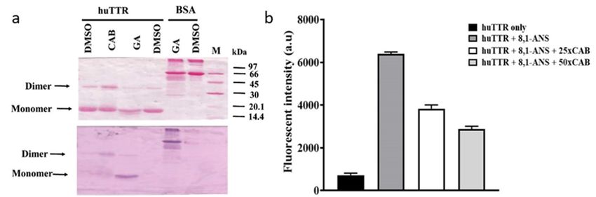
As shown in Figure 1a, Ponceau S staining revealed the presence of huTTR monomers and dimers
based on their positions on the membrane. TTR tetramer normally migrates as an apparent dimer
under non-reduced and non-boiled SDS-PAGE. [42,43]. The membrane was later distained of Ponceau
S. Upon subjecting the membrane to NBT/glycinate staining, purple huTTR bands were detected in the
lanes containing CAB, whereas no color reaction was observed in the absence of CAB (DMSO only)
(Figure 1a). In the presence of gallic acid (positive control), the NBT stain produced a bright formazan
color corresponding to the band position of the huTTR monomer. However, in the presence of CAB,
the formazan color was present at the position representing huTTR dimers and monomers (although,
quite faintly) (Figure 1a). The presence of the formazan color in the lane of CAB-associated huTTR
indicated direct binding interactions. The higher intensity of the formazan color associated with the
dimeric band suggested CAB binding preference for quaternary over tertiary huTTR forms, and that
the CAB–huTTR complex formed is quite stable under reduced SDS-PAGE conditions.bright formazan color corresponding to the band position of the huTTR monomer. However, in the
presence of CAB, the formazan color was present at the position representing huTTR dimers and
monomers (although, quite faintly) (Figure 1a). The presence of the formazan color in the lane of
CAB-associated huTTR indicated direct binding interactions. The higher intensity of the formazan
color associated with the dimeric band suggested CAB binding preference for quaternary over 8 of 18
Biomolecules 2019, 9, 128
tertiary huTTR forms, and that the CAB–huTTR complex formed is quite stable under reduced SDS-
PAGE conditions.
FigureFigure
1. (a)1. Ponceau
(a) Ponceau S stainingofofhuman
S staining human transthyretin
transthyretin (huTTR)
(huTTR) treated or non-treated
treated (vehicle)
or non-treated with with
(vehicle)
bioactives
bioactives afterafter electroblotting
electroblotting ofofSDS-PAGE
SDS-PAGEonto
onto the
the nitrocellulose
nitrocellulose membrane
membrane (above). Protein:
(above). Drug Drug
Protein:
ratio ratio of 1:10. Nitroblue tetrazolium (NBT)/glycinate staining of the membrane above after de-staining
of 1:10. Nitroblue tetrazolium (NBT)/glycinate staining of the membrane above after de-staining
Ponceau S (below). (b) Displacement of bound 8-anilino-1-naphthalene sulfonic acid from huTTR by
increasing concentrations of Centella asiatica (CAB). The bar chart columns and error bars represent the
means of triplicates and standard deviations, respectively.
3.2. CAB Binds to the T4-Binding Sites of huTTR
To further understand the binding interactions between huTTR and CAB, 1,8-ANS binding
displacement assay was performed. The fluorescent dye, 8,1-ANS is very sensitive to its surrounding
chemical environment [44]. In aqueous environments, it produces a weak fluorescence; however,
the fluorescent quantum yield increases significantly with a blue shift in the emission maxima when
the surrounding becomes less polar [44]. ANS binds to the T4-binding sites of TTR tetramers with a
corresponding increase in the magnitude of fluorescent quantum yield and a blue shift from 515 nm
to about 465 nm [34]. Given the hydrophobic nature of the T4-binding pockets, the increase in
fluorescence due to 8,1-ANS binding makes sense. Thus, several studies have utilized the displacement
of 8,1-ANS from the T4-binding site as a probe to determine the binding of small-molecule ligands
to the T4-binding pockets of tetrameric TTR [18,33]. In addition, the ANS fluorescence could serve
as an indicator for the stability and compactness of the TTR core [45]. In this study, the binding of
ANS to huTTR increased the quantum yield (Figure 1b). The addition of CAB reduced the fluorescent
intensity dose-dependently, which suggests the displacement of 8,1-ANS from the T4-binding site
of huTTR tetramers (Figure 1b). These results suggest the binding of CAB at the T4-binding sites of
huTTR tetramers.
3.3. CAB Increases huTTR Structural Stability
To determine the impact of CAB on huTTR kinetic stability in vitro, urea-mediated denaturation
assay was performed. HuTTR was preincubated with or without CAB, and subjected to denaturation
by 7 M urea (final concentration). The intensity of folded huTTR was higher in the presence of CAB than
in its absence (Figure 2a). The percentage of folded CAB-associated huTTR that resisted urea-mediated
dissociation was 89.65 ± 4.6%. In contrast, the percentage of folded huTTR in the absence of CAB
was lower, 58.87 ± 4.37% (Figure 2b). These results demonstrated that CAB improved the stability of
huTTR tetramer against the denaturation stress.assay was performed. HuTTR was preincubated with or without CAB, and subjected to denaturation
by 7 M urea (final concentration). The intensity of folded huTTR was higher in the presence of CAB
than in its absence (Figure 2a). The percentage of folded CAB-associated huTTR that resisted urea-
mediated dissociation was 89.65 ± 4.6%. In contrast, the percentage of folded huTTR in the absence
Biomolecules
of CAB was 2019, 9, 128 58.87 ± 4.37% (Figure 2b). These results demonstrated that CAB improved
lower, 9 of
the18
stability of huTTR tetramer against the denaturation stress.
Figure 2.
Figure (a) Resistance
2. (a) Resistance to
to urea-mediated
urea-mediated denaturation
denaturation ofof huTTR
huTTR in
in the
the presence
presence and
and absence
absence ofof
Centella asiatica bioactive (CAB) components. HuTTR (0.07 µg/µL) was incubated in the presence of
Centella asiatica bioactive (CAB) components. HuTTR (0.07 µ g/µ L) was incubated in the presence of
CAB (6.67 µg/µL) dissolve in GMSO or in the presence of GMSO only. (b) Bar chart represents the
CAB (6.67 µ g/µ L) dissolve in GMSO or in the presence of GMSO only. (b) Bar chart represents the
means of huTTR with and without CAB (protein: CAB mass ratio 1:100) and with their standard errors.
means of huTTR with and without CAB (protein: CAB mass ratio 1:100) and with their standard
* values are significantly different at p < 0.05. (c) Tricine SDS-PAGE gel (10%) of huTTR with increasing
errors. * values are significantly different at p < 0.05. (c) Tricine SDS-PAGE gel (10%) of huTTR with
concentrations of CAB subjected to urea-denaturation stress and cross-linked with glutaraldehyde. M:
increasing concentrations of CAB subjected to urea-denaturation stress and cross-linked with
Protein molecular weight marker; 1: 0 M urea + GMSO only; 2: 7 M urea + GMSO only; 3: 7 M urea +
glutaraldehyde. M: Protein molecular weight marker; 1: 0 M urea + GMSO only; 2: 7 M urea + GMSO
CAB (3.33 µg/µL); 4: 7 M urea + CAB (6.66 µg/µL); 5: 7 M urea + CAB (13.33 µg/µL).
only; 3: 7 M urea + CAB (3.33 µ g/µ L); 4: 7 M urea + CAB (6.66 µ g/µ L); 5: 7 M urea + CAB (13.33 µ g/µ L).
To further understand the potential role of CAB on huTTR amyloidogenesis, we examined the
To further understand the potential role of CAB on huTTR amyloidogenesis, we examined the
quaternary structural changes in CAB-associated huTTR under urea denaturation by glutaraldehyde
quaternary structural changes in CAB-associated huTTR under urea denaturation by glutaraldehyde
cross-linking assay. Human TTR was preincubated without or with varying concentrations of CAB
cross-linking assay. Human TTR was preincubated without or with varying concentrations of CAB
and subjected to denaturation stress. Tricine SDS-PAGE of glutaraldehyde cross-linked huTTR
and subjected to denaturation stress. Tricine SDS-PAGE of glutaraldehyde cross-linked huTTR
samples revealed protein bands of ~60 kDa and ~16 kDa, corresponding to huTTR tetramers and
monomers, respectively (Figure 2c). It is noteworthy that while the intensity of the monomeric
band diminished as the amount of CAB increased, the intensity of huTTR tetrameric band increased
correspondingly (Figure 2c). These findings indicated that CAB increased the quaternary structural
stability of huTTR concentration-dependently.
In vitro, TTR rapidly dissociates when subjected to mildly acidic conditions. This facilitates its
auto-aggregation [11,46]. In order to understand the impact of CAB on the stability of huTTR under
conditions which promote amyloidogenesis, acid-mediated denaturation assay was performed. After
subjecting huTTR samples to denaturation stress, the percentage of huTTR tetramers in the presence
of CAB was higher than in the absence (Figure 3). In the presence of CAB, the percentage of huTTR
tetramers that remained was 68.32 ± 0.81% as opposed to 30.24 ± 0.78% in its absence. These results
indicated that CAB enhanced the quaternary structural stability of huTTR and confirmed the findings
from the urea-mediated denaturation assay.auto-aggregation [11,46]. In order to understand the impact of CAB on the stability of huTTR under
conditions which promote amyloidogenesis, acid-mediated denaturation assay was performed. After
subjecting huTTR samples to denaturation stress, the percentage of huTTR tetramers in the presence
of CAB was higher than in the absence (Figure 3). In the presence of CAB, the percentage of huTTR
tetramers that remained was 68.32 ± 0.81% as opposed to 30.24 ± 0.78% in its absence. These results
Biomolecules 9, 128
2019, that
indicated CAB enhanced the quaternary structural stability of huTTR and confirmed the findings 10 of 18
from the urea-mediated denaturation assay.
(a) (b)
Figure Figure
3. The3.resistance
The resistance of human
of human transthyretin
transthyretin in absence
in the the absence or presence
or presence of CAB
of CAB against
against acid-
acid-mediated
mediated denaturation. HuTTR (0.5 µ g/µ L) was incubated with or
denaturation. HuTTR (0.5 µg/µL) was incubated with or without CAB (50 µg/µL) and subjectedwithout CAB (50 µ g/µ L) and to
subjected to acidic denaturation conditions for 2 weeks. (a) The image represents
acidic denaturation conditions for 2 weeks. (a) The image represents the resolved protein mixtures the resolved protein
mixtures on 10% Tricine SDS-PAGE gels after cross-linking with glutaraldehyde. M: Protein
on 10% Tricine SDS-PAGE gels after cross-linking with glutaraldehyde. M: Protein molecular weight
molecular weight marker; 1: huTTR with GMSO only, pH 4.0; 2: huTTR with CAB, pH 4.0; 3: huTTR
marker; 1: huTTR with GMSO only, pH 4.0; 2: huTTR with CAB, pH 4.0; 3: huTTR with GMSO only,
with GMSO only, pH 7.0. (b) Bar chart represents the extent of huTTR stability, derived from the
pH 7.0. (b) Bar chart
percentage represents
of tetramers theafter
(%) left extent of huTTR* stability,
denaturation. values arederived fromdifferent
significantly the percentage of tetramers
at p < 0.05.
(%) left after denaturation. * values are significantly different at p < 0.05.
3.4. CAB Prevents
Biomolecules 2019,huTTR
9, 128 Fibril Formation 10 of 18
3.4. CAB Prevents huTTR Fibril Formation
The influence
3.6. Chemical of CAB on huTTR
Characterization of CAB fibrillation was determined by examining the morphology of
The influence
the protein of CAB
using on huTTR
transmission fibrillation
electron microscopywas (TEM).
determined
Basedby on examining
the electronthe morphology
micrographs, no of the
protein
huTTR 3.6.1.
using Total fibril
mature Phenolic
transmission wasand Flavonoid
electron
formed in Contents
microscopy
the presence (TEM).
of CAB; Based on the
however, theelectron
formation micrographs, no huTTR
of aggregate was
notfibril
mature impeded
was (Figure in
The formed 4b).
antioxidant theConversely,
presence
activities of many infruits
of the absence
CAB; however,
and ofhave
plants CAB,
thebeennotlargely
only attributed
formation did huTTR
of aggregate transform
to the was notinto
presence impeded
aggregate,
(Figure 4b). but
of Conversely,also produced
phenolics and flavonoids mature
[48]. In of
in the absence fibrils
thisCAB, (Figure
report,not 4c,d).
Folin–Ciocalteu These
only did huTTR findings
and aluminum
transform suggested
chloride that CAB
intocolorimetric
aggregate, but also
prevented
assaysthe
wereformation of mature
used to determine thehuTTR fibrils under
total phenolic moderately
and flavonoid acidic
contents conditions
of CAB, in vitro.
respectively. The The
produced mature fibrils (Figure 4c,d). These findings suggested that CAB prevented the formation of
content
electron of phenolics
micrograph in CAB
of CAB was 43.86
incubated alone ± 0.34
undermgsimilar
of gallic acid equivalent
conditions is shown peringram,
Figurewhile
S4. the
mature huTTR fibrils
flavonoids under
content wasmoderately
13.89 ± 0.23 mgacidic conditions
of quercetin in vitro.
equivalents per gram The electron micrograph of CAB
of CAB.
incubated alone under
3.5. Antioxidant similar
Activity of CABconditions is shown in Figure S4.
3.6.2. TLC Profile of CAB
To determine the antioxidant activity of CAB, DPPH radical scavenging and FRAP assays were
performed. The DPPH assay measures the ability of component(s) to quench the DPPH radical by
transferring H to it. From the DPPH assay, CAB and the antioxidant standard displayed IC50 values
of 28.53 and 2.81 µ g/mL, respectively. While the radical-scavenging effect of CAB was lower than
that of the standard, the abilities of both substances to remove the free radical were of the same
magnitude. The FRAP assay showed that the reducing potential of CAB was 284.17 µ mol Trolox
equivalents per gram of CAB which is considered high [47]. These results demonstrated that CAB
possessed potent antioxidant capacity.
Figure
Figure 4. The 4. The
TEM TEM micrograph
micrograph of huTTRincubated
of huTTR incubated ininthe
thepresence or absence
presence of CAB.of
or absence (a)CAB.
Human (a) Human
transthyretin (TTR) in GF buffer, pH 7.4 supplemented with GMSO and incubated at −20 ◦ C.(b)(b) Human
transthyretin (TTR) in GF buffer, pH 7.4 supplemented with GMSO and incubated at −20 °C.
Human TTR supplemented CAB, final pH 4.0 and incubated at 37 °C for 7 days. (c) Human TTR
TTR supplemented CAB, ◦ C for 7 days. (c) Human TTR supplemented
supplemented withfinal pHpH
GMSO, 4.04.0and
andincubated
incubated atat3737°C for 7 days. (d) Same image as (c) but with
with GMSO, pHmagnification.
higher 4.0 and incubated at 37 ◦ Cmature
Arrows represent for 7 fibrils,
days. filled
(d) Same imagelarge
arrow-heads as (c) but with higher
amorphous
aggregates, and not-filled arrow-heads oligomers.
magnification. Arrows represent mature fibrils, filled arrow-heads large amorphous aggregates, and
not-filled arrow-heads oligomers.
Phytochemical analysis to determine the major chemical profile of CAB was performed by thin
layer chromatography on an aminopropyl-modified silica gel TLC plate. After derivatization with p-
Anisaldehyde-sulfuric acid reagent, emergence of pink, purple, and yellow bands were observed
under white light, indicating the presence of terpenoids and phenolics (Figure S3d). The emergence
of bands that quenched the fluorescence indicator on the TLC plate under ultraviolet light at 254 nm
(Figure S3b), suggested the presence of phenolics. Likewise, the appearance of bright yellow colored
bands under white light, after the TLC plate was saturated with ammonia solution and sprayed with
2% methanolic AlCl3, (Figure S3c) was typical of phenolic acids and flavonoids [49]Biomolecules 2019, 9, 128 11 of 18
3.5. Antioxidant Activity of CAB
To determine the antioxidant activity of CAB, DPPH radical scavenging and FRAP assays were
performed. The DPPH assay measures the ability of component(s) to quench the DPPH radical by
transferring H to it. From the DPPH assay, CAB and the antioxidant standard displayed IC50 values of
28.53 and 2.81 µg/mL, respectively. While the radical-scavenging effect of CAB was lower than that of
the standard, the abilities of both substances to remove the free radical were of the same magnitude.
The FRAP assay showed that the reducing potential of CAB was 284.17 µmol Trolox equivalents per
gram of CAB which is considered high [47]. These results demonstrated that CAB possessed potent
antioxidant capacity.
3.6. Chemical Characterization of CAB
3.6.1. Total Phenolic and Flavonoid Contents
The antioxidant activities of many fruits and plants have been largely attributed to the presence
of phenolics and flavonoids [48]. In this report, Folin–Ciocalteu and aluminum chloride colorimetric
assays were used to determine the total phenolic and flavonoid contents of CAB, respectively. The
content of phenolics in CAB was 43.86 ± 0.34 mg of gallic acid equivalent per gram, while the flavonoids
content was 13.89 ± 0.23 mg of quercetin equivalents per gram of CAB.
3.6.2. TLC Profile of CAB
Phytochemical analysis to determine the major chemical profile of CAB was performed by thin
layer chromatography on an aminopropyl-modified silica gel TLC plate. After derivatization with
p-Anisaldehyde-sulfuric acid reagent, emergence of pink, purple, and yellow bands were observed
under white light, indicating the presence of terpenoids and phenolics (Figure S3d). The emergence of
bands that quenched the fluorescence indicator on the TLC plate under ultraviolet light at 254 nm
(Figure S3b), suggested the presence of phenolics. Likewise, the appearance of bright yellow colored
bands under white light, after the TLC plate was saturated with ammonia solution and sprayed with
2% methanolic AlCl3 , (Figure S3c) was typical of phenolic acids and flavonoids [49].
3.6.3. HPLC-QTOF-MS Analysis of CAB
HPLC-QTOF-MS was used to determine the chemical profile of CAB. The base peak chromatogram
(negative ion mode) is revealed in Figure 5. The tentative identity of the major peaks obtained, retention
times, accurate masses, and predicted molecular formulae are presented in Table 1. Identification of
compounds was accomplished by comparing the accurate masses obtained with those of previously
reported compounds present in C. asiatica found in the literature [40,50] and in online databases including
METLIN and ChemSpider. Eight caffeoylquinic acids (chlorogenic acids) were identified including
3-O-caffeoylquinic acid, 5-O-caffeoylquinic acid, 3,4-O-dicaffeoylquinic acid, 3,5-O-dicaffeoylquinic
acid, 3,5-O-dicaffeoyl-4-malonylquinic, 3-caffeoyl-4-feruloylquinic acid, and 4,5-O-dicaffeoylquinic
acid (Table 1). In addition, seven pentacyclic triterpenoids were present including asiaticoside B,
madecassoside, centellasaponin B, centellasaponin A, asiaticoside, avenacoside A, and soyasaponin I.
The flavonoids quercetin 3-O-glucuronide and eriodictyol 7-(6-galloylglucoside) were also identified
(Table 1). Thus, the prevalent chemical groups in CAB were chlorogenic acids and pentacyclic
triterpenoids. The chemical composition of CAB was similar to those previously reported for C.
asiatica [22,40,50].and 4,5-O-dicaffeoylquinic acid (Table 1). In addition, seven pentacyclic triterpenoids were present
including asiaticoside B, madecassoside, centellasaponin B, centellasaponin A, asiaticoside,
avenacoside A, and soyasaponin I. The flavonoids quercetin 3-O-glucuronide and eriodictyol 7-(6-
galloylglucoside) were also identified (Table 1). Thus, the prevalent chemical groups in CAB were
chlorogenic acids
Biomolecules 2019, and pentacyclic triterpenoids. The chemical composition of CAB was similar
9, 128 12 ofto18
those previously reported for C. asiatica [22,40,50].
Basepeak
Figure5.5.Base
Figure peakchromatogram
chromatogramofofcompounds
compoundsininCAB
CABobtained
obtainedusing
usingHPLC-QTOF-MS
HPLC-QTOF-MSininthe
the
negativemode.
negative mode.
Table 1. Profile of major bioactives detected in CAB by HPLC-QTOF-MS.
Table 1. Profile of major bioactives detected in CAB by HPLC-QTOF-MS.
Retention Observed Accurate Mass Predicted Calculated Tentative Identity of
Peak No. Observed (m/z)
Time Formula Mass (Da) Compound
Peak Retention Predicted Calculated Tentative Identity of
4 2.0 Accurate Mass[M − H]−
503.1527 C21 H28 O14 504.1479 Caffeic acid dihexoside
No. 12 Time 15.1 353.0877 [M − H]− FormulaC16 H18 O9 Mass (Da)
354.0951 Compound
3-O-Caffeoylquinic acid
(m/z) −
14 17.0 353.088 [M − H] C16 H18 O9 354.0951 5-O-Caffeoylquinic acid
21 21.3 693.2790 [M −−H2 O − H]− C34 H48 O16 712.2942 Nominilic acid 17-glucoside
4 22
2.0 21.6
503.1527 [M − H]
477.0685 [M − H]−
C21H28OC14 H O 504.1479
478.0747
Caffeic acid dihexoside
Quercetin 3-O-glucoronide
21 18 13
23 22.4 5151195 [M − H]− C25 H24 O12 516.1268 3,4-O-Dicaffeoylquinic acid
− −
12 24 15.1 22.9 353.0877 [M − H][M − H] C16H18OC925 H24 O12
515.1209 516.1268
354.0951 3,5-O-Dicaffeoylquinic acid
3-O-Caffeoylquinic acid
3,5-O-Dicaffeoyl-4-malonylquinic
25 23.3 601.1226 [M − H]− C28 H26 O15 602.1272
acid
−
14 26 17.0 23.6 353.088 [M − H][M
515.1203 − H]− C16H18OC925 H24 O12 354.0951
516.1268 5-O-Caffeoylquinic acid
4,5-O-Dicaffeoylquinic acid
3,5-O-Dicaffeoyl-4-malonylquinic
27 24.0 693.2790 [M − [M − H]−
601.1209 C28 H26 O15 602.1272
acid isomer
21 21.3 −
C34H48O16 712.2942 Nominilic
Eriodictyol
acid 17-glucoside
28 24.4 H2O −601.1228
H] [M − H] − C28 H26 O15 602.1272
7-(6-galloylglucoside)
24.7 1019.5149 [M +− Formate] −
22 29
21.6 477.0685 [M − H] C21− H18OC1349 H79 O22 478.0747
1019.5063 Asiaticoside B
Quercetin 3-O-glucoronide
30 24.8 1019.5149 [M + Formate] C49 H79 O22 1019.5063 Madecassoside
31 25.1 529.1351 [M − H]− C26 H26 O12 530.1424 3-Caffeoyl-4-feruloylquinic acid
23 32 22.4 25.2 5151195 [M −[M
873.4522 H]+− FormateC−25H24OC1243 H69 O18 516.1268
874.4562 3,4-O-Dicaffeoylquinic
Centellasaponin B acid
H]−
1003.5166 [M + Formate −
25.7 −
24 33 22.9 515.1209 [M − H] H]− C25H24OC1249 H79 O21 1004.5192
516.1268 Centellasaponin A
3,5-O-Dicaffeoylquinic acid
34 26.1 957.5088 [M − H]− C48 H78 O19 958.5137 Asiaticoside
36 27.8 1061.5180 [M − H]−
−
C51 H82 O23 1062.5247 3,5-O-Dicaffeoyl-4-
Avenacoside A
25 39 23.3 29.8 601.1226 [M − H]
987.5213 [M − H] −C 28 H 26 OC15 H O
49 79 20
602.1272
988.5243 Soyasaponin I
40 30.2 571.0881 [M − H2 O − H] − C30 H20 O12 572.0955
malonylquinic
Manniflavanone
acid
26 23.6 515.1203 [M − H]− C25H24O12 516.1268 4,5-O-Dicaffeoylquinic acid
4. Discussion
In this study, we demonstrated that the bioactive compounds in CAB modulated huTTR
amyloidogenesis by binding to the T4-binding sites of the tetramer. This enhanced the quaternary
structural stability of huTTR against denaturation stresses. In addition, CAB prevented the fibrillation
of huTTR under acid-induced aggregation conditions and demonstrated potent antioxidant activity.
Biophysical evidence has shown that dissociation of TTR tetramer is the initial and most critical
step in TTR amyloidogenesis [8,11,12,51–53]. Thus, it is reasoned that enhancing TTR tetramer kinetic
stability would make for an effective therapeutic strategy. Kinetic stabilization of native TTR tetramers
can be accomplished by small-molecule ligands binding to the T4-binding sites of tetrameric TTR, whichBiomolecules 2019, 9, 128 13 of 18
elevates the kinetic barrier for its dissociation [8]. Since TTR amyloidogenesis is a concentration-driven
process, by enhancing tetramer stability, the process is depleted of required amyloidogenic-competent
monomers [54].
Natural products such as curcumin and propolis are now receiving more attention as potential
kinetic stabilizers [18,55]. Commercial bee propolis (brand name: Bio30) extract, rich in caffeoyl
acid phenethyl ester, reportedly stabilized the tetramers and inhibited transthyretin amyloidogenesis
in vitro [18]. Our data demonstrated that CAB preserved the quaternary structural stability of huTTR,
as reflected by the resistance of CAB-associated huTTR against urea- and acid-mediated denaturation
conditions (Figures 2 and 3). These results concurred with previous findings of increased TTR
structural stability by natural products [18,35,55,56]. This often involves, as earlier noted, binding of
the small-molecule ligands at the T4-binding sites of the homotetramers [17]. The two T4-binding sites
are situated in a particularly weak dimer–dimer interface. Electrostatic repulsion by side chains of
symmetric residues (e.g., Lys 15, Lys150 ) located at the surface of this hydrophobic pocket contributes
significantly to the decrease in tetramer stability [53]. Since more than 99.5% of the T4-binding sites of
TTR tetramers in serum are unoccupied under normal physiological conditions [57], an opportunity is
created for small-ligand binding. Binding of small-molecule ligands at the T4-binding sites within the
hydrophobic channel increase interactions and serve as tithers between the two dimers, and as potential
anion shields that reduce the effects of electrostatic repulsion between the dimers. Consequently, the
conformational stability of the tetramer is enhanced [53,56]. Thus, the increase in huTTR stability in this
study could be attributed to the binding of CAB as small ligands at T4-binding sites of the tetramers
as revealed by the ANS-binding displacement assay (Figure 1b). The increased ANS fluorescence
(Figure 1b) is also indicative of the compactness of tetramers and integrity of the protein inner core [45]
upon CAB binding. This was further supported by the preference of CAB binding to folded huTTR as
revealed by the NBT assay (Figure 1a). CAB is phenol-rich. It is plausible that the phenolic components
present in CAB form quinoprotein adducts with huTTR via Schiff base addition involving the Lys15,
Lys150 residues within the T4-binding sites [58,59]. Details of the molecular interactions between CAB
and huTTR could constitute a future investigation.
CAB also binds (albeit, weakly) to huTTR monomers as indicated by the faint formazan color in the
NBT redox-cycling assay (Figure 1a). Non-native monomers are the precursors for TTR aggregation and
fibril formation [30,51]. The binding of CAB to huTTR monomers could partly explain the inhibition of
huTTR amyloid fibril formation under conditions of mild acidity (Figure 4). Previously, it was reported
that the aqueous extract of C. asiatica completely prevented the formation of amyloid-beta aggregates
from monomers, and disintegrated preformed fibrils [23]. While our data showed that CAB prevented
the formation of mature huTTR amyloid fibrils, the formation of aggregates was unabated (Figure 4b).
This could be because huTTR oligomers, after being modified by CAB could no longer proceed in the
amyloid cascade, (i.e., form mature fibrils). While such aggregates could be innocuous as previously
demonstrated [60] in some natural phenolic products, it is yet to be investigated in CAB. The LC-MS
data (Table 1) revealed that the major group of compounds in CAB are triterpenoids and phenolics.
The ability of CAB to modulate TTR amyloidogenesis and fibril formation could be partly explained by
the aromatic rings present in these compounds, and the formation of noncovalent interactions between
them and amino acid residues of the β-sheet-rich core of the amyloidogenic protein [18,23,61].
Pro-oxidation is a feature in the pathophysiology of many neurodegenerative diseases including
TTR amyloidogenesis [15,16]. Cytotoxic TTR comes in various conformations including amyloidogenic
monomers, small soluble aggregates, and oxidatively-modified tetramers [16]. Upon interaction
with cells, amyloidogenic TTR reduces cell viability and induces the release of reactive species [62].
Oxidatively modified TTR reportedly influences amyloidogenicity and age of onset in senile systemic
amyloidosis [16]. In addition, a decrease in serum albumin antioxidant capacity was reported to
accelerate TTR amyloid deposition in FAP patients [63]. Contrariwise, administration of a potent
antioxidant, carvedilol, decreased TTR deposition, and oxidative and ER stresses in a FAP animal
model [64]. All this underscores the importance of pro-oxidants in the promotion of antioxidants inBiomolecules 2019, 9, 128 14 of 18
amelioration of TTR amyloidosis. Our data indicated that CAB possessed potent radical scavenging
activity and reduction ability as demonstrated by IC50 and FRAP values 28.53 µg/mL and 284.17
µmol Trolox equivalents per gram of CAB, respectively. Compared with the previous reports on
44 plant extracts [47,65], CAB possessed excellent antioxidant capacity which could be attributed to
its high phenolic and flavonoid content [48,66] (Pittella et al., 2009). Thus, the mitigation of huTTR
amyloidogenesis by CAB could partly be attributed to its phenolic content [35,60,61] and antioxidative
ability [20,64,67]. In addition, CAB as an antioxidant could potentially enhance oxidative balance by
quenching reactive species and thus, reducing their deleterious effects on not only, huTTR, but also
lipids, genetic materials, and aggregated-TTR-induced oxidative stress [48,64].
A potential drawback of this study is the possibility that a single compound or group of compounds
might be responsible for the observed bioactivity of CAB. However, it has been widely reported
that the bioactivity exerted by phytochemicals in natural products such as plant extract is mainly
due to the additive, synergistic, and complementary effects of many rather than any individual
phytoconstituent [19] (Liu, 2003). Also, CAB as a phytocomplex, rather than an isolated phytochemical,
could make for a better multitarget agent for a complex multifactorial disorder such as TTR amyloidosis.
In this report, we have provided strong evidence that the hydrophilic fraction of C. asiatica contains
potentially useful bioactives or lead compounds that could ameliorate TTR amyloidosis. Since TTR
stability is vital in beta-amyloid clearance [68], CAB could also be relevant in Alzheimer’s disease
therapy. Prior, evidence had been presented for the neuroprotective and cognitive enhancement action
of C. asiatica extracts [69,70]. In addition, C. asiatica bioactives were shown to be neuritogenic [71],
synaptogenic [70], and anxiolytic [72]. Moreover, previous studies indicated that C. asiatica extracts
decreased oxidative damage [73], increased cyclic AMP response element binding protein (CREB)
phosphorylation, modulated the extracellular signal-regulated kinases (ERK) and protein kinase
B signaling pathways [69,71] and was anti-inflammatory [74], which suggests a wide spectrum of
biological activities. However, pertaining to the anti-TTR amyloidogenic activity of CAB, further
studies are still required in order to elucidate the potency and safety in cell and animal models of
the disease.
5. Conclusions
Based on the mechanistic studies presented herein, we determined that CAB might be a potential
candidate or source material for future therapeutic development for TTR amyloidosis and/or related
neurodegenerative diseases.
Supplementary Materials: The following are available online at http://www.mdpi.com/2218-273X/9/4/128/s1.
Figure S1: Purification of huTTR from plasma Figure S2: Preparation of CAB from C. asiatica; Figure S3: Thin layer
chromatography profile of CAB; Figure S4: TEM image of CAB alone.
Author Contributions: F.N.E. planned and conducted experiments, analyzed, and organized the data, generated
figures, interpreted the results, and wrote the manuscript. L.L. assisted in planning the experiments,
co-administrated the project, and reviewed the manuscript. P.P. conceptualized the idea, administrated the project,
supervised the study, planned the experiments, co-interpreted, and reviewed the manuscript.
Acknowledgments: This research was supported by the National Research Council of Thailand, Prince of Songkla
University and the Excellent Biochemistry Program Fund of Prince of Songkla University, Thailand. F.N.E. is a
recipient of scholarship for his PhD research from Thailand’s Education Hub for ASEAN Countries (TEH-AC
055/2014) and the Graduate School of Prince of Songkla University, Thailand.
Conflicts of Interest: The authors declare no conflict of interest. The funders had no role in the design of the
study; in the collection, analyses, or interpretation of data; in the writing of the manuscript, or in the decision to
publish the results.
References
1. Sipe, J.D.; Benson, M.D.; Buxbaum, J.N.; Ikeda, S.; Merlini, G.; Saraiva, M.J.M.; Westermark, P. Amyloid
fibril proteins and amyloidosis: Chemical identification and clinical classification International Society of
Amyloidosis 2016 Nomenclature Guidelines. Amyloid 2016, 23, 209–213. [CrossRef]You can also read