Structure of acetylcholinesterase complexed with E2020 (Aricept ): implications for the design of new anti-Alzheimer drugs
←
→
Page content transcription
If your browser does not render page correctly, please read the page content below
Research Article 297
Structure of acetylcholinesterase complexed with E2020
(Aricept): implications for the design of new anti-Alzheimer drugs
Gitay Kryger1*, Israel Silman2 and Joel L Sussman1,3
Background: Several cholinesterase inhibitors are either being utilized for Addresses: 1Department of Structural Biology,
symptomatic treatment of Alzheimer’s disease or are in advanced clinical trials. Weizmann Institute of Science, Rehovot 76100,
Israel, 2Department of Neurobiology, Weizmann
E2020, marketed as Aricept, is a member of a large family of N-benzylpiperidine-
Institute of Science, Rehovot 76100, Israel and
based acetylcholinesterase (AChE) inhibitors developed, synthesized and 3Department of Biology, Brookhaven National
evaluated by the Eisai Company in Japan. These inhibitors were designed on the Laboratory, Upton, NY 11972, USA.
basis of QSAR studies, prior to elucidation of the three-dimensional structure of
*Corresponding author.
Torpedo californica AChE (TcAChE). It significantly enhances performance in
E-mail: Gitay.Kryger@Weizmann.ac.il
animal models of cholinergic hypofunction and has a high affinity for AChE,
binding to both electric eel and mouse AChE in the nanomolar range. Key words: acetylcholinesterase, Alzheimer’s
disease, crystal structure, drug–protein complex
Results: Our experimental structure of the E2020–TcAChE complex pinpoints Received: 18 November 1998
specific interactions responsible for the high affinity and selectivity Revisions requested: 15 December 1998
demonstrated previously. It shows that E2020 has a unique orientation along Revisions received: 23 December 1998
the active-site gorge, extending from the anionic subsite of the active site, at the Accepted: 11 January 1999
bottom, to the peripheral anionic site, at the top, via aromatic stacking
interactions with conserved aromatic acid residues. E2020 does not, however, Published: 1 March 1999
interact directly with either the catalytic triad or the ‘oxyanion hole’, but only
indirectly via solvent molecules. Structure March 1999, 7:297–307
http://biomednet.com/elecref/0969212600700297
Conclusions: Our study shows, a posteriori, that the design of E2020 took © Elsevier Science Ltd ISSN 0969-2126
advantage of several important features of the active-site gorge of AChE to
produce a drug with both high affinity for AChE and a high degree of selectivity
for AChE versus butyrylcholinesterase (BChE). It also delineates voids within the
gorge that are not occupied by E2020 and could provide sites for potential
modification of E2020 to produce drugs with improved pharmacological profiles.
Introduction The first, and thus far the only, two drugs approved by the
Observations documenting adverse effects of anticholiner- United States Food and Drug Administration (FDA) for
gic drugs on memory [1], taken together with postmortem treatment of AD are both reversible inhibitors of AChE.
data that revealed low cholinergic activities in Alzheimer’s They are tacrine (THA), approved in 1993 and marketed as
disease (AD) patients [2], led to the hypothesis, known as Cognex® [12], and the more potent ChE inhibitor, E2020
the ‘cholinergic hypothesis’, that AD is associated with an ((R,S)-1-benzyl-4-[(5,6-dimethoxy-1-indanon)-2-yl]methyl-
impairment in cholinergic transmission [3–5]. This led to piperidine; Figure 1), also know by its trivial name done-
the suggestion that cholinesterase (ChE) inhibitors would pezil hydrochloride and marketed as Aricept, which was
reverse a putative deficit in acetylcholine (ACh) levels asso- approved in 1996 [13]. E2020 is a member of a large family
ciated with AD, and thus might reverse the memory impair- of N-benzylpiperidine-based AChE inhibitors that were
ments characteristic of the disease [5,6]. Consequently, a developed, synthesized and evaluated by the Eisai Company
number of ChE inhibitors have been considered as candi- in Japan [14], on the basis of QSAR studies [15,16], prior
dates for the symptomatic treatment of AD and have been to elucidation of the three-dimensional (3D) structure of
utilized in clinical trials. They include natural substances, Torpedo californica AChE (TcAChE) [17]. It was shown to
such as physostigmine [6] and huperzine A [7], both of significantly enhance performance in animal models of
which are alkaloids, and synthetic compounds such as SDZ cholinergic hypofunction [18], and to have high affinity for
ENA-713, also known as Exelon [8], and metrifonate [9]. AChE, binding to both electric eel and mouse AChE in the
Recently, evidence was presented that acetylcholinesterase nanomolar range [19]. THA and E2020 share the same target,
(AChE) accelerates assembly of amyloid-β-peptides into but whereas THA must be administered up to four times a
the amyloid fibrils that form the senile plaques characteris- day, and is associated with hepatotoxicity, slow pharmaco-
tic of AD [10]. It was suggested that a hydrophobic environ- kinetics and a high incidence of side effects, E2020 offers
ment close to the peripheral binding site of the enzyme, at the patient significant improvements by being adminis-
or near the entrance to the active-site gorge, might be tered only once a day and having fewer side effects. Fur-
involved in this process [11]. thermore, E2020 displays high selectivity for AChE in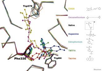
298 Structure 1999, Vol 7 No 3
Figure 1
Schematic drawing of E2020, (R,S)-1-benzyl-
O O24 C17 4-[(5,6-dimethoxy-1-indanon)-2-yl]methyl-
C26
C23 piperidine. *C8 is the chiral carbon.
C12 C22
O25 O C18
C13 N
C6 N14 C21
C7 C19
C1 C5
C28 C2 C4
C8 * C11
C16
C15
C20
C9
C3
C10
O
O27
Dimethoxyindanone Piperidine Benzyl
Structure
comparison to butyrylcholinesterase (BChE); this may be addition, it was possible to model the proximal N-acetyl-
important, as it has been suggested that inhibition of BChE, glucosamine (NAG) moiety at four out of five putative
which is abundant in human plasma, may cause potentiat- glycosylation sites [27], namely at residues Asn59, Asn416
ing side effects [20,21]. The affinity of E2020 for human (where two moieties could be fitted), Asn457 and Asn533.
AChE is ~1000-fold greater than for human BChE, whereas An analysis of the quality of the refined model is summa-
THA has a similar affinity for the two enzymes [22,23]. rized in Table 1.
Attempts to explain the specificity of E2020, and of other All three segments of E2020 interact with AChE
Eisai inhibitors, were made originally using QSAR and by As seen in Figure 5, E2020 makes principal interactions
theoretical conformational analysis. Subsequent to determi- along the active-site gorge of the enzyme through its three
nation of the 3D structure of TcAChE, automated computa- major functional groups: the benzyl moiety, the piperidine
tional techniques based on the known coordinates were nitrogen, and the dimethoxyindanone moiety. These inter-
employed [24,25]. Although the earlier modeling studies actions involve discrete water-mediated contacts that seem
attributed the differential specificity for AChE and BChE to be crucial for binding and specificity.
to differences in the geometry within the active site [26],
the more recent studies suggested that E2020 and the other
Figure 2
Eisai compounds are oriented along the axis of the active-
site gorge and that the differential specificity can be attrib-
uted to structural differences in AChE and BChE at the top
of the gorge, at the ‘peripheral’ anionic site [14,24,25]. Our
experimental structure of the E2020–TcAChE complex
broadly confirms these latter assignments and pinpoints the
specific interactions that are responsible for the high affinity
and selectivity demonstrated previously.
Results and discussion
Overall structure
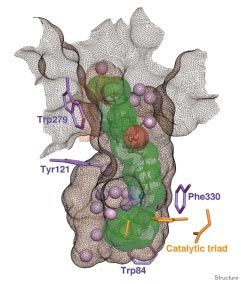
The overall structure of the E2020–TcAChE complex at
2.5 Å resolution is shown in Figure 2. The protein is dis-
played as a coil with the initial difference electron-density
map of E2020 superimposed as a ‘chicken-wire’ net. It can
be clearly seen that E2020 has a unique orientation along
the active-site gorge, extending from the anionic subsite of
the active site, at the bottom near Trp84, to the peripheral
anionic site, at the top near Trp279 (see Figures 3 and 4).
The 3D structure of the complex shows more detail of the
AChE structure than the starting native model (Protein
Initial difference electron-density map, contoured at 4.5σ, based on the
Data Bank [PDB] code 2ACE). Specifically, residues 2
native TcAChE structure (PDB code 2ACE) and the diffraction data for
and 3, at the N terminus, and the 484–490 loop, which the E2020–TcAChE complex.
were not seen in the original model, can be discerned. In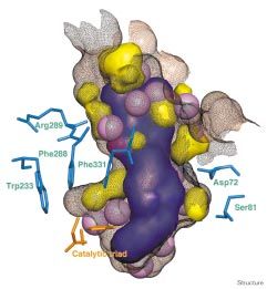
Research Article AChE–E2020 complex structure Kryger, Silman and Sussman   299

Figure 3                                                                Figure 4

                                                                        The E2020–TcAChE complex. Ribbon diagram showing the complex
                                                                        of the drug bound to the enzyme.

                                                                        Ser200 Oγ. Finally, the kinetic evidence showing that
                                                                        E2020 binds to the free and the acylated forms of AChE
E2020 binds along the active site and interacts with the ‘peripheral
anionic’ subsite at the top and with the ‘anionic’ subsite at the
                                                                        [32] is corroborated by our observation that E2020 does
bottom. E2020 is displayed in green semi-transparent CPK and ball-      not interact with the catalytic triad. Other interactions are
and-stick representation, solvent molecules are shown as lilac balls,   shown schematically in Figure 5.
catalytic triad residues are in orange, binding residues are shown as
purple sticks, and the solvent-accessible surface of the gorge is
                                                                        Interactions in the middle of the gorge
shown as a brown net.
                                                                        In the constricted region, halfway up the gorge (Figures 3
                                                                        and 4), the charged nitrogen of the piperidine ring makes
                                                                        a cation–π interaction [33,34] with the phenyl ring of
Interactions at the bottom of the gorge                                 Phe330, with distances of 3.9 Å–4.5 Å between the nitro-
Near the bottom of the gorge, one face of the benzyl ring               gen and the ring carbons. The orientation of the phenyl
displays classic parallel π–π stacking with the six-membered            ring is similar to that seen in the complex of decametho-
ring of the Trp84 indole, similar to the interaction with               nium (DECA) with TcAChE (DECA–TcAChE; PDB code
THA [28]. It thus occupies the binding site for quaternary              1ACL) [28]. The ring nitrogen also makes an in-line 2.9 Å
ligands [28,29], which was also modeled for the quaternary              H bond with WAT 1159, which, in turn, makes H bonds
group of the natural substrate, ACh [17,26]. The ring-to-ring           with Tyr121 OH, with WAT 1158 and with WAT 1160
distances range from 3.7 Å between Trp84 Cδ2 and E2020                  (see above). As already mentioned, the binding site for the
C19 to 4.4 Å between Trp84 Cε2 and E2020 C22.                           quaternary nitrogen of ACh, and for homologous ligands,
                                                                        is the indole ring of Trp84 [17,26,28]. These data suggest
On the opposite face, the benzyl group makes a classic                  that Phe330 may serve as an additional quaternary binding
aromatic hydrogen bond (H bond) [30,31] with a water                    site, midway down the gorge, between the peripheral site
molecule (WAT 1160), with distances to the ring carbons                 and the anionic subsite of the active site.
of 3.5 Å–3.7 Å. This solvent molecule is held firmly, as
assessed by a below-average temperature factor, by an                   Interactions at the entrance to the gorge
H bond to another solvent molecule (WAT 1161) in the                    At the top of the gorge the indanone ring stacks against the
‘oxyanion hole’ and to WAT 1159 (see below). WAT 1161                   indole ring of Trp279, in the peripheral binding site, by a
is another example of a tightly bound water molecule with               classical π–π interaction. Specifically, E2020 C1, C2, C6,
a relatively low temperature factor; it makes an H bond                 O25, C26, O27 and C28 stack against the six-membered
with the residues of the oxyanion hole, namely with                     ring of Trp279, with distances of 3.7 Å–4.2 Å. WAT 1249
Gly118 N, Gly119 N and Gly201 N, as well as with                        lies in the plane of the indanone moiety, and H bonds to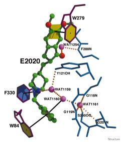
300 Structure 1999, Vol 7 No 3
Table 1
Data collection and refinement statistics.
Data collection
Detector Raxis-II
Source/wavelength (Å) Rigaku FR300 (50 mA, 50 kV), 1.54184 (Cu K∝)
Resolution range (Å) 30.0–2.5
Number of reflections 34,264
Completeness (%) 98.1
Redundancy
value 0 1 2 3 4 >5
accumulated (%) 1.9 15.7 44.3 33.4 4.5 0.2
I/σ
value 0 1 2 3 5 10 20 >20
accumulated (%) 5.5 14.2 23.4 30.7 41.2 57.6 78.1 20.0
Rsym (%) 5
Refinement
Number of protein non-H atoms 4255 = 2137 mainchain + 2118 sidechain
Number of hetero non-H atoms
Water molecules 396
Carbohydrate 70 = 5 × 14 in five NAG groups
Inhibitor 28 in one E2020 group
Resolution (Å) 2.5
Rwork (%) 18.8 (no σ cutoff)
Rfree (%) 22.9 (no σ cutoff)
B factor (Å2); Average / σ / Mininum / Maximum
Protein 28.0 / 12.5 / 2.0 /90.2
Inhibitor 20.4 / 4.8 / 13.2 / 31.6
Carbohydrate 47.6 / 15.0 / 14.5 / 74.5
Water molecules 37.2 / 14.7 / 2.2 / 86.7
Rmsd bond length (Å) 0.005
Rmsd bond angle (°) 1.2
Rmsd dihedral angle (°) 22.9
Rmsd improper angle (°) 0.98
Rsym = ΣIi–/ΣIi. Rwork = Σ|Fo|–|Fc|/ΣFo. Rfree is calculated using 2000 random reflections.
the methoxy group of E2020 O25; it is also H-bonded to maps (see Figure 6). The observed conformation is very
Glu185 Oε1 of the symmetry-related crystal-lattice copy of similar, if one allows for permitted adjustments of dihedral
the enzyme. The carbonyl on the five-membered ring of angles, to the energetically minimized E2020 conformation
the indanone only interacts with AChE via edge-on van der calculated with the InsightII package [35], and it is also
Waals contacts with the aromatic rings of Phe331 and similar to the ‘small molecule’ crystal structure of the pure
Phe290. It also makes indirect contact, via WAT 1254, with R enantiomer (T Steiner, R Boer and J Kroon, personal
Phe288 N. This finding might initially appear puzzling in communication).
view of the fact that a homolog of E2020, which lacks this
carbonyl, was reported to be inactive [15]. Our structure- E2020 analogs
based suggestion is that the van der Waals contacts made by Variation of the inhibitor backbone
the carbonyl function help orient the indanone moiety to Kawakami et al. [14] showed that at least two rotatable
make a favorable interaction with the indole ring of Trp279. bonds on each side of the piperidine are needed to yield
In the homolog which lacks this carbonyl function, the the high affinity displayed by E2020 and some of its
indanone moiety would be less constrained and would con- analogs. We can now corroborate this observation by
sequently make a poorer interaction with Trp279. showing that the two aromatic moieties of E2020 interact
closely with Trp84 and Trp279, while still maintaining the
AChE selects the R form of E2020 Phe330–piperidine-nitrogen interaction. This array of
The reported pharmacological studies on (R,S)-E2020 interactions calls for flexibility along the backbone of the
emphasize that both enantiomers are active; they have inhibitor. The number of rotatable links is also important
similar pharmacological profiles [22,32], but show ~fivefold for optimal positioning of the aromatic systems of the
difference in binding affinity for AChE: Ki = 3.35 nM (R), inhibitor against their enzyme counterparts. Indeed, when
17.5 nM (S) [32]. Although we used the racemate in our two to three additional rotatable bonds are added between
crystallographic study, we were not, therefore, surprised to the indanone and the piperidine, affinity further increases
see only the R form in the experimental electron-density (Table 2, lines 20–22), whereas adding rotatable bonds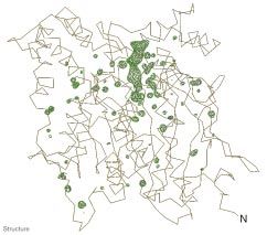
Research Article AChE–E2020 complex structure Kryger, Silman and Sussman        301

Figure 5                                                              Figure 6

                                                                      Two views of E2020 modeled in the initial difference Fourier map.
                                                                      Only one enantiomer is observed in the crystal structure. The shape
                                                                      of the electron density around the chiral carbon and the carbonyl
                                                                      clearly resolve the R/S ambiguity. ‘Side’ and ‘front’ views of E2020
Major interactions between E2020 and TcAChE. Classical H bonds        are shown modeled in the initial difference electron-density map
are shown as dashed lines, aromatic stacking and aromatic H bonds     contoured at 4.5σ.
are shown as black lines connecting matching colored planes.

                                                                      may explain the sensitivity to substitutions of this moiety.
between the piperidine and the benzyl ring lowers it                  The ring carbon at the para position is 3.2 Å from
(Table 2, line 19). This distinction between adding rotat-            Glu199 Oε1; thus a substituent must be both small and able
able bonds ‘above’ and ‘below’ the piperidine is likely to            to make an H bond. Indeed, according to Kawakami et al.
be based on the distances between Phe330 and either                   [14], an analogue with a hydroxyl at this position binds with
Trp84 or Trp279. Any change in the length of the spacer               higher affinity than the corresponding fluorine derivative
between Phe330 and Trp84 would weaken the inter-                      (Table 2, lines 9–12). A substituent at the meta position,
actions of the inhibitor with those sidechains. However,              E2020 C22, will point to a very small space left between
limited elongation of the indanone–piperidine link might              Glu199, His440 and Ser200 (the last two of which are
allow more overlap of the indanone’s aromatic system with             members of the catalytic triad). The other meta position, at
that of Trp279.                                                       E2020 C20, points to a wider space partially bordered by
                                                                      Gly117 and the ‘oxyanion hole’. In our structure, this space
Variation of the benzyl moiety                                        is mostly occupied by solvent molecules and can accommo-
E2020 is highly sensitive to substitutions on the benzyl              date a small, preferably negatively charged, substituent
moiety, as found by Kawakami and coworkers [14], who                  (Table 2, lines 5–7). A cyclohexane in place of a benzyl ring
noticed a general preference for substitution at the meta             cannot make a π–π stacking interaction with Trp84, result-
(E2020 C20, C22) positions in comparison to the ortho                 ing in reduced affinity (Table 2, line 18).
(E2020 C19, C23) and para (E2020 C21) positions (see
Table 2). Although these solution studies could not differ-           Variation of the piperidine moiety
entiate between the two possible ortho/meta substitutions, it         A piperazine in place of piperidine lowers the affinity of
can been seen from the crystal structure how each position            the resulting analog by ~19-fold (Table 2, line 3), whereas a
would experience different environments with respect to               piperidine with a nitrogen at the opposite position (in place
the lining of the gorge and the structural solvent molecules          of E2020 C10) lowers the affinity by ~90-fold (Table 2,
within it. In general there is little space left between the          line 2). A piperazine, although containing a nitrogen at a posi-
benzyl ring and the gorge surface (Figures 3 and 4), which            tion suitable for binding with Phe330, possesses a different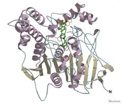
302   Structure 1999, Vol 7 No 3

Table 2

E2020 analogs.

Analog number                       Constant                      Variable R                      Inhibition of AChE IC50 (nM) [14]

                                       O
                           CH3O
                                                                             N
       1.                                  CH2 R CH2                                                                 5.7
                                                                                        (E2020)
                           CH3O

                                                                      N
       2.                                                                                                            480

                                                                      N      N
       3.                                                                                                            94

                                       O                              C H3
                           CH 3 O

       4.                                  C H2        N   R   C H2                                                  10
                           C H 3O
                                                                                 F

       5.                                                      C H2                                                  1

                                                                                 C H3

       6.                                                      C H2                                                  2

                                                                                 N O2

       7.                                                      C H2                                                  4

                                                                                 OMe

       8.                                                      C H2                                                  220

                                                               C H2                     OH
       9.                                                                                                            1.8

                                                               C H2                     F
      10.                                                                                                            9.5

                                                               C H2                     C H3
      11.                                                                                                            40

                                                               CH 2                  NO 2
      12.                                                                                                            100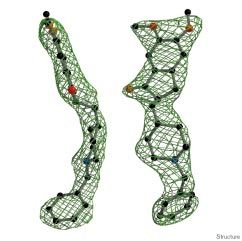
Research Article AChE–E2020 complex structure Kryger, Silman and Sussman   303

Table 2 continued

                                                                               F

   13.                                                                  C H2                                      9.5

                                                                           MeO

   14.                                                                  C H2                                      80

                                                                            O2N

   15.                                                                  C H2                                      160

                                                                       CO
   16.                                                                                                            >10,000

                                                                       H
   17.                                                                                                            5400

                                                                        C H2
   18.                                                                                                            8.9

                                                                        CH 2 CH2
   19.                                                                                                            180

                                         O
                        OH 3C
                                             -R-    N CH2
   20.                                                                                                            0.9
                        OH 3C

   21.                                                                                                            1.5

   22.                                                                                                            3.0

charge distribution, whereas a piperidine with a nitrogen at      entrance to the gorge. Thus, some sites of substitution
the opposite position does not allow a quaternary-π inter-        would point outwards towards the solvent, whereas others
action with Phe330 to occur.                                      would point into the central region of the gorge. However,
                                                                  substitution on the same edge as the carbonyl group
Variation of the indanone moiety                                  (C7 O24), which is juxtaposed to the wall of the gorge,
In general, substitutions on the indanone moiety have a           would disrupt the parallel stacking to Trp279. This is con-
smaller effect than substitutions on the benzyl group [15].       sistent with the observation of Cardozo et al. [15] that a
This is consistent with our crystal structure, as the indanone    cyano substitution on E2020 C6 produces a relatively poor
moiety is located in the wide section of the funnel-like          inhibitor. Suitable substitutions on the other edge of the
304 Structure 1999, Vol 7 No 3
ring (namely E2020 C1, C2, C3 and C9) would be expected that forces the aromatic ring of Phe 330 to swing out of the
to have little effect or, possibly, result in improved affinity. way towards the gorge wall. It is also of interest that the
THA–TcAChE complex displays a different conformation
Comparison of the E2020–TcAChE complex with other for the indole group of Trp279 relative to native AChE and
complexes the other complexes (χ2 value of 30° versus about 90° for
The structures of native TcAChE and of six different com- the others). This provides some support for structural cou-
plexes were overlaid according to their Cα positions, pling along the gorge between the anion subsite of the
namely native AChE (PDB code 2ACE) [36], and the com- active site and the peripheral anionic site, as suggested by
plexes with tacrine (THA; PDB code 1ACJ) [28], DECA Shafferman and coworkers [37]. The conformational flex-
(PDB code 1ACL) [28], m-(N,N,N-trimethylammonio)tri- ibility displayed by the sidechain of Phe330, contrasted
fluoroacetophenone (TMTFA; PDB code 1AMN) [29], with the relative rigidity of the other sidechains lining the
huperzine (HUP; PDB code 1VOT) [36], edrophonium gorge, supports the notion that it contributes to the sig-
(EDR; PDB code 2ACK) and E2020 (PDB code 1EVE). nificantly higher catalytic activity of AChE relative to
Two issues arise from comparison of these structures: the BChE, which lacks this residue. Site-directed mutagene-
role of Phe330, and the possible significance of structurally sis studies [38,39] showed that elimination of this aro-
conserved solvent molecules within the gorge. matic sidechain does not affect the Km for both charged
and uncharged ligands. It does, however, have a signifi-
Phe330 as a ‘swinging gate’ cant effect on kcat, which decreases ~fourfold for cationic
As seen in Figure 7, Phe330 adopts a wide range of con- substrates, although it increases ~twofold for uncharged
formations in the complex structures analyzed. These con- substrates. Thus, via the quaternary-π electron interaction
formations can be assigned, primarily based on their χ1 clearly seen in the E2020 complex, it may serve to guide
values, to three different groups. First, native, TMTFA, ACh towards the active site, while simultaneously isolat-
HUP and EDR (χ1 = –162°, –174°, –171° and –177°, ing the reaction center from the rest of the gorge. In fact,
respectively). Second, THA (χ1 = 157°). Third, E2020 and substrate traffic down the gorge may actually occur con-
DECA (χ1 = –130° and –117°, respectively). In group 1, comitantly with a swinging movement of Phe330. Similar
Phe330 adopts a similar conformation in the native enzyme ideas have been proposed on the basis of molecular dynam-
and in the complexes with ligands that bind near the bottom ics studies [40,41].
of the gorge, with the exception of THA. This ligand
defines Group 2, in which χ1 and χ2 of Phe330 change so as The solvent
to permit its phenyl group to stack on top of the ligand, thus In the E2020–TcAChE complex, 25 ordered waters can be
forming a ‘sandwich’ with the indole of Trp84. The seen within the gorge. If one overlays this structure with
common feature of Group 3 is the gorge-spanning ligand that of the native enzyme and of five other complexes, as
Figure 7
Overlay of native TcAChE and six complexes.
The flexibility of Phe330 in comparison with
the rigidity of the rest of the gorge can be
seen. Backbone trace and key residues are
represented by thin lines, Phe330 by thick
lines and inhibitors are shown in ball-and-stick
representation. *A ChemDraw representation
of ACh is shown for purposes of comparison.Research Article AChE–E2020 complex structure Kryger, Silman and Sussman 305
Figure 8 the gorge (see Figures 4 and 8), which are revealed in the
3D structure of the E2020–AChE complex, point to candi-
date sites on the inhibitor where added functions might
indeed enhance its pharmacological profile. Separation of
enantiomers, which are chemically indistinguishable by
non-chiral environments, is difficult, inefficient and costly.
In the case of E2020, we suggest that introduction of a
second chiral center, with concomitant generation of a
diastereomer system, would facilitate isolation of an active
component from the mixture so generated [42,43]. Inspec-
tion of the empty spaces left within the aromatic gorge by
the ordered solvent and by E2020 reveals a finger-shaped
void at the acyl-binding pocket. This pocket, defined by
Phe288, Phe290, Phe331 and Trp233, could envelop a
non-polar substituent branching from the piperidine ring at
position E2020 C12. Such a substitution, which would fit
into the ‘acyl pocket’, combined with the existing chiral
center, would produce a separable diastereomeric inhibitor.
The recent observations concerning the effect of peripheral-
site ligands on AChE-enhanced amyloid deposition [10],
mentioned above, raise the possibility that E2020, which
our data clearly show as stacking against Trp279, might
also moderate the rate of fibril formation. Many of the com-
pounds synthesized and tested by the Eisai company
‘Empty’ spaces, where no ordered solvent molecule is observed, can involved modification of this segment of the molecule
accommodate a substituent branching from the inhibitor. Voids within [15,16]. Nevertheless, it should be borne in mind that the
the gorge are displayed in yellow, the solvent-accessible surface of
E2020 is in purple, the solvent-accessible surface of the gorge is
screening that they carried out involved assessment of
shown as a brown net, the CPK model of the solvent molecules is in affinity for AChE, together with selectivity for AChE rela-
lilac, residues near voids are in light blue, and the catalytic triad is tive to BChE, but not a possible effect on amyloid fibril
represented by orange sticks. assembly or deposition.
Biological implications
shown in Figure 7, one can see that most of these waters are Acetylcholinesterase (AChE), the enzyme that hydrolyzes
conserved (G Koellner, GK, IS, JLS and T Steiner, unpub- the neurotransmitter acetylcholine (ACh) at cholinergic
lished results). In the E2020 structure, in fact, only three of synapses [44], is the target of the first generation of drugs
the conserved waters seen in the native enzyme are dis- for the treatment of Alzheimer’s disease. E2020 is the
placed. From this one can conclude that a large ligand, such second drug targeted at AChE approved for use by the
as E2020, fits into the gorge by displacing primarily the FDA for treatment of this condition. As design and devel-
unbound solvent molecules. Many of the conserved waters opment of the drug preceded the determination of the 3D
appear to adhere to the gorge wall and may, perhaps, be structure of AChE, it was of interest to use X-ray crystal-
considered as an integral part of the structure of the gorge lography to clearly delineate the structural factors govern-
rather than as voids. This may be especially important in ing its selectivity and specificity. This was particularly
relation to drug design (see below), as well as in molecular important because E2020 bears no structural resemblance
dynamics studies. It should also be noted that water mol- to other anticholinesterase drugs either approved or under
ecules not observed in the native structure are seen in the advanced clinical trial, such as tacrine, huperzine A and
E2020 complex, where they bridge between the inhibitor ENA-713 [45]. Our study shows, a posteriori, that the
and the enzyme. Specifically WAT 1159, WAT 1160 and design of E2020 took advantage of several important fea-
WAT 1254 (see Figure 5) are ‘novel’ structured waters not tures of the active-site gorge of AChE to produce a drug
previously seen in the native structure, while five ordered with both high affinity and a high degree of selectivity for
waters in the native structure are displaced. the enzyme, but not for butyrylcholinesterase (BChE).
The high affinity results from interaction of E2020 with
Structure-based modification of E2020 the aromatic residue involved in recognition of ACh at
Chemical modification of an already effective drug can the bottom of the gorge, Trp84, with a second aromatic
often improve its pharmacological profile in terms of affin- residue at the midpoint of the gorge, Phe330, and with a
ity and specificity. The calculated ‘empty’ spaces within third residue, Trp279, which is part of the peripheral306 Structure 1999, Vol 7 No 3
anionic site at the top of the gorge. The fact that these two analyses were implemented (see Table 1). Voids of minimal continuous
latter residues are conserved in AChE, but absent in volume of one spherical cubic angstrom were calculated using the soft-
ware SurfNet [58], between the inhibitor molecule as one entity and the
BChE, leads to the selectivity that may be an important protein and solvent molecules, taken together, as a second entity.
clinical consideration, as inhibition of BChE may cause
potentiating site effects. Our study also delineates voids Accession numbers
within the gorge of AChE that are not occupied by E2020, The PDB code of the E2020-TcAChe complex is IEVE.
which could serve as sites for modification of E2020 to
produce drugs with greater affinity and/or selectivity for
Acknowledgements
We thank BP Doctor (Walter Reed Army Institute of Research, Washington
AChE. It is worth noting that the 3D structure of Torpedo DC) for generously providing us with E2020, and Terry Lewis (Jealott’s Hill
california AChE (TcAChE) is very similar to those of Research Station, Zeneca Agrochemicals, Bracknell, UK) and Yoshiyuki
Kawakami (Tsukuba Research Laboratories, Eisai Ltd, Tsukuba, Japan) for
both mouse [46] and human AChE [47]. Thus, the con- valuable discussions. This work was supported by the US Army Medical
clusions drawn from the structure of the E2020–TcAChE Research Acquisition Activity under Contract No. DAMD17-97-2-7022, the
European Union IVth Framework in Biotechnology, the Kimmelman Center
complex should be valid for the mammalian enzyme. for Biomolecular Structure and Assembly, Israel, the Nella and Leon
Benoziyo Center for Neurosciences, and the generous support of Tania
Finally, analogs of E2020 are under consideration as a Friedman. IS is Bernstein–Mason Professor of Neurochemistry.
possible new class of insecticides [48]. The structure of
References
Drosophila AChE has recently been solved to 2.7 Å reso- 1. Drachman, D.A. & Leavitt, J. (1974). Human memory and the
lution in our laboratory [49] (M Harel, GK, H Green- cholinergic system. Arch. Neurol. 30, 113-121.
blatt, L Toker, W Mallender, TL Rosenberry, T Lewis, 2. Bowen, D.M., et al., & Davison, A.N. (1983). Biochemical assessment
of serotonergic and cholinergic dysfunction and cerebral atrophy in
IS and JLS, unpublished results). Although Drosophila Alzheimer’s disease. J. Neurochem. 41, 266-272.
AChE shares overall structural features with the Torpedo 3. Bartus, R.T., Dean, R.L., Beer, B. & Lippa, A.S. (1982). The cholinergic
hypothesis of geriatric memory dysfunction. Science 217, 408-414.
and human enzymes, it also displays marked differences. 4. Dunnett, S.B. & Fibiger, H.C. (1993). Role of forebrain cholinergic
Our combined knowledge of the vertebrate and inverte- systems in learning and memory: relevance to the cognitive deficits of
brate enzymes should be valuable in developing effective aging and Alzheimer’s dementia. Prog. Brain Res. 98, 413-420.
5. Weinstock, M. (1997). Possible role of the cholinergic system and
insecticides that combine high specificity for the insect disease models. J. Neural Transm. Suppl. 49, 93-102.
enzyme with low toxicity in humans. 6. Becker, R., Giacobini, E., Elble, R., McIlhany, M. & Sherman, K.
(1988). Potential pharmacotherapy of Alzheimer’s disease. A
comparison of various forms of physostigmine administration. Acta
Materials and methods Neurol. Scand. Suppl. 116, 19-32.
Protein preparation and crystallization 7. Zhang, R.W., et al., & Yang, R.M. (1991). Drug evaluation of huperzine
TcAChE was purified and crystallized as described previously [36]. A in the treatment of senile memory disorders. Acta Pharm. Sinica 12,
250-252.
E2020, as the hydrochloride salt of the pure racemate, was a generous
8. Weinstock, M., Razin, M., Chorev, M. & Enz, A. (1994).
gift from Dr BP Doctor (Division of Biochemistry, Walter Reed Army Insti- Pharmacological evaluation of phenyl-carbamates as CNS-selective
tute of Research, Washington, DC). TcAChE crystals of trigonal morphol- acetylcholinesterase inhibitors. J. Neural Transm. Suppl. 43, 219-225.
ogy were soaked for five days at 4°C in mother liquor (36% PEG 200, 9. Knopman, D.S. (1998). Metrifonate for Alzheimer’s disease: Is the next
10 mM NaCl, 50 mM MES, pH 5.8) containing ~10 mM (R,S) E2020. cholinesterase inhibitor better? Neurology 50, 1203-1206.
10. Inestrosa, N.C., et al., & Garrido, J. (1996). Acetylcholinesterase
X-ray data collection and processing accelerates assembly of amyloid-beta-peptides into Alzheimer’s fibrils:
possible role of the peripheral site of the enzyme. Neuron 16, 881-891.
The X-ray data were collected from a single crystal that was flash cooled 11. Reyes, A.E., et al., & Inestrosa, N.C. (1997). A monoclonal antibody
in a 100K nitrogen stream after exchanging the exterior solvent drop with against acetylcholinesterase inhibits the formation of amyloid fibrils
a coating of Exxon high viscosity motor oil [50]. Data were collected in- induced by the enzyme. Biochem. Biophys. Res. Commun. 232,
house, at the Weizmann Institute, utilizing a Rigaku RAXIS-II image-plate 652-655.
system, and a Rigaku FR300 X-ray generator employing a copper target 12. Davis, K.L. & Powchik, P. (1995). Tacrine. Lancet 345, 625-630.
set at 50 mA and 50 kV. The data-collection scheme was optimized by 13. Nightingale, S.L. (1997). Donepezil approved for treatment of
Alzheimer’s disease. JAMA 277, 10.
use of the software STRATEGY [51], and consisted of 81 frames of 14. Kawakami, Y., Inoue, A., Kawai, T., Wakita, M., Sugimoto, H. &
0.5° rotation and 30 min exposure time each. The diffraction data were Hopfinger, A.J. (1996). The rationale for E2020 as a potent
extracted from the frames using the software package HKL [52], result- acetylcholinesterase inhibitor. Bioorg. Med. Chem. 4, 1429-1446.
ing in a 98.1% complete dataset of 34,264 reflections, overall Rsym of 15. Cardozo, M.G., Imura, Y., Sugimoto, H., Yamanishi, Y. & Hopfinger,
5% and overall I/sigma ratio 13.8 (see Table 1). A.J. (1992). QSAR analyses of the substituted indanone and
benzylpiperidine rings of a series of indanone-benzylpiperidine
inhibitors of acetylcholinesterase. J. Med. Chem. 35, 584-589.
Model refinement and analysis 16. Cardozo, M.G., Kawai, T., Imura, Y., Sugimoto, H., Yamanishi, Y. &
The structure was refined using the starting model of native TcAChE Hopfinger, A.J. (1992). Conformational analyses and molecular-shape
(PDB code 2ACE) with the program X-PLOR version 3.851 [53]. Refine- comparisons of a series of indanone-benzylpiperidine inhibitors of
ment employed all 30–2.5 Å data. Following overall anisotropic B-factor acetylcholinesterase. J. Med. Chem. 35, 590-601.
and bulk-solvent corrections, the structure was first refined as a single 17. Sussman, J.L., et al., & Silman, I. (1991). Atomic structure of
rigid body. Subsequently, refinement was carried out on individual atoms, acetylcholinesterase from Torpedo californica: a prototypic
with restraints, using simulated annealing alternating with positional and acetylcholine-binding protein. Science 253, 872-879.
temperature-factor refinement. The model was fitted to the observed 18. Rupniak, N.M., Tye, S.J. & Field, M.J. (1997). Enhanced performance
of spatial and visual recognition memory tasks by the selective
electron density using the software O [54] on a Silicon Graphics work- acetylcholinesterase inhibitor E2020 in rhesus monkeys.
station. This program was also used to overlay analogous models by Psychopharmacology Berlin 131, 406-410.
least-squares minimization of Cα positions. The intermediate and final 19. Galli, A., Mori, F., Benini, L. & Cacciarelli, N. (1994).
models were analyzed using the software OOPS [55], PROCHECK [56] Acetylcholinesterase protection and the anti-diisopropylfluorophosphate
and WHATCHECK [57], and corrections to the model based on these efficacy of E2020. Eur. J. Pharmacol. 270, 189-193.Research Article AChE–E2020 complex structure Kryger, Silman and Sussman 307
20. Thomsen, T. & Kewitz, H. (1990). Selective inhibition of human 43. Nation, R.L. (1989). Enantioselective drug analysis: problems and
acetylcholinesterase by galanthamine in vitro and in vivo. Life Sci. 46, resolutions. Clin. Exp. Pharmacol. Physiol. 16, 471-477.
1553-1558. 44. Barnard, E.A. (1974). Neuromuscular transmission – enzymatic
21. Loewenstein, Y., Gnatt, A., Neville, L.F. & Soreq, H. (1993). Chimeric destruction of acetylcholine. In The Peripheral Nervous System.
human cholinesterase. Identification of interaction sites responsible for (Hubbard, J.I., ed.), pp. 201-224, Plenum, NY.
recognition of acetyl- or butyrylcholinesterase-specific ligands. J. Mol. 45. Baron, P., Harel, M., Millard, C., Enz, A., Sussman, J.L. & Silman, I.
Biol. 234, 289-296. (1998). Kinetic and X-ray crystallographic studies of the binding of
22. Sugimoto, H., Imura, Y., Yamanishi, Y. & Yamatsu, K. (1995). ENA-713 to Torpedo californica acetylcholinesterase (TcAChE). In
Synthesis and structure-activity-relationships of acetylcholinesterase Structure and Function of Cholinesterases and Related Proteins.
inhibitors – 1-Benzyl-4-[(5,6-dimethoxy-1-oxoindan- (Doctor, BP, Quinn, DM, Rotundo, RL & Taylor, P, eds), pp. 373-374,
2-yl)methyl]piperidine hydrochloride and related compounds. J. Med. Plenum, NY.
Chem. 38, 4821-4829. 46. Bourne, Y., Taylor, P., Kanter, J.R., Bougis, P.E. & Marchot, P. (1998).
23. Cheng, D.H., Ren, H. & Tang, X.C. (1996). Huperzine A, a novel Crystal structure of mouse acetylcholinesterse. In Structure and
promising acetylcholinesterase inhibitor. Neuroreport 8, 97-101. Function of Cholinesterases and Related Proteins. (Doctor, B.P., Quinn,
24. Villalobos, A., et al., & Frost White, W. (1995). 5,7-dihydro-3-[2-[1- D.M., Rotundo, R.L. & Taylor, P., eds), pp. 315-322, Plenum, NY.
(phenylmethyl)-4-piperidinyl]ethyl]-6H- pyrrolo[3,2-f]-1,2-benzisoxazol- 47. Kryger, G., et al., & Sussman, J.L. (1998). 3D structure of a complex of
6-one: a potent and centrally-selective inhibitor of acetylcholinesterase human recombinant acetylcholinesterase with fasciculin-II at 2.7 Å
with an improved margin of safety. J. Med. Chem. 38, 2802-2808. resolution. In Structure and Function of Cholinesterases and Related
25. Pang, Y.P. & Kozikowski, A.P. (1994). Prediction of the binding site of Proteins. (Doctor, B.P., Quinn, D.M., Rotundo, R.L. & Taylor, P., eds),
1-benzyl-4-[(5,6-dimethoxy-1-indanon-2-yl)methyl]piperidine in pp. 323-326, Plenum, NY.
acetylcholinesterase by docking studies with the SYSDOC program. 48. Greenblatt, H., et al., & Sussman, J.L. (1998). Crystal structures of
J. Comput. Aided Mol. Des. 8, 683-693. complexes of E2020 related compounds with Torpedo
26. Harel, M., et al., & Silman, I. (1992). Conversion of acetylcholinesterase. In Structure and Function of Cholinesterases
acetylcholinesterase to butyrylcholinesterase: modeling and and Related Proteins. (Doctor, B.P., Quinn, D.M., Rotundo, R.L. &
mutagenesis. Proc. Natl Acad. Sci. USA 89, 10827-10831. Taylor, P., eds), p. 371, Plenum, NY.
27. Schumacher, M., et al., & Taylor, P. (1986). Primary structure of 49. Kryger, G., et al., & Harel, M. (1998). Structural studies on human and
Torpedo californica acetylcholinesterase deduced from its cDNA insect acetylcholinesterase. Abstract from theSixth International
sequence. Nature 319, 407-409. Meeting on Cholinesterases, La Jolla, CA, p. 14.
28. Harel, M., et al., & Sussman, J.L. (1993). Quaternary ligand binding to 50. Hope, H. (1988). Cryocrystallography of biological macromolecules: a
aromatic residues in the active-site gorge of acetylcholinesterase. generally applicable method. Acta Crystallogr. B 44, 22-26.
Proc. Natl Acad. Sci. USA 90, 9031-9035. 51. Ravelli, R.B.G., Sweet, R.M., Skinner, J.M., Duisenberg, A.J.M. &
29. Harel, M., Quinn, D.M., Nair, H.K., Silman, I. & Sussman, J.L. (1996). Kroon, J. (1997). STRATEGY: a program to optimize the starting
The X-ray structure of a transition state analog complex reveals the spindle angle and scan range for X-ray data collection. J. Appl.
molecular origins of the catalytic power and substrate specificity of Crystallogr. 30, 551-554.
acetylcholinesterase. J. Am. Chem. Soc. 118, 2340-2346. 52. Otwinowski, Z. (1993). Oscillation data reduction program. In
30. Levitt, M. & Perutz, M.F. (1988). Aromatic rings act as hydrogen bond Proceedings of the CCP4 Study Weekend: Data Collection and
acceptors. J. Mol. Biol. 201, 751-754. Processing. (Sawyer, L., Isaacs, N. & Bailey, S., eds), pp. 56-62,
31. Burley, S.K. & Petsko, G.A. (1986). Amino-aromatic interactions in SERC, Daresbury.
proteins. FEBS Lett. 203, 139-143. 53. Brünger, A.T., Kuriyan, J. & Karplus, M. (1987). Crystallographic R
32. Inoue, A., Kawai, T., Wakita, M., Imura, Y., Sugimoto, H. & Kawakami, factor refinement by molecular dynamics. Science 235, 458-460.
Y. (1996). The simulated binding of (+/–)-2,3-dihydro-5,6-dimethoxy- 54. Jones, T.A., Zou, J.Y., Cowan, S.W. & Kjeldgaard, M. (1991). Improved
2-[(1-(phenylmethyl)-4-piperidinyl)methyl]-1H-inden-1-one methods for the building of protein models in electron density maps
hydrochloride (E2020) and related inhibitors to free and acylated and the location of errors in these models. In Crystallographic
acetylcholinesterases and corresponding structure-activity analyses. Computing. (Moras, D., Podany, A.D. & Thierry, J.C., eds), pp. 413-
J. Med. Chem. 39, 4460-4470. 432, Oxford Univ. Press, Oxford.
33. Verdonk, M.L., Boks, G.J., Kooijman, H., Kanters, J.A. & Kroon, J. 55. Kleywegt, G.J. & Jones, T.A. (1996). Efficient rebuilding of protein
(1993). Stereochemistry of charged nitrogen-aromatic interactions structures. Acta Crystallogr. D 52, 829-832.
and its involvement in ligand-receptor binding. J. Comput. Aided Mol. 56. Laskowski, R.A., MacArthur, M.W., Moss, D. & Thornton, J.M. (1993).
Des. 7, 173-182. PROCHECK: a program to check the stereochemical quality of
34. Dougherty, D. (1996). Cation-π interactions in chemistry and biology: protein structures. J. Appl. Crystallogr. 26, 283-291.
A new view of benzene, Phe, Tyr, and Trp. Science 271, 163-168. 57. Vriend, G. (1990). WHAT IF: a molecular modelling and drug design
35. Biosym (1993). InsightII. Biosym Technologies San Diego. program. J. Mol. Graph. 8, 52-56.
36. Raves, M.L., Harel, M., Pang, Y.P., Silman, I., Kozikowski, A.P. & 58. Laskowski, R.A. (1995). SURFNET: a program for visualizing
Sussman, J.L. (1997). Structure of acetylcholinesterase complexed molecular surfaces, cavities and intermolecular interactions. J. Mol.
with the nootropic alkaloid, (-)-huperzine A. Nat. Struct. Biol. 4, Graph. 13, 323-330.
57-63.
37. Shafferman, A., et al., & Ariel, N. (1992). Substrate inhibition of
acetylcholinesterase: residues affecting signal transduction from the
surface to the catalytic center. EMBO J. 11, 3561-3568.
38. Ordentlich, A., et al., & Shafferman, A. (1993). Dissection of the
human acetylcholinesterase active center determinants of substrate
specificity. Identification of residues constituting the anionic site, the
hydrophobic site, and the acyl pocket. J. Biol. Chem. 268,
17083-17095.
39. Radic, Z., Pickering, N.A., Vellom, D.C., Camp, S. & Taylor, P. (1993).
Three distinct domains in the cholinesterase molecule confer
selectivity for acetyl- and butyrylcholinesterase inhibitors. Biochemistry
32, 12074-12084.
40. Antosiewicz, J., Wlodek, S.T. & McCammon, J.A. (1996).
Acetylcholinesterase: role of the enzyme’s charge distribution in steering
charged ligands toward the active site. Biopolymers 39, 85-94.
41. Zhou, H.X., Wlodek, S.T. & McCammon, J.A. (1998). Conformation
gating as a mechanism for enzyme specificity. Proc. Natl Acad. Sci.
USA 95, 9280-9283.
42. Shah, R.R. (1993). Clinical pharmacokinetics: current requirements
and future perspectives from a regulatory point of view. Xenobiotica
23, 1159-1193.You can also read