Study of Symptomatic vs. Silent Brain Infarctions on MRI in Elderly Subjects - Frontiers
←
→
Page content transcription
If your browser does not render page correctly, please read the page content below
ORIGINAL RESEARCH
                                                                                                                                            published: 17 February 2021
                                                                                                                                        doi: 10.3389/fneur.2021.615024

                                               Study of Symptomatic vs. Silent Brain
                                               Infarctions on MRI in Elderly Subjects
                                               Sheelakumari Raghavan 1 , Jonathan Graff-Radford 2 , Eugene Scharf 2 , Scott A. Przybelski 3 ,
                                               Timothy G. Lesnick 3 , Brian Gregg 1 , Christopher G. Schwarz 1 , Jeffrey L. Gunter 4 ,
                                               Samantha M. Zuk 1 , Alejandro Rabinstein 2 , Michelle M. Mielke 2,3 , Ronald C. Petersen 2 ,
                                               David S. Knopman 2 , Kejal Kantarci 1 , Clifford R. Jack Jr. 1 and Prashanthi Vemuri 1*
                                               1
                                                Departments of Radiology, Mayo Clinic, Rochester, MN, United States, 2 Neurology, Mayo Clinic, Rochester, MN,
                                               United States, 3 Health Sciences Research, Mayo Clinic, Rochester, MN, United States, 4 Information Technology, Mayo
                                               Clinic, Rochester, MN, United States

                                               Brain infarctions are closely associated with future risk of stroke and dementia. Our goal
                                               was to report (i) frequency and characteristics that differentiate symptomatic vs. silent
                                               brain infarctions (SBI) on MRI and (ii) frequency and location by vascular distribution
                                               (location of stroke by major vascular territories) in a population based sample. From
                                               Mayo Clinic Study of Aging, 347 participants (≥50 years) with infarcts detected on their
                          Edited by:
              Michael Malek-Ahmadi,
                                               first MRI were included. Infarct information was identified visually on a FLAIR MRI image
          Banner Alzheimer’s Institute,        and a vascular territory atlas was registered to the FLAIR image data in order to identify
                        United States
                                               the arterial territory of infarction. We identified the subset with a clinical history of stroke
                        Reviewed by:
                                               based on medical chart review and used a logistic regression to evaluate the risk factors
                          Arun Bokde,
         Trinity College Dublin, Ireland       associated with greater probability of a symptomatic stroke vs. SBI. We found that 14%
                             Jiu Chen,         of all individuals with infarctions had a history of symptomatic stroke (Silent: n = 300,
     Nanjing Medical University, China
                                               symptomatic: n = 47). Factors associated with a symptomatic vs. SBI were size which
                 *Correspondence:
                   Prashanthi Vemuri
                                               had an odds ratio of 3.07 (p < 0.001), greater frequency of hypertension (odds ratio of
        Vemuri.Prashanthi@mayo.edu             4.12, p = 0.025) and alcohol history (odds ratio of 4.58, p = 0.012). The frequency of
                                               infarcts was greater in right hemisphere compared to the left for SBI. This was primarily
                 Specialty section:
                                               driven by middle cerebral artery (MCA) infarcts (right = 60%, left = 40%, p = 0.005).
       This article was submitted to
    Dementia and Neurodegenerative             While left hemisphere strokes are more common for symptomatic carotid disease and in
                            Diseases,          clinical trials, right hemispheric infarcts may be more frequent in the SBI group.
              a section of the journal
               Frontiers in Neurology          Keywords: silent brain infarction, clinical stroke, vascular distribution, middle cerebral artery, laterality
         Received: 07 October 2020
        Accepted: 01 February 2021
        Published: 17 February 2021            INTRODUCTION
                              Citation:
        Raghavan S, Graff-Radford J,           Brain infarcts are a common cerebrovascular pathology of aging and a common cause of cognitive
 Scharf E, Przybelski SA, Lesnick TG,          impairment (1). The term “Silent brain infarcts” (SBI) (2–5), has been used to refer to infarcts which
   Gregg B, Schwarz CG, Gunter JL,             are observed on conventional MRI or CT but without known clinical symptoms. SBI make up the
   Zuk SM, Rabinstein A, Mielke MM,            majority of infarcts in population-based studies (6–8) compared to clinically recognized infarcts.
          Petersen RC, Knopman DS,             SBI have become an established risk factor for future symptomatic infarcts and cognitive decline.
 Kantarci K, Jack CR Jr and Vemuri P
                                               Several studies have highlighted the prevalence of infarcts detected in the general population
    (2021) Study of Symptomatic vs.
     Silent Brain Infarctions on MRI in
                                               (7, 9, 10). While stroke trials have revealed a greater likelihood of left cerebral hemispheric (LH)
                      Elderly Subjects.        infarctions compared to right hemispheric (RH) infarctions (11, 12), the topography of SBI has been
            Front. Neurol. 12:615024.          understudied. Our objective was to report on the frequency and characteristics that differentiate
    doi: 10.3389/fneur.2021.615024             symptomatic vs. SBI on MRI and also explore their frequency and location by major vascular

Frontiers in Neurology | www.frontiersin.org                                         1                                        February 2021 | Volume 12 | Article 615024
Raghavan et al. Symptomatic vs. Silent Brain Infarctions
territories (vascular distribution). We hypothesized that the SBI extended to the cortical edge with or without involvement of
would be overrepresented in the right hemisphere compared to the underlying white matter. These infarctions were identified on
the left hemisphere because individuals with right hemisphere the T2 FLAIR sequence, with a corresponding T1 hypointensity
strokes may be less likely to recognize non-dominant deficits. required for confirmation. The size of the cortical infarction was
determined by measuring the largest diameter (in mm) of the
METHODS hyperintensity/gliosis on the axial slice by considering the size on
all the slices.
Study Participants Subcortical infarctions were characterized as hyperintense T2
All participant data were selected from the Mayo Clinic Study FLAIR lesions with a dark center, seen in the white matter,
of Aging (MCSA), a population-based study of residents living infratentorial, and central gray-capsular regions. The dark area
in Olmsted County, Minnesota. The Rochester Epidemiology (tissue loss) must be ≥3 mm in diameter as measured on the T2
Project (REP) medical records linkage system was used to FLAIR or T1, whichever image shows the findings more clearly.
enumerate the MCSA population (13, 14), which followed an Subcortical infarcts were distinguished from perivascular spaces
age and sex-stratified design. The REP allowed us to ascertain by size, location, and shape. The size of the subcortical infarction
the history of vascular risk factors and the details have been was determined by measuring the largest diameter (in mm) of the
published previously (15, 16). In MCSA, 1,845 elderly individuals hypointensity/tissue loss on the axial slice by considering the size
(aged ≥ 50 years) had an infarction assessment. We selected 347 on all the slices.
subjects and considered their first scan (FLAIR-MRI and T1-
weighted MPRAGE imaging sequences) in which they had an
infarction for this study. During the corresponding clinical visit Assessment of Vascular Territory and
275 were cognitively unimpaired, 56 were diagnosed with mild
cognitive impairment, and 16 had dementia. The diagnosis of
Laterality on FLAIR MRI
We nonlinearly registered a vascular territory atlas (developed in-
the participants was based on the detailed clinical evaluation by
house, traced in MCALT space) (21) using a textbook reference
the neurologist, assessment of neuropsychological test battery by
(22) onto participants’ T1-weighted image and transformed it
the neuropsychologist, and a clinical dementia rating assessment
onto the rigid-registered FLAIR image using ANTs (23). The
by the study coordinator. The detailed diagnostic criteria for
atlas is divided into 14 regions including bilateral terminal
patients have been previously described (17).
and penetrating anterior cerebral artery (ACA), middle cerebral
Potential Risk Factors artery (MCA), anterior choroidal artery (AChA), posterior
We determined the cardiovascular risk factors including cerebral artery (PCA) and unknown (cerebellum, pons and
hypertension, dyslipidemia, and diabetes mellitus using nurses medulla). We used this method for identifying the anatomical
that abstracted the REP medical-records linkage system as landmarks and then assigning the cortical and subcortical
previously reported (18). Among these, 90% of hypertensive infarctions on the FLAIR image to the specific vascular territory.
individuals were treated for hypertension. The ICD-9 and This technique assigned the anatomical landmarks into atlas
ICD-10 codes were searched also to determine the report of space to distinguish the left and right ACA, MCA and PCA. The
symptomatic strokes. Alcoholism was determined based on the posterior fossa region infarctions were excluded in the vascular
combination of CAGE and the NHIS form. We also assessed territory atlas. We further validated these vascular territory
smoking status based on self-reports of never smoking vs. former assessments with visual inspection by SKR.
or currently smoking (19).
Assessment of Infarcts on FLAIR MRI Statistical Analysis
All MRI images were acquired on 3T GE scanner (GE Medical Statistical analyses were performed with SAS and R. The
Systems, Milwaukee, WI). The 2D T2-weighted FLAIR- MRI continuous variables were summarized as mean and standard
scans were obtained with the following parameters: repetition deviation and categorical variables as frequency and percentage.
time = 11 000 ms, echo time = 147 ms, inversion time = The variables were assessed for normality and log transformed
2,250 ms, 256 × 192 matrix, 24-cm field of view, and voxel for non-normal distribution. These were analyzed with either a
size = 0.86 × 0.86 × 3 mm. The full details of infarct grading two-sample two-sided t-test or chi-squared test. Next, we ran
have been recently published (20). In brief infarcts were graded logistic regression model that included age, gender, education,
on two-dimensional FLAIR MRI that was co-registered with and all of the vascular risk variables as predictors. Then,
an MPRAGE (magnetization-prepared rapid gradient-echo) T1 a step-wise elimination was done to form a parsimonious
MRI. All possible infarcts were initially identified by trained model with the significant predictors of clinical vs. silent
image analysts and subsequently confirmed by a vascular stroke. This model was cross-checked with both forward and
neurologist (JGR) to whom all clinical information was masked. backward elimination that yielded the same final model, and
The intra-rater reliability based on blinded reading of 50 possible odds ratios, associated 95% confidence intervals, and p-values
infarcts on two separate occasions was excellent (κ statistic, 0.92). were reported. The left and right distributions of cerebral
Cortical infarctions were characterized as hyperintense T2 infarcts were compared using McNemar’s test for paired
FLAIR lesions (gliosis) involving cortical gray matter that nominal data.
Frontiers in Neurology | www.frontiersin.org 2 February 2021 | Volume 12 | Article 615024Raghavan et al. Symptomatic vs. Silent Brain Infarctions
TABLE 1 | Characteristics table of infarction subjects with the mean (SD) listed for TABLE 2 | Logistic regression model with clinical stroke as outcome variable.
the continuous variables and count (%) for the categorical variables.
Odds ratio (95% CI) P-value
Silent Symptomatic Total *P-value
n = 300 n = 47 n = 347 Hypertension 4.12 (1.20, 14.21) 0.025
Alcoholism 4.58 (1.40, 15.01) 0.012
Age, yrs 77.6 (8.7) 79.8 (7.9) 77.9 (8.6) 0.093
Maximum diameter 3.07 (1.97, 4.77)Raghavan et al. Symptomatic vs. Silent Brain Infarctions
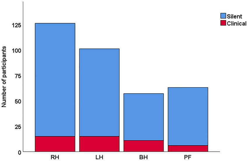
FIGURE 1 | Distribution of overall stroke in silent vs clinical stroke participants. In this cohort, the overall frequency was higher for silent strokes especially in the right
hemisphere. RH, right hemisphere; LH, left hemisphere; BH, both hemisphere; PF, posterior fossa.
TABLE 3 | Hemispheric differences of vascular distribution in overall infarcts TABLE 4 | Hemispheric differences of vascular distribution in silent stroke.
participants.
RH LH Total P Odd’s ratio
RH LH Total P Odds ratio
Total 109 (55.9) 86 (44.1) 195 0.10 1.27
Total 126 (55.5) 101 (44.5) 227 0.097 1.25 ACA 15 (45.5) 18 (54.5) 33 0.60 0.83
ACA 17 (48.6) 18 (51.4) 35 0.87 0.94 MCA 81 (60) 54 (40) 135 0.005 1.5
MCA 93 (59.6) 63 (40.4) 156 0.016 1.47 PCA 18 (45) 22 (55) 40 0.53 0.82
PCA 26 (47.3) 29 (52.7) 55 0.69 0.90
Data values are presented as either n or n(%). ACA, anterior cerebral artery; MCA, middle
Data values are presented as either n or n(%). ACA, anterior cerebral artery; MCA, middle cerebral artery; PCA, posterior cerebral artery. McNemar’s test for paired nominal data.
cerebral artery; PCA, posterior cerebral artery. McNemar’s test for paired nominal data.
common with right hemisphere infarcts (37), particularly
profile studies on silent and symptomatic infarcts (10, 24, 25). those involving the MCA territory (38). Consistent with
Our study along with others (26, 27) detected alcoholism as a this hypothesis, we identified a greater proportion of
strong independent risk factor for stroke, both ischemic and right hemispheric strokes in a population-based study
hemorrhagic. The possible reason for this is that heavy drinking from MCSA with predominantly silent brain infarctions.
and chronic alcoholism is associated with other risk factors that This overrepresentation of non-dominant SBI has been
lead to infarcts such as increase in blood pressure, frequency of observed in 848 subjects with asymptomatic high grade carotid
atrial fibrillation, as well as sleep apnea and cardiomyopathy. stenosis (39).
Since our findings were limited by the lack of dose-response The asymmetric pattern in our study was driven by the
relationship, a detailed study with stroke subtypes may shed greater overall MCA territory distribution in SBI compared
light on the possible mechanisms as suggested by the meta- to ACA and PCA. It is well-known that MCA is the largest
analysis (28). and most prevalent cerebral artery linked with infarcts (40–
42). In contrast with our findings, a similar frequency in the
Laterality of the Silent Strokes left and right MCA territories were reported in the clinically
Accumulating evidence on the hemispheric difference and silent TOAST study participants (43), although they used CT
stroke outcome (29–35) have shown that left hemisphere rather than MRI to define infarction and the TOAST study
strokes are more commonly detected than right hemisphere was not population-based. The findings from the Rotterdam
strokes in clinical trials and hospital based cohorts. This study showed that the atherosclerotic plaque prevalence and
apparent over-representation may be because of greater thickness was greater in left than right (44). This may be
recognition of clinical deficits from the function of the due to a greater composition of calcification in the right-
dominant hemisphere (36). In contrast, anosagnosia is more sided plaques, which are more stable and less vulnerable to
Frontiers in Neurology | www.frontiersin.org 4 February 2021 | Volume 12 | Article 615024Raghavan et al. Symptomatic vs. Silent Brain Infarctions
cerebrovascular complications (45, 46). Previous studies have group which is in contrast to the left hemisphere strokes that
suggested that variability of carotid bifurcation anatomy might are commonly seen with symptomatic carotid disease and in
affect the development of atheroma (47) and ICA stenosis (48) clinical trials.
and possibly affect the preferential laterality of the stroke. In
patients acutely presenting to the hospital Hedna et al. (35) DATA AVAILABILITY STATEMENT
demonstrated greater frequency of left MCA strokes compared
to right MCA strokes and the left MCA strokes presented with a The raw data supporting the conclusions of this article will be
higher NIHSS. made available by the authors, without undue reservation.
Notably, the asymmetric pattern in SBI patients has
been evaluated with cortical thinning (49). By using ETHICS STATEMENT
the large deformation diffeomorphic metric mapping
(LDDMM), Thong et al. (49) identified more widespread The studies involving human participants were reviewed and
and severe RH atrophy patterns than LH in silent lacunar approved by Institutional review board, Mayo Clinic, Rochester.
infarctions. However, the patterns were independent The patients/participants provided their written informed
of the number of infarctions. Speculatively, the RH consent to participate in this study.
dominance either may be due to the age-related damage
(50) or may be the greater impact of associated vascular AUTHOR CONTRIBUTIONS
pathologies in SBI (51). This may be part of our
future research. SR, JG-R, and PV conceived and designed the study. SR, JG-R,
ES, SP, and TL drafted the manuscript and figures. All authors
Limitations participated in data collection and analysis.
The present study has some limitations. Though we had a large
sample of elderly individuals recruited from the population,
FUNDING
the number of participants with symptomatic stroke in the
subsample was smaller and was not sufficient to determine This work was supported by NIH grants R01 NS097495
laterality differences in those with symptomatic stroke. We (PI: PV), U01 AG06786 (PI: RP/MM/CJ), R01 AG56366
accounted for the size of the infarction by using the largest (PI: PV), P50 AG16574 (PI: RP), R37 AG11378 (PI: CJ),
diameter on a slice because it was less time intensive to R01 AG41851 (PIs: CJ and DK); the Gerald and Henrietta
measure but other methods (35) may provide greater accuracy. Rauenhorst Foundation grant, Alzheimer’s Drug Discovery
Another limitation is that nonparticipation in MCSA could Foundation (ADDF), the Alexander Family Alzheimer’s Disease
conceivably have included a greater proportion of individuals Research Professorship of the Mayo Foundation, Liston
with overt infarcts. Had they been included our proportion Award, Elsie and Marvin Dekelboum Family Foundation,
of silent infarcts relative to overt ones might have been a Schuler Foundation, Opus building NIH grant C06 RR018898,
bit lower. and was made possible by Rochester Epidemiology Project
(R01 AG34676).
CONCLUSION
ACKNOWLEDGMENTS
We report on the frequency, location, and characteristics of
clinical vs. silent brain infarctions in a population based sample. We thank all the study participants and staff in the Mayo Clinic
The size and laterality between SBI and clinical infarcts differs Study of Aging, Mayo Alzheimer’s Disease Research Center, and
in the general population. We found evidence for our hypothesis Aging Dementia Imaging Research laboratory at the Mayo Clinic
that right hemispheric infarcts may be more frequent in the SBI for making this study possible.
REFERENCES 5. Shi Y, Wardlaw JM. Update on cerebral small vessel disease: a
dynamic whole-brain disease. Stroke Vasc Neurol. (2016) 1:83–92.
1. Pinter D, Enzinger C, Fazekas F. Cerebral small vessel disease, cognitive doi: 10.1136/svn-2016-000035
reserve and cognitive dysfunction. J Neurol. (2015) 262:2411–9. 6. Das RR, Seshadri S, Beiser AS, Kelly-Hayes M, Au R, Himali
doi: 10.1007/s00415-015-7776-6 JJ, et al. Prevalence and correlates of silent cerebral infarcts
2. Wardlaw JM, Smith C, Dichgans M. Mechanisms of sporadic cerebral small in the Framingham offspring study. Stroke. (2008) 39:2929–35.
vessel disease: insights from neuroimaging. Lancet Neurol. (2013) 12:483–97. doi: 10.1161/STROKEAHA.108.516575
doi: 10.1016/S1474-4422(13)70060-7 7. Vermeer SE, Den Heijer T, Koudstaal PJ, Oudkerk M, Hofman A,
3. Román GC, Erkinjuntti T, Wallin A, Pantoni L, Chui HC. Subcortical Breteler MM. Incidence and risk factors of silent brain infarcts in
ischaemic vascular dementia. Lancet Neurol. (2002) 1:426–36. the population-based Rotterdam Scan Study. Stroke. (2003) 34:392–6.
doi: 10.1016/S1474-4422(02)00190-4 doi: 10.1161/01.STR.0000052631.98405.15
4. Norrving B. Evolving concept of small vessel disease through advanced 8. Saavedra Perez HC, Direk N, Hofman A, Vernooij MW, Tiemeier H, Ikram
brain imaging. J Stroke. (2015) 17:94–100. doi: 10.5853/jos.2015. MA. Silent brain infarcts: a cause of depression in the elderly? Psychiatry Res.
17.2.94 (2013) 211:180–2. doi: 10.1016/j.pscychresns.2012.06.005
Frontiers in Neurology | www.frontiersin.org 5 February 2021 | Volume 12 | Article 615024Raghavan et al. Symptomatic vs. Silent Brain Infarctions
9. Arnold Fiebelkorn C, Vemuri P, Rabinstein AA, Mielke MM, Przybelski 28. Larsson SC, Wallin A, Wolk A, Markus HS. Differing association of alcohol
SA, Kantarci K, et al. Frequency of acute and subacute infarcts consumption with different stroke types: a systematic review and meta-
in a population-based study. Mayo Clinic Proc. (2018) 93:300–6. analysis. BMC Med. (2016) 14:178. doi: 10.1186/s12916-016-0721-4
doi: 10.1016/j.mayocp.2017.11.021 29. Naess H, Waje-Andreassen U, Thomassen L, Myhr KM. High
10. Fan H, Hao X, Yang S, Li Y, Qin W, Yang L, et al. Study on the incidence incidence of infarction in the left cerebral hemisphere among
and risk factor of silent cerebrovascular disease in young adults with first-ever young adults. J Stroke Cerebrovasc Dis. (2006) 15:241–4.
stroke. Medicine. (2018) 97:e13311. doi: 10.1097/MD.0000000000013311 doi: 10.1016/j.jstrokecerebrovasdis.2006.06.003
11. Barnett HJ, Taylor DW, Eliasziw M, Fox AJ, Ferguson GG, Haynes RB, et al. 30. Rodríguez Hernández SA, Kroon AA, van Boxtel MP, Mess WH, Lodder
Benefit of carotid endarterectomy in patients with symptomatic moderate or J, Jolles J, et al. Is there a side predilection for cerebrovascular disease?
severe stenosis. North American Symptomatic Carotid Endarterectomy Trial Hypertension. (2003) 42:56–60. doi: 10.1161/01.HYP.0000077983.66161.6F
Collaborators. N Engl J Med. (1998) 339:1415–25. 31. Woo D, Broderick JP, Kothari RU, Lu M, Brott T, Lyden PD, et al.
12. Randomised trial of endarterectomy for recently symptomatic Does the National Institutes of Health Stroke Scale favor left hemisphere
carotid stenosis: final results of the MRC European Carotid Surgery strokes? NINDS t-PA Stroke Study Group. Stroke. (1999) 30:2355–9.
Trial (ECST). Lancet (London, England). (1998). 351:1379–87. doi: 10.1161/01.STR.30.11.2355
doi: 10.1016/S0140-6736(97)09292-1 32. Fink JN, Selim MH, Kumar S, Silver B, Linfante I, Caplan LR, et al.
13. Rocca WA, Yawn BP, St. Sauver JL, Grossardt BR, Melton LJ. History Is the association of National Institutes of Health Stroke Scale scores
of the Rochester Epidemiology Project: half a century of medical records and acute magnetic resonance imaging stroke volume equal for patients
linkage in a US population. Mayo Clinic Proc. (2012) 87:1202–13. with right- and left-hemisphere ischemic stroke? Stroke. (2002) 33:954–8.
doi: 10.1016/j.mayocp.2012.08.012 doi: 10.1161/01.STR.0000013069.24300.1D
14. St Sauver JL, Grossardt BR, Yawn BP, Melton LJ, Pankratz JJ, Brue 33. Fink JN, Frampton CM, Lyden P, Lees KR. Does hemispheric lateralization
SM, et al. Data resource profile: the Rochester Epidemiology Project influence functional and cardiovascular outcomes after stroke?: an analysis of
(REP) medical records-linkage system. Int J Epidemiol. (2012) 41:1614–24. placebo-treated patients from prospective acute stroke trials. Stroke. (2008)
doi: 10.1093/ije/dys195 39:3335–40. doi: 10.1161/STROKEAHA.108.523365
15. Petersen RC, Roberts RO, Knopman DS, Geda YE, Cha RH, Pankratz 34. Di Legge S, Saposnik G, Nilanont Y, Hachinski V. Neglecting the difference:
VS, et al. Prevalence of mild cognitive impairment is higher in does right or left matter in stroke outcome after thrombolysis? Stroke. (2006)
men. The Mayo Clinic Study of Aging. Neurology. (2010) 75:889–97. 37:2066–9. doi: 10.1161/01.STR.0000229899.66019.62
doi: 10.1212/WNL.0b013e3181f11d85 35. Hedna VS, Bodhit AN, Ansari S, Falchook AD, Stead L, Heilman KM, et al.
16. Roberts RO, Geda YE, Knopman DS, Cha RH, Pankratz VS, Boeve BF, et al. Hemispheric differences in ischemic stroke: is left-hemisphere stroke more
The Mayo Clinic Study of Aging: design and sampling, participation, baseline common? J Clin Neurol. (2013) 9:97–102. doi: 10.3988/jcn.2013.9.2.97
measures and sample characteristics. Neuroepidemiology. (2008) 30:58–69. 36. Foerch C, Misselwitz B, Sitzer M, Berger K, Steinmetz H, Neumann-
doi: 10.1159/000115751 Haefelin T. Difference in recognition of right and left hemispheric
17. St Sauver JL, Grossardt BR, Yawn BP, Melton LJ III, Rocca WA. Use of a stroke. Lancet. (2005) 366:392–3. doi: 10.1016/S0140-6736(05)
medical records linkage system to enumerate a dynamic population over time: 67024-9
the Rochester epidemiology project. Am J Epidemiol. (2011) 173:1059–68. 37. Orfei MD, Robinson RG, Prigatano GP, Starkstein S, Rüsch
doi: 10.1093/aje/kwq482 N, Bria P, et al. Anosognosia for hemiplegia after stroke
18. Vemuri P, Lesnick TG, Przybelski SA, Knopman DS, Lowe VJ, Graff-Radford is a multifaceted phenomenon: a systematic review of the
J, et al. Age, vascular health, and Alzheimer disease biomarkers in an elderly literature. Brain J Neurol. (2007) 130:3075–90. doi: 10.1093/brain/
sample. Ann Neurol. (2017) 82:706–18. doi: 10.1002/ana.25071 awm106
19. Vemuri P, Knopman DS, Lesnick TG, Przybelski SA, Mielke MM, Graff- 38. Kortte KB, McWhorter JW, Pawlak MA, Slentz J, Sur S, Hillis AE.
Radford J, et al. Evaluation of amyloid protective factors and Alzheimer Anosognosia for hemiplegia: the contributory role of right inferior frontal
disease neurodegeneration protective factors in elderly individuals. JAMA gyrus. Neuropsychology. (2015) 29:421–32. doi: 10.1037/neu0000135
Neurol. (2017) 74:718–26. doi: 10.1001/jamaneurol.2017.0244 39. Brott T, Tomsick T, Feinberg W, Johnson C, Biller J, Broderick J, et al. Baseline
20. Graff-Radford J, Aakre JA, Knopman DS, Schwarz CG, Flemming KD, silent cerebral infarction in the Asymptomatic Carotid Atherosclerosis Study.
Rabinstein AA, et al. Prevalence and heterogeneity of cerebrovascular Stroke. (1994) 25:1122–9. doi: 10.1161/01.STR.25.6.1122
disease imaging lesions. Mayo Clinic Proc. (2020) 95:1195–205. 40. Ng YS, Stein J, Ning M, Black-Schaffer RM. Comparison of
doi: 10.1016/j.mayocp.2020.01.028 clinical characteristics and functional outcomes of ischemic
21. Schwarz CG, Gunter JL, Ward CP, Vemuri P, Senjem ML, Wiste HJ, stroke in different vascular territories. Stroke. (2007) 38:2309–14.
et al. The Mayo clinic adult lifespan template: better quantification doi: 10.1161/STROKEAHA.106.475483
across the lifespan. Alzheimers Dement J Alzheimer Assoc. (2018) 13:P792. 41. Feigin VL. Stroke epidemiology in the developing world. Lancet
doi: 10.1016/j.jalz.2017.06.1071 (London, England). (2005) 365:2160–1. doi: 10.1016/S0140-6736(05)6
22. Kretschmann H, Weinrich W. Cranial Neuroimaging and Clinical 6755-4
Neuroanatomy: Magnetic Resonance Imaging and Computed Tomography. 42. Pant S, Deshmukh A, Neupane P. Middle cerebral artery preponderance in
Medicine. Thieme (1992). ischemic stroke: a coincidence or fate? Medical Hypotheses. (2012) 79:63–4.
23. Avants BB, Epstein CL, Grossman M, Gee JC. Symmetric diffeomorphic doi: 10.1016/j.mehy.2012.03.035
image registration with cross-correlation: evaluating automated labeling of 43. Davis PH, Clarke WR, Bendixen BH, Adams HP Jr, Woolson RF, Culebras A.
elderly and neurodegenerative brain. Med Image Anal. (2008) 12:26–41. Silent cerebral infarction in patients enrolled in the TOAST Study. Neurology.
doi: 10.1016/j.media.2007.06.004 (1996) 46:942–8. doi: 10.1212/WNL.46.4.942
24. Kim MH, Moon JS, Park SY, An SA, Kim OJ, Kim NK, et al. Different 44. Selwaness M, van den Bouwhuijsen Q, van Onkelen RS, Hofman A, Franco
risk factor profiles between silent brain infarction and symptomatic lacunar OH, van der Lugt A, et al. Atherosclerotic plaque in the left carotid
infarction. Eur Neurol. (2011) 65:250–6. doi: 10.1159/000324335 artery is more vulnerable than in the right. Stroke. (2014) 45:3226–30.
25. Vermeer SE, Koudstaal PJ, Oudkerk M, Hofman A, Breteler MM. Prevalence doi: 10.1161/STROKEAHA.114.005202
and risk factors of silent brain infarcts in the population-based Rotterdam 45. Hellings WE, Peeters W, Moll FL, Piers SR, van Setten J, Van der Spek
Scan Study. Stroke. (2002) 33:21–5. doi: 10.1161/hs0102.101629 PJ, et al. Composition of carotid atherosclerotic plaque is associated with
26. Hillbom M, Juvela S, Numminen H. Alcohol intake and the risk of stroke. J cardiovascular outcome: a prognostic study. Circulation. (2010) 121:1941–50.
Cardiovasc Risk. (1999) 6:223–8. doi: 10.1177/204748739900600406 doi: 10.1161/CIRCULATIONAHA.109.887497
27. Klatsky AL, Armstrong MA, Friedman GD, Sidney S. Alcohol drinking and 46. Thapar A, Jenkins IH, Mehta A, Davies AH. Diagnosis and management
risk of hospitalization for ischemic stroke. Am J Cardiol. (2001) 88:703–6. of carotid atherosclerosis. BMJ (Clinical research ed). (2013) 346:f1485.
doi: 10.1016/S0002-9149(01)01824-0 doi: 10.1136/bmj.f1485
Frontiers in Neurology | www.frontiersin.org 6 February 2021 | Volume 12 | Article 615024Raghavan et al. Symptomatic vs. Silent Brain Infarctions
47. Schulz UG, Rothwell PM. Major variation in carotid bifurcation anatomy: and unrestricted research grants from Biogen outside the submitted work. CJ
a possible risk factor for plaque development? Stroke. (2001) 32:2522–2529. reported serving on an independent data monitoring board for Roche, serving
doi: 10.1161/hs1101.097391 as consultant for Biogen, for Eli Lilly, and serving as a consultant and speaker
48. Phan TG, Beare RJ, Jolley D, Das G, Ren M, Wong K, et al. Carotid artery for Eisai but receives no personal compensation from any commercial entity; he
anatomy and geometry as risk factors for carotid atherosclerotic disease. also reported receiving research support from the NIH and the Alexander Family
Stroke. (2012) 43:1596–601. doi: 10.1161/STROKEAHA.111.645499 Alzheimer’s Disease Research Professorship of the Mayo Clinic. RP reported
49. Thong JY, Hilal S, Wang Y, Soon HW, Dong Y, Collinson SL, et al. Association receiving consulting fees from Hoffman-La Roche Inc., Merck Inc., Genentech
of silent lacunar infarct with brain atrophy and cognitive impairment. J Neurol Inc., Biogen Inc., GE Healthcare, and Eisai Inc., outside the submitted work.
Neurosurg Psychiatry. (2013) 84:1219–25. doi: 10.1136/jnnp-2013-305310 PV reported receiving grants from the NIH during the conduct of the study. CS
50. Dolcos F, Rice HJ, Cabeza R. Hemispheric asymmetry and aging: right reported receiving funding from the NIH, unrelated to this study.
hemisphere decline or asymmetry reduction. Neurosci Biobehav Rev. (2002)
26:819–25. doi: 10.1016/S0149-7634(02)00068-4 The remaining authors declare that the research was conducted in the absence of
51. Vermeer SE, Longstreth WT Jr, Koudstaal PJ. Silent brain infarcts: a systematic any commercial or financial relationships that could be construed as a potential
review. Lancet Neurol. (2007) 6:611–9. doi: 10.1016/S1474-4422(07)70170-9 conflict of interest.
Conflict of Interest: DK reported serving on a data safety monitoring board for Copyright © 2021 Raghavan, Graff-Radford, Scharf, Przybelski, Lesnick, Gregg,
the DIAN study, serving on a Data Safety monitoring Board for a tau therapeutic Schwarz, Gunter, Zuk, Rabinstein, Mielke, Petersen, Knopman, Kantarci, Jack and
for Biogen, but receives no personal compensation, and serving as an investigator Vemuri. This is an open-access article distributed under the terms of the Creative
in a clinical trials sponsored by Lilly Pharmaceuticals and the University of Commons Attribution License (CC BY). The use, distribution or reproduction in
Southern California, and receiving research support from the National Institutes other forums is permitted, provided the original author(s) and the copyright owner(s)
of Health (NIH) outside the submitted work. JG-R reported receiving research are credited and that the original publication in this journal is cited, in accordance
support from the National Institute on Aging outside the submitted work. MM with accepted academic practice. No use, distribution or reproduction is permitted
reported receiving research support from the NIH, Department of Defense, which does not comply with these terms.
Frontiers in Neurology | www.frontiersin.org 7 February 2021 | Volume 12 | Article 615024You can also read