Study Overview 2019-2021 - Primescan Intraoral Scanner - Status February 2021 - Dentsply Sirona
←
→
Page content transcription
If your browser does not render page correctly, please read the page content below
Primescan™ Intraoral Scanner

Study Overview
2019-2021

Status February 2021

dentsplysirona.com/primescan
02

Overview Primescan™ Studies*
Study Name                            Author/Date          Method     Claims

Accuracy of complete- and             Ender,               In-vitro   In certain aspects, Primescan™ was viewed as
partial-arch impressions of           Zimmermann, Mehl                the most accurate among the tested intraoral
actual intraoral scanning             (2019)                          scanners that were compared in an in-vitro
systems in-vitro                                                      study.

The effect different substrates       Dutton et al.        In-vitro   Primescan™ was ranked number 1 in 11 out of
have on the trueness and              (2019)                          15 categories, for the remaining 4 categories
precision of eight different                                          a top rank was achieved.
intraoral scanners

Do “cut out-rescan”                   Reich, Yatmaz,       In-vitro   Primescan™ ranked top in trueness and
procedures have an impact             Raith (2019)                    precision.
on the accuracy of intraoral
digital scans?

Impact of different scanning          Passos, Meiga,       In-vitro   For trueness and precision of complete-arch
strategies on the accuracy of         Brigagão, Street                scans, group M was the dominant scanning
two current intraoral scanning        (2019)                          strategy in Primescan™, while there was no
systems in complete-arch                                              dominant strategy in Omnicam®. OC and PS
impressions: an in-vitro study                                        had very good results.

In-vitro study on digital splint      Gedrimiene et al.    In-vitro   Primescan™ showed the best results of
effect to the accuracy of digital     (2019)                          trueness and precision of distance and angle
dental implant impression                                             measurements.

Local accuracy of actual              Zimmermann,          In-vitro   Results showed that PS had higher trueness
intraoral scanning systems            Ender, Mehl (2020)              and values were statistically significantly
for single-tooth preparations                                         different from the other IOS systems, except
in-vitro                                                              TRIOS®.

Accuracy of digital and               Schmidt,             In-vivo    Primescan™ yielded the lowest deviation for
conventional full-arch 2              Klussmann,                      digital impressions in-vivo.
impressions in patients:              Wöstmann,
an update                             Schlenz (2020)

Digital versus conventional           Schlenz, Schubert,   In-vivo    Primescan™ can display a higher percentage
impression taking focusing on         Schmidt,                        of Interdental Areas (IA) than CVI. Amongst
interdental areas: a clinical trial   Wöstmann, Ruf,                  the powder-free IOS, Primescan™ displayed
                                      Klaus (2020)                    the highest percentage of IA together with
                                                                      Carestream CS 3600.

Congruence between meshes             Mangano, Lerner,     In-vitro   Primescan™ showed the lowest mean
and library files of implant          Margiani, Solop,                absolute deviation. The difference to the
scanbodies: an in-vitro study         Latuta, Admakin                 other IOS systems was statistically
comparing five intraoral              (2020)                          significant, except Carestream CS-3700.
scanners

Accuracy of intraoral scanning        Schimmel, Akino,     In-vitro   The accuracy of Primescan™ for partially and
in completely and partially           Srinivasan,                     completely edentulous arches in in-vitro
edentulous maxillary and              Wittneben, Yilmaz,              settings was high. The operator’s experience
mandibular jaws: an in-vitro          Abou-Ayash                      with intraoral scanners had small influence on
analysis                              (2020)                          the accuracy of the scans.

Accuracy of three intraoral           Cao, Chen, Deng,     In-vitro   The precision of CEREC Primescan™ scanner
scans for primary impressions         Wang, Sun, Zhao                 was significantly better than that of the other
of edentulous jaws                    (2020)                          two scanners for maxilla. There was no
                                                                      significant difference in trueness of the three
                                                                      scanners when scanning the maxilla and
                                                                      mandible.
03

Overview Primescan™ Studies*
 Study Name                                  Author/Date                 Method        Claims

 Trueness of 12 intraoral                    Mangano,                    In-vitro      Primescan™ belonged to the group of IOS
 scanners in the full-arch implant           Admakin,                                  with the highest accuracy (together with
 impression: a comparative                   Bonacina, Lerner,                         iTero® Elements® 5D, Carestream CS 3700,
 in-vitro study                              Rutkunas,                                 Carestream CS 3600, TRIOS® 3, Medit i-500)
                                             Mangano (2020)                            In the analysis of the overall trueness with
                                                                                       the nurbs/nurbs method Primescan™
                                                                                       belonged to the three best IOS (together
                                                                                       with iTero® Elements® 5D and TRIOS® 3)

 Comparing the accuracy of six               Diker, Tak                  In-vitro      Primescan™ showed statistically the highest
 intraoral scanners on prepared              (2020)                                    trueness. The highest precision value was
 teeth and effect of scanning                                                          also measured for Primescan™ but with no
 sequence                                                                              statistically significant difference to TRIOS®,
                                                                                       iTero®, and Omnicam®.

 In-vitro analysis of intraoral              Kim, Son, Lee, Kim,         In-vitro      The overall accuracy of digital impressions
 digital impression of inlay                 Park (2020)                               with Primescan™ for inlay preparations was
 preparation according to tooth                                                        clinically acceptable. Small differences were
 location and cavity type                                                              observed depending on tooth location
                                                                                       (< 2 µm) and inlay cavity type (< 1 µm).

 Accuracy and repeatability of               Ebeid, Sabet, Bona          In-vitro      There was no statistical difference for shade
 different intraoral scanners on             (2020)                                    detection between Primescan™, Omnicam®
 shade determination                                                                   and TRIOS® 3.

 Effect of pulp chamber depth                Gurpinar, Tak               In-vitro      CEREC Primescan™ was found to have the
 on the accuracy of endocrown                (2020)                                    best trueness and precision among the
 scans made with different                                                             evaluated IOSs (P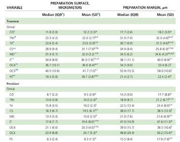
04

Accuracy of complete- and partial-arch impressions
of actual intraoral scanning systems in-vitro
Study Background                                            Talking Points
• In-vitro study with local and global accuracy             • In certain aspects, Primescan™ was viewed as the
• Translucent, ceramic tooth model was used                   most accurate among the tested intraoral scanners
                                                              that were compared in an in-vitro study
• Primescan™, Omnicam®, TRIOS® 3, Medit i500,
  Carestream CS3600, iTero®                                 • In the peer group of intraoral scanners, which did not
                                                              cover several systems commercially available today,
                                                              Primescan™ showed the best median and mean values
                                                              across complete arch, anterior and posterior segments,
                                                              few statistical limitations apply
                                                            • Omnicam® results have significantly improved with the
                                                              latest CEREC SW 5

  Abstract
  Objective                                                 were evaluated using a three-dimensional (3D)
  Intraoral scanners (IOSs) are widely used for obtaining   superimposition method with special 3D difference
  digital dental models directly from the patient.          analysis software (GOM Inspect) using (90-10)/2
  Additionally, improvements in IOSs are made from          percentile values. Statistical analysis was performed
  generation to generation. The aim of this study was to    using either one-way analysis of variance (ANOVA) or
  evaluate the accuracy of new and actual IOS devices       Kruskal-Wallis test (α = 0.05). Results are given as
  for complete- and partial-arch dental impressions in an   median and interquartile range [IQR] values in µm.
  in-vitro setup.
                                                            Results
  Materials and methods                                     Statistically significant differences were found between
  A custom maxillary complete-arch cast with teeth          test groups for complete- and partial-arch impression
  made from feldspar ceramic material was used as the       methods in-vitro (p < 0.05). Values ranged from
  reference cast and digitized with a reference scanner     16.3 [2.8] µm (CO) up to 89.8 [26.1] µm (OC4) for
  (ATOS III Triple Scan MV60). One conventional             in-vitro trueness, and from 10.6 [3.8] µm (CO) up to
  impression technique using polyvinylsiloxane (PVS)        58.6 [38.4] µm (iT) for in-vitro precision for the
  material (President) served as the control (CO), and      complete-arch methods. The best values for trueness
  eight different IOS devices comprising different          of partial-arch impressions were found for the posterior
  hardware and software configurations (TRn: TRIOS® 3;      segment, with 9.7 [1.2] µm for the conventional
  TRi: TRIOS® 3 insane; Carestream CS: Carestream           impression method (CO), and 21.9 [1.5] µm (PS) for the
  Dental Carestream CS 3600; MD: Medit i500; iT: iTero®     digital impression method.
  Element® 2; OC4: CEREC Omnicam® 4.6.1; OC5: CEREC
  Omnicam® 5.0.0; PS: Primescan™) were used to take         Conclusion
  complete-arch impressions from the reference cast.        Within the limitations of this study, digital impressions
  The impressions were repeated 10 times (n = 10) for       obtained from specific IOSs are a valid alternative to
  each group. Conventional impressions were poured          conventional impressions for partial-arch segments.
  with type IV gypsum and digitized with a laboratory       Complete-arch impressions are still challenging for IOS
  scanner (inEos X5). All datasets were obtained in         devices; however, certain devices were shown to be
  standard tessellation language (STL) file format and      well within the required range for clinical quality.
  cut to either complete-arch, anterior segment, or         Further in-vivo studies are needed to support these
  posterior segment areas for respective analysis. Values   results.
  for trueness and precision for the respective areas

Go to study: https://ijcd.quintessenz.de/ijcd_2019_01_s0011.pdf                               Back to Table of Contents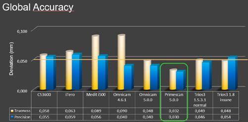
05
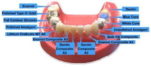
The effect different substrates have on the trueness
and precision of eight different intraoral scanners
Study Background Talking Points
• In-vitro study with local and global accuracy • Except for TRIOS® 3, substrate influences trueness and
• Primescan™, Omnicam®, TRIOS® 3, Element2, Medit precision -> doesn’t say anything about the level of
i500, Emerald™, Emerald™ S accuracy
• Dentin, Enamel, Gold, Amalgam, Resin, Zirconia, • Different scanners show different accuracy for same
Lithium Disilicate, Enamel/Dentin Composite, White/ substrate
Blue Core, Bulk Fill Composite • Latest generation scanners more accurate than older
• 3D best fit alignment scanners
• Average of the absolute values of the average positive • Primescan™ ranked #1 in 11 out of 15 categories
and negative deviations of the IOS data. • Amongst those the important categories: Enamel,
Dentin, Cross arch
• Primescan™ ranked within top 4 for remaining
4 categories
• Omnicam® was used with an old SW version, results
are expected to be significantly better with latest
version
• Study supports the proven accuracy of Primescan™
once again
Abstract
Objective Results
This in-vitro study compares the newest generation of For all scanners tested, except TRIOS® 3, the substrate
intraoral scanners to their older counterparts, and tests does influence the trueness and precision of the scan.
whether material substrates affect the trueness and Furthermore, differences exist when comparing the
precision of intraoral scanners (IOS). same substrate across different scanners with some of
the latest generation scanners clearly leaping ahead of
Material and methods the older generation regarding both trueness and
A custom model, used as the reference standard, was precision.
fabricated with teeth composed of different dental
materials. The reference standard scan was obtained Conclusions
using a three dimensional (3D) optical scanner, the Substrate type affects the trueness and precision of a
ATOS III. Experimental scans were obtained using eight scan. Active Triangulation scanners are more sensitive
different IOS, operated by experienced clinicians, using to substrate differences than their parallel confocal
the manufacturer‘s recommended scanning strategy. counterparts. Some scanners scan certain substrates
A comprehensive metrology program, Geomagic better, but in general the new generation of scanners
Control X, was used to compare the reference standard outperforms the old, across all substrates.
scan with the experimental scans.
Clinical significance
The substrates being scanned play an import role in
the trueness and precision of the 3D model. The new
generation of scanners is remarkably accurate across
all substrates and for complete arch scanning.
Go to study: https://onlinelibrary.wiley.com/doi/full/10.1111/jerd.12528 Back to Table of Contents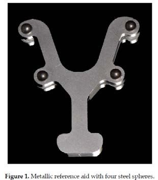
06

Do “cut out-rescan” procedures have an impact on
the accuracy of intraoral digital scans?
Study Background                                              Talking Points
• Complete-arch scan data of a maxillary master cast          • The t test revealed statistically significant differences
  were generated 10 times with 3 intraoral scanners:            among the different scanners
  TRIOS® 3 [TR], CEREC Primescan™ [PR], and                   • The comparison of the trueness values of the
  CEREC Omnicam® [OM].                                          complete arch scan data with those of the
• For the “cut-out-rescan”:                                     corresponding “cut out-rescanned” data of each
 •	all complete arch scans were duplicated                     scanner system did not reveal statistically significant
                                                                differences in any scanner system
 •	the posterior area from the right lateral incisor
    was cut out from the duplicated scan data and             • Significant differences were found between the
    rescanned                                                   precision results of the OM and PR as well as for the
                                                                pairs OM_rs/TR_rs and TR_rs/PR_rs
 •	superimposition of the rescanned area onto the
    cut-out scan ([TR_rs], [PR_rs], [OM_rs])
• As reference the master cast was scanned with a high
  precision industrial structured light scanner
• Evaluation of trueness and precision
• To evaluate statistical differences, either the Mann-
  Whitney U test or the t test was used (α=.05)

  Abstract
  Statement of problem                                        Results
  The software of digital intraoral scanners typically        The median precision values of the complete-arch scan
  offers the option to cut out areas from 3D casts, to do     data was 19 μm for [OM] and [TR], whereas the median
  rescans, and to merge them with the initial scan.           for [PR] was 14 μm. In the “cut out-rescanned” data
  However, evidence of whether this procedure has an          group, the values were 25 μm for [OM_rs], 16 μm for
  impact on the accuracy of the scan is lacking.              [TR_rs], and 14 μm for [PR_rs]. Statistically significant
                                                              differences were found among the scanners [OM]/
  Purpose                                                     [PR], [OM_rs]/[TR_rs], and [TR_rs]/[PR_rs]. The mean
  The purpose of this study was to determine whether          ± standard deviation values of trueness for the
  “cut out-rescan” procedures change the accuracy of a        complete-arch scan data were 54 ±4 μm for [OM],
  3D cast.                                                    42 ±5 μm for [TR], and 30 ±2 μm for [PR]. In the group
                                                              of the “cut out-rescanned” data, the mean trueness
  Material and methods                                        results were 55 ± 6 μm for [OM_rs], 38 ±5 μm for
                                                              [TR_rs], and 31 ±5 μm for [PR_rs]. Significant
  A maxillary master cast was digitized with an industrial    differences were found among the complete-arch scan
  structured light scanner to obtain a digital reference      data and the “cut out-rescanned” data of the different
  cast. This master cast was repeatedly scanned by 3          scanners, but not between the complete-arch scan
  intraoral scanners: TRIOS® 3 [TR], CEREC Primescan™         data and the “cut out-rescanned” data within one
  [PR], and CEREC Omnicam® [OM]. The scan data were           scanning system.
  duplicated, and the posterior area from the right lateral
  incisor was cut out and rescanned to obtain complete-
  arch casts containing the rescanned data [TR_rs],           Conclusions
  [PR_rs], and [OM_rs]. The trueness and precision of         Significant differences were found among the scanners,
  the scans were evaluated by superimposing procedures        but “cut out-rescan” procedures did not affect the
  of the relevant data sets. To evaluate statistical          accuracy within each scanning system.
  differences, either the Mann-Whitney U test or the t
  test was used (α=.05).

Go to study: https://www.sciencedirect.com/science/article/abs/pii/S0022391319307553             Back to Table of Contents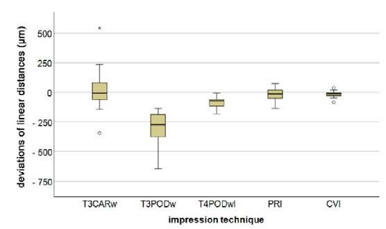
07

Impact of different scanning strategies on the
accuracy of two current intraoral scanning systems
in complete-arch impressions: an in-vitro study
Study Background                                               Talking Points
• A customized complete-arch maxillary cast was                • This scan strategy has very good value and is easy
  scanned                                                        to use.
• A master reference scan was obtained through an              • Primescan™ featured a better trueness index
  ATOS III Triple Scan 3D optical scanner                        (4.79 µm) than that of Omnicam® (19.13 µm).
• Omnicam® (CEREC SW 5.1.0) and Primescan™                       Primescan™, also featured a better precision
  (CEREC SW 5.0.2) were used for complete-arch                   index (4.67 µm) than Omnicam®, group B (16.75 µm),
  scanning with 13 different scanning strategies                 with a statistically significant difference.

• Best fit alignment of the scans with master scan
• Evaluation of trueness and precision
• Statistical analyses utilized Welch‘s unequal variances
  t test

  Abstract
  Aim                                                          Results
  To determine the scanning strategy that obtains the          Group M exhibited the lowest trueness and precision
  most accurate results for two intraoral scanners (IOS)       values (P < 0.05) for Primescan™ (47.5% of the average
  in complete-arch digital impressions. Scan time was          among all other groups) and the lowest trueness value
  evaluated and correlated with scan strategies.               (P < 0.05) for Omnicam® (53.4% of the average among
                                                               all other groups), where group B exhibited the lowest
  Materials and method                                         precision value (65.6% of the average among all other
  A custom model used as the reference standard was            groups) with P < 0.05. Primescan™ featured a better
  fabricated with teeth having dentin- and enamel-             trueness index (4.79 µm) than that of Omnicam®
  identical refractive indices simulating natural dentition.   (19.13 µm), with a statistically significant difference
  A reference scan of the custom typodont was obtained         (P < 0.00001). Primescan™, group M, also featured a
  using an ATOS III Triple Scan 3D optical scanner. Two        better precision index (4.67 µm) than Omnicam®,
  IOS setups – Omnicam® v 5.1.0 and Primescan™ v 5.0.2         group B (16.75 µm), with a statistically significant
  – were used for complete-arch scanning, each using 13        difference (P < 0.00001).
  scanning strategies, obtaining 260 digital files (n = 10
  per group), recording each scan time, converting all         Conclusion
  experimental scans to standard tessellation language         For both IOS systems, group M provided the lowest
  (STL) format, and using a comprehensive metrology            scanning times. For trueness and precision of
  program to compare the reference standard scan with          complete-arch scans, group M was the dominant
  the experimental scans. Statistical analyses utilized        scanning strategy in Primescan™, while there was no
  Welch‘s unequal variances t test.                            dominant strategy in Omnicam®. Group M had the best
                                                               scanning time for both IOS systems.

Go to study: https://www.ncbi.nlm.nih.gov/pubmed/31840139                                      Back to Table of Contents
08
In-vitro study on digital splint effect to the accuracy
of digital dental implant impression
Abstract
Background of hardened Fuji Plus® cement was glued in edentulous
Digital implant impressions (DII) with intraoral scanners areas to form digital splint and all models were scanned
(IOS) are a relatively novel, but continuously improving with three different IOS. Scanning data were exported
technique. Since IOS devices can only capture part of in standard tessellation language format for analysis.
the object at a time, images have to be stitched
together to form a 3D object and therefore it is the Results
source of possible errors of the scan. Digital splinting Trueness of distance and angle in Carestream partially
at edentulous areas can possibly improve the accuracy edentulous models was 185 μm in the group with splint
of DII. and 280 μm without one and 0.22° in the group with
splint and 0.29° in the group without respectively.
Aim/Hypothesis Precision of distance and angle measurements in the
The aim of this in-vitro study was to compare the splint groups were 87 μm and 0.13°, in the groups
trueness and precision of three different IOS scanning without −202 μm and 0.25°. In fully edentulous models
partially and fully edentulous models with 2 or 4 trueness of distance varied 53–106 μm in the groups
implants with attached scan bodies and digital splints. with splint and 67–8 μm in the groups without. Trueness
of Primescan™ in partially edentulous models with
Material and Methods splints was 21 μm and 0.16° in distance and angular
Two types of maxilla models were printed with Asiga™ measurements. Without splints −27 μm and 0.21°. For
Max 3D printer. The first model was missing both fully edentulous models trueness and precision of
premolars and molars on the right side, so Straumann distance and angle was better n groups with splint
BL dental implants were inserted instead first premolar than without. Trueness of distance and angle of TRIOS®
(straight) and second molar (tilted 20° mesially). Four 3 in partially edentulous splinted models was 15 μm
implants were inserted in the second edentulous model and 0.3°;53 μm and 0.11° in unsplinted models
symmetrically at second incisors (straight) and first respectively.
molar areas (tilted 20° mesially). Scan bodies were
attached to the implants and models were scanned Conclusion and Clinical Implications
with Nikon Altera 10.7.6. coordinate measurement Primescan™ showed the best results of trueness and
machine (CMM) to form a reference scan. DII was taken precision of distance and angle measurements. Since
with a Primescan™ (version 5.0.1), Carestream CS 3600 digital splints improve the accuracy of DII, the impact
(version 3.1.0), TRIOS® 3 (version 1.18.2.10) IOS ten times of their forms and materials should be more researched.
each (n = 10) without digital splint. After that, tablets
Carestream CS 3600 (version 3.1.0), TRIOS® 3 (version 1.18.2.10), Asiga Max™ and Fuji Plus® are not registered trademarks of Dentsply Sirona Inc.
Go to study: https://onlinelibrary.wiley.com/doi/pdf/10.1111/clr.322_13509 Back to Table of Contents09
Local accuracy of actual intraoral scanning systems
for single-tooth preparations in-vitro
Study Background Talking Points
The authors evaluated the local accuracy of intraoral “We found statistically significant differences of CO for
scanning (IOS) systems for single-tooth preparation all IOS systems except PS. Among the IOS systems, our
impressions with an in-vitro setup. results showed that the PS group had higher trueness
for SU parameter, with median (IQR) of 19.4 (5.0) mm;
values were statistically significantly different from the
other IOS systems, except TRn and TRi.”
Abstract
Background Results
The authors evaluated the local accuracy of intraoral The authors found statistically significant differences
scanning (IOS) systems for single-tooth preparation for MA and SU among different test groups for both
impressions with an in-vitro setup. trueness and precision (P < .05). Median (interquartile
range) trueness values ranged from 11.8 (2.0) μm (CO)
Methods up to 40.5 (10.9) μm (CEREC Omnicam®, Version 5.0.0)
The authors digitized a mandibular complete-arch for SU parameter and from 17.7 (2.6) μm (CO) up to
model with 2 full-contour crowns and 2 multisurface 55.9 (15.5) μm (CEREC Omnicam®, Version 5.0.0) for
inlay preparations with a highly accurate reference MA parameter.
scanner. Teeth were made from zirconia-reinforced
glass ceramic material to simulate toothlike optical Conclusions
behavior. Impressions were obtained either IOS systems differ in terms of local accuracy.
conventionally (PRESIDENT Micosystem™, Coltène) or Preparation MA had higher deviations compared with
digitally using the IOS systems TRIOS® 3 and TRIOS® 3 preparation SU for all test groups.
using insane scan speed mode (3Shape), Medit i500,
Version 1.2.1 (Medit), iTero® Element® 2, Version 1.7 Practical implications
(Align Technology), Carestream CS 3600, Version 3.1.0 Trueness and precision values for both MA and SU of
(Carestream Dental), CEREC Omnicam®, Version 4.6.1, single-unit preparations are equal or close to CO
CEREC Omnicam®, Version 5.0.0, and Primescan™ impression for several IOS systems.
(Dentsply Sirona). Impressions were repeated 10 times
per test group. Conventional (CO) impressions were
poured with type IV gypsum and digitized with a
laboratory scanner. The authors evaluated trueness
and precision for preparation margin (MA) and
preparation surface (SU) using 3-dimensional
superimposition and 3-dimensional difference analysis
method using (95% – 5%) / 2 percentile values.
Statistical analysis was performed using Kruskal-Wallis
test. Results were presented as median (interquartile
range) values in micrometers.
Go to study: https://www.sciencedirect.com/science/article/abs/pii/S0002817719307664 Back to Table of Contents10 Accuracy of digital and conventional full-arch impressions in patients: an update Study Background Talking Points • Five patients with a complete lower dental arch were • For the two short distances in the posterior segments included in this invivo study. (i.e., spheres D1_2 and D3_4), digital had more precise • Four bearing steel spheres with a diameter of 5 mm results were found using digital compared with were reversibly luted on the teeth of the lower jaw conventional impressions. using a flowable composite • For long-span distances, the CVI technique provided • Subsequently, in every patient four digital full-arch the lowest deviation, although no significant difference impressions were taken using TRIOS® 3 Cart wired, was demonstrated for PRI and T4PODwl. TRIOS® 3 Pod wired, TRIOS® 4 Pod wireless and • Hardware components of the TRIOS® scanner Primescan™ as well as a high precision conventional exhibited an influence on accuracy. impression was taken • Distances between the single spheres were compared Abstract The aim of this clinical study was to update the deviation (SD) transfer accuracy ranged from 24.6 ± available data in the literature regarding the transfer 17.7 µm (CVI) to 204.5 ± 182.1 µm (TRIOS® 3 Pod). The accuracy (trueness/precision) of four current intraoral Primescan™ yielded the lowest deviation for digital scanners (IOS) equipped with the latest software impressions (33.8 ± 31.5 µm), followed by TRIOS® 4 versions and to compare these data with conventional Pod (65.2 ± 52.9 µm), TRIOS® 3 Cart (84.7 ± 120.3 µm), impressions (CVI). A metallic reference aid served as a and TRIOS® 3 Pod. Within the limitations of this study, reference dataset. Four digital impressions (TRIOS® 3 current IOS equipped with the latest software versions Cart, TRIOS® 3 Pod, TRIOS® 4 Pod, and Primescan™) demonstrated less deviation for short-span distances and one CVI were investigated in five patients. Scan compared with the conventional impression technique. data were analyzed using three-dimensional analysis However, for long-span distances, the conventional software and conventional models using a coordinate impression technique provided the lowest deviation. measurement machine. The transfer accuracy between Overall, currently available IOS systems demonstrated the reference aid and the impression methods were improvement regarding transfer accuracy of full-arch compared. Differences with p < 0.05 were considered scans in patients. to be statistically significant. Overall, mean ± standard Go to study: https://www.ncbi.nlm.nih.gov/pubmed/32143433 Back to Table of Contents
11

Digital versus conventional impression taking
Focusing on interdental areas: a clinical trial
Study Background                                              Talking Points
• Overcome limitations of in-vitro study                      • IOS can display higher percentage of IAs then CVI
• Compare the ability of one conventional and four            • IAs in the anterior area of the jaw are better displayed
  digital impression techniques to reproduce Interdental        than in the posterior area by IOS
  Areas (IA) of periodontally compromised dentitions          • A higher percentage of IA was displayed for class III
  (PCD)                                                         PCD
• In-vivo, 30 patients, 1 experienced operator                • True definition displayed a higher percentage of IAs
• Four digital impressions were taken for each jaw with         but requires application of optical powder for
  3M True Definition, Primescan™, Carestream CS 3600,           impression taking
  TRIOS® 3                                                    • Primescan™ and Carestream CS 3600 displayed the
• Comparison against digitized conventional impression          highest percentage of IA amongst the powder-free
• 3D best-fit alignment                                         IOS

• Calculation of percentage of displayed IA in relation to    • TRIOS® 3 displayed the lowest percentage of IA
  absolute IA                                                   compared to all other IOS

  Abstract
  Due to the high prevalence of periodontitis, dentists       Primescan™ (PRI), Carestream CS 3600 (CAR), and
  have to face a larger group of patients with periodon-      TRIOS® 3 (TIO). The gypsum models of the CVIs were
  tally compromised dentitions (PCDs) characterized by        digitalized using a laboratory scanner. Subsequently,
  pathologic tooth migration and malocclusion. Impres-        the percentage of the displayed IAs in relation to the
  sion taking in these patients is challenging due to seve-   absolute IAs was calculated for the five impression
  ral undercuts and extensive interdental areas (IAs). The    techniques in a three-dimensionalmeasuring software.
  aim of this clinical trial was to analyze the ability of    Significant differences were observed among the
  analog and digital impression techniques to display         impression techniques (except between PRI and CAR,
  the IAs in PCDs. The upper and the lower jaws of 30         p-value < 0.05). TRU displayed the highest percentage
  patients (n = 60, age: 48–87 years) were investigated       of IAs, followed by PRI, CAR, TIO, and CVI. The results
  with one conventional impression (CVI) using polyvinyl      indicated that the IOSs are superior to CVI regarding
  siloxane and four digital impressions with intraoral        the ability to display the IAs in PCDs.
  scanners (IOSs), namely 3M True Definition (TRU),

Go to study: https://www.mdpi.com/1660-4601/17/13/4725                                         Back to Table of Contents
12

Congruence between meshes and library files of
implant scanbodies: an in-vitro study comparing
five intraoral scanners
Study Background                                            Talking Points
• Assess and compare reliability of five different IOS in   • Primescan™ and Carestream CS 3700 showed the
  the capture of implant Scanbodies (SB)                      highest congruence between SB MEs and LF, with the
• Verify dimensional congruence between meshes of SB          lowest mean absolute deviations
  captured during scan of a complete arch model with        • Statistically significant difference between these two
  six implants and the corresponding library file             scanners and the other three
• In-vitro                                                  • Primescan™ was the IOS with the lowest mean
• Gypsum cast representing a fully endentulous maxilla        absolute deviation but the difference to Carestream
  with 6 implant was scanned with: Primescan™,                CS 3700 was statistically not significant
  Carestream CS 3700, Medit i-500, iTero® Elements® 5D,
  Emerald™ S
• 3D analysis of the congruence between scanned mesh
  of SB and SB library file, best fit alignment
• Calculation of quantitative and qualitative deviation
  between scanned mesh of SB and SB library file

  Abstract
  Purpose                                                   Primescan™ showed a significantly higher congruence
  To compare the reliability of five different intraoral    than Medit i-500 (29.8 ± 4.8 μm, p < 0.0001), iTero®
  scanners (IOSs) in the capture of implant scanbodies      Elements® 5D (34.2 ± 9.3 μm, p < 0.0001), and Emerald™
  (SBs) and to verify the dimensional congruence            S (38.3 ± 7.8 μm, p < 0.0001). Carestream CS 3700 had
  between the meshes (MEs) of the SBs and the               a significantly higher congruence than Medit i-500 (p
  corresponding library file (LF).                          = 0.0004), iTero® Elements® 5D (p < 0.0001), and
                                                            Emerald™ S (p < 0.0001). Significant differences were
  Methods                                                   also found between Medit i-500 and iTero® Elements®
                                                            5D (p < 0.0001), Medit i-500 and Emerald™ S (p <
  A gypsum cast of a fully edentulous maxilla with six      0.0001), and iTero® Elements® 5D and Emerald™ S (p <
  implant analogues and SBs screwed on was scanned          0.0001). Significant differences were found among
  with five different IOSs (Primescan™, Carestream CS       different SBs when scanned with the same IOS. The
  3700, Medit i-500, iTero® Elements® 5D, and Emerald™      deviations of the IOSs showed different directions and
  S). Ten scans were taken for each IOS. The resulting      patterns. With Primescan™, iTero® Elements® 5D, and
  MEs were imported to reverse engineering software         Emerald™ S, the MEs were included inside the LF; with
  for 3D analysis, consisting of the superimposition of     Carestream CS 3700, the LF was included in the MEs.
  the SB LF onto each SB ME. Then, a quantitative and       Medit i-500 showed interpolation between the MEs
  qualitative evaluation of the deviations between MEs      and LF, with no clear direction for the deviation.
  and LF was performed. A careful statistical analysis
  was performed.                                            Conclusions

  Results                                                   Statistically different levels of congruence were found
                                                            between the SB MEs and the corresponding LF when
  Primescan™ showed the highest congruence between          using different IOSs. Significant differences were also
  SB MEs and LF, with the lowest mean absolute              found between different SBs when scanned with the
  deviation (25.5 ± 5.0 μm), immediately followed by        same IOS. Finally, the qualitative evaluation revealed
  Carestream CS 3700 (27.0 ± 4.3 μm); the difference        different directions and patterns for the five IOSs.
  between them was not significant (p = 0.1235).

Go to study: https://pubmed.ncbi.nlm.nih.gov/32660070/                                       Back to Table of Contents
13
Accuracy of intraoral scanning in completely and
partially edentulous maxillary and mandibular jaws:
an in-vitro analysis
Study Background Talking Points
• Analyze the accuracy (trueness and precision) of IOS • Overall median trueness comprising of all digital scans
in completely and partially edentulous maxillary and by the two operators was 24.2 μm (IQR 20.7 μm–27.4
mandibular models μm)
• Evaluated the influence of the operators’ experience • Significantly higher trueness was found in the scans of
with this new generation IOS device on the scan the edentulous mandibular model by the
accuracy and scan time inexperienced operator
• Resin models: edentulous and partially edentulous, • No differences were detected among the other scans
mandibular and maxillary models • Overall median precision was 18.3 μm
• Digital scans were performed by two specialist (IQR14.4–22.1 μm)
prosthodontists, one experienced and one • A significantly higher precision was found for the
inexperienced in IOS. Neither of the clinicians had ever scans of the edentulous maxillary model by the
used the tested IOS device before inexperienced operator
• For the reference data, all models were digitized using • No differences were detected among the other scans
an industrial high-precision scanner
• Overall median scan time was 100.5 s (IQR 72.0,139.2 s)
• Determination of trueness and precision
• Scans of experienced operator were faster than the
scans of inexperienced operator
• Longer scan times could be associated with a higher
level of trueness
Abstract
Objectives Results
New generation intraoral scanners are promoted to be The median overall trueness and precision were 24.2
suitable for digital scans of long-span edentulous μm (IQR 20.7-27.4 μm) and 18.3 μm (IQR 14.4-22.1 μm),
spaces and completely edentulous arches; however, respectively. The scans of the inexperienced operator
the evidence is lacking. The current study evaluated had significantly higher trueness in the edentulous
the accuracy of intraoral scanning (IOS) in partially mandibular model (p = 0.0001) and higher precision in
and completely edentulous arch models and analyzed the edentulous maxillary model (p = 0.0004).
the influence of operator experience on accuracy.
Conclusion
Materials and methods The accuracy of IOS for partially and completely
Four different resin models (completely and partially edentulous arches in in-vitro settings was high.
edentulous maxilla and mandible) were scanned, using Experience with IOS had small influence on the
a new generation IOS device (n = 20 each). Ten scans accuracy of the scans.
of each model were performed by an IOS-experienced
and an inexperienced operator. An industrial high- Clinical relevance
precision scanner was employed to obtain reference IOS with the tested new generation intraoral scanner
scans. IOS files of each model-operator combination, may be suitable for the fabrication of removable
their respective reference scan files (n = 10 each; total dentures regardless of clinician‘s experience in IOS.
= 80), as well as the IOS files from each model
generated by the same operator, were superimposed
(n = 45; total = 360) to calculate trueness and precision.
An ANOVA for mixed models and post hoc t tests for
mixed models were used to assess group-wise
differences (α = 0.05).
Go to study: https://pubmed.ncbi.nlm.nih.gov/32812098/ Back to Table of Contents14
Accuracy of three intraoral scans for primary
impressions of edentulous jaws
Abstract
Objective μm for mandible. There was no significant difference in
To provide a reference for using intraoral scanners for trueness of the three scanners when scanning the
making clinical diagnostic dentures of edentulous jaws maxilla and mandible (P>0.05). The precision of the
by comparing the accuracy of three intraoral scanners three scanners was 147.65 (156.30) μm, (147.54±83.33)
for primary impression and jaw relation record of μm, and 40.30 (32.80) μm for maxilla; (90.96±30.77)
edentulous jaws. μm, (53.73±23.56) μm, and 37.60 (93.93) μm for
mandible. The precision of CEREC Primescan™ scanner
Methods was significantly better than that of the other two
scanners for maxilla (P15
Trueness of 12 intraoral scanners in the full-arch
implant impression: a comparative in-vitro study
Study Background Talking Points
• Assessment and comparison of the trueness of • Primescan™ belonged to the group of IOS with the
12 different IOSs in full arch (FA) impression: (iTero® highest accuracy (together with iTero® Elements® 5D,
Elements® 5D, Primescan™, Omnicam®, Carestream CS Carestream CS 3700, Carestream CS 3600, TRIOS® 3,
3700, Carestream CS 3600, TRIOS® 3, Medit i-500, Medit i-500)
Emerald™ S, Emerald™, Virtuo Vivo™, DWIO, RUNEYES – With average intrinsic error < 40 μm with the mesh/
QUICKSCAN) mesh method and < 25 μm with the nurbs/nurbs
• Using a type IV gypsum model representing a totally method, representing a theoretically compatible
edentulous maxilla with 6 implant analogues and solution for taking impressions for FA restorations.
PEEK ScanBodies screwed on • In the analysis of the overall trueness with the nurbs/
• Reference virtual models in STL were aquired by a nurbs method Primescan™ belonged to the three best
desktop scanner IOS (together with iTero® Elements® 5D and TRIOS® 3)
• A single operator captured the scans with each of the – With no statistically significant difference between
IOSs the IOS (for α=00.05)
• Evaluation of overall generall trueness via mesh/mesh • The best absolute performance with mesh/mesh
and nurbs/nurbs method method was obtained by Carestream CS 3700, iTero®
• The evaluation of the linear and cross distances Elements® and Medit i-500
between the different SBs, for analysis of the local – Only iTero® Elements® 5D was significantly different
trueness of the intraoral scanning models to Primescan™ (for α=00.05) with a mean difference
of 7 µm
– Carestream CS 3700 and Medit i-500 were not
significantly different to Primescan™ (for α=00.05)
• For the cross-distance method, the distance category
S2-S4 is missing which could cause a bias in the
results.
• Primescan™ has the lowest mean error value in „Linear
distances method“ (see table 5)
• Best performance for the cross-distance method was
obtained by iTero® Elements® 5D and Medit i-500 but
with no statistically significant difference to
Primescan™ (for α=00.05)
• In general, the selected model type (gypsum) enables
good scanning results for all intraoral scanners applied
in this study
• Other factors are important in determining the
reliability of an optical impression including the
operator, patient, environnemental conditions and SB.
Further studies are therefore necessary to understand
the weight of each factor.
Go to study: https://bmcoralhealth.biomedcentral.com/track/pdf/10.1186/s12903-020-01254-9 Back to Table of Contents16
Trueness of 12 intraoral scanners in the full-arch
implant impression: a comparative in-vitro study
Abstract
Objective Results
The literature has not yet validated the use of intraoral With the mesh/mesh method, the best results were
scanners (IOSs) for full-arch (FA) implant impression. obtained by Carestream CS 3700 (mean error 30.4
Hence, the aim of this in-vitro study was to assess and μm) followed by iTero® Elements® 5D (31.4 μm), Medit
compare the trueness of 12 different IOSs in FA implant i-500 (32.2 μm), TRIOS® 3 (36.4 μm), Carestream CS
impression. 3600 (36.5 μm), Primescan™ (38.4 μm), Virtuo Vivo™
(43.8 μm), RUNEYES® (44.4 μm), Emerald™ S (52.9
Materials and methods μm), Emerald™ (76.1 μm), Omnicam® (79.6 μm) and
A stone-cast model of a totally edentulous maxilla with DWIO® (98.4 μm). With the nurbs/nurbs method, the
6 implant analogues and scanbodies (SBs) was best results were obtained by iTero® Elements® 5D
scanned with a desktop scanner (Freedom UHD®) to (mean error 16.1 μm), followed by Primescan™ (19.3
capture a reference model (RM), and with 12 IOSs μm), TRIOS® 3 (20.2 μm), Medit i-500 (20.8 μm),
(iTero® Elements® 5D; Primescan™ and Omnicam®; Carestream CS 3700 (21.9 μm), Carestream CS 3600
Carestream CS 3700 and Carestream CS 3600; TRIOS® (24.4 μm), Virtuo Vivo™ (32.0 μm), RUNEYES® (33.9
3; Medit i-500; Emerald™ S and Emerald™; Virtuo Vivo™ μm), Emerald™ S (36.8 μm), Omnicam® (47.0 μm),
and DWIO®; RUNEYES QUICKSCAN®). Ten scans were Emerald™ (51.9 μm) and DWIO® (69.9 μm). Statistically
taken using each IOS, and each was compared to the significant differences were found between the IOSs.
RM, to evaluate trueness. A mesh/mesh method and a Linear and cross distances between the SBs (local
nurbs/nurbs method were used to evaluate the overall trueness analysis) confirmed the data that emerged
trueness of the scans; linear and cross distances from the overall trueness evaluation.
between the SBs were used to evaluate the local
trueness of the scans. The analysis was performed Conclusion
using reverse engineering software (Artec Studio Different levels of trueness were found among the
software, Geomagic software, and Materialise Magics IOSs evaluated in this study. Further studies are needed
software). A statistical evaluation was performed. to confirm these results.
Go to study: https://bmcoralhealth.biomedcentral.com/track/pdf/10.1186/s12903-020-01254-9 Back to Table of Contents17
Comparing the accuracy of six intraoral scanners
on prepared teeth and effect of scanning sequence
Study Background Talking Points
• Using a maxillary complete arch model, right and left • The statistically higher trueness was obtained from
canine teeth prepared for single crowns Primescan™ (25 μm), followed by TRIOS® (40.5 μm),
• Using a highly accurate industrial reference scanner to Omnicam® (41.5 μm), Virtuo Vivo™ (52 μm), iTero®
create digital reference (70 μm), and Planmeca Emerald™ (73.5 μm)
• Six IOSs (TRIOS® 3, iTero® Element® 2, CEREC – There was no statistically significant difference
Omnicam®, Planmeca Emerald™, Primescan™, Virtuo between TRIOS®, Omnicam®, Virtuo Vivo™, and
Vivo™) were used to investigate precision and trueness iTero® (P > .003)
• Ten scans were taken of the model using each intraoral • The highest precision was obtained from Primescan™
scanner. The first 5 scans started from the right (10 ± 2 μm), followed by TRIOS® (11 ± 3 μm), iTero®
maxillary quadrant (Scan Right-ScanR) and the (12 ± 3 μm),Omnicam® (18 ± 5 μm), Virtuo Vivo™
following 5 scans started from the left maxillary (37 ± 19 μm), and Planmeca Emerald™ (60 ± 27 μm).
quadrant (Scan Left- ScanL) to evaluate effect of – There was no statistically significant difference
scanning sequence between Primescan™, TRIOS®, iTero®, and Omnicam®.
• For trueness, models were superimposed on the – The difference between Primescan™ and Planmeca
reference model using a best-fit algorithm Emerald™ and Virtuo Vivo™ was statistically
• For precision, a two-way pairwise comparison was significant.
performed • No significant difference was found between the
precision and trueness values of the ScanR and ScanL
obtained from each IOS for the prepared teeth
Abstract
Objective analyzing software (Geomagic Studio 12, 3D Systems).
The aim of this study was to evaluate the accuracy of The Kruskal Wallis and Mann-Whitney U statistical
six recently introduced intraoral scanners (IOSs) for tests for trueness analysis and the One-way ANOVA
single crown preparations isolated from the complete test for precision analysis were performed (α=.05).
arch, and to determine the effect of scanning sequence
on accuracy. Results
The trueness and precision values were the lowest with
Materials and methods the Primescan™ (25 and 10 µm), followed by TRIOS®
(40.5 and 11 µm), Omnicam® (41.5 µm and 18 µm),
A complete arch with right and left canine preparations Virtuo Vivo™ (52 and 37 µm), iTero® (70 and 12 µm) and
for single crowns was used as a study model. The Planmeca Emerald™ (73.5 and 60 µm). Regarding
reference dataset was obtained by scanning the trueness, iTero® showed more deviation when scanning
complete arch using a highly accurate industrial started from the right (P=.009).
scanner (ATOS Core 80, GOM GmbH). Six different
IOSs (TRIOS®, iTero®, Planmeca Emerald™, CEREC Conclusion
Omnicam®, Primescan™, and Virtuo Vivo™) were used
to scan the model ten times each. The scans performed The accuracy of digital impressions varied depending
with each IOS were divided into two groups, based on on the IOS and scanning sequence used. Primescan™
whether the scanning sequence started from the right had the highest accuracy, while Planmeca Emerald™
or left quadrant (n=5). The accuracy of digital showed the most deviation in accuracy for single
impression was evaluated using three-dimensional crown preparations.
Go to study: https://www.ncbi.nlm.nih.gov/pmc/articles/PMC7604233/ Back to Table of Contents18
In-vitro analysis of intraoral digital impression of
inlay preparation according to tooth location and
cavity type
Study Background Talking Points
• Evaluate influence of tooth location and inlay cavity • Overall trueness for tooth 16 (average deviation:
type on the accuracy of digital intraoral impression 10.43 µm ± 0.39 µm) was higher than for tooth 46
• Teeth with inlay cavities were screw-retained on four (12.42 µm ± 0.59 µm)
typodont sets which were mounted on a phantom • Precision was similar between the teeth (tooth 16:
head during the scanning procedure 3.08 µm ± 0.92 µm; tooth 46: 3.08 µm ± 0.76 µm)
• 10 scans of each tooth with Primescan™ • The cavity type affected the trueness and precision
• Reference scan data was obtained by scanning with but with differences < 1 µm
a laboratory scanner (E3, 3Shape) which has an • In contrast to other in-vitro studies intraoral scanning
accuracy of 7 µm. was performed on the phantom head what might have
• Assessment of accuracy by trueness and precision. permitted less freedom while placing the scanning
walls. A greater degree of freedom ensures a direct-
• Best fit alignment line of sight, favorable angle of incidence which can
affect the quality of scan.
• The overall accuracy of digital impressions for inlay
preparations was clinically acceptable, but positive
deviations were observed at the margins of the
proximal boxes
Abstract
Objective Results
This study aimed to evaluate the influence of tooth The overall results showed that the trueness for 16
location and inlay cavity type on the accuracy of (10.43 ± 0.39 μm) was higher than that for 46 (12.42 ±
intraoral digital impressions. 0.59 μm) (p < 0.05), while the precision was similar
between 16 (3.08 ± 0.92 μm) and 46 (3.08 ± 0.76 μm).
Materials and methods The cavity type affected the accuracy of the digital
Class II inlay preparation was performed on anatomical impressions. The highest deviation was observed in
models of the maxillary first molar (16) and mandibular positive directions at the margins of the proximal
first molar (46). Mesio-occlusal and disto-occlusal boxes regardless of the cavity type.
cavities were prepared, such that the axial wall of
the proximal box measured 1 mm or 2 mm in height. Conclusion
Thus, four types of inlay cavities were prepared in 16 Tooth location and cavity type affected the accuracy
and 46, respectively. Ten digital impressions of each of intraoral digital impressions. Positive deviations
cavity were obtained using CEREC Primescan™ were observed at the margins of the proximal boxes.
(Dentsply Sirona).
Reference scans were obtained with a laboratory
scanner (E3, 3Shape). All scan data were exported for
comparative analysis of the three-dimensional models.
Mean absolute deviation values were calculated to
evaluate the trueness and precision of the digital
models. Color-coded maps were used for the
qualitative analysis of deviations.
Go to study: https://www.jstage.jst.go.jp/article/jpr/advpub/0/advpub_JPR_D_20_00169/_article Back to Table of Contents19
Accuracy and repeatability of different intraoral
scanners on shade determination
Study Background Talking Points
• Evaluate the accuracy and repeatability of different • No statistical difference was found on the overall
intraoral scanners on shade determination in accuracy between the spectrophotometer Easyshade®
comparison to a dental spectrophotometer V (78%) and the scanner 3Shape TRIOS® (66%)
• Ten different shades (A1, A2, A3, A3.5, A4, B2, B3, C2, (p > 0.05), with the latter being similar to the other
C3, and D3) of VITABLOCS® Mark II monochromatic scanners Primescan™ (63%) and Omnicam® (57%)
CAD-CAM block were used (p > 0.05)
• One disc-shape specimen per ceramic block was • Scanner‘s accuracy was only significantly different on
milled and polished reading a specific shade (A4), with the Primescan™
(90%) showing greater accuracy than 3Shape TRIOS®
• Color measurements (n = 10) were performed to each (50%)
specimen using an intraoral spectrophotometer (VITA
Easyshade® V) and three intraoral scanners (3shape • There was no statistical difference on the overall
TRIOS®, CEREC Omnicam®, CEREC Primescan™) repeatability for the evaluated devices, ranging from
44.3% for Easyshade® V to 51.9% for Omnicam®
Abstract
Objective Results
To evaluate the accuracy and repeatability of different There was a significant difference in the instrumental
intraoral scanners on shade determination. accuracy for shade determination (p < 0.001). There
was no statistical difference between the Easyshade®
Materials and methods V (78%) and the 3Shape TRIOS® (66%) (p > 0.05), with
Ten different shades of VITABLOCS® Mark II the latter being similar to the other scanners
monochromatic CAD-CAM block were used. A disc- Primescan™ (63%) and Omnicam® (57%) (p > 0.05). No
shape specimen (10 mm in diameter and 1 mm thick) significant difference was found (p > 0.05) when
per ceramic block was fabricated. Ten color different shades were evaluated by the same
measurements per specimen were performed by each instrument. Similar repeatability was found for the
instrument (VITA Easyshade® V [control], 3shape different devices, ranging from 44.3% for VITA
TRIOS®, CEREC Omnicam®, CEREC Primescan™) and Easyshade® V to 51.9% for Omnicam®.
recorded in VITA Classic color system. The number of
correct shade match per instrument for each shade Conclusion
was recorded. Instrumental accuracy was compared The evaluated instruments showed less than expected
using Cochran Q test and repeatability was analyzed repeatability and accuracy on measuring different
using Cronbach‘s alpha. dental shades. Therefore, caution should be exercised
when using instrumental shade determination, which
should be accompanied by experienced human visual
assessment.
VITABLOCS® Mark II and VITA Easyshade® V are not registered trademarks of Dentsply Sirona Inc.
Go to study: https://pubmed.ncbi.nlm.nih.gov/33227179/ Back to Table of Contents20
Effect of pulp chamber depth on the accuracy of
endocrown scans made with different intraoral
scanners versus an industrial scanner: an in-vitro
study
Study Background Talking Points
• Evaluate the effect of different pulpal chamber • A statistically significant difference in the accuracy of
extension depth (PCEDs; 2, 3.5, 5 mm) and IOSs on endocrown cavities with different PCEDs was found
the scanning accuracy of endocrown preparations among compared IOSs, and PCED affected the
• Master reference scans of a model with specimens scanning accuracy significantly
were created by using an industrial structured blue • For all PCEDs evaluated, Primescan™ was found to
light 3D scanner (ATOS; GOM Technologies) have the best results among the tested IOSs with
• Experimental scans were made with 6 IOSs (TRIOS® 3, regard to trueness and precision
Primescan™, Omnicam®, iTero® Element® 2, Planmeca – Trueness and precision of Primescan™ were
Emerald™, Virtuo Vivo™, Rhinoceros®, Telio® and ATOS) significantly different in all cases.
• Trueness and Precision measurement Increasing the pulpal chamber extension depth of
endocrown preparations can reduce scanning accuracy.
CEREC Primescan™ appears to be the best IOS choice
for scanning endocrowns with deep pulpal chamber
extensions.
Abstract
Objective Results
The purpose of this in-vitro study was to assess the CEREC Primescan™ was found to have the best
effect of pulpal chamber extension depth (PCED) on trueness and precision among the evaluated IOSs
scanning accuracy and to compare the accuracy of (P21
Influence of preparation design, marginal gingiva
location, and tooth morphology on the accuracy
of digital Impressions for full-crown restorations:
an in-vitro investigation
Study Background Talking Points
• Analyze the influence of different finish lines for • The overall accuracy for all abutment teeth was
complete crown preparations, their locations related very high, without significant differences in the
to the gingival margin, and tooth morphology on the performance of 3Shape TRIOS® 3 Pod versus
accuracy of digital impressions Primescan™
• Maxillary dental training model was used as reference, • The supragingival finishing lines were captured
a maxillary central incisor (FDI 11) represented the significantly better than the epigingivally located
anterior tooth morphology, a first maxillary molar margins using IOS. If the clinical situation allows, a
(FDI 16) represented posterior sites supragingival margin should be chosen accordingly
• Prepared typodonts were digitized with a laboratory • The tooth morphology seems to be a negligible factor
desktop scanner and served as the basis for the digital for IOS accuracy in terms of single-unit complete
designs of the virtual modifications to create the test crown restorations
specimens, involving four different finish-line designs
for both morphologies
• 16 virtual tooth preparations were 3D-printed and
mounted in the reference model
• Scanning with Primescan™ and TRIOS® 3.5 times
• Accuracy determination
Abstract
Objective Results
Intraoral optical scanning (IOS) has gained increased Analysis revealed homogenous findings with high
importance in prosthodontics. The aim of this in-vitro accuracy for intra- and inter-group comparisons for
study was to analyze the IOS accuracy for treatment both IOS systems, with mean values of 80% quantiles
with full crowns, considering possible influencing from 20 ± 2 μm to 50 ± 5 μm. Supragingival finishing
factors. lines demonstrated significantly higher accuracy than
epigingival margins when comparing each preparation
Materials and methods (p < 0.05), whereas tangential preparations exhibited
Two tooth morphologies, each with four different similar results independent of the gingival location.
finish-line designs for tooth preparation and epi- or Morphology of anterior versus posterior teeth showed
supragingival locations, were digitally designed, slightly better results in favor of molars in combination
3D-printed, and post-processed for 16 sample with shoulder preparations only.
abutment teeth. Specimens were digitized using a
laboratory scanner to generate reference STLs Conclusion
(Standard Tessellation Language), and were secondary- The clinical challenge for the treatment with full crowns
scanned with two IOS systems five times each in a following digital impressions is the location of the pro-
complete-arch model scenario (TRIOS® 3 Pod, spective restoration margin related to the distance to
Primescan™ AC). For accuracy, a best-fit algorithm the gingiva. However, the overall accuracy for all abut-
(Final Surface) was used to analyze deviations of the ment teeth was very high; thus, the factors tested are
abutment teeth based on 160 IOS-STLs compared to unlikely to have a strong clinical impact.
the reference STLs (16 preparations × 2 IOS-systems ×
5 scans per tooth).
Go to study: https://www.ncbi.nlm.nih.gov/pmc/articles/PMC7763051/ Back to Table of ContentsYou can also read