Studying nuclear disassembly in vitro using Xenopus egg extract
←
→
Page content transcription
If your browser does not render page correctly, please read the page content below
Methods 39 (2006) 284–290
                                                                                                                       www.elsevier.com/locate/ymeth

          Studying nuclear disassembly in vitro using Xenopus egg extract
                                Meda M. Higa, Katharine S. Ullman ¤, Amy J. Prunuske
                   Department of Oncological Sciences, Huntsman Cancer Institute, University of Utah, Salt Lake City, UT 84112, USA

                                                                   Accepted 14 June 2006

Abstract

   Xenopus egg extract provides an extremely powerful approach in the study of cell cycle regulated aspects of nuclear form and function.
Each egg contains enough membrane and protein components to support multiple rounds of cell division. Remarkably, incubation of egg
extract with DNA in the presence of an energy regeneration system is suYcient to induce formation of a nuclear envelope around DNA. In
addition, these in vitro nuclei contain functional nuclear pore complexes, which form de novo and are capable of supporting nucleocytoplas-
mic transport. Mitotic entry can be induced by the addition of recombinant cyclin to an interphase extract. This initiates signaling that leads
to disassembly of the nuclei. Thus, this cell-free system can be used to decipher events involved in mitotic remodeling of the nuclear envelope
such as changes in nuclear pore permeability, dispersal of membrane, and disassembly of the lamina. Both general mechanisms and individual
players required for orchestrating these events can be identiWed via biochemical manipulation of the egg extract. Here, we describe a proce-
dure for the assembly and disassembly of in vitro nuclei, including the production of Xenopus egg extract and sperm chromatin DNA.
© 2006 Elsevier Inc. All rights reserved.

Keywords: Xenopus egg extract; Sperm chromatin DNA; Mitosis; Nuclear envelope breakdown; Disassembly; Prophase; Nuclear membrane; Nuclear
pore; Lamina; Nucleus

1. Introduction                                                                tion promoting factor. This factor is responsible for
                                                                               initiating the phosphorylation of nuclear pore proteins,
   In eukaryotic cells, genomic DNA is separated from the                      lamin proteins, and inner nuclear membrane proteins.
cytoplasm by the nuclear envelope [1]. This envelope is both                   Phosphorylation is accompanied by changes in pore perme-
the boundary and passageway between the nuclear and                            ability, disassembly of the nuclear lamina, microtubule-
cytoplasmic compartments. Selective traYcking occurs                           mediated gathering and tearing of the nuclear envelope,
through nuclear pore complexes (NPCs), which facilitate                        and dispersal of nuclear membranes [1–7]. Although these
movement of macromolecules between the two environ-                            basic events have been identiWed, we are still far from
ments. The nuclear envelope consists of an outer mem-                          understanding all the factors necessary for nuclear disas-
brane, which is continuous with the endoplasmic reticulum                      sembly and how these factors coordinate successful pro-
(ER), and an inner membrane. Directly beneath the inner                        gression into mitosis.
membrane is the nuclear lamina, a meshwork of lamin pro-                          In vitro studies of cell cycle processes and their regula-
teins that contributes to the structural integrity of the                      tion in eukaryotic cells have beneWted extensively from
nuclear envelope.                                                              Xenopus laevis egg extracts. Unfertilized Xenopus eggs are
   Higher eukaryotes have an open mitosis, in which                            arrested at metaphase of the second meiotic division and
nuclear structures are disassembled and then reassembled                       contain large stores of soluble proteins and membranes [8].
during the cell cycle. Nuclear envelope breakdown is initi-                    When eggs are crushed under conditions that promote
ated by activated Cdk1-cyclin B, also referred to as matura-                   cyclin degradation, the resulting egg extract is “interphase”
                                                                               in nature. When DNA is introduced into such an extract,
 *
     Corresponding author. Fax: +1 801 585 0900.                               along with an energy regeneration system, functional nuclei
     E-mail address: Katharine.ullman@hci.utah.edu (K.S. Ullman).              form. Many dynamic processes are recapitulated in this

1046-2023/$ - see front matter © 2006 Elsevier Inc. All rights reserved.
doi:10.1016/j.ymeth.2006.06.004
M.M. Higa et al. / Methods 39 (2006) 284–290 285
system, including nuclear assembly, DNA replication, and use a “crude” interphase egg extract, adding in cyclohexi-
nuclear transport. If the egg extract is not held in interphase mide to prevent new cyclin synthesis. This extract has gone
by the presence of translation inhibitors, nuclei will also through only a low speed spin, and is sometimes referred to
undergo disassembly as the extract cycles into mitosis. as an LSS, or “low speed supernatant” extract. The proto-
Alternatively, cyclin can be added to an extract arrested in col below incorporates our experiences and Wndings into
interphase in order to initiate mitotic events. Indeed, this previously published procedures for generating a crude egg
cell-free system has been utilized extensively by many labs extract.
to identify and characterize many cell cycle events. In our
laboratory, cell cycle studies using this in vitro technique 2.2. Materials
have focused on the mechanism and events surrounding
nuclear envelope breakdown at mitosis. • 3–4 Female frogs (from Nasco, or other Xenopus
There are several advantages to the use of this cell-free distributor).
system. Xenopus eggs and sperm chromatin are relatively • 6 L Tubs and lids (with several breathing holes in lid).
easy to obtain in large quantities, providing a constant • 250 ml Glass beakers.
and abundant source of protein, membranes, and DNA. • 14 ml Round-bottom Falcon tubes (Fisher, C/N
Egg extracts and chromatin can also be stored for use in 14-959-11B).
multiple experiments. These extracts are useful not only • 5 ml Round-bottom Falcon tubes (Fisher, C/N 14-959-
for functional studies but for biochemical dissection as 11A).
well, providing multiple avenues by which to approach a • IEC Centra CL2 Clinical centrifuge (or equivalent).
particular question. The nuclear assembly and disassem- • Beckman JS13.1 rotor.
bly assay allows cell cycle-dependent events to be probed • Beckman Avanti J-25 I centrifuge.
in a highly synchronous population. Particularly useful is • 3 ml or 10 ml Syringes (BD Biosciences, C/N BD309585
the ability to introduce dominant-negative proteins, anti- or BD309604).
bodies, or drugs in the assay to probe the role of particu- • 18 Gauge, 1 1/2 needle (Fisher C/N 305-196).
lar proteins. The ability to immuno-deplete a protein and • Human chorionic gonadotrophin (HCG) (Sigma, C/N
assess the eVect on speciWc processes is an additional ben- CG-10); 5 U/l in sterile distilled water.
eWt of this system. • 1£ MMR: 100 mM NaCl, 2 mM KCl, 1 mM MgSO4,
Naturally, there are some limitations to this system as 2 mM CaCl2, 0.1 mM EDTA, and 5 mM Hepes, pH 7.8.
well. Egg quantity and egg extract quality can be variable. • 0.25£ MMR.
Also, although it provides a robust recapitulation of • ELB (Egg lysis buVer): 2.5 mM MgCl2, 50 mM KCl,
nuclear events during the cell cycle, the egg extract system is 10 mM Hepes, pH 7.6, 250 mM sucrose, pH 7.4–7.5.
clearly a step away from the in vivo situation. Although it • 2% L-cysteine hydrochloride, pH 7.8–7.9 (prepare imme-
may initially be an advantage to study nuclear function diately before dejellying and pH with NaOH) (Fisher, C/
without the full complexity of the cellular milieu, any Wnd- N BP376-100).
ings should be examined in intact cells as well. It is also • 1 M dithiothreitol (DTT) (Fisher, C/N BP172-5).
worth bearing in mind that egg extract most closely mimics • Aprotinin and leupeptin (10 mg/ml) (Roche Applied
cellular conditions within the early embryo, and thus some Science, C/N 10 236 624 001 and 11 017 101 001).
diVerences may arise when comparing to somatic cells. • Cycloheximide in sterile distilled water (10 mg/ml)
Overall, as egg extracts are amenable to manipulation in (Calbiochem, C/N 239764).
ways not possible in intact cells, the insight gained about • Cytochalasin B in DMSO (5 mg/ml) (Calbiochem, C/N
basic mechanisms using these assays often provides valu- 250233).
able information not readily obtainable elsewhere. • Glycerol.
• Liquid nitrogen.
2. Description of method • Standard plastic bulb transfer pipette (Fisher, C/N
13-711-7).
2.1. Xenopus interphase egg extracts • 0.5 ml Microfuge tubes.
Cell-free extracts from Xenopus eggs are a useful tool in 2.3. Frog care and maintenance
studying many events such as DNA replication, spindle
assembly, nuclear assembly and disassembly, and nuclear Frogs are stored in municipal water which has been run
import and export. There are various procedures for mak- through a carbon Wlter, a water softener and a reverse
ing egg extracts depending on the experimental question osmosis Wlter. Synthetic ocean mix is added for sodium and
and strategy [9–13]. “Ultra-S” or high speed supernatants other minerals, and sodium bicarbonate to adjust the pH to
are more highly fractionated, CSF-arrested extracts hold 6.9–7.1 and the conductivity to 1.0–1.5 micro siemens. We
conditions at meiosis rather than interphase, and “cycling” also test weekly for nitrite and ammonia levels, which rep-
extracts permit several rounds of cell cycle driven by endog- resent the level of unprocessed waste. Frogs are stored at a
enous components. For our nuclear disassembly assay, we temperature of between 17 and 20 °C.286 M.M. Higa et al. / Methods 39 (2006) 284–290
2.4. Protocol 9. Add aprotinin, leupeptin, and cytochalasin B (each to
a Wnal concentration of 5 g/ml, estimated from the
1. In preparation for egg laying, frogs are primed ini- volume of packed eggs) to the top of the eggs in the
tially with 100 l of 1.6 U/l HCG subcutaneously in tube and centrifuge in a Beckman JS13.1 rotor at
dorsal lymph sacs (a shallow injection near top of 10,000 rpm (15,680g) for 15 min at 4 °C [note: the
thigh). After injection, frogs are placed into tanks rotor itself is held at room temperature until the spin
containing 100 mM NaCl to reduce the potential for starts]. This spin serves to crush the eggs and several
infection at the site of injection. At least seven days layers will form in the tube. The top will be a yellow
are needed to allow HCG priming to take eVect and lipid cap, followed by the crude cytoplasmic layer that
frogs should be used within one month. is generally tan to brown. Underneath that will be a
2. A day prior to making egg extracts, inject frogs sub- thin layer of pigment granules and dark yolk beneath
cutaneously in dorsal lymph sacs with 100 l of 5 U/l that (Fig. 1).
HCG. Place each injected frog in its own tank of frog 10. To retrieve the crude egg extract, an 18 gauge needle
water supplemented with 100 mM NaCl at 17–22 °C. is attached to a 3 or 10 ml syringe, depending on the
3. After 16–22 h, move frogs into new tanks and collect volume to be collected, and is inserted directly into
eggs from each frog in separate beakers, pouring oV the side of the round-bottom tube above the pigment
the excess frog water. We generally keep eggs from granules (see red arrowhead, Fig. 1), being careful not
each frog separate throughout the entire process or at to puncture both sides. If the needle gets plugged with
least until the crushing step, since the egg quality can plastic in this process, remove it and promptly place a
vary between frogs. Eggs should be fairly uniform in new needle with syringe into the same hole—only a
appearance with well deWned hemispheres. Bloated, few drops of extract will be lost. With the bevel side of
XuVy, or strings of eggs are not ideal and should be the needle facing up, the egg extract can be carefully
discarded with a plastic transfer pipette. taken up into the syringe stopping when the lipid
4. After removing excess water and bad eggs, add layer begins to be taken up. The syringe is also used to
freshly made 2% cysteine for no longer than 5 min, measure the amount of crude extract obtained.
swirling gently to facilitate the dejellying process. 11. Transfer the crude extract into a chilled 14 ml round-
Generally, about 100 ml of cysteine are used for every bottom tube. As before, add aprotinin, leupeptin, and
25 ml of eggs. When eggs are completely dejellied, cytochalasin B (each to a 5 g/ml Wnal concentration)
they pack tightly against each other and often the to the top of the sample and centrifuge as above in
buVer gets cloudy and contaminated with sediment. Beckman JS13.1 rotor at 10,000 rpm (15,680g) for
5. Pour cysteine oV and wash eggs three times with 15 min at 4 °C.
0.25£ MMR and once with 1£MMR, swirling 12. Aspirate oV any lipid remaining on the top and repeat
between each wash and allowing eggs to settle before the syringe extraction or simply remove crude extract
pouring oV the buVer. These washes should be com- with pipette tip and transfer into a clean, chilled 5 or
pleted as quickly as possible, so that eggs do not sit 14 ml round-bottom tube. Add glycerol to 5% Wnal
long in the cysteine or MMR buVers. concentration and mix by rotating at 4 °C until well
6. Rinse eggs once with ELB, pour oV buVer and add combined (5–10 min).
fresh ELB. At this point, any bad eggs should be 13. Make single-use aliquots of less than 100 l in 0.5 ml
removed with a plastic pipette. Bad eggs include those tubes on liquid nitrogen. Store in liquid nitrogen. Ali-
that are activated (mostly white) or XuVy and/or Xoat- quots can also be stored at ¡80 °C, with a shorter
ing. Rinse once more with ELB. shelf-life. Yields will vary but typically a good yield is
7. Exchange eggs into ELB supplemented with 1 mM 2–4 ml of egg extract per frog.
DTT and 50 g/ml cycloheximide. Decant as much
ELB as possible before swirling the eggs gently into 2.5. Notes on procedure
the remaining buVer and transferring them to a 14 ml
round-bottom tube (divide evenly between several 1. Our IACUC-approved protocol requires that frogs be
tubes if necessary). At this point, good batches of eggs returned to a Wltered tank system no more than 24 h
can be combined, if desired. After the eggs settle, from the time they were put into the transfer tank. Frogs
remove excess buVer from the top of the tube with then recover for at least 30 days before reuse. For best
plastic transfer pipette. results, wait at least 60+ days for recuperation. The
8. Spin eggs in a clinical centrifuge for 10–15 s at usual fertile lifespan for the Xenopus is 2–3 years, if they
800 rpm (»110g) and remove excess buVer that col- are allowed 3–4 months of oV time.
lects on top with a plastic transfer pipette to avoid 2. Success of egg extracts in further application is essen-
dilution of the egg extract. Pipette tips or a syringe tially deWned by the quality of the eggs after all washes.
with needle can also be used to aspirate excess buVer, Although this has not been exhaustively examined, we
but care must be taken to avoid breaking or losing the generally abandon egg batches that have spontaneously
eggs. activated during the course of washing (visualized byM.M. Higa et al. / Methods 39 (2006) 284–290 287
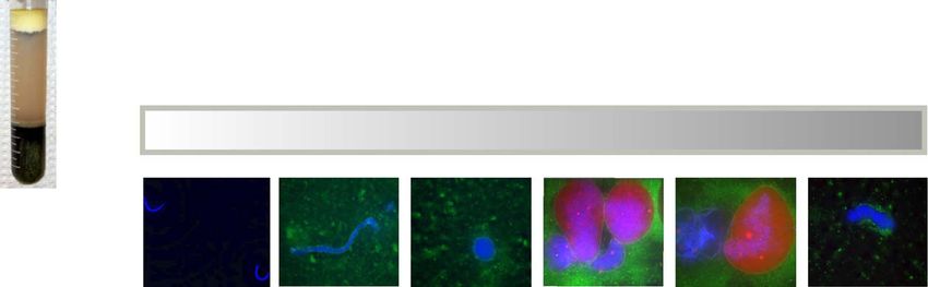
Fig. 1. Timeline for the nuclear assembly and disassembly assay. Xenopus eggs are fractionated into lipids (A), crude egg extract (B), and pigment granules
and yolk proteins (C). To collect the crude extract, a syringe is inserted into the bottom of the crude layer (indicated by the red arrowhead). For the assem-
bly assay, egg extract is Wrst pre-incubated with protein or drug of interest. In order to initiate assembly, an energy regeneration system and sperm chroma-
tin DNA are added to the extract (0⬘). Early in assembly the DNA (blue) decondenses and membrane vesicles (green) bind to the DNA (15⬘). As assembly
progresses, the DNA becomes more rounded and vesicles fuse to form a nuclear envelope (45⬘). Import substrate (red) is added at 60 min. In the next
30 min, the import substrate accumulates within the nuclei, which continue to add more membrane leading to signiWcant growth in size (90⬘). After 90 min
of assembly, cyclin is added to shift the extract into mitosis. Breakdown begins sometime around 135⬘ and is complete at 165⬘ as is indicated by condensa-
tion of the DNA, dispersal of the membrane, and release of the import substrate. These images were acquired with a Deltavision microscope and decon-
volved for 10 cycles. The scale bar indicates 15 M.
hyper-retraction of the pigment) or have more than 5% protocols suggest using crude extracts fresh. This alter-
bad eggs. native is Wne but it is worth noting that the kinetics of
3. Since a calcium-dependent signal must be activated dur- disassembly will likely be faster than in an extract that
ing the crushing step that ultimately leads to cyclin deg- has been frozen.
radation, we do not chill the eggs, the buVer, or the rotor
prior to the crushing step. The centrifuge is chilled so 3. Demembranated sperm chromatin
that by the end of the spin, the egg lysate is cold and we
keep it cold from this point on. An alternative method is Xenopus males are an excellent source of chromatin for
to pre-activate the eggs prior to crushing, either by incu- use in nuclear disassembly assays. Male frogs are smaller in
bation with calcium ionophore or electrical stimulation size and generally darker in pigment than their female
[13]. counterparts. A beneWt of using Xenopus sperm as a source
4. It is important to remove as much excess buVer as possi- of DNA is that, as external fertilizers, their testes contain
ble from the eggs after the packing step but before crush- large stores of easily harvested sperm. In addition, they are
ing. Alternatively, one can also spin eggs through the oil, obtained from the same suppliers as females and can be
Versilube F-50, to help pack the eggs and separate them housed similarly to females (though in separate tanks from
from the buVer [14]. Since this is not easily obtained, we females). Since each frog will yield about 1–2 £ 107 sperm
use Nyosil M25 (TAI Lubricants Inc., Hockessin, DE), heads and a single nuclear assembly reaction requires only
which is a commercially available equivalent. We have 100–1000 sperm heads per microliter of egg extract, this
found, however, that this is not necessary if time and protocol should provide an ample supply. The protocol
care is taken to remove as much buVer as possible with a below has been adapted from a previous protocol [15].
transfer pipette.
5. When obtaining the crude extract layer after the egg 3.1. Materials
crushing step, it is preferable to sacriWce complete recov-
ery of this fraction to avoid taking the pigmented layer • 2–4 Male X. laevis frogs (from Nasco, or other Xenopus
directly below. distributor).
6. To aliquot extracts on liquid nitrogen, we set up an • 60 mm Glass petri dish.
open-bottom rack on top of a shallow Styrofoam con- • Dissection scissors (Fine Science Tools C/N 14068-12).
tainer Wlled with enough liquid nitrogen to submerge the • 2 Pairs sharp forceps (Fine Science Tools C/N 11231-30
bottoms of the tubes. We then aliquot the egg extract or 11021-12).
into the partially submerged tubes and transfer directly • Hemacytometer (Hausser ScientiWc C/N 1492).
into storage in liquid nitrogen. • Liquid nitrogen.
7. Crude egg extracts have a limited time in which they are • 15 ml Conical polypropylene tubes (Fisher 14-959-49B).
functional for disassembly assays. In our hands, we have • 14 ml Round-bottom Falcon tubes (Fisher 14-959-11B).
found that good crude egg extracts can last for approxi- • IEC Centra CL2 clinical centrifuge (or equivalent).
mately 3 months when stored in liquid nitrogen. Some • Beckman TL-100 ultracentrifuge.288 M.M. Higa et al. / Methods 39 (2006) 284–290
• Beckman ultraclear thin-wall centrifuge tubes (TLS-55, attached by a small amount of tissue to the surround-
2.5 ml) (Beckman, C/N 347356). ing fatty material.
• Beckman JS 13.1 swinging bucket rotor (or equivalent). 5. Cut the testes free of adherent tissue with either for-
• 1.5 ml Microfuge tubes. ceps or scissors. Place testes in a 60 mm glass petri
• Eppendorf centrifuge 5415C (or equivalent). dish, containing a small amount (»3 ml) of cold
• Ultra-pure sucrose (Fisher, C/N BP220-1). extraction buVer (no supplements).
• Triton X-100 (10%) (Fisher, C/N BP151-100). 6. Shred testes into very small pieces of a few mm2 or
• Aprotinin and leupeptin (10 mg/ml) (Roche Applied Sci- less with 2 pairs of sharp forceps, and transfer them
ence, C/N 10 236 624 001 and 11 017 101 001). with buVer into a 15 ml conical tube.
• Dithiothreitol (DTT) (1 M) (Fisher, C/N BP172-5). 7. Vortex the minced testes vigorously and pellet the
• 10% Ethyl p-aminobenzoate (Benzocaine) solubilized in larger pieces by gentle centrifugation in a clinical cen-
EtOH (Sigma, C/N E-1501). trifuge at »1000 rpm (»200g) for 10–20 s.
• Bovine serum albumin (BSA) (Fisher, C/N BP1605-100). 8. Remove the supernatant containing the sperm to a
• 0.5 ml Microfuge tubes. new 15 ml conical tube and add 3 ml of extraction
• 10£ Extraction BuVer:100 mM Hepes, pH 7.4, 800 mM buVer supplemented with 200 mM sucrose to the tes-
KCl, 150 mM NaCl, 50 mM MgCl2, 10 mM EDTA. tes pellet. Vortex and spin as before.
9. Collect supernatants together and repeat the process
3.2. Reagent preparation with the testes pellet 2–3 times or until the superna-
tant is no longer that cloudy.
It is easiest to prepare reagents and solutions the day 10. Centrifuge the combined supernatants at 2600 rpm
before and store at 4 °C. It will take a while for the sucrose (»1200g) in a clinical centrifuge for 50 s to pellet any
to completely dissolve into the buVers, and rotation at remaining pieces of tissue. Transfer the supernatants to
room temperature for several hours is not uncommon, a 14 ml round-bottom tube and pellet sperm at
particularly for the 2.5 M sucrose buVer. Prepare 50 ml of 4100 rpm (»2600g) for 10 min in Beckman JS13.1 rotor.
1£ BuVer for use in preparation of other buVers. Also pre- 11. Prepare sucrose gradients for sperm separation as fol-
pare 20 ml of 2.5 M sucrose buVer by adding 17.12 g of lows: Add 0.2 ml 2.5 M sucrose extraction buVer into
ultrapure sucrose to 2 ml 10£ BuVer plus ddH2O to a Wnal each of four 2.5 ml tubes (Beckman TLS55 ultraclear
volume of 20 ml. Prepare the remaining buVers as outlined thin-wall). Overlay with 1.7 ml of 2.3 M sucrose
(Table 1). BuVers should be made without aprotinin/leu- extraction buVer.
peptin, BSA, and DTT, adding these reagents just before 12. Resuspend sperm pellet in 0.8 ml of 2 M sucrose
using. extraction buVer and overlay gently on top of the
sucrose gradients (0.2 ml per tube). Stir the interface
3.3. Protocol between the sperm and top sucrose layer by gently
swirling with a pipette tip. Centrifuge sucrose gradi-
1. Anesthetize male frogs by immersion in 0.05% benzo- ents at 33,000 rpm (»73,000g) in a swinging bucket
caine for 15 min, until frog is limp. TLS55 rotor for the TL100 table top ultracentrifuge
2. Lay the frog on its back. Using dissection scissors, cut for 25 min at 4 °C.
through the skin Wrst from the center of the abdomen 13. Aspirate the top half of the gradient, which contains
in a semi-circle up both sides. Repeat incision into the the majority of contaminating red blood cells. The
peritoneum. majority of sperm heads are on top of the 2.5 M
3. Lift Xap up, push liver aside, and clip heart. sucrose cushion; however, it is best to keep the entire
4. By removing the yellowish fatty material, the testes lower half of the gradient and remove this into a 14 ml
should emerge on either side of the midline. Testes are round-bottom tube.
small, oval, and pink/tan in coloration, and are 14. Dilute the sperm to 12 ml with 0.2 M sucrose extraction
buVer and pellet the sperm by centrifugation in a
Table 1 swinging bucket rotor at 5100 rpm (»4100g) in a Beck-
BuVer preparation for demembranated sperm chromatin protocol man JS13.1 swinging bucket rotor for 10 min at 4 °C.
BuVer 2.5 M Sucrose buVer (ml) 1£ BuVer (ml) 15. Decant supernatant and resuspend the pellet in 1 ml
2.3 M Sucrose 9.2 0.8 of 0.2 M sucrose extraction buVer containing 5 g/ml
2.0 M Sucrose 2 0.5 aprotinin, 5 g/ml leupeptin, 1 mM DTT, and 0.4%
0.5 M Sucrosea 0.5 2 Triton X-100. Incubate on ice for 30 min, being care-
0.2 M Sucrose 4 46
ful to avoid a longer incubation. This step will dem-
0.2 M Sucroseb Use 1.80 ml of 0.2 M sucrose buVer —
0.2 M sucrosec Use 3.00 ml of 0.2 M sucrose buVer — embranate the sperm.
16. Prepare sucrose cushions in two 1.5 ml microfuge
a
Add 1.25 l aprotinin and leupeptin, 2.5 l DTT, and 75 mg BSA fresh.
b
Add 1 l aprotinin and leupeptin, 2 l DTT, and 75 l 10% Triton tubes as follows: Add 0.5 ml of 0.5 M sucrose extrac-
X-100 fresh. tion buVer with 5 g/ml aprotinin, 5 g/ml leupeptin,
c
Add 1.5 l aprotinin and leupeptin, 3 l DTT, and 90 mg BSA fresh. 1 mM DTT, and 3% BSA. Overlay each sucroseM.M. Higa et al. / Methods 39 (2006) 284–290 289
cushion with half of the sperm prep and centrifuge in • Sperm chromatin DNA (100–1000 sc/l of egg extract).
a microcentrifuge (such as Eppendorf Centrifuge • 5 mg/ml Creatine Kinase (Calbiochem, C/N 2384);
5415C) at »3300 rpm (870g) for 10 min at room tem- Resuspend in 50% glycerol, 10 mM Hepes, pH 7.8,
perature to pellet the sperm. 100 mM NaCl.
17. Remove the supernatant and carefully resuspend the • 0.2 M ATP (Calbiochem, C/N 1191).
sperm pellet in 0.1 ml of 0.2 M sucrose extraction • 1 M Phosphocreatine (Calbiochem, C/N 2380); Resus-
buVer with 3% BSA, 5 g/ml aprotinin, 5 g/ml leu- pend in 10 mM Wltered potassium phosphate buVer,
peptin, and 1 mM DTT. Avoid washing residual Tri- pH 7.0.
ton X-100 from the sides of the tube. Transfer the • ELB (egg lysis buVer): 2.5 mM MgCl2, 50 mM KCl,
resuspended sperm to a clean microfuge tube. 10 mM Hepes, pH 7.6, 250 mM sucrose.
18. Further resuspend the sperm chromatin pellet up to • 0.5 ml Microfuge tubes.
approximately 0.25 ml of 0.2 M sucrose extraction • Pipette tips with »3 mm removed from the end with a
buVer with 3% BSA, 5 g/ml aprotinin, 5 g/ml leu- razor blade.
peptin, and 1 mM DTT. • NLS-RITC import substrate [15].
19. To count the sperm, make a 1:500 dilution of an ali- • 16% Paraformaldehyde (Polysciences, C/N 18814).
quot of the sperm and load on a hemacytometer. • 90 Cyclin [17].
Count sperm at 10£ magniWcation. Sperm will appear • Fix: 12% paraformaldehyde, 0.2 M sucrose, 10 mM
black, thin, and curvy or curly. After counting, dilute Hepes, pH 7.8, 1 g/ml Bisbenzimide H 33258 Hoechst
the sperm appropriately to 50,000–100,000 sperm/l. (Calbiochem, C/N 382061), and 10 g/ml 3,3⬘-dihexylox-
20. Mix diluted sperm gently but thoroughly. Make 5 l ali- acarbocyanine (DHCC; Fisher, C/N AC407601000).
quots, snap freeze in liquid nitrogen, and store at ¡80 °C. • Microscope (such as Zeiss Axiophot).
Typical yield is between 1 and 2 £107 sperm/frog.
4.2. Procedure
3.4. Notes on procedure
1. For each reaction, 28 l of freshly thawed crude egg
1. Dissection of frogs is accompanied by a large amount of extract can be pre-incubated if desired with antibody,
blood and other Xuids. We generally lay frogs on paper recombinant protein fragment, or drug (this volume
towels or bench paper, which we can use afterwards to should be less than 10% the volume of the reaction) for
wrap the frog before putting into a rubber glove (hold 15–30 min at room temperature in 0.5 ml microfuge tube.
frog with gloved hand and invert glove) for disposal in 2. Make up an energy regeneration mix: 0.5 M phosphocre-
the animal facility. atine, 50 mM ATP, and 1.25 mg/ml creatine phosphoki-
2. Taking time to mince testes well can signiWcantly nase, to maintain ATP in the reaction. The components
increase the yield of sperm chromatin obtained. of the energy mix should be stored in small aliquots to
3. Sperm are much more susceptible to shearing after the avoid multiple freeze thaw cycles.
demembranation step and care should be taken to avoid 3. Add 1 l of energy mix and 2 l of sperm chromatin to
over-pipetting or excessive handling of the sperm after initiate in vitro nuclear assembly. The sperm chromatin
that point. is pre-diluted in ELB to obtain the desired concentra-
4. Sperm chromatin has a tendency to settle during the ali- tion. The pre-diluted sperm chromatin should be thor-
quoting process, so should be mixed by inversion oughly mixed to avoid clumping, but after the sperm
throughout aliquoting. chromatin is added to the egg extract all mixing must be
done with a cut pipette tip in a gentle manner.
4. Nuclear assembly/disassembly assay 4. After 60 min of assembly, add in 1 l of NLS-RITC
import substrate and store the tubes in the dark. At this
Our protocol for the assembly and disassembly of nuclei time, a closed nuclear envelope containing nuclear
(Fig. 1) has evolved from several earlier protocols [15,16]. pore complexes has formed around the sperm chroma-
The nuclei are Wrst assembled from interphase egg extracts tin. Import of the nuclear localization signal containing
containing cycloheximide in order to prevent the synthesis substrate will test for eYcient nuclear assembly and
of cyclin, required for mitotic entry. We then initiate mito- also serves as an indicator of nuclear envelope
sis by adding exogenous cyclin, which associates with and breakdown.
activates the mitotic kinase Cdk1. The form of recombinant 5. After 90 min of assembly, a 9 l interphase sample is
cyclin B that we use (90) results in a stable shift into mito- Wxed in 3 l of 16% paraformaldehyde. These interphase
sis since it lacks the destruction box required for degrada- samples are taken with a cut pipette tip and are gently
tion by the proteasome. mixed. Then recombinant cyclin is added to the reaction
to induce disassembly. We titrate the cyclin such that the
4.1. Materials amount added triggers nuclear envelope breakdown
after »75–90 min. After cyclin addition, the reaction is
• Xenopus crude egg extract. carefully mixed once with a cut pipette tip.290 M.M. Higa et al. / Methods 39 (2006) 284–290
6. 75–90 min after cyclin addition, 9 l samples are Wxed in Wxation [18]. These samples can also be Wxed for electron
3 l of 16% paraformaldehyde. As the nuclei undergo microscopy [19]. To maximize the number of nuclei pres-
disassembly, they become very fragile so mixing is ent, undiluted sperm chromatin (1000 sperm chromatin/
avoided. Mitotic time points are taken with a cut pipette l of egg extract) is used for these techniques.
tip from the middle of the sample and mixed only once
in paraformaldehyde. 5. Concluding remarks
7. From each Wxed timepoint, 4 l is added to 1 l of Wx on a
microscope slide. A coverslip is placed very carefully over The mechanisms required for nuclear envelope break-
the sample (lower slowly at an angle using forceps with down are not well understood. This assay is a powerful
very Wne tips). These samples can be imaged with Xuores- biochemical tool to dissect the steps required for this pro-
cence microscopy using a set-up such as a Zeiss Axiophot cess. Mechanisms of interest can easily be interfered with by
optimally with a Plan-Apochromat 63£/1.40 Oil objec- the addition of drugs, depletion of proteins, or the addition
tive (Zeiss, C/N 440762) or Deltavision system. Disassem- of recombinant proteins. This assay has enabled us to iden-
bly of the nuclei is visualized by condensation of the tify a requirement for the nuclear pore and COPI complex
DNA (blue, Hoechst), loss of the import substrate (red), in the remodeling of the membrane during mitotic nuclear
and dispersal of the membrane (green, DHCC) (Fig. 1). disassembly [18,20].
8. To quantitate disassembly, the number of nuclei in two
4 l aliquots of each Wxed sample is determined and aver- Acknowledgments
aged. The percent of intact nuclei at a given mitotic time-
point is the average number of nuclei at that time We thank Ammon Fager and Jin Liu for initial work in
divided by the average number of nuclei present at the our lab on this method, and Darin Coombs for help with
interphase timepoint, multiplied by 100. When nuclei are protocols pertaining to the care of Xenopus colonies. This
quantitated, the lower amount of sperm chromatin (100 work was supported by the National Institutes of Health
sperm chromatin/l of egg extract) is added to the (GM 61275 to K.S.U.), National Institute of Health train-
reaction. ing grant (5 T32 GM07464 to M.M.H.), and predoctoral
fellowships from the National Science Foundation and the
4.3. Notes on Procedure University of Utah (A.J.P).
1. An egg extract of high quality with eYcient import is References
required. Egg extract and sperm chromatin freeze-
thawed more than once will not work in the assay. The [1] A.J. Prunuske, K.S. Ullman, Curr. Opin. Cell Biol. 18 (2006) 108–116.
kinetics with which disassembly occurs can vary between [2] B. Burke, J. Ellenberg, Nat. Rev. Mol. Cell Biol. 3 (2002) 487–497.
egg extracts. To see eYcient protection from disassem- [3] Y. Gruenbaum, A. Margalit, R.D. Goldman, D.K. Shumaker, K.L.
bly, we prefer slower kinetics, with the process of disas- Wilson, Nat. Rev. Mol. Cell Biol. 6 (2005) 21–31.
[4] M.W. Hetzer, T.C. Walther, I.W. Mattaj, Annu. Rev. Cell Dev. Biol.
sembly taking 75–90 min. The kinetics of disassembly 21 (2005) 347–380.
can be controlled to some degree by adjusting the [5] P. Lenart, J. Ellenberg, Curr. Opin. Cell Biol. 15 (2003) 88–95.
amount of cyclin added. [6] A. Margalit, S. Vlcek, Y. Gruenbaum, R. Foisner, J. Cell Biochem. 95
2. The import substrate can be added at later time points. It (2005) 454–465.
takes 20–30 min depending on the egg extract to achieve [7] I.W. Mattaj, Nat. Rev. Mol. Cell Biol. 5 (2004) 65–69.
[8] M.J. Lohka, Y. Masui, Exp. Cell Res. 148 (1983) 481–491.
signiWcant accumulation within the nuclei. [9] A.J. Crowe, M.C. Barton, Methods 17 (1999) 173–187.
3. The potential inhibitor of disassembly may be added to [10] C.J. Hutchison, in: P. Fantes, R. Brookes (Eds.), The Cell Cycle: A
the reaction at a later time point if there is a concern that Practical Approach, IRL Press, Oxford, 1994, pp. 177–195.
assembly may be inhibited. In addition, we have found [11] G.H. Leno, R.A. Laskey, Methods Cell Biol. 36 (1991) 561–579.
that imidazole disrupts assembly and glycine interferes [12] D.D. Newmeyer, K.L. Wilson, Methods Cell Biol. 36 (1991) 607–634.
[13] C. Smythe, J.W. Newport, Methods Cell Biol. 35 (1991) 449–468.
with Wxation in paraformaldehyde, so recombinant pro- [14] S. Kornbluth, J. Yang, M. Powers, in: J. Bonifacino, M. Dasso, J. Lip-
tein and antibodies that are to be added to the reaction pincott-Schwartz, J.B. Harford, K.M. Yamada (Eds.), Current Proto-
are dialyzed into PBS or ELB. cols in Cell Biology, John Wiley, New York, 2001 (Chapter 11.11).
4. Rather than adding exogenous cyclin, disassembly of the [15] M. Powers, E.K. Evans, J. Yang, S. Kornbluth, in: J. Bonifacino, M.
assembly reaction can also be induced by adding mitotic Dasso, J. Lippincott-Schwartz, J.B. Harford, K.M. Yamada (Eds.),
Current Protocols in Cell Biology, John Wiley, New York, 2001
egg extract, which is made in the presence of a calcium (Chapter 11.10).
chelator and a general phosphatase inhibitor [14]. [16] M.J. Lohka, Y. Masui, Science 220 (1983) 719–721.
5. To process samples for immunoXuorescence, nuclei can [17] A.W. Murray, M.J. Solomon, M.W. Kirschner, Nature 339 (1989)
be Wxed in 2 mM Ethylene glycol bis[succinimidylsucci- 280–286.
nate] (Pierce, C/N 21565) or 4% paraformaldehyde and [18] J. Liu, A.J. Prunuske, A.M. Fager, K.S. Ullman, Dev. Cell 5 (2003)
487–498.
spun through a 30% sucrose cushion onto a coverslip. [19] C. Macaulay, D.J. Forbes, J. Cell Biol. 132 (1996) 5–20.
Alternatively, they can be Wxed onto a coverslip through [20] A.J. Prunuske, J. Liu, S. Elgort, J. Joseph, M. Dasso, K.S. Ullman,
a quick-freeze in liquid nitrogen followed by a methanol Mol. Biol. Cell (2005).You can also read