Subunit Organization of a Synechocystis Hetero-Oligomeric Thylakoid FtsH Complex Involved in Photosystem II Repair C W OA
←
→
Page content transcription
If your browser does not render page correctly, please read the page content below
The Plant Cell, Vol. 24: 3669–3683, September 2012, www.plantcell.org ã 2012 American Society of Plant Biologists. All rights reserved.

Subunit Organization of a Synechocystis Hetero-Oligomeric
Thylakoid FtsH Complex Involved in Photosystem II Repair                                                                                 C W OA

Marko Boehm,a,1,2 Jianfeng Yu,a,1 Vendula Krynicka,b Myles Barker,a Martin Tichy,b Josef Komenda,b
Peter J. Nixon,a,3 and Jon Nieldc
a Divisionof Molecular Biosciences, Imperial College London, London SW7 2AZ, United Kingdom
          of Microbiology, Academy of Sciences, 37981 Treboň, Czech Republic
b Institute
c School of Biological and Chemical Sciences, Queen Mary University of London, London E1 4NS, United Kingdom

                                                                                                                                                       Downloaded from https://academic.oup.com/plcell/article/24/9/3669/6100557 by guest on 13 July 2021
FtsH metalloproteases are key components of the photosystem II (PSII) repair cycle, which operates to maintain photosynthetic
activity in the light. Despite their physiological importance, the structure and subunit composition of thylakoid FtsH complexes
remain uncertain. Mutagenesis has previously revealed that the four FtsH homologs encoded by the cyanobacterium
Synechocystis sp PCC 6803 are functionally different: FtsH1 and FtsH3 are required for cell viability, whereas FtsH2 and
FtsH4 are dispensable. To gain insights into FtsH2, which is involved in selective D1 protein degradation during PSII repair, we
used a strain of Synechocystis 6803 expressing a glutathione S-transferase (GST)–tagged derivative (FtsH2-GST) to isolate
FtsH2-containing complexes. Biochemical analysis revealed that FtsH2-GST forms a hetero-oligomeric complex with FtsH3.
FtsH2 also interacts with FtsH3 in the wild-type strain, and a mutant depleted in FtsH3, like ftsH22 mutants, displays impaired D1
degradation. FtsH3 also forms a separate heterocomplex with FtsH1, thus explaining why FtsH3 is more important than FtsH2
for cell viability. We investigated the structure of the isolated FtsH2-GST/FtsH3 complex using transmission electron microscopy
and single-particle analysis. The three-dimensional structural model obtained at a resolution of 26 Å revealed that the complex is
hexameric and consists of alternating FtsH2/FtsH3 subunits.

INTRODUCTION                                                                   To maintain PSII activity, irreversibly damaged subunits are
                                                                               replaced by newly synthesized copies in the so-called PSII
The ability of oxygenic photosynthetic organisms, such as                      repair cycle (Adir et al., 2003; Nixon et al., 2005, 2010). This
cyanobacteria and green plants, to convert solar energy into                   process involves partial disassembly of the damaged PSII
chemical energy via photosynthesis is inhibited at high irra-                  complex, degradation and replacement of the damaged
diances in a process termed photoinhibition (Aro et al., 1993;                 subunit by a newly synthesized copy, reattachment of dis-
Adir et al., 2003). Such inhibition is considered to be a major                assembled, undamaged PSII subunits, and light-driven as-
factor in limiting crop yields, especially when plants are ex-                 sembly of the Mn 4Ca water-splitting catalyst (Nixon et al.,
posed to other types of abiotic stress (Murata et al., 2007;                   2010).
Takahashi and Badger, 2011). At a molecular level, a major target                 A crucial step in PSII repair is the recognition and degradation
for photodamage is the photosystem II (PSII) complex of the                    of the damaged D1 subunit. Recent work has identified a critical
thylakoid membrane, which functions as the light-driven water:                 role for FtsH proteases in both cyanobacteria and chloroplasts
plastoquinone oxidoreductase of the photosynthetic electron                    (Nixon et al., 2005; Kato and Sakamoto, 2009). This class of
transport chain (Wydrzynski and Satoh, 2005). Although PSII                    protease is embedded in the membrane via one or two trans-
contains 20 subunits or more, depending on the species, the D1                 membrane helices in the N-terminal region of the molecule and
reaction center polypeptide, which binds many of the cofactors                 contains an AAA+ (ATPase associated with diverse cellular ac-
involved in electron transfer through the complex, is most prone               tivities) module followed by a protease domain (PD) harboring
to irreversible oxidative damage in the light (Adir et al., 2003).             a Zn2+ binding site (Erzberger and Berger, 2006). FtsH proteases
                                                                               are found in bacteria as well as in chloroplasts and mitochondria
1 These  authors contributed equally to this work.
                                                                               and catalyze the degradation of both membrane-bound and
2 Current address: National Renewable Energy Laboratory, 16253 Denver          soluble proteins (Ito and Akiyama, 2005; Koppen and Langer,
West Parkway, Field Test Laboratory Building-(190-04B), Mailstop 3313,         2007). FtsH can also form supercomplexes with members of
Golden, CO 80401.                                                              the Band 7 (or SPFH [for stomatin/prohibitin/flotillin/HflKC])
3 Address correspondence to p.nixon@imperial.ac.uk.
                                                                               family of proteins, such as the prohibitins in mitochondria (Steglich
The author responsible for distribution of materials integral to the findings
presented in this article in accordance with the policy described in the       et al., 1999).
Instructions for Authors (www.plantcell.org) is: Peter J. Nixon (p.nixon@         In the case of the cyanobacterium Synechocystis sp PCC
imperial.ac.uk).                                                               6803 (hereafter Synechocystis 6803), four FtsH homologs,
C
  Some figures in this article are displayed in color online but in black and   designated FtsH1-4, encoded by slr1390, slr0228, slr1604, and
white in the print edition.
W
  Online version contains Web-only data.
                                                                               sll1463, respectively, have been identified from analysis of the
OA
   Open Access articles can be viewed online without a subscription.           genome sequence (Mann et al., 2000). FtsH2 and FtsH4 are
www.plantcell.org/cgi/doi/10.1105/tpc.112.100891                               dispensable, whereas FtsH1 and FtsH3 are required for cell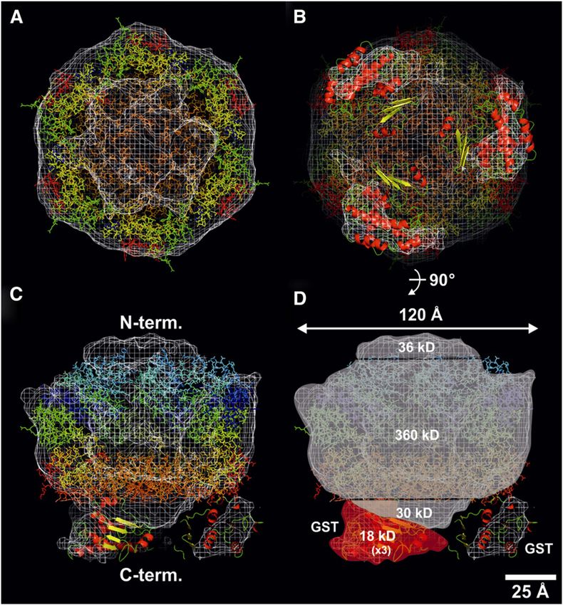
3670     The Plant Cell

viability (Mann et al., 2000). Studies on mutants lacking FtsH2         strain was able to grow under high-light conditions (Figure 1A),
have provided compelling evidence for a physiological role for this     with a cellular pigment content indistinguishable from the wild-
FtsH subunit in the degradation of D1 during PSII repair in vivo        type strain (WT-G) (see Supplemental Figure 1 online), and to
(Silva et al., 2003; Komenda et al., 2006). FtsH2 is also involved in   repair PSII as effectively as WT-G as deduced by the ability to
the removal of D1 following damage by heat (Kamata et al.,              maintain PSII activity upon exposure to high irradiances of
2005) and UV-B irradiation (Cheregi et al., 2007) and degrades          white light (Figure 1B). The rate of damage to PSII, assessed
unassembled and mutant PSII subunits in the thylakoid mem-              by determining loss of PSII activity in cells exposed to linco-
brane (Komenda et al., 2006). FtsH2 also regulates, directly or         mycin to block protein synthesis, was similar in the WT-G,
indirectly, the level of the soluble enzyme, glucosyl-glycerol          SynFtsH2GST, and the SynFtsH2GENT strains (Figure 1B).
phosphate synthase, involved in osmoprotection (Stirnberg               Additionally, the GST-tagged strain, like the WT-G strain, was
et al., 2007).                                                          unable to grow in liquid culture in the presence of 300 mM
   Nine FtsH subunits are also targeted to the Arabidopsis              maltose (Figure 1C). By contrast, SynFtsH2GENT was able to

                                                                                                                                              Downloaded from https://academic.oup.com/plcell/article/24/9/3669/6100557 by guest on 13 July 2021
thaliana chloroplast (Sakamoto et al., 2003). Mutants lacking           grow because of a perturbation in osmoregulation due to a defect
At-FtsH2 and At-FtsH5 show impaired rates of D1 degradation             in degrading the soluble enzyme glucosyl-glycerol phosphate
and PSII repair (Bailey et al., 2002; Kato et al., 2009). Genetic and   synthase involved in the synthesis of the compatible solute
coimmunoprecipitation data suggest that FtsH subunits might             glucosyl-glycerol (Stirnberg et al., 2007).
form both homo-oligomeric (Sakamoto et al., 2003) and hetero-              Control immunoblots confirmed the presence of the FtsH2-
oligomeric complexes in chloroplasts (Sakamoto et al., 2003; Yu         GST fusion in the membrane and, importantly, that there was no
et al., 2004; Zaltsman et al., 2005), but as yet no chloroplast FtsH    detectable accumulation of FtsH2 liberated by cleavage of the
complex has been isolated and characterized in terms of subunit         larger fusion protein (Figure 1D). Accumulation of FtsH1 and
composition and organization.                                           FtsH4, determined using FtsH-specific antibodies, was not sig-
   Information on the structure of intact FtsH complexes, rather        nificantly affected by inactivation of FtsH2 (Figure 1D), whereas
than Escherichia coli–expressed soluble fragments, is sparse,           a dramatic reduction in the amount of FtsH3 was observed in
and it is only recently that a hexameric structure was confirmed         SynFtsH2GENT, which was restored to wild-type levels in the
for the heteromeric m-AAA complex of the yeast mitochondrial            SynFtsH2GST strain (Figure 1D). Together, these data sug-
inner membrane by cryo–electron microscopy (cryo-EM) (Lee               gested that (1) the FtsH2-GST fusion protein was functional in
et al., 2011). The resolution of the structure, determined at 12        vivo and still retained the ability to degrade both membrane
Å, was sufficient to confirm the presence of six FtsH proto-              proteins and soluble targets and (2) accumulation of FtsH3
mers within each complex, obtained by fitting the atomic                 was heavily dependent on FtsH2, possibly through formation
structures of cytosolic fragments of FtsH into the cryo-EM              of a common complex.
model (Bieniossek et al., 2006, 2009), but was insufficient to
determine the structural organization of the two types of subunit
                                                                        Purification of FtsH2-GST
within the ring.
   Here, we report the isolation of FtsH2 from Synechocystis            GST-tagged FtsH2 was isolated from detergent-solubilized
6803 using a glutathione S-transferase (GST) tagging approach           membrane extracts by binding to glutathione-agarose resin and
(Barker et al., 2008). Our results have revealed that FtsH2 forms       eluting with reduced glutathione (Figure 2A). SDS-PAGE fol-
a hetero-oligomeric complex with the FtsH3 subunit. Using the           lowed by Coomassie blue staining revealed the presence of two
GST tag as a marker for the FtsH2 subunit, we have been able to         major protein bands in the eluate (Figure 2A). Some minor
provide direct evidence from negative stain electron microscopy         copurifying proteins could be visualized by silver staining (Figure
that FtsH2-GST and FtsH3 subunits alternate within a hexameric          2C), but the D1 subunit was not detected (Figure 2B). The upper
ring structure.                                                         band migrating at ;100 kD was detected by antibodies specific
                                                                        for FtsH2, GST, and the Strep II tag (Figures 2B and 2C) and
                                                                        was assigned to the full-length FtsH2-GST fusion protein. This
RESULTS                                                                 was confirmed by microsequencing that yielded the sequence
                                                                        MKFSXXXALL (where X is an unidentified amino acid),
Phenotype of a GST-Tagged FtsH2 Derivative of                           which matched the predicted N-terminal sequence of FtsH2,
Synechocystis 6803                                                      encoded by slr0228 (MKFSWRTALL). The lower band cross-
                                                                        reacted with antibodies to FtsH but not with antibodies to FtsH2,
Previous work has shown that E. coli FtsH can tolerate the ad-          indicating the presence of a different FtsH subunit(s) (Figure 2C).
dition of an affinity tag at the C terminus (Akiyama et al., 1995;       N-terminal sequencing for this band yielded the sequence
Shotland et al., 1997). Consequently, to aid the purification of         SKNNKKXXNA (where X is an unidentified amino acid), which
FtsH2, we constructed a strain of Synechocystis 6803, termed            corresponds to the first 10 predicted amino acid residues of
SynFtsH2GST, in which a GST affinity tag that also included              FtsH3, encoded by slr1604, after removal of the N-terminal Met
a C-terminal Strep II tag, was fused to the C terminus of FtsH2         residue (MSKNNKKWRNA). This assignment was confirmed in
(see Methods). To probe for potential effects of the GST tag on         immunoblots using specific antibodies (Figure 2C). No other
FtsH2 function, we tested growth of the GST-tagged strain under         FtsH subunit was found in this band by mass spectrometry
conditions that are dependent on FtsH2 activity. In contrast            (data not shown). Overall, these data indicated that FtsH2-
with strain SynFtsH2GENT lacking FtsH2, the SynFtsH2GST                 GST and FtsH3 form hetero-oligomeric complexes in vivo.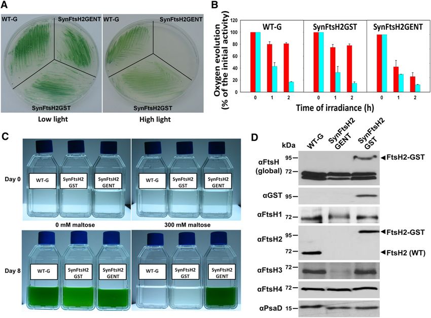
Structure of Thylakoid FtsH Complex            3671

                                                                                                                                                                Downloaded from https://academic.oup.com/plcell/article/24/9/3669/6100557 by guest on 13 July 2021
Figure 1. SynFtsH2GST Expressing FtsH2-GST Behaves Like WT-G.
(A) Growth of WT-G, SynFtsHGENT (lacking FtsH2), and SynFtsH2GST on BG-11 plates under low (5 mE m22 s21) and high light (100 mE m22 s21).
(B) PSII repair activity of WT-G, SynFtsH2GST, and SynFtsH2GENT assessed by measuring PSII oxygen-evolving activity in cells exposed to 300 mE
m22 s21 white light at 29°C with or without lincomycin (cyan and red columns, respectively), a protein synthesis inhibitor; initial rates of oxygen evolution
were 731 6 46, 802 6 60, and 545 6 43 mmol O2 mg chlorophyll21 h21 for WT-G, SynFtsH2GST, and SynFtsH2GENT, respectively (means of two to
three measurements for each of the two biological replicates 6 SE).
(C) Growth of WT-G, SynFtsHGENT, and SynFtsH2GST in BG-11 in either the presence or absence of 300 mM maltose.
(D) Immunoblotting analysis of the different strains using antibodies specific for all FtsH subunits (global FtsH), GST, and each of the FtsH subunits.
PsaD was used a loading control. Protein samples were separated by SDS-PAGE using 8% (w/v) polyacrylamide gels lacking urea.

Interestingly, low levels of the prohibitin homolog, Phb1, encoded               sulfur-containing amino acid residues in each subunit (19 in
by slr1106, were also detected immunochemically (Figures 2B                      FtsH2 and 12 in FtsH3).
and 2C).                                                                           The significant reduction in the steady state level of FtsH3,
                                                                                 to ;10% of wild-type levels in the absence of FtsH2 as as-
FtsH2 also Interacts with FtsH3 in the Wild Type                                 sessed in immunoblotting experiments (Figure 3B), is largely due
                                                                                 to destabilization of FtsH3 in the membrane as determined in
To confirm that FtsH2 and FtsH3 formed a complex in the pa-                       radioactive pulse-chase experiments (see Supplemental Figure
rental wild-type strain (WT-G), immunoprecipitation experiments                  2 online), consistent with loss of a stabilizing binding partner for
using FtsH2- and FtsH3-specific antibodies were performed. To                     FtsH3. FtsH2 also comigrated with FtsH3 when solubilized
improve the sensitivity of detection, [35S] radiolabeled samples                 membrane extracts were analyzed by two-dimensional Clear
of WT-G and SynFtsH2GST were used. As expected, GST-tagged                       Native/SDS-PAGE (Figure 3C). The most abundant complex
FtsH2 and FtsH3 were immunoprecipitated from SynFtsH2GST                         migrated with an apparent mass of ;650 kD, larger than the
extracts using both sets of antibodies (Figure 3A). In the case of               size of 410 kD predicted for a hexamer, which might reflect the
WT-G, both FtsH2 and FtsH3 were again coimmunoprecipitated                       presence of additional subunits in the complex in vivo. Smaller
(Figure 3A). It must be noted that the relative intensity of the                 amounts of even larger FtsH complexes were also detected as
bands detected by autoradiography is not a direct measure of                     well as a 550-kD complex that comigrated with dimeric PSII
the stoichiometry of FtsH2 and FtsH3 in the complex as it is                     [RCC(2)]. The larger FtsH complexes comigrated with Phb1
dependent on a number of factors, including the number of                        and might represent FtsH/Phb1 supercomplexes (arrowed in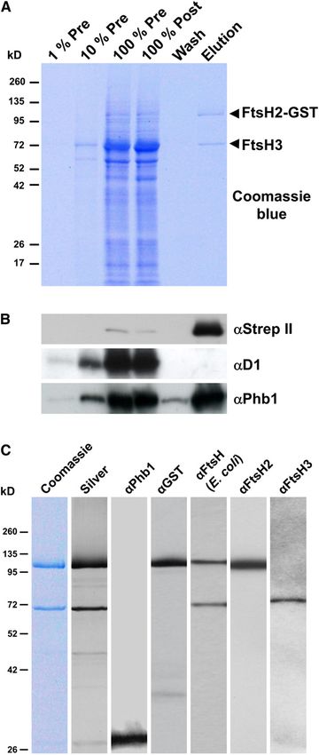
3672      The Plant Cell

                                                                              Figure 3C). Overall, these data indicated that untagged FtsH2
                                                                              also forms a complex with FtsH3 in the wild type.

                                                                              Analysis of a Mutant Deficient in FtsH3

                                                                              The ftsH3 gene is vital for cell viability in Synechocystis 6803
                                                                              (Mann et al., 2000). To gain insights into the physiological role
                                                                              of FtsH3, we constructed a strain, SynFtsH3reg, lacking the
                                                                              chromosomal copy of ftsH3 but containing a plasmid-borne
                                                                              copy of ftsH3 whose expression was driven by the nirA pro-
                                                                              moter, which can be downregulated by the inclusion of NH4+ in
                                                                              the culture medium (Qi et al., 2005). Addition of 13 mM NH4+ to

                                                                                                                                                         Downloaded from https://academic.oup.com/plcell/article/24/9/3669/6100557 by guest on 13 July 2021
                                                                              the medium led to the parallel loss of both FtsH2 and FtsH3
                                                                              within 3 d, whereas no such disappearance was observed in
                                                                              WT-G cultivated under identical conditions (Figure 4A). Cells of
                                                                              SynFtsH3reg depleted of FtsH3 nearly stopped growing after
                                                                              4 d of cultivation in the presence of 13 mM NH4+ (see Supplemental
                                                                              Figure 3A online) and showed a decrease in the cellular content
                                                                              of chlorophyll a (see Supplemental Figure 3B online) and in PSII
                                                                              oxygen-evolving activity (see Supplemental Figure 3C online). They
                                                                              were also not able to efficiently degrade the D1 protein during 120
                                                                              min of exposure to high irradiances of visible light in the presence
                                                                              of chloramphenicol (Figure 4B). Instead, the electrophoretically
                                                                              resolved D1 band became smeared, indicating its oxidation
                                                                              (Lupínková and Komenda, 2004). A similar impairment of growth
                                                                              upon addition of NH4+ was also displayed by ftsH22 mutants (data
                                                                              not shown; Drath et al., 2008). By contrast, under the same con-
                                                                              ditions, WT-G cells grew normally (see Supplemental Figure 3A
                                                                              online) and their pigment content did not change substantially (see
                                                                              Supplemental Figure 3B online), despite decreased oxygen evo-
                                                                              lution (see Supplemental Figure 3C online), and cells showed an
                                                                              approximate 50% decrease in the amount of D1 after 30 min of
                                                                              high-light treatment (Figure 4B). Overall, these data provide further
                                                                              support for a close relationship between FtsH2 and FtsH3 and
                                                                              evidence for a role for FtsH3 in degrading damaged D1 during PSII
                                                                              repair.
                                                                                 To assess the global impact of FtsH3 depletion, membrane
                                                                              proteins isolated from SynFtsH3reg were compared with WT-G.
                                                                              While the two-dimensional pattern of membrane proteins in WT-
                                                                              G cultivated for 5 d in the presence of 13 mM NH4+ was similar
                                                                              to that of the control SynFtsH3reg cells cultivated without NH4+,
                                                                              the protein profile of SynFtsH3reg grown in the presence of 13
                                                                              mM NH4+ (Figure 4C) showed marked similarities to that of
                                                                              a previously characterized ftsH22 strain (Komenda et al., 2006):
                                                                              The level of the PSII subcomplex lacking CP43, termed RC47,
                                                                              accumulated in both strains due to inhibition of PSII repair-
                                                                              related D1 degradation, and expression of the SbtA protein, which
                                                                              is involved in bicarbonate uptake and whose expression is
                                                                              dependent on FtsH2 (Zhang et al., 2007), was noticeably less
Figure 2. Affinity Purification of FtsH2-GST.
                                                                              than in WT-G and in SynFtsH3reg grown without NH4+. These
(A) Detergent-solubilized thylakoid membranes from SynFtsH2GST                data show that downregulation of ftsH3 expression caused
before chromatography (Pre), the extract after binding to the glutathi-
one resin (Post), the wash fraction just before elution (Wash), and the
fraction eluted by glutathione (Elution) were separated by SDS-PAGE.
Arrowheads indicate the positions of the FtsH2-GST and FtsH3 pro-
teins.                                                                        (C) Analysis of purified sample by Coomassie blue and silver staining and
(B) Analysis of column fractions by immunoblotting with antibodies            by immunoblotting with antibodies specific for Prohibitin (Phb1), GST,
specific for the Strep II tag (aStrep II), D1 (aD1), and Prohibitin (aPhb1).   E. coli FtsH, FtsH2, and FtsH3.
100% Pre corresponds to 1 mg chlorophyll a.                                   [See online article for color version of this figure.]
Structure of Thylakoid FtsH Complex 3673
Downloaded from https://academic.oup.com/plcell/article/24/9/3669/6100557 by guest on 13 July 2021
Figure 3. FtsH2 Interacts with FtsH3 in WT-G.
(A) Immunoprecipitation analysis performed on radiolabeled crude thylakoid membranes (TM) isolated from SynFtsH2GST and the wild type (WT-G)
using the indicated antibodies were analyzed by immunoblotting (Blot) and autoradiography (Autorad). The positions of the FtsH2-GST, FtsH2, and
FtsH3 proteins are indicated, * and ** indicate non-FtsH related cross-reactions.
(B) Crude thylakoid membrane proteins isolated from WT-G and mutant slr0228:cmR lacking FtsH2 (FtsH22) were separated by SDS-PAGE (amounts
corresponding to 2.0 µg [100%], 1.0 mg [50%], and 0.5 mg [25%] of chlorophyll a were loaded) and immunoblotted using antibodies specific for FtsH2
and FtsH3. Correct sample loadings were confirmed by detection of cytochrome f.
(C) Membrane proteins isolated from WT-G were separated by 2D-Clear Native (CN)/SDS-PAGE and blotted onto PVDF membrane. The blot was
stained by Sypro Orange (Stained blot) and then sequentially probed with antibodies specific for FtsH2, FtsH3, and Phb1 (Blots). Arrows indicate3674 The Plant Cell
a similar phenotype to ftsH22 consistent with participation of angular reconstitution (Van Heel, 1987). Figure 6B shows five
a hetero-oligomeric FtsH2/FtsH3 in PSII repair and induction of typical exemplar class averages, taken from a total of 263
CO2-concentrating mechanisms. class averages, representing 2964 single two-dimensional par-
One notable feature seen just in the NH4+-treated SynFtsH3reg ticles used to construct the final three-dimensional map, pro-
cells was a decrease in the level of NDH-1 complexes (Figure 4C), gressing from a view attributed to the top elevation through to
which also participate in the delivery of CO2/bicarbonate into the a view assigned to the side. This three-dimensional map is
cells (Zhang et al., 2004). No additional changes to the two- shown in Figure 6C as a surface-rendered molecular envelope,
dimensional protein profile were detected when SynFtsH3reg each at the same orientation as the class averages given in
was incubated for up to 7 d in NH4+ (data not shown). Figure 6B. According to Fourier shell correlation analysis (see
Supplemental Figure 4B online), the three-dimensional map has
Isolation of a FtsH1/FtsH3 Heterocomplex a resolution of ;26 Å assuming asymmetry and Fourier-shell
correlation at 3s with a conservative correlation coefficient of
Downloaded from https://academic.oup.com/plcell/article/24/9/3669/6100557 by guest on 13 July 2021
Previous mutagenesis experiments have shown that ftsH2 and
0.5. It is apparent from the comparison of the two-dimensional
ftsH4 are dispensable for cell viability, whereas ftsH1 and ftsH3
class averages (Figure 6B) with the post-three-dimensional re-
are crucial (Mann et al., 2000). Given that our data showed the
construction, two-dimensional reprojection images (Figure 6D)
formation of FtsH2/FtsH3 complexes in vivo and that FtsH2, and
that a majority of orientations were incorporated into the three-
hence the FtsH2/FtsH3 complex, is not required for cell viability
dimensional map.
(Mann et al., 2000), we investigated the possibility that FtsH3
To aid interpretation of the three-dimensional reconstruction,
might participate in more than one type of FtsH complex. To
the crystallographically derived hexameric structure of a soluble
test this, a fully segregated GST-tagged derivative of FtsH3, as
fragment of FtsH, containing residues 146 to 603 of the 610–
well as GST-tagged versions of FtsH1 and FtsH4, and a FtsH2
amino acid sequence of Thermotoga maritima FtsH (Bieniossek
control, were isolated by affinity chromatography and analyzed by
et al., 2009), encompassing the conserved AAA domain and
SDS-PAGE and immunoblotting using FtsH-specific antibodies
PD, was modeled by visual inspection into the molecular
(Figure 5). The results indicated that FtsH3 forms an FtsH1/FtsH3
envelope, contoured at a threshold of 2.5 s (Figure 7A). The
heterocomplex as well as the previously described FtsH2/FtsH3
crystal structure fitted remarkably well into the central core,
heterocomplex, and that FtsH4 forms a homo-oligomeric com-
providing clear evidence to support the presence of six FtsH
plex. N-terminal sequencing of the FtsH4-GST band yielded the
subunits in the GST-tagged FtsH complex. The model shows
sequence AIKPQP that matched the predicted N-terminal se-
a central ball-shaped complex of ;120 Å in diameter, with
quence of FtsH4, encoded by sll1463 (MAIKPQP). No N-terminal
three minor densities extending out from one side, which
sequence data could be obtained from FtsH1-GST, possibly
could be attributed to the presence of three GST tags, which
because the N terminus was blocked.
were fitted using an atomic structure (Figures 7B and 7C). The
Electron Microscopy and Single-Particle Analysis of the location of the GST tags with respect to the central body of
FtsH2-GST/FtsH3 Complex the complex suggests an alternating arrangement of three
FtsH2-GST and three FtsH3 subunits in a hexameric complex
We used transmission electron microscopy of negatively stained (Figure 7B).
samples to determine the overall dimensions of the isolated The fusion of the GST tag to the C terminus of FtsH2 also
FtsH2-GST/FtsH3 complex, the number of protomers present in allowed us to determine the orientation of the complex with
the complex, and their arrangement within the complex using respect to the N and C termini (Figure 7C). The central core of
the GST tag as a means of differentiating the FtsH2 subunits the complex has a maximum diameter of 120 Å, close to the
from FtsH3. Although there was some aggregation of the value of 130 Å determined by cryo-EM for the m-AAA protease
sample on the grid, possibly due to interactions between GST (Lee et al., 2011). By assuming a protein density of 0.844 Å3 per
tags on different complexes (Natalello et al., 2008), a sufficient Dalton (van Heel et al., 1996), masses of 360 kD could be as-
number of negatively stained protein complexes could be vi- signed to the central portion containing the AAA module and PD,
sualized in the 18 micrographs used to allow detailed analysis. 36 kD to the N-terminal cap consisting of the transmembrane
An asymmetric structural map was obtained by analyzing all helices and lumenal loops, 18 kD to the three outlying domains
discrete two-dimensional projection views observed in the assigned to the 27-kD GST tag, leaving a C-terminal bottom
electron micrographs of negatively stained protein complexes region of 30 kD (Figure 7D) likely to contain the C-terminal region
(Figure 6A). These views covered a broad range of orientation, of FtsH, which is relatively disordered and not present in the T.
including top and side elevations, as well as views that could maritima crystal structure used in the fitting, plus possibly some
be attributed to tilting particles (see the Euler angle distribution of the GST tag. Thus, the overall mass of the isolated FtsH2-GST/
in Supplemental Figure 4A online). We took advantage of this FtsH3 complex using the above assumptions was estimated to be
to calculate a three-dimensional map, or reconstruction, by 480 kD, close to the theoretical mass of 489 kD.
Figure 3. (continued).
possible FtsH/Phb1 supercomplexes. Positions of unassembled proteins (U.P.), PSII reaction center core lacking CP43 (RC47), monomeric [RCC(1)],
dimeric [RCC(2)], and supercomplexes (RCCS1) of PSII are indicated.Structure of Thylakoid FtsH Complex 3675
DISCUSSION
Previous work has identified an important role for FtsH2 in PSII
repair and in other quality control processes in the thylakoid
membrane of Synechocystis 6803 (reviewed in Nixon et al.,
2010). We show here that FtsH2 forms a hetero-oligomeric
complex with FtsH3. This conclusion is based on a number of
independent lines of evidence, including copurification of FtsH2
and FtsH3 by affinity chromatography (Figure 2), coimmunopre-
cipitation of FtsH2 and FtsH3 (Figure 3A), comigration of FtsH2
and FtsH3 by native gel electrophoresis (Figure 3C), and an en-
hanced rate of degradation of FtsH3 in the absence of FtsH2 (see
Downloaded from https://academic.oup.com/plcell/article/24/9/3669/6100557 by guest on 13 July 2021
Supplemental Figure 2 online), leading to reduced accumulation
of FtsH3 (Figure 3B). Repression of FtsH3 synthesis also led to
decreased FtsH2 accumulation in the membrane (Figure 4A) and
a similar impairment in D1 degradation as observed earlier in
ftsH22 mutants (Figure 4B). Overall, these data indicate that
the major FtsH complex involved in degrading D1 during PSII
repair in Synechocystis 6803 is an FtsH2/FtsH3 heterocomplex.
Other physiological processes currently assigned to FtsH2, such
as a role in osmoregulation and carbon concentrating mecha-
nisms, are also likely to be dependent on the activity of the FtsH2/
FtsH3 hetero-oligomer.
In addition, our structural analysis of the FtsH2-GST/FtsH3
heterocomplex (Figure 7) has provided new insights into the
subunit organization of this class of FtsH complex. The com-
bination of structural data obtained by single-particle electron
microscopy and the available high-resolution crystal struc-
tures for the conserved soluble domains of FtsH allowed us to
conclude that the number of FtsH subunits within the complex
was six. The additional GST tag of ;27 kD was of sufficient
size for it to be detected in the transmission electron mi-
croscopy reconstructions of the complex, thereby providing
information on the location of the FtsH2 subunits within the
complex. A similar experimental strategy involving the use of
yellow fluorescent protein fusions has also been exploited to
study the location of subunits within the cyanobacterial NDH-
1 complex (Birungi et al., 2010). Antibodies can also be used
to identify subunits during single-particle analysis, but there
can be significant experimental drawbacks, including partial
Figure 4. Analysis of the SynFtsH3reg Strain.
decoration of the complex (Perálvarez-Marín et al., 2011). Our
(A) Immunoblot analysis of crude thylakoid membranes isolated from structural model suggests that the FtsH2-GST and FtsH3
WT-G and SynFtsH3reg at various times following addition of 13 mM subunits alternate within the complex. Given the averaging
NH4Cl to the growth medium using antibodies specific for FtsH3 and procedure used to generate the final structure, a random
FtsH2. Detection of cytochrome f was used to confirm equal sample positioning of the subunits within the hexamer would not have
loadings. Mutant slr0228:cmR lacking FtsH2 (FtsH22) was used as a con- given rise to the three additional densities that we assign to
trol. All strains were grown in BG11 medium in the presence of 13 mM
the GST tag but instead would have been averaged out over
NH4Cl and 5 mM Glc at an irradiance of 10 mE m22 s21. A total of 2 mg of
chlorophyll a was loaded per lane.
(B) High light–induced D1 degradation in WT-G and SynFtsH3reg. Cells
of WT-G and SynFtsH3reg grown for 7 d in BG11 medium containing 13
mM NH4Cl at an irradiance of 10 mE m22 s21 were exposed to high light transferred to the BG11 medium containing 13 mM NH4Cl and cultivated
(500 mE m22 s21) in the presence of chloramphenicol (50 mg mL21), and for 5 d (WT-G 5d and FtsH3reg 5d) at an irradiance of 40 mmol photons
cells were harvested after various times of illumination for protein anal- m22 s21. Complexes are designated as in Figure 3C. A total of 5 mg of
ysis using antibody specific for the D1 protein. A total of 2 mg of chlo- chlorophyll a was loaded per lane. The arrows designate the RC47
rophyll a was loaded per lane. Detection of cytochrome f was used to complex, which accumulates more in the mutant than the wild type
confirm equal sample loadings. (arrow 1), the SbtA spot, that accumulates more in the wild type than the
(C) Two-dimensional analysis of PSII complexes in cells of WT-G and mutant (arrow 2), and the NdhH spots of two NDH complexes that ac-
SynFtsH3reg mutants. WT-G and FtsH3reg cells were grown in BG11 cumulate less in the mutant than the wild type (arrows 3 and 4). U.P.,
medium without ammonium ions (WT-G 0d and FtsH3reg 0d) and then unassembled proteins.3676 The Plant Cell
membrane. Consequently, FtsH2 and FtsH3 are likely to contain
two transmembrane helices in the N-terminal region connected
by short lumenally exposed loops of ;80 to 100 residues, which
might be involved in recognizing substrates, such as the D1
subunit (Bailey et al., 2002). The recent structural model for the
m-AAA protease obtained by cryo-EM has highlighted the fact
that the short loops lying on the opposite side of the membrane
to the AAA+ and PD are actually splayed out. This feature is
absent in our structural model most likely due to a combination
of limited resolution and the fact that the loop regions seem
to be more flexible than the rest of the complex (Lee et al.,
2011).
Downloaded from https://academic.oup.com/plcell/article/24/9/3669/6100557 by guest on 13 July 2021
There are reports of mitochondrial FtsH subunits participating
in both homo- and hetero-oligomeric complexes (Koppen et al.,
2007; Piechota et al., 2010) with the relative proportions in some
cases dependent on cell type (Koppen et al., 2007). Conse-
quently, we do not exclude the possibility that under certain
growth conditions FtsH2 might form significant amounts of
other types of FtsH2 complex, such as homocomplexes, in
addition to the FtsH2/FtsH3 complex described here. The fact
that FtsH3 is required for cell viability whereas FtsH2 is dis-
pensable (Mann et al., 2000) suggests that FtsH3 is not re-
stricted to forming complexes with FtsH2. The data in Figure 5
show that FtsH3 also forms a complex with FtsH1. As accu-
mulation of FtsH3 is reduced to muchStructure of Thylakoid FtsH Complex 3677
Downloaded from https://academic.oup.com/plcell/article/24/9/3669/6100557 by guest on 13 July 2021
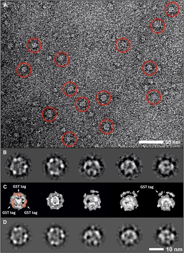
Figure 6. Single-Particle Analysis of the GST-Tagged FtsH2/FtsH3 Complex.
(A) Typical region in a micrograph of negatively stained FtsH2-GST complexes. FtsH particles are circled. Additional images showing this complex can
be viewed in Barker et al. (2008). Bar = 50 nm.
(B) A selection of five characteristic two-dimensional views of the FtsH2-GST complex, taken from a total of 263 different class averages.
(C) Surface-rendered views of the final three-dimensional map calculated by angular reconstitution viewed from the same angles as presented in (B).
Positions of density attributed to three GST tags are indicated by arrows and colored in pink for one of the views.
(D) Two-dimensional reprojections of the views shown in (C).
Bar in (D) = 10 nm for (B) to (D).3678 The Plant Cell
Downloaded from https://academic.oup.com/plcell/article/24/9/3669/6100557 by guest on 13 July 2021
Figure 7. Three-Dimensional Modeling of the GST-Tagged FtsH2/FtsH3 Complex.
An isomesh rendered molecular envelope, contoured with a threshold of 2.5 s for the final calculated asymmetric three-dimensional map, is shown in
white. The modeling in of the crystallography-derived hexameric soluble fragment of apoFtsH from T. maritima (3KDS.pdb) was done by visual
inspection with atoms colored as a spectral rainbow from the N terminus (blue) to the C terminus (red).
(A) A 5-nm-thick cross section viewed from the C-terminal PDs of 3KDS.pdb downwards, revealing its hexameric nature.
(B) A 4-nm-thick cross section, viewed from the GST tags upwards, toward the C-terminal domains of the hexameric biological assembly of 3KDS.pdb.
(C) A 5-nm-thick cross section, viewed from the side; the best fit for the six FtsH protomers is shown with the N-terminal AAA+ domain uppermost.
(D) Four regions shown include an N-terminal cap, whose volume is calculated to be ;36 kD (assuming 0.844 Å3 per D at 2.5 s), a central core of 360
kD, a C-terminal bottom of 30 kD, and three smaller outlying domains of 18 kD. In (B) to (D), these outlying domains, closest to the C-terminally
orientated central core 3KDS.pdb file, are assigned to the GST tag (1GTA.pdb). Three such domains are observed, even though no threefold symmetry
operators were applied during angular reconstitution. The maximum diameter of the complex was observed to be 120 Å. Bar = 25 Å.
that chloroplast FtsH heterocomplexes might be composed of FtsH hexameric complexes can form larger supercomplexes,
two type A and four type B subunits, but deviations from a 1:1 of unknown function, with members of the Band 7 family of
stoichiometry because of contamination by FtsH monomers or proteins, including HflK/C in E. coli and prohibitins in mitochondria
partially assembled FtsH complexes could not be excluded (Ito and Akiyama, 2005). Preliminary data presented here (Figures
(Moldavski et al., 2012). The single-particle approach described 2B and 3C) suggest that FtsH/Phb1 supercomplexes might also
here helps overcome this potential problem. accumulate in Synechocystis 6803. The relatively low levels of
By contrast, the FtsH4 subunit of Synechocystis 6803 ap- Phb1 found in the affinity-purified FtsH2-GST/FtsH3 prepara-
pears to form homocomplexes (Figure 5). Phylogenetic analyses tion could reflect the transient nature of the complex or sen-
suggest that FtsH4 is most closely related to the At-FtsH7 and sitivity of the complex to detergent treatment (Boehm et al.,
At-FtsH9 nucleus-encoded chloroplast-targeted FtsH subunits 2009).
(Sakamoto et al., 2003; Yu et al., 2004), about which little is How FtsH complexes function is still unclear. The recent cryo-
currently known. The advantage of expressing heterocomplexes EM data obtained for the yeast m-AAA complex (Lee et al., 2011)
rather than homocomplexes remains to be clarified, although hint toward the possibility that the hexameric complexes might
a role in determining substrate specificity has been suggested contain a 13-Å gap between the N-terminal transmembrane
(Koppen et al., 2007). segment and the AAA+ domain through which unfolded, but notStructure of Thylakoid FtsH Complex 3679
folded, polypeptides could be guided to the central pore loops the intermediate p0228TEVHISSTREP plasmid was produced from
of the AAA ring. The distance between a proposed initial sub- p0228 as follows. The coding sequence for a tobacco etch virus pro-
strate binding site and the central pore region (Lee et al., 2011) is tease site (TEV; encoding ENLYFQG) followed by a nona-His tag (His9)
compatible with a requirement for target substrates to possess and a STREP-II affinity tag (encoding WSHPQFEK) were introduced
after the penultimate slr0228 codon by overlap extension PCR (Ho et al.,
an unfolded segment of 20–amino acid residues or more (Chiba
1989). The primers in the first two separate reactions were 0228fd
et al., 2000). Once engaged with the FtsH complex, ATP-driven
with 0228tevhisstprv (59-CACGTGATGGTGATGGTGATGGTGATGGTGGC-
conformational changes within the AAA ring would drive the CTTGAAAATAGAGATTTTCAGCGCTTAGTTGGGGAATTAACTGTTC-39) and
movement of the unfolded target protein into the proteolytic 0228tevhisstpfd (59-TATTTTCAAGGCCACCATCACCATCACCATCACCAT-
chamber for subsequent highly processive degradation into CACGTGGTATACGGATCCAATTGGTCCCATCCC-39) with 0228rv using the
small peptides, which then exit through ;25-Å lateral open- p0228 plasmid as template DNA in both reactions. The second PCR
ings within the body of the complex (Lee et al., 2011). We have reaction used the 0228fd and 0228rv primers with the previous two PCR
shown that degradation of D1 during PSII repair in Synechocystis reaction products as DNA template yielding p0228TEVHISSTREP after
Downloaded from https://academic.oup.com/plcell/article/24/9/3669/6100557 by guest on 13 July 2021
6803 is also dependent on the length of the N-terminal tail of ligation into the pGEM-T Easy vector. In the last step, the p0228TEV-
D1 (Komenda et al., 2007). Consequently, our current view is HISSTREP plasmid was digested in a two-step process to replace the
sequence between the Eco47III and BamHI restriction sites with the gst
that D1 degradation in Synechocystis 6803 is mainly initiated
gene. The gst gene was amplified from the pGEX-6P-3 vector (GE
from the N terminus and proceeds in a highly processive manner
Healthcare) using PCR with the GSTfd (59-GGGGTATACATGTCCCCTA-
without formation of distinct degradation products (Nixon et al., TACTAGGTTAT-39; introduces blunt cutter Bst1107I at the 59-end) and
2010). GSTrv (59-GGGGGATCCATCCGATTTTGGAGGATGGTC-39; introduces
In summary, we isolated a hetero-oligomeric FtsH complex of BamHI at the 39-end) primer pair. The resulting PCR fragment was digested
the thylakoid membrane involved in PSII repair. Our data confirm with Bst1107I and BamHI restriction enzymes. Also, p0228TEVHISSTREP
that the isolated complex is hexameric and provide direct evi- was digested using BamHI and AlwNI (cuts once in the pGEMTeasy
dence for an alternating arrangement of the two subunits within vector) to yield three fragments of 2146, 1638 (fragment a), and 739 bp.
the complex. Ultimately, a detailed understanding of the com- In a second reaction, AlwNI and Eco47III (blunt cutter) were used to
plex will require a high-resolution structure probably determined digest p0228TEVHISSTREP into two fragments of 2824 (fragment b)
and 1699 bp. After these digestions, the fragments a and b and the gst
by x-ray crystallography. Given the low amounts of the natural
gene were ligated together, yielding the p0228GSTSTREP vector. This
complex that can be purified, this will most likely require high-
process was necessary due to the presence of two BamHI sites in the
level coexpression of FtsH2 and FtsH3 in a heterologous p0228TEVHISSTREP plasmid. After transformation of the p0228GSTSTREP
system. vector into Syn0228GENT, transformants were selected for restored ability
to grow under high-light conditions (100 µE m22 s21). A resulting high-light
METHODS resistant strain, termed SynFtsH2GST, was confirmed by PCR and re-
striction digestion to contain the correct slr0228:gst fusion (see Supplemental
Cyanobacterial Strains and Growth Conditions Figure 6 online).
All strains used in this study are derived from the Glc-tolerant Syn-
Construction of the Erythromycin-Resistant GST-Tagged Strains
echocystis sp PCC 6803 strain (WT-G) (Williams, 1988). Strain Syn-
of FtsH
FtsH2GENT is equivalent to the strain Syn0228GENT that contains
a deletion of the 39 end of the ftsH2 (slr0228) gene and replacement by A universal tagging cassette (see Supplemental Figure 7B online) con-
a gentamycin resistance cassette (Komenda et al., 2006). Strain slr0228: sisting of the coding sequences for the thrombin cleavage site, GST, and
cmR contains the insertion of a chloramphenicol resistance cassette into the strep II tag and a selectable marker conferring erythromycin resistance
ftsH2 (slr0228) gene (Komenda et al., 2006). Unless stated otherwise, strains was made to enable a simple, one-step tagging strategy. The gst gene
were grown in liquid BG-11 mineral medium or on solid BG-11 plates was amplified from pGEX-6P-3 (GE Healthcare) using the GST-F/R primer
containing 1.5% (w/v) agar, both containing 5 mM TES-KOH, pH 8.2, and pair (see Supplemental Table 1 online) and cloned into pGEM-T Easy
supplemented with 5 mM Glc, at a light intensity of 20 µE m22 s21 white (Promega) to yield the intermediate vector pGST (see Supplemental
fluorescent light and at 29°C. Light sensitivity growth assays were con- Figure 7A online). An erythromycin resistance marker (Elhai and Wolk,
ducted on plates at low (5 µE m22 s21) and high (100 µE m22 s21) light 1988) was then ligated into pGST via the HpaI site to generate pGST-
intensities, and osmoregulation was assessed in liquid cultures by the ErmA (see Supplemental Figure 7C online).
addition of 300 mM maltose to BG-11 mineral medium. To construct the transformation vectors, the full-length ftsH open
reading frame and 655 bp of downstream DNA sequence were used as
the flanking sequence to facilitate homologous recombination into the
Construction of the FtsH2-GST–Tagged Strain (SynFtsH2GST)
genome. To enable insertion of the gst-tagging cassette, overlap-
To tag FtsH2 with GST, strain SynFtsH2GENT was transformed with extension PCR was used to introduce an EcoRV and an XbaI site im-
plasmid p0228GSTSTREP that contains an in-frame fusion of the coding mediately before the STOP codon (see Supplemental Figure 8A online). In
sequence for GST and a STREP II tag to the 39-end of ftsH2 (slr0228), plus the first step of overlap extension PCR, two separate reactions were
flanking DNA to facilitate homologous recombination into the genome. performed using primer sets FtsH-F/FtsH-OE-R and FtsH-OE-F/FtsH-R for
Plasmid p0228GSTSTREP was constructed in three steps (see Supplemental each ftsH gene (see Supplemental Table 1 online), with genomic DNA from
Figure 5 online). First, the last third of the slr0228 gene plus some down- WT-G Synechocystis 6803 as template. In the second step, PCR fragments
stream region (genome coordinates: 2530235-2531612) were amplified using from the previous reactions were used as DNA template, together with
the 0228fd (59-GGGGGATCCGGACCGGGTGGTAGCTGGTAT-39) and the primer set FtsH-F/R, with the resulting PCR products then cloned into
0228rv (59-GGGGGTACCATGGCATCCTCCGTTGCAATT-39) primers. The pGEM-T Easy vector to create pGEMFtsHx vectors where FtsHx is the
resulting DNA fragment was cloned into the pGEM-T Easy vector particular FtsH subunit (see Supplemental Figures 8B and 8C online).
(Promega) to yield p0228. To generate the p0228GSTSTREP plasmid, Consequently, the gst-tagging cassette, released from pGST-ErmA using3680 The Plant Cell
EcoRV and SpeI, was ligated into each of the pGEMFtsHx vectors via over a period of 120 min either in the presence or absence of the protein
EcoRV and XbaI sites to yield the four final transformation vectors synthesis inhibitor lincomycin (100 µg/mL). The PSII oxygen-evolving
pFtsHxGSTery, where FtsHx represents the particular FtsH subunit (see activity of samples taken during the time course was measured in the
Supplemental Figures 8D and 8E online). After transformation of WT-G, presence of artificial electron acceptors (see above).
transformants were selected for erythromycin resistance (15 µg mL21)
and complete segregation was confirmed by PCR (see Supplemental
SDS-PAGE and Immunoblotting
Figure 9 online). The resulting mutants are termed SynFtsHxGSTery,
where FtsHx represents the particular FtsH subunit. Unless stated otherwise, membrane protein samples and selected
fractions of the GST affinity purification procedure were separated on
10% (v/v) denaturing one-dimensional SDS-PAGE gels containing 6 M
Construction of the SynFtsH3reg Strain
urea as described previously (Boehm et al., 2009). Gels were either
For regulatable expression of FtsH3 under control of the nirA promoter stained with Coomassie Brilliant Blue R 250 or by silver staining (Blum
from Synechococcus sp PCC 7942, the ftsH3 (slr1604) gene was cloned in et al., 1987) or electroblotted onto polyvinylidene difluoride (PVDF)
Downloaded from https://academic.oup.com/plcell/article/24/9/3669/6100557 by guest on 13 July 2021
frame into the pCER20 plasmid using NdeI and XbaI restriction sites (Qi membrane using the iBlot system (Invitrogen). Immunoblotting analyses
et al., 2005). The resulting plasmid (pFtsH3reg), able to replicate au- were performed using specific primary antibodies and anti-rabbit or
tonomously in Synechocystis, was transformed into the WT-G strain of anti-mouse horseradish peroxidase–conjugated secondary antibodies
Synechocystis 6803 via triparental mating (Elhai and Wolk, 1988), and respectively (both GE Healthcare). Signals were visualized using
transformants were selected on medium supplemented with gentamycin a chemiluminescent kit (SuperSignal West Pico; Pierce). Primary an-
at 10 mg mL21. tibodies used in this study were as follows: (1) a polyclonal antibody
To delete the wild-type copy of the ftsH3 (slr1604) gene, a linear specific for Escherichia coli–expressed recombinant GST from Schis-
deletion construct was prepared replacing most of the ftsH3 gene (nu- tosoma japonicum generated in rabbit (Sigma-Aldrich), at 1 in 2000
cleotides 161 to 1691) with an erythromycin resistance cassette using dilution; (2) a monoclonal antibody specific for Strep-tag II generated in
a megaprimer PCR method as described by Dobáková et al. (2009). This mouse (Qiagen), at 1 in 2000 dilution; (3) a polyclonal antipeptide an-
construct containing 600 bp upstream and downstream regions of the tibody raised against residues 297 to 312 of E. coli FtsH (Tomoyasu
ftsH3 gene with the erythromycin resistance cassette (obtained from et al., 1993), which is potentially cross-reactive with all Synechocystis
plasmid pPV142 of Staphylococcus simulans) in the middle was con- sp PCC 6803 FtsH homologs, kindly provided by Teru Ogura (University
structed in two steps using long fusion primers complementary to the of Kumamoto, Japan); (4) polyclonal antipeptide antibodies raised
ftsH3 gene in one direction and the erythromycin resistance cassette in against residues 578 to 592 of FtsH1, residues 98 to 115 of Syn-
the other. This linear deletion construct was used for transformation of echocystis FtsH2, residues 59 to 75 of Synechocystis FtsH3, all at 1 in
Synechocystis 6803 WT-G containing plasmid pFtsH3reg. Transformants 1000 dilution, and residues 556 to 574 of Synechocystis FtsH4, at 1 in
were selected and segregated on erythromycin-containing agar plates, 10,000 dilution; (5) a rabbit polyclonal antiserum (#304-F) raised against
and full segregation was confirmed by PCR. Unlike in the wild type, residues 325 to 353 of precursor D1 from pea (Pisum sativum; Nixon
deletion of ftsH3 was possible in the strain containing plasmid pFtsH3reg, et al., 1990) at a dilution of 1 in 5000; (6) a rabbit polyclonal antiserum (global
showing functional expression of FtsH3 from this plasmid. The obtained FtsH) raised against residues 286 to 304 of FtsH2, which are conserved in all
mutant was designated SynFtsH3reg. To suppress expression of FtsH3, four FtsH subunits, at a dilution of 1 in 10,000; (7) a rabbit antiserum raised
the strain was initially grown in BG-11 medium containing no ammonium ions against the prohibitin homolog, Phb1, encoded by slr1106 (Boehm et al.,
(ferric ammonium citrate replaced by ferric citrate) and after reaching an OD750 2009); (8) a rabbit antiserum raised against Chlamydomonas reinhardtii
of 0.5 (exponential phase of growth), the culture was diluted with BG-11 PsaD, kindly provided by Jean-David Rochaix, used at a dilution of 1 in
medium containing 17 mM NH4Cl instead of NaNO3 to give a final con- 5000. The cytochrome f subunit of the cytochrome b6f complex was de-
centration of ;13 mM NH4Cl. The culture was diluted every day with this tected by directly incubating unblocked blots with chemiluminescent
medium to OD750 0.2 in order to maintain sufficient ammonium in the medium. reagents.
Preparation of Crude Thylakoid Membranes Two-Dimensional Gel Electrophoresis
Crude Synechocystis thylakoid membranes for gel electrophoresis (SDS- Isolated membranes were solubilized with 1% n-dodecyl-b-D-maltoside
PAGE) analyses were prepared by glass bead (212 to 300 µm in diameter) (b-DM) and analyzed by two-dimensional electrophoresis. The first step
breakage at 4°C followed by differential centrifugation (Boehm et al., 2009). was performed at 4°C through a 5 to 14% polyacrylamide gel using a clear
Chlorophyll a content was determined by extraction into methanol and variant of the blue-native PAGE described by Schägger and von Jagow
absorption measurements at 666 and 750 nm (Komenda and Barber, 1995). (1991) in which Coomassie Brilliant Blue was omitted from all solutions
and the upper electrophoresis buffer contained instead 0.05% sodium
Spectroscopic and Polarography Methods deoxycholate and 0.02% b-DM. Samples containing 5 mg of chlorophyll
were loaded in each lane. The whole lane was excised from the native gel,
Measurement of chlorophyll concentrations after methanol extraction and incubated for 30 min in 25 mM Tris-HCl, pH 7.5, containing 1% SDS, and
absorption spectra of cells in vivo were measured using a Shimadzu UV3000 placed on the top of a denaturing linear 12 to 20% gradient polyacrylamide
spectrophotometer, and the light-saturated steady state rate of oxygen gel containing 7 M urea (Komenda et al., 2005). Proteins separated in the gel
evolution in cell suspensions was assessed polarographically in a thermo- were either stained by Coomassie Brilliant Blue or transferred onto a PVDF
stated chamber at 29°C in the presence of artificial electron acceptors membrane for immunoblotting.
p-benzoquinone (0.5 mM final concentration) and potassium ferricyanide (1
mM final concentration), all as described by Komenda et al. (2007).
Affinity Purification of FtsH2-GST
The cells of a 10-liter culture of SynFtsH2GST, grown at ;100 µE m22 s21
Assessment of PSII Repair Efficiency
and bubbled with air, were first concentrated to ;1 liters using a tangential
Cultures (50 mL) diluted to 2 µg chlorophyll/mL were shaken in 250-mL flow cell concentrator and then harvested by centrifugation (Sorvall RC6+;
Erlenmeyer flasks and exposed to photoinhibitory light (300 µE m22 s21) F10; 9000 rpm; 15 min; 4°C). The cell pellet was washed in 500 mL KPNStructure of Thylakoid FtsH Complex 3681
buffer (40 mM K-phosphate, pH 8.0, 100 mM NaCl, and 1 mM DTT) and Protein Sequencing
finally resuspended in 50 mL KPN buffer (chlorophyll a concentration = 1
N-terminal protein sequencing was performed by the University of Leeds
mg/mL) supplemented with one tablet of complete EDTA-free protease
Protein Sequencing Facility, the Medical Research Council Laboratory of
inhibitor (Roche Diagnostics). Cells were broken by being passed twice
Molecular Biology, and the University of Cambridge Protein and Nucleic
through a French Press at 1250 p.s.i., and intact cells were removed by
Acid Chemistry Facility, according to their instructions.
centrifugation (Sorvall RC6+; F21; 5000 rpm; 5 min; 4°C). Crude thylakoid
membranes were then harvested by ultracentrifugation (Beckmann L8-
70M; Ti45; 38,500 rpm; 60 min; 4°C) and resuspended in buffer A (50 mM
Single-Particle Analysis
HEPES, pH 7.2, 1.2 M betaine, 5% [v/v] glycerol, 100 mM NaCl, 5 mM
MgCl2, 10 µM ZnCl2, and 1 mM DTT; chlorophyll a concentration = 1 mg/ Samples were applied to glow-discharged copper grids and negatively
mL). A 10% (w/v) b-DM stock solution in buffer A was added to solubilize stained with 2% (w/v) uranyl acetate. Images were recorded at room
the membrane protein complexes at a final concentration of 1% (w/v) temperature using a Philips CM100 TEM, operating at 80 kV and 350,850
b-DM for 30 min on ice with occasional gentle agitation. Insolubilized magnification. Micrographs were chosen for minimal astigmatism/drift
Downloaded from https://academic.oup.com/plcell/article/24/9/3669/6100557 by guest on 13 July 2021
material was pelleted (Beckmann L8-70M; Ti70; 31,700 rpm; 30 min; 4°C) and scanned using a Nikon LS9000 densitometer. Fourier power spectra
and the chlorophyll a concentration determined. Swollen glutathione resin for each micrograph displayed first minima in the range of 19 to 21 Å. A
(5 mL; Sigma-Aldrich), equilibrated with 60 mL buffer A, was then added to data set of ;3400 particles was compiled using “boxer” of the EMAN
the sample. After an incubation period of 3 h on a rotating wheel, the software package (Ludtke et al., 2004). Further processing was performed
sample was transferred to a Proteus one-step batch midi spin column using Imagic-5 (Image Science) at a sampling frequency of 2.5 Å/pixel on
(Generon) and washed with GST washing buffer (50 mM HEPES, pH 7.2, the specimen scale. Reference-free alignment, multivariate statistical
5% [v/v] glycerol, 100 mM NaCl, 5 mM MgCl2, 10 µM ZnCl2, 1 mM DTT, analysis, and iterative refinement resulted in two-dimensional class
and 0.03% [w/v] b-DM) until the washes were clear. A last wash was averages (Ruprecht and Nield, 2001). Eulerian angles were then as-
performed with 10 mL GST washing buffer for one-dimensional SDS- signed a priori by angular reconstitution (Van Heel, 1987) (see Supplemental
PAGE analysis. FtsH2-GST protein complexes were eluted twice with 5 Figure 4 online) and iterative refinements implemented. The resolution of the
mL GST elution buffer (50 mM Tris/HCl, pH 8.0, 80 mM NaCl, 5 mM final three-dimensional map, which comprised 263 class averages, merged
MgCl2, 12.5 µM ZnCl2, 0.005% [w/v] b-DM, 25 mM reduced glutathione, from a broad range of relative orientation subpopulations representing 2964
and 1.4 mM b-mercaptoethanol, added freshly). The eluted fractions particles, was estimated conservatively (0.5 correlation coefficient) by
were then pooled and concentrated to 1 mL using a Vivaspin 20 protein Fourier shell correlation (van Heel and Schatz, 2005). Reprojections were
concentrator (Sartorius Stedim Biotech) with a 100 kD molecular mass taken from the final three-dimensional model and used to identify atypical
cutoff. views and further refine averages.
Coordinate data sets were obtained from the Research Collaboratory
Structural Bioinformatics Data Bank (www.rcsb.org) for 3KDS.pdb
Radioactive Labeling (structure of the cytosolic region of the Thermotoga maritima FtsH pro-
tease at 2.60 Å; Bieniossek et al., 2009) and 1GTA.pdb (structure of GST at
For coimmunoprecipitation, cells were radioactively labeled according to
2.4 Å; McTigue et al., 1995). Structures were modeled into the final
Komenda et al. (2005). An amount of cells containing 75 mg of chlorophyll
calculated three-dimensional map using PyMol (DeLano, 2008). Surface-
a was resuspended in 250 all of BG-11, incubated under shaking at 60 mE
rendered views are shown with a threshold of 2.5 s.
m–2 s–1 for 30 min and then mixed with L-[35S]Met and L-[35S]Cys (trans-
label, MP Biochemicals, B; >1000 Ci mmol–1; final activity 400 mCi mL–1).
The suspension was then exposed to 500 mE m–2 s–1 at 29°C for 30 min,
Accession Numbers
and after the incubation period, cells were frozen in liquid nitrogen to be
used for crude thylakoid isolation. Pulse-chase experiments were per- Sequence data from this article can be found in the GenBank/EMBL
formed as described by Komenda et al. (2006). databases under the following accession numbers: FtsH1 (Slr1390),
NC_000911.1; FtsH2 (Slr0228), Q55700.1; FtsH3 (Slr1604), NP_440330.1;
and FtsH4 (Sll1463), NC_000911.1.
Coimmunoprecipitation of FtsH2 and FtsH3 from Crude
Thylakoid Membranes
Supplemental Data
The coimmunoprecipitation experiment was performed using a modified
The following materials are available in the online version of this article.
method described by Komenda et al. (2005). Isolated membranes (10 mg
of chlorophyll) were resuspended in 25 mM MES-NaOH, pH 6.5, con- Supplemental Figure 1. Absorption Spectra of Photoautotrophically
taining 10 mM CaCl2, 10 mM MgCl2, and 25% glycerol (B buffer) and Grown Cells.
solubilized by the addition of 10% b-DM (b-DM to chlorophyll a ratio 20:1 Supplemental Figure 2. Enhanced Degradation of FtsH3 in Strains
[w/w], 0.4% b-DM final concentration). After pelleting insolubilized ma- Lacking FtsH2.
terial, the supernatant was incubated overnight with antibody raised
Supplemental Figure 3. Phenotype of SynFtsH3reg.
against FtsH2 or FtsH3 (dilution 153) at 4°C. Samples were then in-
cubated with protein A–Sepharose 4B (Sigma-Aldrich) for 1 h at 4°C and Supplemental Figure 4. Image Processing Euler Map.
protein A–Sepharose bound protein immunoglobulin complexes were
Supplemental Figure 5. Plasmids Used to Construct SynFtsH2GST.
pelleted. The resin was washed twice with 1 mL of B buffer containing
0.1% (w/v) b-DM and twice with B buffer. Protein complexes were Supplemental Figure 6. Genotype Analysis of the FtsH2 Mutants.
eluted from the resin at 50°C with 13 SDS sample buffer (25 mM Tris- Supplemental Figure 7. Construction of gst Tagging Cassette.
HCl, pH 7.5, 1 M Suc, 2% [w/v] SDS, and 2% [w/v] DTT) and analyzed by
Supplemental Figure 8. Construction of Transformation Vectors to
SDS-PAGE. Separated proteins were transferred onto a PVDF mem-
Make SynFtsHxGSTery Mutants.
brane, which was then dried and exposed to a phosphor imager plate
(GE Healthcare) for 3 d. The membrane was then used for immunodetection Supplemental Figure 9. Genotype Analysis of the Four Syn-
of FtsH2 and FtsH3. FtsHxGSTery Mutants.You can also read