SUPERMAN, a regulator of floral homeotic genes in Arabidopsis
←
→
Page content transcription
If your browser does not render page correctly, please read the page content below
Development 114, 599-615 (1992)                                                                                               599
Printed in Great Britain © The Company of Biologists Limited 1992

SUPERMAN, a regulator of floral homeotic genes in Arabidopsis

JOHN L. BOWMAN1, HAJIME SAKAI1, THOMAS JACK1, DETLEF WEIGEL1, ULRIKE MAYER2 and
ELLIOT M. MEYEROWTTZ1
1
     Division of Biology 156-29, California Institute of Technology, Pasadena, California 91125, USA
2
    lnstitut fiir Genetik und Mikrobiologie, UniversitUt Miinchen, Maria-Ward-Strasse la, 8000 MUnchen 19, Germany

Summary

We describe a locus, SUPERMAN, mutations in which         stamens. In contrast, in supermanflowers,APETALA3
result in extra stamens developing at the expense of the  expression expands to include most of the cells that
central carpels in the Arabidopsis thaliana flower. The would normally constitute the fourth whorl. This ectopic
development of supermanflowers,from initial primor- APETALA3 expression is proposed to be one of the
dium to mature flower, is described by scanning electron  causes of the development of the extra stamens in
microscopy. The development of doubly and triply          superman flowers. The spatial pattern of AGAMOUS
mutant strains, constructed with superman alleles and     expression remains unaltered in superman flowers as
previously identified homeotic mutations that cause       compared to wild-type flowers. Taken together these
alterations in floral organ identity, is also described.  data indicate that one of the functions of the wild-type
Essentially additive phenotypes are observed in super-    SUPERMAN gene product is to negatively regulate
man agamous and superman apetala2 double mutants. APETALA3 in the fourth whorl of the flower. In
The epistatic relationships observed between either       addition, superman mutants exhibit a loss of determi-
apetala3 or pistillate and superman alleles suggest that nacy of the floral meristem, an effect that appears to be
the SUPERMAN gene product could be a regulator of         mediated by the APETALA3 and PISTILLATA gene
these floral homeotic genes. To test this, the expression products.
patterns of AGAMOUS and APETALA3 were examined
in superman flowers. In wild-type flowers, APETALA3
expression is restricted to the second and third whorls   Key words:flowerdevelopment, Arabidopsis, homeotic
where it is required for the specification of petals and  genes.

Introduction                                                             genes (AGAMOUS, APETALA2, APETALA3, and
                                                                         PISTILLATA) whose mutant phenotype includes hom-
Flowers of Arabidopsis thaliana originate as small                       eotic conversions of floral organs. Based on a series of
outgrowths of cells on the flanks of florally-induced                    genetic experiments, it has been proposed that these
shoot apical meristems. These cells divide and differen-                 genes, acting alone and in combination, determine the
tiate to produce a precise pattern of four different types               specification of floral organ identity (Bowman et al.,
of floral organs (sepals, petals, stamens and carpels),                  1991b).
with each type being confined to one of four concentric                     Two of these genes, AGAMOUS (Yanofsky et al.,
whorls that constitute the wild-type flower. During this                 1990) and APETALA3 (Jack et al., 1992), have been
process, the cells of the flower primordium learn their                  cloned. Both encode proteins with regions that share
relative position and subsequently differentiate appro-                  similarity with the DNA-binding domains of transcrip-
priately. One approach to understanding how cells                        tional factors from humans (SRF; Norman et al., 1988)
realize their fates in developingflowersis to study genes                and yeast (MCM1; Passmore et al., 1988) suggesting
whose wild-type products are necessary for proper                        that both act as transcription factors. The spatial and
pattern formation in Arabidopsis flowers.                                temporal expression patterns of both genes, at the level
   Mutations in several genes that disrupt flower pattern                of in situ hybridization to RNA, are consistent with
in Arabidopsis have been described (Pruitt et al., 1987;                 their proposed role in cell fate specification within the
Komaki et al., 1988; Bowman et al., 1988; 1989; 1991b;                   developing flower (Drews et al., 1991; Jack et al., 1992).
Haughn and Somerville, 1988; Hill and Lord, 1989;                        They are expressed in wild-type flowers in those whorls
Kunst et al., 1989; Meyerowitz et al., 1989; Irish and                   that are disrupted by mutations in the genes, and during
Sussex, 1990; Yanofsky et al., 1990; Schultz and                         the developmental stage when the specification of floral
Haughn, 1991). Recent focus has been on four of these                    organ identity is thought to take place. That each of the
600 /. L. Bowman and others
genes is initially expressed in a well-defined spatial and strains homozygous for individual mutations. Since agamous
temporal pattern early in flower development, suggests alleles are sterile when homozygous we used heterozygotes as
that the homeotic genes are responding to earlier-acting parents. The resulting Fx plants were allowed to self-
genes or signals in the flower primordium. It has been pollinate, and double and triple mutants were selected from
shown that the establishment of the proper initial the F2 plants.
spatial pattern of AGAMOUS expression is dependent Segregation data for the F2 progeny of Fj self-crosses was as
follows: for sup-1 outcrossed to Landsberg erecta, 90 wild
upon the wild-type activity of the floral homeotic gene type, 75 sup-1; tor sup-1 ap2-l, 76 wild type, 21 ap2-l, 7Asup-
APETALA2 (Drews et al., 1991), as was suggested by 1, 2 sup-1 ap2-l; for sup-1 ap2-2, 62 wild type, 22 ap2-2, 38
genetic experiments (Bowman et al., 1991b). While it is sup-1, 7 sup-1 ap2-2; for sup-1 ag-2, 73 wild type, 10 ag-2 (3
clear that cross-regulatory interactions between the erecta, 1 ERECTA), 13 sup-1, 3 sup-1 ag-2 (all ERECTA).
floral homeotic genes help define their spatial domains The sup-1 ag-1 ap2-l plant was identified in the cross pi-l/pi-
of activity, these interactions do not completely explain T,ag-l/AG;ap2-l/AP2 x sup-l/sup-1. The segregation num-
the localization of homeotic gene activity to specific bers were: 30 wild type, 7 sup-1, 14 pi-1 + sup-1 pi-1
whorls. (phenotypes indistinguishable), A ag-1, A ap2-l, lsup-1 ag-1,3
ag-1 pi-1 + sup-1 ag-1 pi-1 (phenotypes indistinguishable), 2
In this paper we describe a gene, SUPERMAN, that ap2-l pi-1, 3 ag-1 ap2-l, 1 sup-1 ag-1 ap2-l, 2 ag-1 ap2-l pi-1
acts as a regulator of floral homeotic genes, superman + sup-1 ag-1 ap2-l pi-1 (phenotypes indistinguishable), sup-1
mutants have extra stamens which form at the expense ag-1 plants were identified in the above cross as well aspi-l/pi-
of the central carpels (Bowman and Meyerowitz, 1991; l;ag-l/AG x sup-l/sup-1. The segregation numbers were: 91
Meyerowitz et al., 1991). Analysis of the development wild type, 46pi-1 + sup-1 pi-1 (phenotypes indistinguishable),
of superman flowers and of double mutant combi- 34 ag-1, 7 sup-1, 9 ag-1 pi-1 + sup-1 ag-1 pi-1 (phenotypes
nations of superman with floral homeotic mutants indistinguishable), 4 sup-1 ag-1.
indicates that the pattern defect in superman flowers is Since no plants in the F2 generation from a cross between
not a simple homeotic conversion of carpels to stamens. sup-1 homozygotes and pi-1 homozygotes produced flowers
The genetic data suggest that one role of the wild-type with a phenotype different from sup-1 and pi-1 flowers, the
SUPERMAN product is to repress the activities of the double mutant plants were identified by crossing five plants
APETALA3 and PISTILLATA products in the fourth with a phenotype indistinguishable from pi-1 single mutants to
sup-1 homozygotes. One cross generated 100% (8/8) progeny
whorl of the developing flower. We show that APE- with the sup-1 phenotype, indicating that this particular plant
TALA3 RNA expression, which is restricted to the had been homozygous for both pi-1 and sup-1. To prove this
second and third whorls in wild-type flowers (Jack et conclusively, 8 plants of the genotype sup-l/sup-1 ;pi-l/PI
al., 1992), expands into the fourth whorl in superman were allowed to self-fertilize. The progeny resulting from this
flowers, supporting the contention that SUPERMAN is cross, all of which were homozygous tor sup-1, segregated 3:1
a regulator of the initial spatial expression patterns of (99:32), phenotypically sup-1: phenotypically pi-1, indicating
the floral homeotic gene APETALA3. that the double mutant phenotype is the same as that oipi-1.
Similarly, to identify sup-1 ap3-l double mutants, 22 ap3-l-
like F2 progeny were obtained from ap3-l/+^up-l/+ selfed
Materials and methods plants. Five of these F2 progeny had a distinct phenotype,
while 17 had the normal ap3-l phenotype. All plants were
Genetic materials crossed with sup-1 pollen, and all but one of the distinct ones
superman (sup)-l, -2, -3, and -4 are recessive and were produced seeds. Of the 17 normal ones, 7 failed to segregate
generated by mutagenesis of seeds with ethylmethane sulfo- sup, and thus were SUP/SUP; 9 segregated sup and wild type,
nate (EMS), sup-1 was isolated in the Landsberg ecotype, and thus were sup-1/SUP. All 4 distinct ones segregated 100%
homozygous for the erecta mutation, while sup-2, -3 and -4 sup (at least 12 progeny), and were thus inferred to have been
were isolated in the Columbia ecotype. sup-1 was isolated in sup-l/sup-1.
the lab of Gerd Jurgens (University of Munich, Munich, Seeds were planted on a peat moss/potting soil/sand (3:3:1,
Germany) and has been briefly described in Bowman and v:v:v) mixture. The plants were grown in incubators under
Meyerowitz (1991) and Meyerowitz et al. (1991); sup-2 constant cool-white fluorescent light at 25°C (unless otherwise
(Schultz and Haughn, 1990) was a gift from Elizabeth Schultz stated) and 70% relative humidity.
and George Haughn (University of Saskatchewan, Saska-
toon, Saskatchewan), its isolation number was flolO; sup-3 Microscopy
was a gift from Russell Malmberg (University of Georgia, For scanning electron microscopy (SEM), young primary
Athens); and sup-4 was a gift from John Alvarez and David inflorescences were collected, fixed, dissected, coated and
Smyth (Monash University, Melbourne, Australia). All other photographed as previously described (Bowman et al., 1989,
strains have been previously described (Bowman et al., 1989; 1991b; Smyth et al., 1990).
1991). Wild-type alleles are symbolized in block capitals and
italics; mutant alleles in lower case italics. Individual mutant In situ hybridization
alleles are designated by a number that follows the mutant Individual flowers or a cluster offlowerbuds at stages 1-14 of
symbol and a hyphen. development were dissected andfixedin 3.7% formaldehyde,
Complementation tests were done with pollen from 5% acetic acid, 50% ethanol. Fixed tissue was dehydrated
homozygous sup plants, which was used to fertilize hetero- with ethanol, cleared with xylene, and embedded in paraffin
zygotes of another allele. Allelism was inferred from a 1:1 (Paraplast Plus). Embedded tissue was sliced into serial 8 /an
segregation ratio in the progeny. Allelism of sup-1 with the sections with a Sorvall JB-4 microtome and attached to
other alleles was established by crossing all alleles to sup-1. microscope slides that were coated with poly-L-lysine
When possible, doubly and triply mutant strains were (Sigma). The in situ hybridizations were carried out as
constructed by manual cross-pollination, using as parents described by Cox and Goldberg (1988). Both AG and AP3SUPERMAN, a regulator of floral homeotic genes in Arabidopsis 601
contain a putative DNA-binding region termed the MADS appearance of a flower primordium to post-anthesis
box (Schwarz-Sommer et al., 1991) that is present in at least (Miiller, 1961; Bowman et al., 1989; Hill and Lord,
12 genes of Arabidopsis (Yanofsky et al., 1990; Ma et al., 1989; Smyth et al., 1990; Fig. 1A-D). Briefly, flower
1991; Jack et al., 1992; Koji Goto, personal communication; primordia arise in a phyllotactic spiral on the flanks of
Martin Yanofsky and Hong Ma, personal communication).
To avoid the possibility of cross-hybridization with the other an indeterminate meristem (stages 1-2), individual
genes, the MADS box sequences were removed from the organ primordia form in whorls from each flower
plasmids used to make the probes. The probes used in these primordium (stages 2-5), and finally the organ primor-
experiments have been described previously (Drews et al., dia morphologically differentiate during stages 7-12. It
1991; Jack et al., 1992). In situ hybridization of pH]poly(U) to is during stages 2-7 when the identities of the floral
inflorescence sections results in a uniform signal over the organ primordia are thought to be specified (Bowman
tissue indicating that the hybridizations shown here reflect et al., 1991b). This spans the time when the organ
relative AP3 and AG RNA concentrations rather than overall primordia arise, but before they begin to morphologi-
poly(A)+ RNA distributions or probe accessibility. Both anti- cally differentiate. The inflorescence is a raceme, and
sense and control sense strand probes produced a signal at the an individual inflorescence may contain a complete
outer edge of the sepals in flowers older than stage 7 and, developmental series of flowers, from the youngest
thus, this signal is background.
primordium at the apex, to mature fruits toward the
RFLP mapping base.
RFLP mapping was done as described by Chang et al. (1988).
The two parents used in the mapping cross were Landsberg superman
erecta sup-1 and Columbia glabral (gll). F t progeny from this Similar phenotypes are observed for each of four
cross were allowed to self-fertilize to produce F2 individuals. recessive mutant alleles identified for the SUPERMAN
Linkage of the SUPERMAN locus to gll on the third locus. Each of the superman mutations causes defects in
chromosome was inferred since no recombinants (sup-1 gll floral pattern primarily in the inner whorls of the
homozygotes) were observed in 367 F2 progeny. 74 sup-1 F2 flower, where there are alterations in both numbers and
progeny of the cross were further analyzed with respect to types of floral organs. Interior to the second whorl, an
genetic linkage to RJrLP markers. DNA was prepared from
each of the 74 sup-1 F 2 plants and digested with BgHl and excess of staminoid organs with a partial or complete
EcoRI and probed with RFLP markers generated by Chang et loss of the gynoecium is observed. The result is a flower
al. (1988) and others. Linkage observed with KG-23 (Koji with four sepals and four petals in the first and second
Goto, unpublished), AbatlO5 and pCITf7P was 6 (6 recombi- whorls, a large and variable number of stamens
nants/94 meioses), 6 (5/80), and 9 (11/122) centimorgans (cM) developing interior to the second whorl (Fig. 1E-P),
respectively. KG-23 exhibits linkage to Abat433 of about 1 cM and in the center, an ovary of variable size and
(Koji Goto and E. M. M. unpublished) and pCITf7P distal to morphology. Since the phenotypes of each of the four
AbatlO5 of about 3 cM. As the recombinant progeny pattern mutations are similar, a detailed analysis is presented
of AbatlO5 was the same as that of pCITf7P, but not of KG-23, only for superman-1 (sup-1).
the SUPERMAN locus maps distal to Abat433 and proximal to
AbatlO5 on the third chromosome. The linkage analysis was Flowers of plants homozygous for the sup-1 mutation
performed using MAPMAKER (Lander et al., 1987). have pattern defects interior to the third whorl (Fig. 1E-
P; Bowman and Meyerowitz, 1991; Meyerowitz et al.,
1991). In wild-type flowers, six stamens and the central
Results gynoecium occupy the region interior to the second
whorl. In contrast, in sup-1 flowers, between 8 and 26
Wild type stamens (average 14.6; 876 stamens in 60flowers)and a
Wild-type Arabidopsis thaliana flowers (Fig. 1A-D) gynoecium that is reduced and variable in structure are
contain four concentric regions (whorls), each occupied present interior to the second whorl (Fig. 1I-M). The
by a different organ type (Smyth et al., 1990). The first number of stamens produced decreases acropetally
(outermost) whorl of the wild-type flower contains four such that the first few flowers often have more than
sepals, two medial and two lateral (with respect to the twenty stamens while the later ones may have only 8-10
stem of the inflorescence). The second whorl contains stamens. Nectaries may be found at the base of the
four petals, which are in alternate positions with the innermost as well as the outermost stamens.
sepals. The third whorl includes six stamens, four long The carpelloid organs at the center of sup-1 flowers
medial ones, and two short lateral ones. The fourth were examined in 60 flowers (the first fifteen flowers
whorl is occupied by the gynoecium, which consists of a produced on four different plants). The extremes
two-chambered ovary topped with a short style, and ranged from a complete absence of carpelloid tissue in
capped with stigmatic papillae. Nectaries, which appear some flowers, to flowers with a nearly normal gynoe-
as small mounds with stomata on top, are formed at the cium interior to the extra stamens. There does not
base of the stamens, though their presence is variable appear to be a correlation between the number of
(Smyth et al., 1990). The individual cells that constitute stamens produced and the amount or type of carpelloid
each organ are characteristic of the organ type, so that tissue that develops in the first 15 flowers. However,
both overall structure and cellular identity can be used normal gynoecia were observed most commonly in the
as criteria for organ type. more acropetal (after approximately 30 flowers were
The development of theflowershas been described in produced by an inflorescence meristem) flowers. No
detail, and divided into fourteen stages from the first carpel tissue was observed interior to the extra stamens602 /. L. Bowman and others in 13/60 flowers. Gynoecia consisting of one (26/60 (45/46 carpelloid organs from above) and may be fused flowers) or two (10/60 flowers) carpelloid organs are with the innermost stamens. In these organs, the often present. These organs usually also have staminoid regions of stamen tissue and carpel tissue are typically characteristics (see below). Alternatively, filamentous visible in large distinct sectors (Fig. 1O), with only two carpels lacking ovules and capped with stigmatic longitudinal sectors (one of each type) in the organ or, papillae (10/60 flowers; Fig. 1L) can develop. Filamen- alternatively, small sectors of stamen tissue flanking a tous organs with cellular morphology similar to stamen central carpelloid region. In the latter case ovules are filaments, but capped with stigmatic papillae, are also produced not from the margin of the carpel tissue, but observed (Fig. IN). Occasionally, phenotypically in a column in its center. These types of mosaic organs nearly-normal gynoecia develop (1/60 flowers), allow- have a well denned boundary between stamen and ing for self-fertilization. carpel tissue. Occasionally, the sectors of stamen and The carpelloid organs that develop interior to the carpel tissue are smaller and interspersed (Fig. IP), stamens are usually mosaics of carpel and stamen tissue similar to the third whorl organs of ap3-l flowers
SUPERMAN, a regulator of floral homeotic genes in Arabidopsis 603
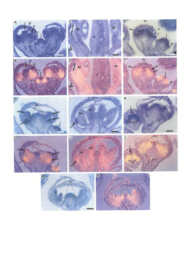
Fig. 1. Scanning electron micrographs depicting the remaining floral meristem, if there is any, develops into
development of wild-type and superman-1 Arabidopsis carpelloid tissue (Fig. 1L, N-P). Often the carpel
flowers. In many cases the outer whorls of the flowers have primordium is irregularly shaped and is congenitally
been dissected away to reveal the inner whorls. (A- fused to one of the innermost presumptive stamen
D) Wild type. (A) Inflorescence meristem and flowers in
stages 1-4 (Smyth et al., 1990). The sepal primordia have primordia, resulting in the formation of a mosaic organ
emerged on the two oldest flower buds. (B) Stage 6 flower. (Fig. 1O,P).
At this time all floral organ primordia have formed; the
second whorl organ primordia are not visible in this view. Double and triple mutants of superman-1 and floral
(C) Stage 7-8 flower. Second whorl petal (P), third whorl homeotic mutants
lateral stamen (LS), third whorl medial stamen (MS), and superman-1 pistillata-1
fourth whorl gynoecium (G) primordia are indicated. It is The phenotype of sup-1 pi-1flowersis the same as that
thought that the identity of each of the floral organ of pi-1 flowers (Bowman et al., 1989). The first whorl
primordia is specified during stages 2-7. (D) Stage 12
flower showing the differentiated floral organs. (E- contains four wild-type sepals, while the second whorl is
P) superman-1. (E) Inflorescence meristem and flowers of occupied by four smaller sepals. The remaining floral
stages 1-5. (F) Stage 6 flower. Up to this stage of meristem, which in wild-type flowers gives rise to the
development, sup-1 flowers are morphologically third and fourth whorl organs, instead gives rise to an
indistinguishable from wild-type. (G) Stage 7-8 flower. enlarged gynoecium composed of 2 to 4 carpels (mean
Four organ primordia are developing in a whorled fashion 3.1, 198 carpels/63 flowers).
on the flanks of the cells that would normally give rise to As shown in Fig. 2A-C, the development of sup-1 pi-1
the gynoecium. (H) Stage 9-10 flower. Interior to the six flowers is indistinguishable from that of pi-1 flowers
third whorl stamens, a ring of five stamens and another (Fig. 2D-E). The first whorl primordia are initiated in
ring of three stamens are visible. (I) Six stamens occupy
their normal positions in the third whorl (3). Eleven their normal positions and differentiate into sepals. The
additional stamens are evident, six forming a ring interior second whorl organ primordia are also initiated in the
to the third whorl (4) and five more forming an additional correct positions, but they differentiate inappropriately
ring (5). (J) Stage 11 flower. Six stamens, in addition to into small sepals, like those of pi-1flowers(Fig. 2B, D).
the normal third whorl of stamens, are present. (K) Stage The remaining floral meristem, which ordinarily gives
12 flower. Four distinct rings of stamens are visible. rise to the third and fourth whorl primordia, is
(L) Mature superman-1flower.Ten stamens and a central incorporated into the developing ovary (Fig. 2C), as is
gynoecium lacking most of the ovary tissue are visible. observed in pi-1 flowers (Fig. 2E). This results in a
(M) The outer two whorls, the sepals and petals have been gynoecium that is irregularly shaped and is composed of
removed to reveal the thirteen stamens occupying the more than two carpels. Nectaries may form in the
central region of the flower. (N) A filamentous structure,
whose epidermal morphology is similar to that of stamen region between the second whorl organs and the
filaments, capped with stigmatic tissue occupies the center gynoecium. Thus, pi-1 is epistatic to sup-1 and the loss
of this flower. Nectaries at the base of the third whorl and of determinacy observed in sup-1 flowers is eliminated
inner stamens are visible (arrows). (O) A mosaic organ in a pi-1 background.
with stamen and carpel sectors. Each sector is large, well-
defined, and longitudinal. (P) Mosaic organ in which the superman-1 apetala3-l
sectors are small and less well-defined than those in O. An allelic series of apetala3 mutations has been
Bar=10 fan in A, B, C, E, F, and G: 30 fim in H; 100 fan described with ap3-3 (as well as ap3-4 and ap3-5)
in D, I, J, K, M, N, O, and P; 300 fan in L.
flowers representative of the most severe phenotype
(Jack et al., 1992). ap3-3 flowers consist of two outer
whorls of sepals, as in pi-1 flowers, while the organs of
(Bowman et al., 1989). Usually, only one or two of the inner two whorls, all of which are carpels, appear
these mosaic organs occur in the same flower, although congenitally fused (Fig. 2F-G; Jack et al., 1992). In the
three or more have been observed infrequently. weaker, temperature-sensitive ap3-l allele (Fig. 2H),
The development of the first three whorls of sup-1 the third whorl organs develop as staminoid carpels and
flowers is usually normal: four sepals, four petals, and the fourth whorl develops into a normal gynoecium
six stamens form in their wild-type positions (Fig. when grown at the restrictive temperature (Fig. 21;
1E,F). However, the third whorl organ primordia Bowman et al., 1989). The molecular lesions of these
occasionally vary in number (5-7), size and position. two ap3 alleles are consistent with the severity of their
Interior to the third whorl, more primordia are phenotypes. ap3-3 is a nonsense mutation in the first
produced from the region that in wild type develops coding exon, while ap3-l is a mis-sense mutation in a
into the gynoecial cylinder. The number (3-6) and conserved protein domain (Jack et al., 1992).
position of these extra organ primordia is somewhat Flowers of plants homozygous for both sup-1 and
irregular (Fig. 1G-I). This process continues for a ap3-l grown at 25°C have two outer whorls of sepals
variable period of time; only a single additional ring or surrounding several fused carpelloid structures that
as many as four to five extra sequentially-formed rings arise from the inner two whorls (Fig. 2J-O). The outer
of organ primordia, all of which develop into stamens two whorls develop as described for ap3-l, ap3-3 and
(or sometimes carpelloid stamens in the case of the sup-1 pi-1 flowers (above; Bowman et al., 1989). Cells
innermost organs), may be produced. Following the that would normally form the medial third whorl organ
production of primordia that develop into stamens, the primordia appear to be incorporated into an enlargedCD
o
a.
Fig. 2. Scanning electron micrographs depicting the development of sup-l pi-l cells that normally constitute the medial third whorl organ primordia (m) are
and sup-l ap3-l flowers. In many cases the outer whorls of the flowers have congenitally fused with some fourth whorl tissue. The lateral third whorl
been dissected away to reveal the inner whorls. (A-C) sup-l pi-l flowers. primordia remain distinct (I). Compare this flower with F and H.
(A) Inflorescence meristem and flowers in stages 1-4. (B) The phenotype of this (L) Approximately stage 8 flower. Adjacent medial third whorl primordia have
approximately stage 7-8 sup-l pi-l flower is indistinguishable from that of a pi-l fused and are enveloping the fourth whorl. The second whorl primordia are
flowers of the same age shown in D. The cells that would normally constitute developing characteristics of sepals. (M) Approximately stage 9 flower. The
the third whorl organ primordia are incorporated into the developing gynoecial medial third whorl organs (m) have fused as in L while the lateral third whorl
cylinder. (C) Mature sup-l pi-l flower, again similar in phenotype to the pi-l organs (1) are separate and staminoid. (N) Stage 10-11 flower. Adjacent medial
flower shown in E. (D-E) pi-l flowers. (D) Stage 7-8 flower. (E) Mature flower. third whorl organs are fused along entire length and are carpelloid with
(F-G) ap3-3 flowers. (F) Stage 7 flower. The cells that would normally stigmatic tissue at the top of the structure and rudimentary ovules along the
constitute the medial third whorl organ primordia fuse together and envelop the margins. Nectaries are visible below the fused structure (arrows). (O) Mature
fourth whorl. (G) Mature flower. The third and fourth whorl organs are fused flower. The fused medial third whorl organs retain some staminoid
together. (H-I) ap3-l flowers. (H) Stage 7 flower. The third and fourth whorl characteristics such as the filamentous base (double arrow), but are mostly
organ primordia are distinct. (I) Mature flower. The third whorl is occupied by carpelloid and have fused with fourth whorl tissue. Nectaries are visible at the
solitary carpels and carpelloid stamens. (J-O) sup-l ap3-l flowers. base of the third whorl (arrows). Compare this flower with G and I. Bar=20
(J) Inflorescence meristem and flowers in stages 1-4. (K) Stage 7 flower. The pan in A, B, D, F, H, J, K, L, and M; 100 fun in C, E, G, \, O, and N.00
c
m
2
2
&
I
Fig. 3. Scanning electron micrographs depicting the development of superman Stipules are present at the base of the first whorl organs. (H) Interior to the o
apetala2 flowers. In many cases the outer whorls of the flowers have been third whorl are two organs whose epidermal morphology is carpelloid.
dissected away to reveal the inner whorls. (A-E) sup-] ap2-2 flowers. (I) Interior to the third whorl are three organs, one of which is a stamen. 1
(A) Inflorescence meristem and flowers of stages 1-5. (B) Stage 7 flower. (J) Mature sup-l ap2-l flower. Four cauline leaf-like organs are present in the
(C) Developing ovules are visible along the margins of the first whorl carpels. first whorl, four staminoid petals in the second whorl, five stamens in the third
Three stamens and a carpelloid organ are visible interior to the first whorl. whorl, and another four stamens are present interior to the third whorl. The 1
(D) Mature sup-l ap2-2 flower. Two medial first whorl carpels and three inner center of the flower is occupied by a filamentous structure that has developing
whorl stamens (arrows) are visible. (E) Occasionally, sup-l ap2-2 flowers consist stigmatic tissue at its tip. Bar=10 (an in A, B, F, and G; 100 (tin in C, D, E,
of merely a two-carpelled gynoecium as shown here. (F-J) sup-l ap2-l flowers. H, and I; 300 (im in J.
(F) Inflorescence meristem and flowers in stages 1-4. (G) Stage six flower. B3
a;
a.
o
•a606 /. L. Bowman and others
organ primordium that consists of the four medial third structures (17/78) or carpelloid leaves (3/78); again this
whorl organ primordia as well as those cells that usually is similar to what is observed in ap2-2 flowers. No
constitute the fourth whorl gynoecium (Fig. 2K). Thus, second whorl organs are present.
the medial third whorl organ primordia are usually The central region of sup-1 ap2-2 flowers is highly
fused to each other and are frequently fused to the variable in phenotype in terms of number of organs
fourth whorl organ primordia (Fig. 2K-O). All of this present. On average 3 organs (118 organs/39 flowers)
tissue differentiates into carpelloid structures resulting occupy this region of the flower, but from 0 to 6 organs
in an abnormal gynoecium. were observed. The phenotypes of these organs are
The fate of the lateral third whorl primordia is stamens (60/118 organs), carpelloid stamens (12/118),
somewhat different. The lateral third whorl organ staminoid carpels (20/118), solitary carpels (14/118),
primordia usually remain distinct from the rest of the and filamentous fused carpels lacking all internal
third whorl and the fourth whorl structures and structures and capped with stigmatic papillae (12/118).
differentiate into staminoid carpels (Fig. 2K,M, and N). These carpelloid organs are similar to the carpelloid
However, in the later flowers produced by an inflor- organs present in the center of sup-1 flowers. Normal,
escence, the lateral third whorl organ primordia may be fully fused gynoecia were not observed. The outermost
fused with medial third whorl organ primordia and the of these organs, those in the region that would normally
fourth whorl organ primordia. Nectaries may be found be the third whorl, tend to be stamens, while the organs
at the base of the fused third whorl organs (Fig. 2N-O). arising from the region that would ordinarily develop
The extent of fusion of the medial third whorl and into the gynoecium are usually mosaics of both stamen
fourth whorl organs increases acropetally on the and carpel tissue. The mosaic organs resemble those
inflorescence, as does the extent of carpellody of the observed in sup-1 flowers. If more than one carpelloid
lateral third whorl organs. Similar trends are also organ is present, the carpelloid organs are usually fused
observed in ap3-l flowers; the lateral third whorl organs to each other along the carpel tissue. The reduction in
are more staminoid than the medial, and the extent of the numbers of organs occupying the inner whorls of
carpellody of all third whorl organs increases acro- ap2-2 sup-1flowersparallels that in singly mutant ap2-2
petally. flowers, which have a severe reduction in the number of
The development of sup-1 ap3-l doubly mutant third whorl organs.
flowers differs from ap3-l flowers, where the third and The development of the outer whorl organs in sup-1
fourth whorl organs remain distinct (Fig. 2H-I). ap2-2 flowers parallels that observed for ap2-2 flowers
Rather, it resembles what is observed in ap3-3 flowers. (Fig. 3A-E; Bowman et al., 1991b). The development
The third and fourth whorl organ primordia of sup-1 of the central organs, in the third and fourth whorls, is
ap3-l flowers congenitally fuse, with the third whorl variable. The organ primordia of these whorls are not
organs often enveloping the fourth whorl organs (Fig. produced in a consistent pattern and they are often
2F-G). Thus, the sup-1 mutation enhances the weak, fused congenitally (Fig. 3B-D). The development of
partial loss-of-function ap3-l phenotype, causing the these organs is similar to that observed for the
sup-1 ap3-l double mutant to resemble closely the innermost stamens and stamen-carpel mosaic organs of
severe ap3-3 phenotype. Similar to the case for sup-1 pi- sup-1 flowers. Occasionally, when no second, third, or
1 flowers, the loss of determinacy in sup-1 flowers is fourth whorl primordia are produced, the medial first
eliminated in an ap3-l background. whorl carpels are congenitally fused, resulting in a
flower that consists only of a two- carpelled gynoecium
superman-1 apetala2-2 (Fig. 3E). This has also been observed in ap2-2 flowers
Doubly mutant flowers of plants homozygous for both (Bowman et al., 1991b).
sup-1 and ap2-2 exhibit a nearly additive phenotype
(Fig. 3A-E). The outer two whorls resemble those of
ap2-2 flowers (Bowman et al., 1991b), while the inner superman-1 apetala2-l
whorls resemble those of sup-1 flowers. The outer The outer two whorls of ap2-l sup-1 flowers resemble
whorl of ap2-2 has carpelloid and phylloid organs; the those of ap2-l, while the inner whorls resemble those of
second whorl has no organs. The third whorl of ap2-2 sup-1 flowers (Fig. 3F-J). Thus, as in the other double
flowers is occupied by a severely reduced number of mutant combination involving ap2 and sup alleles, the
stamens (0.25 stamens/flower) and the fourth whorl is phenotype of sup-1 ap2-l homozygotes is essentially an
occupied by a two-carpelled gynoecium that is usually addition of the effects of the two single mutations. The
unfused. outer two whorls of ap2-l flowers are the same as
The medial first whorl organs of sup-1 ap2-2 flowers described below for the double mutant. The third and
are solitary carpels (52/78 positions counted in 39 fourth whorls of ap2-l flowers are phenotypically
flowers), phylloid carpels (2/78), or mosaic organs with normal except for reduced numbers and altered
sectors of carpel and stamen tissue (24/78). The stamen positions of third whorl stamens.
sectors of the mosaic organs always occupy the margins, The first whorl of sup-1 ap2-l flowers consists of four
while the carpel sectors occupy the central regions of cauline leaf-like organs that may develop carpelloid
the organs, as is observed in ap2-2 flowers. The lateral features such as stigmatic papillae at their tips and
first whorl organs are most often absent (43/78), but rudimentary ovules along their margins, while the
may be cauline leaf-like organs (15/78), filamentous second whorl is occupied by organs with features ofSUPERMAN, a regulator of floral homeotic genes in Arabidopsis 607
both stamens and petals, as in ap2-l flowers. Interior to behaving like another floral meristem as occurs in ag-1
the outer two whorls, 4 to 9 stamens develop (average flowers, continues to sequentially produce rings of
6.8; 204 stamens/30 flowers), fewer than are observed in organ primordia that subsequently develop into petals
sup-1 flowers. This parallels the reduced number of (Fig. 4C-E). The organ primordia in each of these later
stamens found in ap2-l singly mutant flowers (4.9 per rings are variable in number (4-8) and position (Fig. 4C-
flower; Bowman et al., 1989). The innermost stamens E). This process continues indeterminately, resulting in
often have stigmatic tissue at their tips. The remainder an extreme double flower phenotype, and the ultimate
of the flower consists of a variable amount carpelloid ornamental Arabidopsis. Close examination of fully
tissue similar to that observed in sup-1 flowers. This developed flowers reveals that some of the inner
tissue is usually a mosaic between carpel and stamen organs, while primarily petaloid in character, may have
tissue (42 staminoid carpels/30 flowers). In only one of some sepaloid characteristics such as stomata, and some
30 flowers scored was no carpelloid organ present. sepaloid epidermal cells (Fig. 4H). These sepaloid
The development of sup-1 ap2-l flowers is also characters are random in their frequency and position,
similar to that of ap2-l and sup-1 flowers (Fig. 3F-J). in contrast to the large longitudinal sepaloid sectors of
The outer two whorls develop as has been described for ag-1 singly mutant flowers, which occur every third
ap2-l flowers (Bowman et al., 1989). Third whorl whorl in ag-1flowers(Bowman et al., 1989).
primordia develop in a similar fashion to that observed Both single mutations, sup-1 and ag-1, cause indeter-
in ap2-l flowers: there may be fewer than six stamens, minate growth to varying extents. In sup-1 ag-1 flowers,
and they may arise in ectopic positions (Fig. 3G). Each fasciation of the floral meristem may occur with the
of these primordia develops into a stamen. Interior to meristem becoming enlarged (over 100 /zm in width)
these stamens, usually 3 or 4 (range 0-4) organ and elongated in shape. Organ primordia, all of which
primordia arise (Fig. 3I-J), each of which develops into develop into petals, are produced along the entire
a stamen, although these organs often develop carpel- margin of the fasciated meristem (Fig. 4F-G). Thus, the
loid characteristics, such as stigmatic tissue at their tips. two mutations interact synergistically to cause greatly
Development interior to this is variable. In most cases increased, indeterminate growth of the floral meristem.
one or two staminoid carpels or a filamentous structure sup-1 ag-1 double mutant flowers are easily dis-
capped with stigmatic papillae is produced, although tinguished from ag-1 flowers due to the differences in
occasionally the floral meristem stops proliferating growth rates of sepals and petals. In ag-1 flowers, the
following the development of the second ring of sepals that arise in the fourth whorl rapidly dwarf the
stamens (Fig. 3J). Thus, the number of stamens in both adjacent developing petals and grow to cover the floral
sup-1 ap2-2 and sup-1 ap2-l is intermediate to that meristem forming a structure resembling an internal
observed in sup-1 and the respective ap2 allele. flower bud (Bowman et al., 1989). In contrast, all of the
primordia (except those of the first whorl) of sup-1 ag-1
superman-1 agamous-1 flowers develop into petals, and all have the same slow
Mutations at the AGAMOUS locus cause indetermi- growth rate characteristic of petals. The result is that
nate growth of the floral meristem as well as organ the floral meristem is not covered by developing organs,
identity transformations. The outer two whorls of and it is exposed even in relatively old flowers (Fig. 4F).
agamous flowers are phenotypically normal sepals and
petals. In whorl 3, six petals develop in the positions superman-1 agamous-2
normally occupied by stamens. In whorl 4, the cells that
would normally give rise to the gynoecium instead form Mutations at the ERECTA locus have a profound effect
another ag flower. This process repeats itself, resulting on the overall morphology of ag flowers (Bowman et
in the formation of an indeterminate number of whorls al., 1991b). In an erecta mutant background, there is
of floral organs in the pattern (sepals, petals, petals)n little elongation of the pedicel between whorls of ag
(Bowman et al., 1989; 1991b). flowers. However, in a wild-type ERECTA back-
ground, the pedicel elongates after every third floral
Flowers of plants homozygous for both sup-1 and ag- whorl, or just prior to the whorls of sepals, in ag flowers
1 consist of a first whorl of sepals followed by an (Yanofsky et al., 1990). To determine whether the
indeterminate number of petals. Observations of ERECTA locus has an effect on the elongation of the
developing flowers elucidate the developmental basis of pedicel of sup ag flowers, a sup-1 ag-2 double mutant
this phenotype (Fig. 4A-H). The first and second whorl was constructed in a wild-type ERECTA background.
organ primordia arise in the correct positions and The phenotype of sup-1 ag-2 ERECTA flowers is the
numbers, as in sup-1 or ag-1 single mutants (Fig. 4A-B). same as that of sup-1 ag-2 erecta flowers. Thus, in
These organ primordia subsequently differentiate into contrast to ag-2flowers,there is no pedicel elongation
wild-type sepals and petals, respectively. The pro- between any of the floral organs in sup-1 ag-2 flowers,
duction of third whorl organ primordia, although corroborating the conclusion that no new flower is
usually normal, may be altered in a manner similar to formed in the fourth whorl in sup-1 ag-2 flowers.
that of the third whorl primordia of sup-1flowers.As in
ag-1 flowers, the organ primordia that arise in the
second and third whorls are approximately the same superman-1 agamous-1 apetala2-l
size and each of these primordia develops into a petal The overall architecture of sup-1 ag-1 ap2-l flowers is
(Fig. 4B). The remaining floral meristem, rather than similar to sup-1 ag-1flowers,but with minor differences608 J. L. Bowman and others Fig. 4. Scanning electron micrographs depicting the development of sup-1 ag-1 and sup-1 ag-1 ap2-l flowers. In many cases the outer whorls of the flowers have been dissected away to reveal the inner whorls. (A-H) sup-1 ag-1 flowers. (A) Inflorescence meristem and flowers in stages 1-4. (B) Stage 6 flower. At this stage, sup-1 ag-1flowersresemble wild type except that the second and third whorl organ primordia are similar in size, as observed in ag-1 flowers. (C) Six organ primordia are present interior to the third whorl and the floral meristem is pentagonal. (D) Eight organ primordia are present along the margins of the irregularly shaped floral meristem. (E) The floral meristem continues to produce organ primordia along its margins. (F) sup-1 ag-1flowerof late developmental stage. (G) The floral meristem of flower shown in F has become enlarged and elongated, producing organ primordia along its entire circumference. (H) Close up of inner organs of sup-1 ag-1 flower. Many stomata are visible as well as a sector of sepaloid tissue (arrow). (I-L) sup-1 ag-1 ap2-l flowers. (I) Inflorescence meristem and flowers in stages 1-5. (J) Stage 6-7 flower. Organ primordia of the third whorl are not in the normal positions and the floral meristem is enlarged. (K) The first whorl cauline leaf-like organs have developed stellate trichomes, while all the inner organs are staminoid petals. (L) Mature sup-1 ag-1 ap2-l flower. Bar=10 /mi in A, B, C, I, and J; 50 fim in D, E, and K; 100 /mi in G and H; 300 /an in F and L. (Fig. 4I-L). For instance, the second whorl primordia whorls of ag-1 ap2-l flowers are not present in the triply do not always form in the triply mutant flowers, mutant flowers. The first whorl organs of sup-1 ag-1 probably due to the ap2-l mutation, since loss of second ap2-l flowers are cauline leaves with some carpelloid whorl organs also occurs in ap2-l and ap2-l ag-1 characteristics, as is observed in the first whorl of ag-1 flowers. Additionally, the pattern (numbers and pos- ap2-l flowers (Fig. 4F,L). All organs interior to the first itions) of third whorl, and subsequent, organ primordia whorl (a large, indeterminate number) in the triply formation is more often irregular in sup-1 ag-1 ap2-l mutant flowers are staminoid petals, like those occupy- flowers (Fig. 4J; altered third whorl positions are also ing the second and third whorls of ag-1 ap2-l flowers observed in ap2-l and ap2-l ag-1 flowers). (Fig. 4F,L; Bowman et al., 1989). Thus, the pattern of The identity of the organs in sup-1 ag-1 ap2-l flowers organs in the triple mutant is similar to that in sup-1 ag- resembles those of ag-1 ap2-l flowers (Bowman et al., 1 flowers, while the identity of the organs is like that 1989) except that the leaf-like organs that occur in inner observed in ag-1 ap2-l flowers.
SUPERMAN, a regulator of floral homeotic genes in Arabidopsis              609

Expression patterns of floral homeotic genes in              form interior to the third whorl (Fig. 5I-J); each of these
superman-1 flowers                                           primordia differentiates into a stamen. This pattern
The phenotype of superman flowers suggests that the          continues, with expression interior to the youngest
SUPERMAN gene product may be a regulator of floral           organ primordia commencing just before the next organ
homeotic genes. The epistatic interactions oiap3 and pi      primordia morphologically differentiate from the floral
mutants with sup mutants and the synergistic interac-        meristem (similar to initial AP3 expression in the
tion between ag mutants and sup mutants identify these       second and third whorls of wild-type flowers), until the
genes as candidates for genes regulated by SUPER-            floral meristem ceases to proliferate (Fig. 5G-H). Thus,
MAN. To determine if the expression patterns of the          the spatial pattern of AP3 expression expands into the
two cloned Arabidopsis floral homeotic genes, AP3            fourth whorl to include the extra stamens in superman-1
(Jack et al., 1992) and AG (Yanofsky et al., 1990), are      flowers.
altered in a superman genetic background, we per-               The spatial and temporal pattern of AP3 RNA
formed tissue in situ hybridizations to sections of          detected in sup-1 ag-1flowersis similar to that observed
developing flowers using AP3 and AG probes. The              for sup-1 flowers. During stages 3-6, AP3 RNA is
SEM micrographs in Figures 1-4 provide a visual guide        detected in a pattern indistinguishable from that
for the tissue sections of Figures 5 and 6.                  described for sup-1 flowers. The signal is associated
                                                             with the second and third whorl primordia, as well as
   The expression of APETALA3 is altered in                  the more abaxial cells of the floral meristem interior to
   superman-1 flowers                                        the third whorl (Fig. 5K-L). Following this, the floral
The expression pattern of the AP3 gene in both wild-         meristem produces an indeterminate number of organ
type and homeotic mutant flowers has been described          primordia, all of which exhibit a hybridization signal
by Jack et al. (1992). Briefly, in wild-type flowers, AP3    that appears on the flanks of the floral meristem before
expression commences during stage 3 (see Fig. 1A),           the primordia arise and is uniform throughout the organ
before the appearance of the second and third whorl          primordia once formed (Fig. 5M-N). Each of the organ
primordia, and is restricted to floral whorls two and        primordia that develops interior to the first whorl
three, the whorls affected in ap3 mutants (Fig. 5A-D;        differentiates into a petal (Fig. 4F). The central region
Jack et al., 1992). During stages 3-4, a uniform signal is   of the indeterminate floral meristem of sup-1 ag-1
detected throughout the region of the floral meristem        flowers, one to several rows of cells wide, exhibits no
that will later give rise to the second and third whorl      signal above background throughout flower develop-
organ primordia (Fig. 5A-B). When the second and             ment, similar to that observed for the floral meristem of
third whorl organ primordia morphologically differen-        sup-1 flowers (compare Fig. 5F and 5H to 5L and 5N).
tiate from the floral meristem during stage 5, the signal
is confined to those organ primordia and cells directly         AGAMOUS expression is unaltered in superman-1
underlying the primordia (Fig. 5A-B). As differen-             flowers
tiation of the second and third whorl organ primordia        The floral specific expression pattern of the AG gene, in
progresses, into petals and stamens, respectively,           both wild-type and homeotic mutant flowers, has been
(stages 7-14), the uniform spatial expression pattern is     described by Drews et al. (1991) and Bowman et al.
maintained, although the intensity of the signal dimi-       (1991a). Briefly, in wild-type flowers, AG expression is
nishes compared to that of stage 3 (Fig. 5C- D), with the    restricted to whorls three and four, the whorls affected
expression in the second whorl petals persisting longer      in ag mutants, and commences during stage 3 (see Fig.
than that in the third whorl stamens.                        1A), before the appearance of the third and fourth
   The initial spatial pattern (stages 3-4) of AP3 RNA in    whorl primordia (Fig. 6A-B; Drews et al., 1991). A
superman-1 flowers is strikingly different from that         uniform signal is detected throughout the region of the
observed in wild-type flowers. In addition to those cells    floral meristem that will give rise to the third and fourth
developing into the second and third whorls, the             whorl organ primordia during stages 3-4. When the
hybridization signal is expanded to include most of the      third and fourth whorl organ primordia emerge from
cells that constitute the fourth whorl in wild-type          the floral meristem during stage 5, the signal is confined
flowers (Fig. 5E-F). A narrow band of cells, one to four     to those organ primordia and cells directly underlying
cellular rows wide, at the center of the floral meristem     the primordia. As cellular differentiation of the third
has no detectable signal above background. Thus, the         and fourth whorl organ primordia progresses, into
initial inner boundary of AP3 expression has been            stamens and carpels, respectively, (stages 9-14), AG
shifted towards the center of the floral meristem.           expression is localized to those organs (Fig. 6C-D) and
During stages 5-6, when the second and third whorl           becomes progressively restricted to certain cell types
organ primordia emerge, AP3 RNA is detected in these         within these organs, such as stigmatic papillae, endo-
primordia (Fig. 5E-F). However, AP3 RNA is also              thelial cells and endothecial cells (Bowman et al.,
detected in cells interior to the third whorl primordia,     1991a).
which develop into gynoecial tissue in wild-type                The spatial and temporal pattern of AG expression is
flowers, but develop into additional stamens in sup-1        not significantly altered in sup-1flowers.During stages
flowers (Fig. 5E-H). Again, no signal is detected at the     3-4, the distribution of AG RNA is relatively uniform in
center of the floral meristem. Following stage 6, AP3        the floral meristem in those regions that will give rise to
RNA is detected in the additional organ primordia that       the organs interior to the second whorl, the same
610 /. L. Bowman and others
Fig. 5. Expression of A PETALA3 RNA in wild-type, sup-1 (G and H) In this approximately stage 8 flower, AP3 RNA
and sup-1 ag-1 flowers. Each section was photographed in is detected in the second whorl petal (p) and third whorl
two ways: bright field exposure (A, C, E, G, I, K, M) and stamen (st) primordia. In addition, AP3 RNA is detected
bright field - dark field double exposure (B, D, F, H, J, L, in regions of the floral meristem (fm) from which
N) using a yellow filter during the dark field exposure additional stamen primordia will form prior to their
causing the silver grains (RNA hybridization signal) to emergence. In the lower right of the panel AP3 RNA is
appear yellow. All flowers are oriented with the apex of detected nearly throughout the floral meristem (f) of a
the flower towards the top. In E-H and K-N the floral stage 4 flower. (I and J) This series shows a section
meristem is indicated (fm) and is the region between the through a sup-1 flower in which morphological
arrows.(A-D) Wild-type flowers. (A and B) This series differentiation has commenced in the third whorl (3st) and
shows a section through stage 4 and stage 6 wild-type additional (4st) stamens. AP3 RNA is present in all of the
flowers. AP3 RNA is detected in the regions of the floral stamens as well as the second whorl petals (p). A lower
meristem (f) destined to give rise to the second and third signal is associated with the organ primordia in the center
whorl organs, but not in the sepal primordia (sp) of the of theflower.The fate of these central organ primordia
stage 4 flower. At stage 6, AP3 RNA is detected in the varies from stamens to carpels, with mosaic organs
petal (p) and stamen (st) primordia, occupying the second occurring most frequently. (K-N) sup-1 ag-1flowers.(K
and third whorls respectively, but not in the sepals (se) and and L) This series shows a section through a stage 5 flower
carpels (c) in the first and fourth whorls. A similar pattern (left) and an older flower (right). AP3 RNA is detected
is seen in the other stage 6 flower in the right side of the nearly throughout the floral meristem (f) of the stage 5
frame. (C and D) Late in development, after flower as well as all the organ primordia (p) present
morphological differentiation of the floral organs has interior to the first whorl sepals. Simlarly, AP3 RNA is
commenced, AP3 RNA is detected in the maturing petals observed in all the organ primordia interior to the first
(p) and stamens (st), but not in the sepals (se) or carpels whorl in the older flower as well as the region of the floral
(c). (E-J) sup-1 flowers. (E and F) This series depicts stage meristem (fm) from which the next organ primordia will
4 (right) and stage 6 (left) sup-1 flowers. The inner emerge. All organ primordia, except the first whorl, will
boundary of AP3 RNA expression is shifted towards the differentiate into petals. (M and N) This series shows a
center of the floral meristem (f) of the stage 4 flower, such section through an approximately stage 8 flower. Two petal
that AP3 RNA is detected in cells interior to those that are primordia (p), occupying the second and third whorls are
the precursors of the second and third whorl organs. This visible in addition to very small primordia nestled between
is in sharp contrast to what is seen in wild-type (A and B) the third whorl organ primordia and the floral meristem
where the entire floral meristem interior to the third whorl (fm). AP3 RNA is detected in all organs and organ
organ primordia consists of non-^4P5-expressing cells. As in primordia interior to the first whorl, as well as theflanksof
wild-type, no signal is seen in the sepal primordia (sp). In the floral meristem (fm). The floral meristem is
the stage 6 flower, AP3 RNA is detected in the second indeterminate, continuing to produce organ primordia, all
whorl petal (p) and third whorl stamen (st) primordia. In of which differentiate as petals. In nearly all cases AP3
addition, AP3 RNA is present in cells on the flank of the RNA is detected in those cells on the flank of the floral
remaining floral meristem (fm) which in wild type gives rise meristem from which the organ primordia are produced.
to the fourth whorl carpels but in sup-1 flowers gives rise Bars=50 um.
to extra stamens. No signal is detected in the sepals (se).
pattern seen in wild-type flowers at this stage of not required for carpel development. Rather, the
development (Fig. 6E-F). As the third and additional genetic and molecular data show that SUPERMAN has
interior rings of organ primordia are produced in sup-1 a role in the spatial regulation of at least one floral
flowers, AG RNA continues to be detected in the homeotic gene.
additional whorls of organ primordia and throughout
the entire floral meristem from which they are derived Homeotic genes
(Fig. 6E-H). Later in development, AG RNA is Based on a series of genetic experiments, it has been
detected in the same cell types with which it is proposed that the four floral homeotic genes, AGA-
associated in wild-type flowers. A high signal is present MOUS, APETALA2, APETALA3 and PISTILLATA,
in nectaries, the connectives and endothecia of anthers, act alone and in combination to determine in large part
and the occasional ovules and stigmatic tissue that the specification of floral organ identity (Bowman et al.,
develop on carpelloid organs at the center of sup-1 1991b). The precise spatial expression patterns of the
flowers. floral homeotic genes are proposed to represent
positional information within the developing flower.
Discussion Each of the four genes is proposed to act in two
adjacent whorls of the flower, and thus, falls into one of
In contrast to the floral homeotic mutants, the alter- three classes: those that affect the outer two whorls
ations in flower structure observed in superman mutants (APETALA2), those that act upon the second and third
are not strictly homeotic conversions of floral organs. whorls (APETALA3 and PISTILLATA), and those
Although additional stamens develop at the expense of that exert their influence on the inner two whorls
carpels in sup-1 single mutants, the fact that carpels (AGAMOUS). The three classes demonstrate the
develop in the first whorl of sup-1 ap2-2 doubly mutant division of the flower primordium into four regions with
flowers indicates that the SUPERMAN gene product is each region, or whorl, having in wild type a uniqueI* c se
SUPERMAN, a regulator of floral homeotic genes in Arabidopsis 611
Fig. 6. Expression of AGAMOUS RNA in wild-type and              istic interactions between AG and AP2 (Bowman et al.,
sup-1 flowers. Each section was photographed in two ways:       1991b), which have been shown to be executed at the
bright field exposure (A, C, E, G) and bright field - dark      RNA level for AG (Drews et al., 1991), result in
field double exposure (B, D, F, H) using a yellow filter        helping to define the spatial boundary of expression of
during the dark field exposure causing the silver grains        AG. However, cross-regulatory interactions are insuf-
(RNA hybridization signal) to appear yellow. All flowers
are oriented with the apex of the flower towards the top.       ficient to account for the localization of the AP3 and PI
In E and F the floral meristem is indicated (fm) and is the     activities. That AP3 is initially expressed in a well-
region between the arrows. (A-D) Wild-type flowers. (A.         defined temporal and spatial manner in wild type as well
and B) This series shows a section of a stage 4 flower          as in other homeotic mutants (Jack et al., 1992),
(right) and an older flower (left). AG RNA is detected in       suggests that it may be responding to factors that are
the regions of the floral meristem (f) that will give rise to   present prior to the other homeotic gene products. The
the third and fourth whorl organs, but not in the first         phenotype of sup flowers suggests that SUP is at least
whorl sepal primordia (sp) of the stage 4 flower. In the        partially responsible for defining the initial expression
older flower, AG RNA is restricted to the stamens (st) and      patterns of AP3 and/or PI.
carpels (not shown in this section) while no signal is
detected in the first whorl sepals (se) and second whorl
petals (p). (C and D) Late in development, after                The role of SUPERMAN
morphological differentiation of the floral organs has          If the primary role of SUPERMAN is to suppress
commenced, AG RNA is detected in the third whorl                AP3/PI activity in the fourth whorl, certain predictions
stamens (st) and fourth whorl carpels (c), but not in the       can be made about doubly mutant strains with sup and
first whorl sepals (se) and second whorl petals (p). Note       the homeotic mutations, and these are shown in Fig. 7.
that AG RNA is already becoming restricted to certain cell      Since ap2 mutations primarily affect organ specification
types as there is no AG RNA detectable in the                   in the outer two whorls and SUP acts in the fourth
sporogenous tissue (s) of the stamens. (E-H) sup-1 flowers.
(E and F) This series shows a section through stage 3 and       whorl, additive interactions should be observed in sup
5 flowers as well as the inflorescence meristem. Similar to     ap2 flowers. This is the case in sup-1 ap2-2flowers.The
AG expression in wild-type flowers, AG RNA is detected          outer two whorls resemble those of ap2-2flowerswhile
in the regions of the floral meristem (f) that will give rise   the inner whorls are occupied by an increased number
to the third whorl and inner organs, but not in the sepal       of stamens (relative to the number in ap2-2flowers)and
primordia (sp) of the stage 3 flower. In the stage 5 flower,    a reduced amount of carpel tissue. Since ap2 mutations
AG RNA is detected in the third whorl stamen primordia          cause a severe reduction in the number of third whorl
(st) and throughout the floral meristem (fm) which will         organs, the number of staminoid organs produced in
give rise to the extra stamens, but not in the first whorl      sup-1 ap2 flowers is intermediate between the number
sepals (se). As in wild-type (Drews et al., 1991), no signal    produced in sup-1 flowers and the number produced in
is detected in the inflorescence meristem. (G and H) Later      ap2 flowers (Bowman et al., 1991b).
in development, AG RNA is detected in all organs interior
to the second whorl. In this section, AG RNA is detected           In sup ag flowers, we predict that AP2 is active in all
in the third whorl (3st) and interior (4st) stamens, but not    whorls due to the absence of AG activity, and AP3/PI
in the first whorl sepals and second whorl petals (p). In       activity expands into all whorls except the outermost
both the third whorl and inner stamens, AG RNA is               whorl due to the absence of SUP activity. Therefore,
becoming restricted to the same cell types as in wild type.     sup agflowersare expected to consist of an outer whorl
For example, there is no detectable signal in the               of sepals with an indeterminate number of whorls (due
sporogenous tissue (s). Bars=50 /zm.                            to the indeterminate nature of ag flowers) of petals
                                                                interior to the sepals. These predictions are validated
                                                                by the phenotype of sup agflowers(Fig. 4A-H) and the
combination of homeotic gene products present. Each             expression pattern of AP3 in sup agflowers(Fig. 5K-N;
of these regions then follows an organ-specific develop-        see below). Similar arguments can be made to predict
mental pathway directed by different combinations of            flowers with an outer whorl of leaves and an indetermi-
floral homeotic gene products as shown in Fig. 7. In            nate number of whorls of staminoid petals for sup ap2
support of this model, the expression patterns of the           ag triple mutants (Fig. 7).
two cloned homeotic genes, AGAMOUS and APE-                        In contrast, an epistatic relationship is observed
TALA3, are consistent with their proposed role in this          between the sup-1 and pi-1 mutations, with pi-1 being
model of floral organ specification; in wild-type flowers       epistatic to sup-1; sup-1 pi-1 flowers are morphologi-
AG expression is restricted to the third and fourth             cally indistinguishable from pi-1 flowers. This is the
whorls, and AP3 expression is restricted to the second          predicted result if SUP acts as an upstream negative
and third whorls (Yanofsky et al., 1990; Drews et al.,          regulator of PI.
1991; Jacket al., 1992).                                           According to the arguments presented above, ap3
   A major question that arises is, how do these floral         mutations should also be epistatic to sup mutations.
homeotic genes come to be expressed in their precise            This is observed with the strong ap3-3 allele; sup-2 ap3-
spatially and temporally restricted patterns? It is clear       3 flowers are indistinguishable from ap3-3 flowers (H.
that cross-regulatory interactions between the homeotic         Sakai and E. Meyerowitz, unpublished). However, this
genes are in part responsible for defining the spatial          is not the case for all ap3 alleles, such as the weaker ap3-
domains of activity of some of the floral homeotic              1 allele. In sup-1 ap3-l flowers the sup-1 mutation has a
genes. For example, the proposed mutually antagon-              phenotypic effect in the third whorl in the ap3-l
You can also read