Targeting T cell activation in immuno-oncology - Current ...
←
→
Page content transcription
If your browser does not render page correctly, please read the page content below
REVIEW ARTICLE

Targeting T cell activation
in immuno-oncology
S.D. Saibil md phd*† and P.S. Ohashi phd*‡

   ABSTRACT
   The years since 2009 have seen tremendous progress in unlocking the curative potential of the immune system for
   the treatment of cancer. Much of that revolution in immuno-oncology has been fueled by the clinical success of
   immune checkpoint inhibitors, particularly those targeting the PD-1 axis. Unfortunately, many patients still fail to
   benefit from checkpoint blockade or other immunotherapies. An inability to fully activate antitumour T cells con-
   tributes in part to the failure of those therapies. Here, we review the basic biology of T cell activation, with particular
   emphasis on the essential role of the dendritic cell and the innate immune system in T cell activation. The current
   understanding of the multiple factors that govern T cell activation and how they impinge on tumour immunotherapy
   are also discussed. Lastly, treatment strategies to potentially overcome barriers to T cell activation and to enhance
   the efficacy of immunotherapy are addressed.

   Key Words Immuno-oncology, T cell activation, immune checkpoint inhibitors

   Curr Oncol. 2020 April:27(S2)98–105                                                                         www.current-oncology.com

INTRODUCTION                                                                     We also highlight opportunities and therapeutic strategies
                                                                                 that aim to increase T cell activation, thereby enhancing
Immune checkpoint inhibitors (icis)—antibodies targeting                         the efficacy of icis and other immunotherapies.
either ctla-4 (CD152), PD-1 (CD279), or PD-L1 (CD274)—
have revolutionized immuno-oncology. Patients with                               MECHANISMS
tumours of varying histologies have experienced impres-
sive survival advantages after treatment using ici agents                        T Cell Tolerance: A Fine Balance
compared with treatment using the previous standard-of-                          The fundamental challenge of T cell immunity is for the
care agents1. Despite that success, the reality is that most                     system to respond to a universe of pathogens while lim-
patients treated with icis will not achieve a significant                        iting autoreactivity. The immune system must be able to
clinical response.                                                               develop a repertoire of T cells that encode antigen receptor
     Although frustrating from a clinical perspective, the                       specificities sufficiently diverse to recognize the wide range
lack of response to ici monotherapy in most patients is                          of antigens potentially present in pathogens. Conversely,
perhaps not surprising in the context of T cell biology. It is                   the T cell response must be constrained from reacting to
now appreciated that myriad intricate regulatory mech-                           antigens present in self-proteins. For T lymphocytes, this
anisms control T cell functioning, particularly in the                           balancing act begins in thymus, during a process called
context of antitumour immunity 2. A key factor governing                         “central tolerance” or “thymic selection”4.
the successful activation of the T cell arm of the adaptive                            In the thymus, immature T cells randomly rearrange
immune system is the requirement for the coordinated                             the variable, diversity, and joining segments of their T cell
activation of the innate immune system, which consists                           receptor (tcr) genes to generate a vast array of tcr speci-
of cells such as macrophages, innate lymphoid cells, nat-                        ficities5. For a T cell to mature and leave the thymus, the
ural killer cells, and dendritic cells (dcs). In particular,                     variable, diversity, and joining rearrangements must result
T cell activation is highly dependent on the functioning                         in a tcr that is capable of recognizing peptide antigens
of dcs, which present antigens to T cells and bridge the                         presented in the context of the individual’s own major
activation of the innate immune system to the adaptive                           histocompatibility complex [mhc (human leucocyte anti-
immune system3. Here, we review the regulation of T cell                         gen)] molecules. This process is called “positive selection,”
activation within the context of tumour immunotherapy.                           because it selects T cells expressing receptors appropriate

Correspondence to: Samuel Saibil, Department of Medical Oncology and Hematology, Princess Margaret Cancer Centre, 610 University Avenue, Toronto, Ontario
M5G 2M9.
E-mail: Sam.Saibil@uhn.ca n DOI: https://doi.org/10.3747/co.27.5285

S98                                                                                     Current Oncology, Vol. 27, Supp. 2, April 2020 © 2020 Multimed Inc.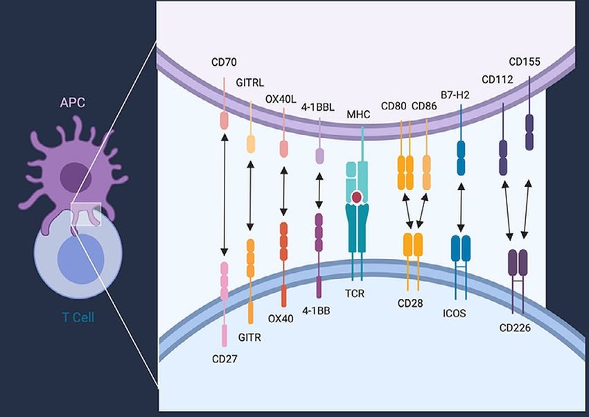
TARGETING T CELL ACTIVATION IN IMMUNO-ONCOLOGY, Saibil and Ohashi

to survival and differentiation. Alternatively, T cells ex-
pressing tcrs that react with high affinity to the self-peptide
mhc complexes present in the thymus are instructed, in a
process called “negative selection,” to undergo cell death.
Thus, negative selection is a mechanism to prevent T cells
that are strongly autoreactive from leaving the thymus and
potentially causing autoimmunity.
     Unfortunately, negative selection is not perfect, and
mature self-reactive cells can be found outside the thymus,
even in individuals who do not demonstrate any signs
of autoimmune disease 6. Accordingly, other regulatory
mechanisms are required to prevent the induction of au-
toimmunity by the self-reactive clones that have avoided
thymic deletion. Other cells of the immune system—such
as specialized T regulatory cells (Tregs), and cells of the
innate immune system such as dcs —play pivotal roles in                     FIGURE 1 The maturation status of dendritic cells (DCs) determines T cell
regulating activation or induction of immune tolerance for                  activation or tolerance. Mature DCs provide T cells with signals through
clones that have left the thymus.                                           the T cell receptor (TCR) and through co-stimulatory receptors (co-stim)
                                                                            and cytokines, resulting in T cell activation. Immature DCs provide signals
T Cell Activation                                                           only through the TCR, which results in T cell tolerance. Maturation of DCs
Stimulation of a tcr in a mature T cell with a cognate                      requires signals from the pattern recognition family of receptors.
antigen– mhc complex can lead to opposing outcomes.
Signals from the tcr can induce T cell activation when                           Upon activation, dcs provide multiple costimulatory
delivered with the appropriate additional costimulatory                     signals to T cells. Those signals can be delivered by ligation
signals. Conversely, signals from the tcr can induce T cell                 of specialized costimulatory receptors expressed on the
tolerance by causing deletion of the T cells or induction of                T cell or by provision of inflammatory cytokines such as
cells that display a hyporesponsive state called “anergy,”                  interleukin 12, or both12. Multiple receptors expressed on
the hallmarks of which are lack of proliferation and low                    T cells have been demonstrated to provide costimulatory
production of interleukin 2 (il-2)7,8. The functional state                 signals for T cell activation13. Most of those receptors be-
of dcs has emerged as a key determinant of the decision                     long either to the immunoglobulin superfamily (such as
between T cell activation and tolerance. Immature or                        CD28) or the tumour necrosis factor receptor superfamily
non-activated dcs have low levels of mhc complexes and                      [such as 4-1BB (CD137 or tnfrsf9) and OX40 (tnfsf4)]. All
costimulatory ligands. Upon maturation, dcs dramatically                    of those costimulatory receptors contain intracellular
increase the expression of mhc peptide complexes and of                     signalling domains that synergize with signals from the
the costimulatory ligands in addition to T cell–stimulating                 tcr and that induce T cell proliferation, effector function,
cytokines. Thus, the current model of T cell activation is                  and survival. Figure 2 outlines a selection of costimulatory
that immature dcs are tolerogenic and induce T cell toler-                  receptors expressed on T cells and their known cognate
ance through deletion or anergy and that mature dcs are                     ligands expressed on dcs. The precise role that the individ-
activating and induce a robust immune response against                      ual costimulatory receptor and ligand pairs play in T cell
the antigens they present 9,10 (Figure 1).                                  activation and differentiation is still not fully elucidated
     The pattern recognition receptor (prr) family of recep-                and is currently an area of active investigation.
tors plays a central role in mediating the activation of dcs.
The prr family members are expressed on immature dcs                        THERAPEUTIC OPPORTUNITIES
and induce dc maturation and activation upon binding of
their ligand3. The toll-like receptors (tlrs) constitute one of             Targeting DC and T Cell Activation Pathways
the best-characterized groups of prr family members. The
tlrs recognize unique bacteria or viral molecules, called                   Targeting PRR Family Receptors
pathogen-associated molecular patterns (pamp s ). More                      In the context of tumour immunotherapy, there has been
recently, however, it has been discovered that, in addition                 interest in using pamps and damps to increase dc activation
to pamps, certain endogenous molecules can activate pamps.                  and antitumour immunity. Unlike pathogens, tumours do
Those molecules, called damage-associated molecular                         not express traditional viral or bacterial pamps to induce dc
patterns (damps), can be molecules typically located in                     activation. The most direct strategy to overcome the lack
the nucleus (such as dna or histones), cytosolic molecules                  of conventional pamps is through the direct introduction of
(such as atp), and molecules derived from the extracellular                 those molecules. The concept was first demonstrated clini-
matrix (such as biglycan)11. The damps can be recognized                    cally near the start of the 1980s, with the local immunologic
by a variety of prr family members, including some of the                   response generated against bladder cancer by the intra-
tlrs and other receptors such as mda5 and the cgas – sting                  vesicular instillation of the attenuated bacteria bacillus
pathway, resulting in the induction of local inflammation                   Calmette–Guérin14. More recent reports from preclinical
and dc maturation. The exposure of dcs to pamps and damps                   models have suggested that intratumoural treatment with
therefore influences the activation status of the dcs and,                  tlr ligands can boost antitumour immunity both locally
subsequently, T cell activation or tolerance.                               and at tumour sites distant to the injection, particularly

Current Oncology, Vol. 27, Supp. 2, April 2020 © 2020 Multimed Inc.                                                                               S99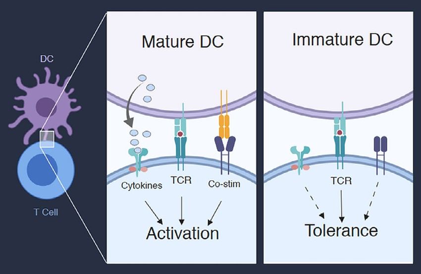
TARGETING T CELL ACTIVATION IN IMMUNO-ONCOLOGY, Saibil and Ohashi
particularly in the treatment of metastatic disease21. How-
ever, the increased understanding of dc and T cell biology
has led to new vaccine designs with better selection of the
tumour target antigens and the vaccine adjuvants that
trigger pamps, ensuring dc maturation 22. There has been
great interest in exploring the use of tumour “neoantigens”
as vaccine targets.
Tumour neoantigens arise because of nonsynonymous
mutations that result in amino acid substitutions and the
generation of novel peptides that can potentially be recog-
nized by the immune system. The clinical excitement about
neoantigens arises from the observation that tumours with
a high tumour mutational burden—and hence more neoan-
tigens—show increased responsiveness to ici therapy23,24. A
recent study demonstrated a correlation of increased tumour
mutational burden with patient survival after ici therapy in
multiple tumour types25. However, it is still unclear whether
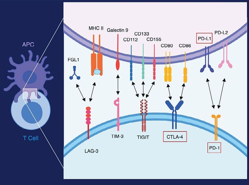
FIGURE 2 Selection of co-stimulatory receptors and their ligands.
Members of the immunoglobulin superfamily co-stimulatory receptors the neoantigens are themselves the direct targets of antitu-
and their ligands depicted are CD28, inducible T cell co-stimulator mour immunity, as has been suggested by some findings in
(ICOS, CD278), and CD226. Members of the tumour necrosis factor preclinical models of ici therapy26, or if they are a surrogate
receptor superfamily co-stimulatory receptors and their ligands depicted marker for tumours with genetic instability, for which high
are CD27 (TNFRSF7), GITR (TNFRSF18), OX40 (CD134/TNFSF4), and tumour mutational burden might trigger the innate immune
4-1BB (CD137/TNFRSF9). Abbreviations or other names for the ligands system by neoantigen-independent mechanisms. Despite
are B7-1 (CD80), B7-2 (CD86), ICOS ligand (ICOSL/B7-H2/CD275), the uncertainty, personalized vaccine strategies targeting
OX40 ligand (OX40L/CD252), 4-1BB ligand (CD137L), GITR ligand patient-specific neoantigens have shown some promise in
(GITRL/TL6), and CD70 (TNFSF7). APC = antigen presenting cell.
early-phase trials27,28. Furthermore, the improved vaccine
platforms used in the relevant studies hold a promise to
when combined with other immune-stimulating treat- induce dc and T cell activation and to bolster antitumour
ments such as checkpoint blockade. In mouse models, immunity to multiple different target antigens.
such intratumoural injections can activate dc s in the
tumour microenvironment, which, upon activation, mi- Oncolytic Viruses
grate to regional lymph nodes to prime the antitumour Other therapeutic agents that potentially act by inducing
T cell response. Hence, the responses generated by the dc and T cell activation include modified viruses. These
injections can result in tumour regression at sites distant engineered (“oncolytic”) viruses were originally developed
to the injected lesions15,16 . Currently, multiple clinical as agents to induce direct lysis of tumour cells. However,
trials investigating the clinical efficacy of intratumoural further research has indicated that a major mechanism
injections of tlr agonists, mainly in combination with other of the antitumour effect of the viruses is to stimulate
immuno-oncology agents, are underway17. the antitumour immune response29. Oncolytic viruses
An alternative strategy to boost dc activation has stimulate the antitumour immune response by multiple
been to produce synthetic ligands for the pamps. Of all the mechanisms. Given that they are viruses, they encode viral
receptors studied so far, the cgas – sting signalling pathway pamps that can potentially activate dcs directly. They also
has appeared particularly attractive for this approach. A infect tumour cells and result in cell lysis, thereby promot-
cytosolic prr, cgas is activated downstream of cytosolic dna. ing the release of damps and tumour antigens. Oncolytic
Once activated, cgas activates sting, which results in the viruses can also be engineered to express inflammatory
activation of dcs and the expression of pro-inflammatory cytokines and chemokines. For example, talimogene
cytokines such as type i interferons18. Recent work in mice laherparepvec (t-vec) is a modified herpes simplex virus
has suggested that activation of sting within the dcs in the type 1, designed to selectively replicate in and lyse tumour
tumour microenvironment is crucial for the production of cells. The virus also encodes the gene for encoding human
type i interferons and generation of an antitumour T cell granulocyte–macrophage colony–stimulating factor to
response19. Importantly, recently identified sting agonist attract and activate dcs. Talimogene laherparepvec is the
compounds that can be administered systemically instead first-in-class oncolytic virus to be approved for clinical use
of by intratumoural injection were found to mediate im- by the U.S. Food and Drug Administration. That approval
pressive sting-dependent tumour regression in a preclini- was granted based on a demonstrated therapeutic benefit
cal model 20. These agents hold the promise of a therapeutic of the injections in patients with unresectable stages iiib–iv
modality that can induce dc maturation even in tumours melanoma 30. Trials with other oncolytic viruses are on-
not amenable to intratumoural treatments. going, and those agents might prove to be useful tools to
induce an antitumour T cell response29.
Vaccine Approaches
Vaccination is another therapeutic strategy to induce dc Cytokines
maturation and T cell immunity. Historically, tumour vac- In many cases, the immune response has already been
cines have not demonstrated significant clinical efficacy, triggered in patients through natural immune surveillance,
S100 Current Oncology, Vol. 27, Supp. 2, April 2020 © 2020 Multimed Inc.TARGETING T CELL ACTIVATION IN IMMUNO-ONCOLOGY, Saibil and Ohashi
leading to detectable infiltration of CD8+ T cells into the Negative Regulation of T Cells
tumour. Early immune therapies, such as provision of cy-
tokines, acted to improve those natural tumour-specific Checkpoint Inhibitors: Beyond PD-1 and PD-L1
surveillance mechanisms. Historically, high-dose il-2 and Counteracting the activity of the costimulatory recep-
interferon alpha have been used in the treatment of renal tors are multiple negative regulatory receptors (such as
cell carcinoma and melanoma, with limited benefit31. More PD-1) that are expressed by T cells. Many of the negative
recently, multiple other cytokines that improve T cell and regulatory receptors, also called immune checkpoints or
natural killer cell survival and function—such as il-12, co-inhibitory receptors, are expressed on T cells only after
il-15, and il-21—have been tested in early clinical trials 32. activation. The co-inhibitory receptors (akin to the costim-
The toxicity of systemic administration of those cytokines ulatory receptors) also belong mainly to either the immuno-
has been a major issue in most of their trials. In the attempt globulin superfamily or the tumour necrosis factor receptor
to minimize toxicity but maintain efficacy, modified ver- superfamily and have at least 1 identified ligand13. Figure 3
sions of the cytokines are being developed. For example, depicts a selection of those receptors and their ligands.
bempegaldesleukin, a polyethylene glycol–conjugated Ligation of the co-inhibitory receptors results in the
recombinant il-2, has demonstrated antitumour activity intracellular activation of signalling molecules such as
but acceptable toxicity in animal models33 and is currently phosphatases that oppose T cell activation in part by tar-
being tested in the clinic in combination with icis. geting signals from costimulatory receptors. For example,
ligation of PD-1 on activated T cells has been demonstrated
Costimulatory Agonists: Investigating New Targets to antagonize activating signals downstream of costimula-
Given the importance of costimulatory signals for T cell tory receptors, particularly CD2840. Interestingly, although
activation, agonist antibodies targeting receptors such as co-inhibitory receptors block signals from costimulatory re-
4-1BB and OX40 have been investigated as an additional ceptors, they are not primarily associated with induction of
strategy to enhance T cell activation. In preclinical models, T cell anergy or deletion. In the context of persistent antigen
agonistic antibodies targeting many of these costimulatory and inflammation, signals downstream of the inhibitory
receptors have demonstrated the ability to increase T cell receptors have been found to limit T cell activation by in-
activation and promote tumour eradication, which has led ducing a hypofunctional state called T cell exhaustion (Tex).
to many being tested in clinical trials34. A current challenge Tex is a distinct cellular state that can be defined by a
is to fully understand the context-dependent role the each unique metabolic, epigenetic, and transcriptional signa-
of these costimulatory receptors plays in the T cell response ture and that is clearly distinct from both the activated and
against various tumour types and to discover which patient anergic states41,42. The functional characteristics of Tex are
populations would potentially benefit from treatment best described in the CD8+ T cell compartment, because
with a given agonistic antibody. Toxicity has also been a they were first defined in the noncytopathic lymphocytic
problem with the clinical development of some of these choriomeningitis virus clone 13 chronic infection model. In
agents. For instance, hepatic toxicity was a major concern mice with a chronic version of that infection, virus-specific
in the development of the agonistic anti–4-1BB antibody
urelumab35. Thus, further studies are required to define
and optimize the clinical utility of those agents as tools
to promote T cell activation and antitumour immunity.
Strategies of Adoptive T Cell Therapy
Instead of attempting to promote T cell activation in vivo,
another therapeutic strategy is the transfer of ex vivo acti-
vated, tumour-specific T cells in an adoptive cell therapy
approach. Early trials using polyclonal T cells expanded
from tumour-infiltrating lymphocytes demonstrated im-
pressive response rates in patients with melanoma before
the era of icis 36. More recent iterations of those cellular
therapies include the use of modified T cells that have been
genetically engineered to recognize tumour cells. Such
technologies include chimeric antigen receptor T cells,
which hold the promise to revolutionize the treatment
of hematologic malignancies 37. Tumour regression has
also been achieved in solid tumours using T cells that are
engineered to express a high-affinity tcr that recognizes FIGURE 3 Selection of co-inhibitory receptors and their ligands.
a peptide derived from a protein whose expression is en- Co-inhibitory molecular interactions include LAG-3 (CD223), TIM-3
riched for certain tumours, such as the cancer/testis anti- (HAVCR2/CD366), TIGIT (VSTM3), CTLA-4 (CD152), and PD-1 (CD279).
gen New York esophageal squamous cell carcinoma 138,39. Abbreviations or other names for the ligands are FGL1, B7-1 (CD80),
These cellular treatment protocols also offer the exciting B7-2 (CD86), PD-L1 (B7-H1/CD274), and PD-L2 (B7-DC/CD273). Red
opportunity to use genetic or pharmacologic means to boxes indicate molecules for which antibodies that block the interaction
with the molecule’s target ligand are approved for clinical use. APC =
further enhance T cell activation ex vivo before the T cells
antigen presenting cell.
are infused.
Current Oncology, Vol. 27, Supp. 2, April 2020 © 2020 Multimed Inc. S101TARGETING T CELL ACTIVATION IN IMMUNO-ONCOLOGY, Saibil and Ohashi
CD8+ T cells that accumulated over the course of infection T cells developed a multi-system autoimmune disease.
demonstrated reduced functionality43,44. Those Tex showed Subsequently, it was discovered that foxp3 was the lineage-
a hierarchal loss of effector function that depended on the defining transcription factor governing the development
persistence of cognate antigen and the accumulation of of Tregs and that mice and humans with mutations in the
expression of negative regulatory receptors. That sequence gene encoding foxp3 also develop a profound autoim-
of functional loss, which starts with reduced production munity50–52. Mechanistically, Tregs appear to have mul-
of il-2 and then proceeds to defects in tumour necrosis tiple ways to prevent autoimmunity. Tregs can suppress
factor α production and finally to interferon γ production T cell activation by inhibiting the activation of dcs. Tregs
and cytotoxicity, is a hallmark feature of the development can also directly suppress T cell activation using both
of Tex in both chronic infections and cancer. The Tex are poorly defined cell contact-dependent mechanisms and
believed to lose function as a mechanism of peripheral contact-independent mechanisms, such as the production
tolerance that prevents immunopathology in the face of of immunosuppressive cytokines such as transforming
persistent antigen. Conversely, the functional loss in Tex growth factor β53.
is also likely a barrier to productive antitumour immunity. Within the context of immunotherapy and tumour
The discovery that the negative regulatory receptors biology, Tregs have also been found to play an important
provided “druggable” targets to modulate T cell function role. Depletion of Tregs from tumours can lead to en-
was a major advance in the field of immuno-oncology. hanced tumour clearance in murine tumour models54,55.
Seminal work using a tumour model and the noncytopath- In patients, a low ratio of CD8+ T cell effectors to Tregs in
ic lymphocytic choriomeningitis virus infection model the tumour microenvironment has been linked with poor
demonstrated that using blocking antibodies to one of the prognosis in multiple tumour types56. Collectively, work by
immune checkpoint receptors, PD-1, or its ligand, PD-L1, many researchers has demonstrated a critical role of CD4+
to prevent signals to CD8+ T cells could improve T cell ef- foxp3+ Tregs in influencing the function of T cells in the
fector function and promote tumour or viral clearance45,46. context of both autoimmunity and cancer.
However, a major challenge in the immuno-oncology Depletion of negative regulatory cells is an attractive
field is to fully understand the function of all the immune therapeutic strategy to increase T cell activity. It has been
checkpoint receptors. As discussed earlier, in addition to proposed that depletion of Tregs in the tumour microenvi-
the PD-1/-L1 signalling axis, T cells express multiple other ronment through antibody-dependent cellular cytotoxicity
negative regulatory receptors under conditions of activa- forms part of the antitumour effect of the anti– ctla-4 anti-
tion and exhaustion (Figure 3). Therapeutic antibodies body ipilimumab57,58, although that mechanism of action
against many of the negative regulatory receptors—for remains a matter of debate59. An important aim of immuno-
example, lag-3 and tigit—have already been developed therapy research is to discover agents that can specifically
and are currently in clinical trials. deplete Tregs in the tumour microenvironment and thereby
Unfortunately, a mechanistic understanding of the augment the antitumour immune response. The challenge
complex biology of each of the inhibitory receptors is still with the development of such agents is avoid systemically
lacking. For example, although anti–lag-3 antibodies are depleting Tregs and inducing autoimmunity. Targeting the
currently in phase ii/iii clinical trials, a recent report has chemokine receptor ccr8 might have such potential, be-
just identified a novel ligand for lag-3, fgl-1, revealing a cause two recent studies suggested that ccr8 is specifically
more complex biology to be understood47. Accordingly, expressed on Tregs in the tumour microenvironment60,61.
designing clinical trials based on rational combinations of Studies have also shown that an anti-ccr8 antibody can
the antibodies has proved to be challenging because the deplete Tregs in the tumour microenvironment and im-
precise role for each of the negative receptors in modulating prove the immunoresponse62. Targeting other molecules,
T cell function (or other cells) remains to be elucidated. including CD25 and ccr4, to deplete Tregs in the tumour
Importantly, combining agents that target different in- microenvironment is also being actively pursued55,63.
hibitory receptors remains a practical approach, with the
most striking example being the synergy demonstrated Innate Immune Cells
between anti–PD-1 and anti– ctla-4 agents in the treatment In addition to Tregs, other cells found in the tumour
of various tumours 48. Thus, further work is required to microenvironment can modulate T cell activation and
determine the optimal combinations for the therapeutic function. Cells of the myeloid lineage, such as macro-
agents already in various phases of clinical trial. phages and myeloid-derived suppressor cells ( mdsc s ),
have important roles in regulating T cell activation with-
Tregs in the tumour microenvironment 64,65. One mechanism
In addition to co-inhibitory receptors, multiple regula- that mdscs and tumour-associated macrophages (tams )
tory cell populations oppose T cell activation. One of the both use to suppress T cell activation is production of the
best-characterized of the regulatory cell populations is immuno-regulatory enzymes ido and arginase 1. Those
the CD4+ T cell subset that express the lineage-defining enzymes degrade and starve T cells of, respectively, the
transcription factor foxp3 in addition to constitutively high amino acids tryptophan and arginine, which are required
levels of CD25 (il-2 receptor α chain) and ctla-4. Those to sustain T cell activation66. In addition, ido results in the
cells—the Tregs—were first discovered by Sakaguchi and production of kynurenine and its metabolites, molecules
colleagues49 to have an important role in promoting per- with emerging immunomodulatory functions.
ipheral tolerance and preventing autoimmunity. Sakaguchi In addition to tams and mdscs, novel subsets of cells
et al. demonstrated that mice depleted of CD4+CD25+ with regulatory properties continue to be described. In
S102 Current Oncology, Vol. 27, Supp. 2, April 2020 © 2020 Multimed Inc.TARGETING T CELL ACTIVATION IN IMMUNO-ONCOLOGY, Saibil and Ohashi
patients with high-grade serous ovarian cancer and non- SUMMARY
small-cell lung cancer, recent studies have described a
population of innate lymphoid cells that demonstrated The regulation of T cell activation is complex, with multiple
an ability to suppress T cells ex vivo 67,68. Further work is levels of control, many of which involve the activation of
required to better define the role of all of those suppressive the innate immune system—and specifically dcs. Many
cell types in tumour biology and in peripheral tolerance. It of the regulatory mechanisms evolved to prevent the
is clear that multiple immune cell populations of the innate activation of self-reactive T cells and the development of
immune system can regulate the immune response, par- autoimmunity. However, in immunotherapy, those same
ticularly in the context of the tumour microenvironment. mechanisms limit T cell activation and the curative poten-
As in Treg-depleting therapeutics, reagents are being tial of immuno-oncology treatment strategies. Moreover,
developed to deplete tams and mdscs from the tumour mi- many of the mechanisms regulating T cell activation are
croenvironment69,70. For example, an agonistic antibody dynamic, and blocking one might result only in the induc-
targeting trail-r 2 was recently shown to be able to deplete tion of another. Accordingly, combination therapies that
mdscs from tumours in about half the patients treated on a
target multiple aspects of T cell activation are the future of
phase i trial71. Additionally, as opposed to depleting tams immunotherapy. The challenge will be to devise treatment
and mdscs from the tumour microenvironment, reagents to strategies that can lead to T cell activation without causing
inhibit the enzymatic activity of arginase 1 and ido are also intolerable systemic autoimmunity. Striking that balance
being evaluated66. Great clinical excitement attended the will require an increased understanding of all the cellular
early-phase efficacy data for a combination of an anti–PD-1 and host factors that control the activation of T cells.
agent and the ido inhibitor epacadostat (aka INCB24360),
CONFLICT OF INTEREST DISCLOSURES
particularly for the treatment of melanoma. Unfortunately, We have read and understood Current Oncology’s policy on
the phase iii trial echo-301/keynote-252 (see NCT02752075 disclosing conf licts of interest, and we declare the following
at https://ClinicalTrials.gov/) was halted because of a lack interests: SDS has participated on an advisory board for Janssen;
of added clinical benefit for the combination compared PSO receives funding from emd Serono and participates on advis-
with anti–PD-1 alone. Further trials are needed to test ory boards for Myst Therapeutics, Providence, Symphogen, and
whether other ido inhibitors that potentially have more Tessa Therapeutics.
potent enzymatic inhibition will result in clinical benefit or
whether a biomarker that will predict clinical response to AUTHOR AFFILIATIONS
ido inhibition can be defined. However, targeting negative *Princess Margaret Cancer Centre, †Department of Medical On-
cology and Hematology, Princess Margaret Cancer Centre, Uni-
regulatory cells or their effector mechanisms remains an
versity of Toronto, and ‡Department of Immunology, University
attractive therapeutic avenue. of Toronto, Toronto, ON.
Emerging Considerations for T Cell Activation: REFERENCES
Microbiome and Metabolism 1. Wallis CJD, Butaney M, Satkunasivam R, et al. Association
Mammals are colonized by billions of commensal bacteria, of patient sex with efficacy of immune checkpoint inhibitors
particularly within the gastrointestinal tract. It was first and overall survival in advanced cancers: a systematic review
and meta-analysis. JAMA Oncol 2019;5:529–36.
noted in preclinical models that mice with different intes-
2. Chen DS, Mellman I. Elements of cancer immunity and the
tinal microbiota displayed differing responses to anti–PD-1 cancer-immune set point. Nature 2017;541:321–30.
therapy when implanted with the same tumour72. That 3. Steinman RM, Hemmi H. Dendritic cells: translating in-
finding was then extended to humans, because patients nate to adaptive immunity. Curr Top Microbiol Immunol
with melanoma who responded to anti–PD-1 therapy were 2006;311:17–58.
discovered to have a more diverse gut microbiome than did 4. Daley SR, Teh C, Hu DY, Strasser A, Gray DHD. Cell death and
patients who did not respond to treatment73. Similarly, in thymic tolerance. Immunol Rev 2017;277:9–20.
a retrospective analysis, depletion of the microbiome with 5. Davis MM. T Cell receptor gene diversity and selection. Annu
antibiotics was found to decrease the efficacy of ici therapy Rev Biochem 1990;59:475–96.
6. Theofilopoulos AN, Kono DH, Baccala R. The multiple path-
in multiple tumour histologies74. Collectively, those data
ways to autoimmunity. Nat Immunol 2017;18:716–24.
indicate an important relationship between the intestinal 7. Schwartz RH. T Cell anergy. Annu Rev Immunol 2003;21:305–34.
bacteria and the immune system. However, the precise 8. Miller JF, Morahan G. Peripheral T cell tolerance. Annu Rev
mechanisms through which the commensal bacteria affect Immunol 1992;10:51–69.
ici therapy efficacy has not been determined. 9. Osorio F, Fuentes C, Lopez MN, Salazar-Onfray F, Gonzalez
Interestingly, commensal bacterial have been im- FE. Role of dendritic cells in the induction of lymphocyte
plicated in regulating the circulating level of multiple tolerance. Front Immunol 2015;6:535.
metabolites, particularly short-chain fatty acids such as 10. Audiger C, Rahman MJ, Yun TJ, Tarbell KV, Lesage S. The im-
acetate, butyrate, and propionate75. In mouse models, all portance of dendritic cells in maintaining immune tolerance.
J Immunol 2017;198:2223–31.
of those molecules have been found to affect T cell activa-
11. Schaefer L. Complexity of danger: the diverse nature of
tion76,77. Further studies are required to investigate if those damage-associated molecular patterns. J Biol Chem 2014;289:
microbial-regulated metabolites do indeed contribute to 35237–45.
the influence of the microbiome on ici therapy. However, 12. Croft M, Dubey C. Accessory molecule and costimulation
it seems likely that future adjuncts to ici will be aimed at requirements for CD4 T cell response. Crit Rev Immunol
altering the microbiome itself or its metabolic products. 2017;37:261–90.
Current Oncology, Vol. 27, Supp. 2, April 2020 © 2020 Multimed Inc. S103TARGETING T CELL ACTIVATION IN IMMUNO-ONCOLOGY, Saibil and Ohashi
13. Chen L, Flies DB. Molecular mechanisms of T cell co- 36. Hinrichs CS, Rosenberg SA. Exploiting the curative po-
stimulation and co-inhibition. Nat Rev Immunol 2013;13: tential of adoptive T-cell therapy for cancer. Immunol Rev
227–42. 2014;257:56–71.
14. Morales A. bcg : a throwback from the stone age of vaccines 37. June CH, O’Connor RS, Kawalekar OU, Ghassemi S, Milone
opened the path for bladder cancer immunotherapy. Can J MC. car T cell immunotherapy for human cancer. Science
Urol 2017;24:8788–93. 2018;359:1361–5.
15. Sagiv-Barfi I, Czerwinski DK, Levy S, et al. Eradication of 38. Robbins PF, Morgan RA, Feldman SA, et al. Tumor regression
spontaneous malignancy by local immunotherapy. Sci Transl in patients with metastatic synovial cell sarcoma and mel-
Med 2018;10:pii:eaan4488. anoma using genetically engineered lymphocytes reactive
16. Sato-Kaneko F, Yao S, Ahmadi A, et al. Combination im- with ny-eso-1. J Clin Oncol 2011;29:917–24.
munotherapy with tlr agonists and checkpoint inhibitors 39. D’Angelo SP, Melchiori L, Merchant MS, et al. Antitumour
suppresses head and neck cancer. JCI Insight 2017;2:pii:93397. activity associated with prolonged persistence of adoptively
17. Iribarren K, Bloy N, Buque A, et al. Trial watch: immunostim- transferred ny-eso-1 c259T cells in synovial sarcoma. Cancer
ulation with toll-like receptor agonists in cancer therapy. Discov 2018;8:944–57.
Oncoimmunology 2015;5:e1088631. 40. Hui E, Cheung J, Zhu J, et al. T Cell costimulatory receptor
18. Barber GN. sting. Infection, inflammation and cancer. Nat CD28 is a primary target for PD-1–mediated inhibition. Sci-
Rev Immunol 2015;15:760–70. ence 2017;355:1428–33.
19. Woo SR, Fuertes MB, Corrales L, et al. sting-dependent cy- 41. McLane LM, Abdel-Hakeem MS, Wherry EJ. CD8 T cell ex-
tosolic dna sensing mediates innate immune recognition of haustion during chronic viral infection and cancer. Annu Rev
immunogenic tumors. Immunity 2014;41:830–42. Immunol 2019;37:457–95.
20. Ramanjulu JM, Pesiridis GS, Yang J, et al. Design of amido- 42. Crespo J, Sun H, Welling TH, Tian Z, Zou W. T Cell anergy,
benzimidazole sting receptor agonists with systemic activity. exhaustion, senescence, and stemness in the tumor micro-
Nature 2018;564:439–43. environment. Curr Opin Immunol 2013;25:214–21.
21. Melero I, Gaudernack G, Gerritsen W, et al. Therapeutic vac- 43. Zajac AJ, Blattman JN, Murali-Krishna K, et al. Viral immune
cines for cancer: an overview of clinical trials. Nat Rev Clin evasion due to persistence of activated T cells without effector
Oncol 2014;11:509–24. function. J Exp Med 1998;188:2205–13.
22. van der Burg SH, Arens R, Ossendorp F, van Hall T, Melief CJ. 44. Gallimore A, Glithero A, Godkin A, et al. Induction and
Vaccines for established cancer: overcoming the challenges exhaustion of lymphocytic choriomeningitis virus-specific
posed by immune evasion. Nat Rev Cancer 2016;16:219–33. cytotoxic T lymphocytes visualized using soluble tetrameric
23. Rizvi NA, Hellmann MD, Snyder A, et al. Cancer immunology. major histocompatibility complex class i–peptide complexes.
Mutational landscape determines sensitivity to PD-1 block- J Exp Med 1998;187:1383–93.
ade in non–small cell lung cancer. Science 2015;348:124–8. 45. Barber DL, Wherry EJ, Masopust D, et al. Restoring function in
24. Yarchoan M, Hopkins A, Jaffee EM. Tumor mutational exhausted CD8 T cells during chronic viral infection. Nature
burden and response rate to PD-1 inhibition. N Engl J Med 2006;439:682–7.
2017;377:2500–1. 46. Iwai Y, Ishida M, Tanaka Y, Okazaki T, Honjo T, Minato N.
25. Samstein RM, Lee CH, Shoushtari AN, et al. Tumor mutational Involvement of PD-L1 on tumor cells in the escape from
load predicts survival after immunotherapy across multiple host immune system and tumor immunotherapy by PD-L1
cancer types. Nat Genet 2019;51:202–6. blockade. Proc Natl Acad Sci U S A 2002;99:12293–7.
26. Gubin MM, Zhang X, Schuster H, et al. Checkpoint blockade 47. Wang J, Sanmamed MF, Datar I, et al. Fibrinogen-like pro-
cancer immunotherapy targets tumour-specific mutant tein 1 is a major immune inhibitory ligand of lag -3. Cell
antigens. Nature 2014;515:577–81. 2019;176:334–347.e312.
27. Li L, Goedegebuure SP, Gillanders WE. Preclinical and 48. Esin E. Clinical applications of immunotherapy combination
clinical development of neoantigen vaccines. Ann Oncol methods and new opportunities for the future. Biomed Res
2017;28(suppl 12):xii11–17. Int 2017;2017:1623679.
28. Keskin DB, Anandappa AJ, Sun J, et al. Neoantigen vaccine 49. Sakaguchi S, Sakaguchi N, Asano M, Itoh M, Toda M. Im-
generates intratumoral T cell responses in phase ib glioblas- munologic self-tolerance maintained by activated T cells
toma trial. Nature 2019;565:234–9. expressing il-2 receptor alpha-chains (CD25). Breakdown
29. Lawler SE, Speranza MC, Cho CF, Chiocca EA. Oncolytic vi- of a single mechanism of self-tolerance causes various au-
ruses in cancer treatment: a review. JAMA Oncol 2017;3:841–9. toimmune diseases. J Immunol 1995;155:1151–64.
30. Andtbacka RH, Kaufman HL, Collichio F, et al. Talimogene 50. Bennett CL, Christie J, Ramsdell F, et al. The immune dysregu-
laherparepvec improves durable response rate in patients lation, polyendocrinopathy, enteropathy, X-linked syndrome
with advanced melanoma. J Clin Oncol 2015;33:2780–8. (ipex) is caused by mutations of foxp3. Nat Genet 2001;27:20–1.
31. Waldmann TA. Cytokines in cancer immunotherapy. Cold 51. Wildin RS, Ramsdell F, Peake J, et al. X-Linked neonatal
Spring Harb Perspect Biol 2018;10:pii:a028472. diabetes mellitus, enteropathy and endocrinopathy syn-
32. Floros T, Tarhini A A. Anticancer cytokines: biology and drome is the human equivalent of mouse scurfy. Nat Genet
clinical effects of interferon-alpha2, interleukin (il)–2, il-15, 2001;27:18–20.
il-21, and il-12. Semin Oncol 2015;42:539–48. 52. Brunkow ME, Jeffery EW, Hjerrild KA, et al. Disruption of a
33. Charych DH, Hoch U, Langowski JL, et al. nktr-214, an engi- new forkhead/winged-helix protein, scurfin, results in the
neered cytokine with biased il 2 receptor binding, increased fatal lymphoproliferative disorder of the scurfy mouse. Nat
tumor exposure, and marked efficacy in mouse tumor mod- Genet 2001;27:68–73.
els. Clin Cancer Res 2016;22:680–90. 53. Josefowicz SZ, Lu LF, Rudensky AY. Regulatory T cells: mech-
34. Mayes PA, Hance KW, Hoos A. The promise and challenges anisms of differentiation and function. Annu Rev Immunol
of immune agonist antibody development in cancer. Nat Rev 2012;30:531–64.
Drug Discov 2018;17:509–27. 54. Shimizu J, Yamazaki S, Sakaguchi S. Induction of tumor
35. Segal NH, Logan TF, Hodi FS, et al. Results from an integrated immunity by removing CD25+CD4+ T cells: a common basis
safety analysis of urelumab, an agonist anti-CD137 monoclo- between tumor immunity and autoimmunity. J Immunol
nal antibody. Clin Cancer Res 2017;23:1929–36. 1999;163:5211–18.
S104 Current Oncology, Vol. 27, Supp. 2, April 2020 © 2020 Multimed Inc.TARGETING T CELL ACTIVATION IN IMMUNO-ONCOLOGY, Saibil and Ohashi
55. Arce Vargas F, Furness AJS, Solomon I, et al. FC-optimized 66. Lemos H, Huang L, Prendergast GC, Mellor AL. Immune
anti-CD25 depletes tumor-infiltrating regulatory T cells control by amino acid catabolism during tumorigenesis and
and synergizes with PD-1 blockade to eradicate established therapy. Nat Rev Cancer 2019;19:162–75.
tumors. Immunity 2017;46:577–86. 67. Crome SQ, Nguyen LT, Lopez-Verges S, et al. A distinct innate
56. Shang B, Liu Y, Jiang SJ, Liu Y. Prognostic value of tumor- lymphoid cell population regulates tumor-associated T cells.
infiltrating foxp3+ regulatory T cells in cancers: a systematic Nat Med 2017;23:368–75.
review and meta-analysis. Sci Rep 2015;5:15179. 68. Picard E, Godet Y, Laheurte C, et al. Circulating NKp46 +
57. Simpson TR, Li F, Montalvo-Ortiz W, et al. FC-dependent natural killer cells have a potential regulatory property and
depletion of tumor-infiltrating regulatory T cells co-defines predict distinct survival in non–small cell lung cancer. On-
the efficacy of anti– ctla-4 therapy against melanoma. J Exp coimmunology 2019;8:e1527498.
Med 2013;210:1695–710. 69. Poh AR, Ernst M. Targeting macrophages in cancer: from
58. Romano E, Kusio-Kobialka M, Foukas PG, et al. Ipilimumab- bench to bedside. Front Oncol 2018;8:49.
dependent cell-mediated cytotoxicity of regulatory T cells 70. Anani W, Shurin MR. Targeting myeloid-derived suppressor
ex vivo by nonclassical monocytes in melanoma patients. cells in cancer. Adv Exp Med Biol 2017;1036:105–28.
Proc Natl Acad Sci U S A 2015;112:6140–5. 71. Dominguez GA, Condamine T, Mony S, et al. Selective tar-
59. Sharma A, Subudhi SK, Blando J, et al. Anti-ctla-4 immuno- geting of myeloid-derived suppressor cells in cancer patients
therapy does not deplete foxp3+ regulatory T cells (Tregs) in using DS-8273a, an agonistic trail-r 2 antibody. Clin Cancer
human cancers. Clin Cancer Res 2019;25:1233–8. Res 2017;23:2942–50.
60. De Simone M, Arrigoni A, Rossetti G, et al. Transcriptional 72. Sivan A, Corrales L, Hubert N, et al. Commensal Bifido-
landscape of human tissue lymphocytes unveils unique- bacterium promotes antitumour immunity and facilitates
ness of tumor-infiltrating T regulatory cells. Immunity anti–PD-L1 efficacy. Science 2015;350:1084–9.
2016;45:1135–47. 73. Gopalakrishnan V, Spencer CN, Nezi L, et al. Gut microbiome
61. Plitas G, Konopacki C, Wu K, et al. Regulatory T cells ex- modulates response to anti–PD-1 immunotherapy in mela-
hibit distinct features in human breast cancer. Immunity noma patients. Science 2018;359:97–103.
2016;45:1122–34. 74. Routy B, Le Chatelier E, Derosa L, et al. Gut microbiome
62. Villarreal DO, L’Huillier A, Armington S, et al. Targeting ccr8 in- influences efficacy of PD-1–based immunotherapy against
duces protective antitumour immunity and enhances vaccine- epithelial tumors. Science 2018;359:91–7.
induced responses in colon cancer. Cancer Res 2018;78:5340–8. 75. Levy M, Thaiss CA, Elinav E. Metabolites: messengers be-
63. Wing JB, Tanaka A, Sakaguchi S. Human foxp3 + regulatory tween the microbiota and the immune system. Genes Dev
T cell heterogeneity and function in autoimmunity and 2016;30:1589–97.
cancer. Immunity 2019;50:302–16. 76. Balmer ML, Ma EH, Bantug GR, et al. Memory CD8 + T cells
64. Yang M, McKay D, Pollard JW, Lewis CE. Diverse functions of require increased concentrations of acetate induced by stress
macrophages in different tumor microenvironments. Cancer for optimal function. Immunity 2016;44:1312–24.
Res 2018;78:5492–503. 77. Luu M, Weigand K, Wedi F, et al. Regulation of the effector
65. Gabrilovich DI. Myeloid-derived suppressor cells. Cancer function of CD8 + T cells by gut microbiota–derived metab-
Immunol Res 2017;5:3–8. olite butyrate. Sci Rep 2018;8:14430.
Current Oncology, Vol. 27, Supp. 2, April 2020 © 2020 Multimed Inc. S105You can also read