Testing Toxicity and Antidote Effect of Selenium Nanoparticles with Paramecium caudatum
←
→
Page content transcription
If your browser does not render page correctly, please read the page content below
Open Journal of Animal Sciences, 2021, 11, 532-542
                                                                                                 https://www.scirp.org/journal/ojas
                                                                                                            ISSN Online: 2161-7627
                                                                                                              ISSN Print: 2161-7597

Testing Toxicity and Antidote Effect of
Selenium Nanoparticles with
Paramecium caudatum

Khandsuren Badgar1,2* , József Prokisch1,2
1
 Institute of Animal Science, Biotechnology and Nature Conservation, Faculty of Agricultural and Food Sciences and
Environmental Management, University of Debrecen, Debrecen, Hungary
2
 Doctoral School of Animal Science, University of Debrecen, Debrecen, Hungary

How to cite this paper: Badgar, K. and          Abstract
Prokisch, J. (2021) Testing Toxicity and
Antidote Effect of Selenium Nanoparticles       A simple method for assessment of the toxicity and antidote effect of sele-
with Paramecium caudatum. Open Journal          nium nanoparticles with Paramecium caudatum is presented. Light micro-
of Animal Sciences, 11, 532-542.
https://doi.org/10.4236/ojas.2021.114036
                                                scopy in combination with computerized video tracking is employed for the
                                                determination of the survival time of P. caudatum. Up to 800 mg/L, selenium
Received: July 26, 2021                         nanoparticles are not acutely toxic. With respect to a potential antidote effect,
Accepted: August 29, 2021
                                                the lethality of silver nanoparticles, silver nitrate, sodium hydrogen selenite,
Published: September 1, 2021
                                                and sodium selenite to P. caudatum was decreased and survival time was ex-
Copyright © 2021 by author(s) and               tended upon pre-treatment with selenium nanoparticles. Taken together, these
Scientific Research Publishing Inc.             findings suggest that administration of selenium nanoparticles attenuates ex-
This work is licensed under the Creative
                                                posure to toxicants. Selenium nanoparticles could be a good functional addi-
Commons Attribution International
License (CC BY 4.0).                            tive for food management in animals.
http://creativecommons.org/licenses/by/4.0/
               Open Access                      Keywords
                                                Selenium Nanoparticles, Toxicity, Antidote Effect, Paramecium caudatum

                                              1. Introduction
                                              Protozoan cells are often used as bioindicators of chemical pollution and their
                                              toxicity in an aqueous environment [1]. Paramecium caudatum is one of the
                                              widely used ciliate models as bioindicators for scientific research. Komala re-
                                              ported the acute toxicity test of P. caudatum is highly sensitive to investigate the
                                              direct toxicity of chemical compounds [2]. Basically, Paramecium simplifies the
                                              study of physiological processes and effects of water pollutants such as mineral
                                              oil, pesticides, metals, and others by monitoring locomotor behavior, morphol-

DOI: 10.4236/ojas.2021.114036 Sep. 1, 2021                         532                             Open Journal of Animal Sciences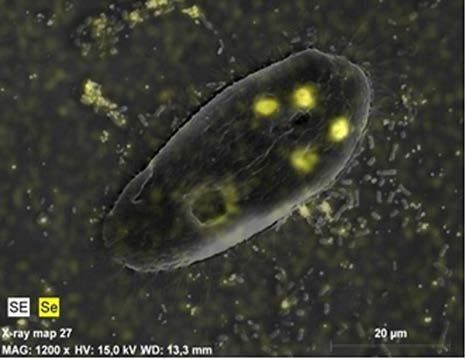
K. Badgar, J. Prokisch

                                ogy and mortality [3] [4] [5] [6] [7]. A sensitive measure of stress by water con-
                                tamination [8] is locomotion, depending on the movement of the cilia mostly con-
                                trolled by the action potential of the cell membrane [9] [10]. In this study, the an-
                                tidote effect of selenium nanoparticles on P. caudatum is evaluated in a simple
                                fashion.
                                  Generally, selenium is a functional microelement that is involved in many
                                physiological functions, and it has several therapeutic effects by participating in
                                the basic composition of some enzymes and amino acids. Earlier in our experi-
                                ments, we investigated how probiotic yogurt bacteria transform the inorganic
                                selenium compounds into organic compounds. It has been found that certain
                                bacteria have been defending against the toxicity of selenite ion; elemental sele-
                                nium was produced within the cell and stored as small, nano-sized spheres
                                (SeNPs). During the fermentation, the transparent nutrient solution becomes
                                red in the selenium nanoparticles produced by the proliferating bacteria. The
                                nanoparticles formed in the bacterium can be recovered and used after purifica-
                                tion of the cell wall after purification. The spheres produced measured 100 to
                                500 nanometers depending on the bacterial species. With animal studies with
                                sheep, chicken and fish, and human studies, it was proved that this selenium
                                form has a significantly better antioxidant effect than other selenium com-
                                pounds; it cannot be overdosed and is the least toxic form of selenium [11] [12]
                                [13]. Selenium has long been known for its ability to reduce the harmful effects
                                of metals [14] [15]. For example, Hao et al. reported that selenium supplement
                                prevented abnormal changes in the levels of reduced glutathione, mitochondrial
                                membrane potential, and Ca2+-ATPase activity of chromium (VI)-induced dam-
                                age in chicken brain [16]. Also, selenium is able to alleviate the symptoms of
                                mercury toxicity in cell culture [17], fish [18], and adult mice [19]. Selenium sup-
                                plementation reduces hepatic oxidative stress induced by silver nanoparticles
                                (AgNP) in rats [20]. Besides, selenium nanoparticles protect against As(III)-
                                induced cell death and DNA damage by reducing the production of As(III)-
                                induced reactive oxygen species [21]. Recently, there has been an increasing
                                ecological and global public health concern associated with environmental con-
                                tamination by some metals. Therefore, the studies of antidote substances are in-
                                creasing in scientific research. Based on the information mentioned above, the
                                authors focused on a simple and rapid method to determine the antidote effect
                                of biosynthesized selenium nanoparticles.

                                2. Materials and Methods
                                Selenium nanoparticles were produced according to Eszenyi et al. and Prokisch
                                and Zommara [22] [23]. In the preparation, 20 mL of 10,000 mg/L sodium sele-
                                nite stock solution was added into 980 mL MRS broth, then 10 mL of L. acido-
                                philus culture was added, and the mixture was incubated at 37˚C for 24 - 36 h.
                                The culture was centrifuged at 6000 rpm for 15 min, and the pellets were washed
                                with water. Finally, cells were lysed by hydrochloric acid 37% (m/m) for 5 days

DOI: 10.4236/ojas.2021.114036                        533                              Open Journal of Animal Sciences
K. Badgar, J. Prokisch
at room temperature. The mixture was centrifuged at 6000 rpm for 15 min, washed
with water and filtered by vacuum filtration. After purification, the suspension
of selenium nanoparticles contained 800 mg/kg selenium in the form of 250 nm
size red elemental selenium (Figure 1).
Stock cultures of P. caudatum were cultured in tap water supplemented with
yogurt powder (1 g/L). Yogurt was made by starter culture (Lyofast Y 250) con-
taining Streptococcus thermophilus and Lactobacillus delbrueckii ssp. bulgaricus
were obtained from SACCO Srl (Italy). Fresh cultures were initiated by seeding
100 ml of freshwater with 1 mL of a stationary phase paramecium culture. The
cultures were maintained at room temperature for 15 - 30 days.
2.1. Determination of Toxic Level of Selenium Nanoparticles and
Toxicants
A solution of silver nanoparticles with a concentration of 20 mg/L and particle
size of 10 - 20 nm was purchased from Dr. Juice (Miskolc, Hungary). Silver ni-
trate (AgNO3) was obtained from Reanal Laborvegyszer. (Budapest, Hungary).
Sodium hydrogen selenite (NaHSeO3) was obtained from VWR International (Lut-
terworth, Leics, UK) and sodium selenate decahydrate (Na2SeO4∙10H2O) were ob-
tained from Scharlau Chemie (Barcelona, Spain).
Stock solutions of 20 mg/L of silver nanoparticles and silver nitrate, 80.0 mg/L
of sodium hydrogen selenite, and sodium selenate decahydrate were prepared
and then diluted in serial dilutions (until 10−10). 20 µL fresh culture of P. cauda-
tum was exposed with 20 µL solutions on the glass slide in control groups. LC95
values of every toxicant were determined. Surviving time and the effect of con-
centration are applied to calculate the LC50 concentration values. The NOEL (No
Observed Effect Level) concentration was defined as the concentration when the
surviving is longer than 20 min.
2.2. Determination of Antidote Effects of Selenium Nanoparticles
500 µL fresh culture of P. caudatum was treated with 500 µL selenium nanopar-
ticles (800 mg/L) at room temperature for 2 h in experimental groups. At the
end of the treatment, 20 µL of the treated culture was placed on the middle of
Figure 1. The scanning electron microscopic (SEM) image of applied selenium nanopar-
ticles.
DOI: 10.4236/ojas.2021.114036 534 Open Journal of Animal SciencesK. Badgar, J. Prokisch
the glass slide, and while observing under a microscope 20 µL toxicants were
added. Then, the survival time, locomotor behavior, and morphological changes
of P. caudatum are continuously observed under the microscope for 20 min. Af-
ter more than 20 min, the solution on the glass slide became dry out and the cells
were started to die. The location of selenium nanoparticles inside the cell is de-
scribed by light microscopy attached to a CCD camera, and scanning electron
microscope (Hitachi S-4300).
3. Results and Discussion
3.1. The Toxic Level of Selenium Nanoparticles and Toxicants
The concentration of selenium nanoparticles higher than 800 mg/L was shown
to exert no effects on locomotion and morphology of P. caudatum. Within 5 min
of treatment, selenium nanoparticles started to be accumulated inside the cells.
Figure 2 shows the black spots are inside P. caudatum which are red with a
proper illumination (white light from the side). Also, scanning electron micro-
scopic image with X-ray mapping shows the location of selenium nanoparticles
in the cell (Figure 3). These results indicated no morphological changes in P.
caudatum.
Figure 2. Light microscopic image of P. caudatum treated with selenium nanoparticles
(800 mg/L).
Figure 3. Scanning electron microscopic (SEM) image of P. caudatum treated with sele-
nium nanoparticles. The X-ray fluorescent mapping shows the location of selenium in the
pictures. The selenium spheres and Lactobacillus cells are visible as well.
DOI: 10.4236/ojas.2021.114036 535 Open Journal of Animal SciencesK. Badgar, J. Prokisch
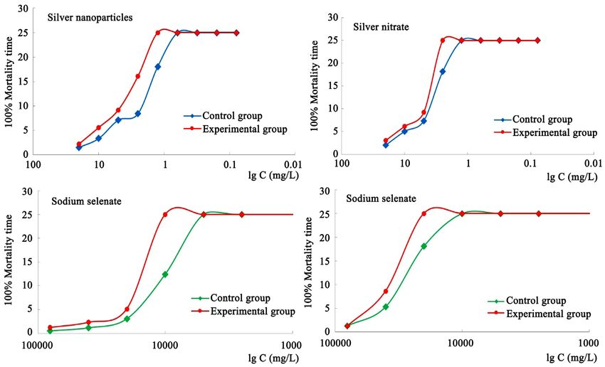
For toxicants, results showed that ≥1.25 mg/L of silver nanoparticles, ≥2.5
mg/L of silver nitrate, ≥10.0 mg/L of sodium hydrogen selenite, and ≥20.0 mg/L
of sodium selenate have a lethal effect on P. caudatum in the control groups as
presented in Figure 4. The survival time vs. concentrations has a special shape,
which is similar to the pH titration curve. Its inflection point gives the LC50
value. All P. caudatum died within 18 min due to cell lysis. In the present expe-
riments, we observed that these toxicants affected the test organism, P. cauda-
tum in a concentration-dependent manner.
Silver especially AgNPs are known for their antibacterial effect and are used in
various nanomedicine and biomedical applications. However, there is increasing
concern related to the biological impact of the use of AgNPs on a large scale, and
the possible toxicity to the environment and health [20]. Several works show that
silver nanoparticles exert an inhibitory effect in all biological systems such as the
virus [24], bacteria [25], protozoa [26], algae [27], and fungi [28] regardless of
their structural or physiological characteristics, at the cellular level. For instance,
30 - 50 µg/L of silver nanoparticles have a rapid toxic effect on Paramecium within
15 min [29]. In other words, AgNPs have also the capacity to kill cells with differ-
ent levels of complexity, both prokaryotic and eukaryotic. Most of the reports
show an effective concentration near 0.1 mg/L of silver [27] [28]. It is classified
that sodium selenite is highly toxic, and sodium selenate is moderately toxic to
fingerlings of tilapia [30]. Generally, selenium bioavailability depends on their
Figure 4. The survival time of P. caudatum and the concentration of toxicants on a logarithmic scale. Experimental groups were
pre-treated with selenium nanoparticles, and control groups were not treated.
DOI: 10.4236/ojas.2021.114036 536 Open Journal of Animal SciencesK. Badgar, J. Prokisch
size, chemical type, and dose in the source. The elemental selenium nanopar-
ticles have very good bioavailability as indicated by enhancement of serum anti-
oxidant status and selenium concentration in blood and tissues [31], and much
lower toxicity as indicated by median lethal dose, acute liver injury, survival rate,
and short-term toxicity [32] compared to other chemical types.
3.2. The Antidote Effects of Selenium Nanoparticles
The lethal concentration of toxicants was significantly decreased by 2 times in
the experimental groups which were pre-treated with selenium nanoparticles
(800 mg/L). Namely, the lethal concentrations were found as ≥2.5 mg/L, ≥5.0
mg/L, ≥20.0 mg/L, and ≥40.0 mg/L for silver nanoparticles, silver nitrate, so-
dium hydrogen selenite, and sodium selenate, respectively (Figure 4). There
were no changes in locomotion and morphology of P. caudatum in the experi-
mental groups under the exposure of previously determined lethal concentra-
tions of the toxicants. Particularly, P. caudatum did not exhibit any alteration in
their shape, blebbing, lysis, and swimming. And their survival time was extended
by the administration of selenium nanoparticles (Figure 4). The changes in the
experimental group are compared with the control groups in Figure 4. These
results are theoretically consistent with the results of other research methods.
For instance, bulk selenium (selenite) treatment in a mouse model was more
toxic than selenium nanoparticles in terms of deleterious effects on mice growth,
liver functions, and hepatic lipid peroxidation [33], and the selenium nanopar-
ticles dramatically reduce the death incurred by acute toxicity associated with
bulk selenium up to 4 times in a rodent model [34]. Also, selenium nanoparticles
protect against As(III)-induced cell death and DNA damage [21], and supple-
mentation of selenium with lead reduced the lead level in serum [19]. Selenium
antagonizes the toxicity of arsenic and cadmium mainly through sequestration
of these elements into biologically inert complexes and/or through the action of
selenium-dependent antioxidant enzymes [35]. Besides, Ansar et al., reported
selenium supplementation reduces the AgNP-induced hepatotoxicity in rats
[20]. Basically, the toxicity of AgNPs is explained due to the release of Ag ions in
the system or extensive systemic distribution of Ag in tissues causing oxidative
stress, protein or DNA damage, and apoptotic cell death [36] [37] [38]. Selenium
restores the activity of important antioxidant enzymes such as glutathione pe-
roxidase and thioredoxin reductase [39]. Therefore, the antidote effect of sele-
nium nanoparticles can be explained by the restoration of target selenoprotein
activity and restoring the intracellular redox environment. Basically, impairment
of the thioredoxin and glutaredoxin systems allows for proliferation of cytosol
and mitochondrial reactive oxygen and nitrogen species which lead to mito-
chondrial injury/loss, lipid peroxidation, calcium dyshomeostasis, impairment of
protein repair, and apoptosis [40]. For instance, mercury binds to the seleno-
cysteine binding site of thioredoxin reductase 1 in the cytosol and thioredoxin
reductase 2 in the mitochondria dramatically inhibiting their function [41]. Bas-
ically, metals have a lower affinity for thiol groups and a higher affinity for sele-
DOI: 10.4236/ojas.2021.114036 537 Open Journal of Animal SciencesK. Badgar, J. Prokisch
nium containing groups. In this case, selenium supplementation, with limita-
tions, may have a beneficial role in restoring adequate selenium status and miti-
gating the toxicity of mercury [42].
Different changes in the locomotion and morphology of P. caudatum were
observed for the exposure of the silver and bulk selenium during the experiment.
P. caudatum affected by silver mainly showed alteration of their shape by devel-
oping irregular blebbing of the cell membrane before cell lysis. There were oc-
curred single or multiple blebs in the cell membrane. The process of blebbing is
a common phenomenon during apoptosis. At the same time, fragmentation and
disintegration of macronucleus were also observed with increased time of expo-
sure. These morphological changes in the experimental group lasted longer than
the control group. Similar kind of changes on the cell membrane of P. caudatum
was observed with xenobiotics, monocroptophos (>90 mg/L), and fenthion in P.
caudatum. The complete cell lysis occurred within 17 min on average ranging
from 5 to 20 min [6] [43] [44].
The effect of bulk selenium mainly showed the locomotion changes. Particu-
larly, due to the high dose, the swimming of P. caudatum stopped completely,
but his ciliates kept moving until cell lysis. As the concentrations decreased, se-
quential changes such as the swimming speed and changes (circular movement),
immobile, and cell lysis were observed. In other words, P. caudatum died after
they became immobile. Gradual decrease in the swimming speed, with the in-
creased time of exposure, is due to the effect of the toxicants on the cellular me-
tabolism and morphological changes. Similar changes like reduction in the swim-
ming speeds, altered morphology, and generation times were observed when P.
caudatum was exposed to a higher concentration (>350 mg/L) of acephate [7]
[44].
4. Conclusion
Through our study, we found P. caudatum is a suitable and practical microor-
ganism for testing toxicological research. The locomotion, morphology, and
death of P. caudatum are easily detectable under a microscope. We proved that
biosynthesized selenium nanoparticles are antidotes against silver and bulk sele-
nium induced toxicity, the P. caudatum can tolerate 2 times higher concentra-
tions of tested toxicants if they were pre-treated with selenium nanoparticles.
The developed method is simple, rapid, and economical. This method will be
suitable to test the acute toxicity of toxic metals, metalloid compounds, myco-
toxins, and insecticides also.
Acknowledgements
This study was funded by the Stipendium Hungaricum Scholarship Program.
The authors wish to thank Dr. Lajos Daróczy (Department of Solid State
Physics, University of Debrecen) for help with the scanning electron microscop-
ic pictures.
DOI: 10.4236/ojas.2021.114036 538 Open Journal of Animal SciencesK. Badgar, J. Prokisch
Conflicts of Interest
The authors declare no conflicts of interest regarding the publication of this pa-
per.
References
[1] Mortuza, M.G., Takahashi, T., Tatsuya, U., Kosaka, T., Michibata, H. and Hosoya,
H. (2005) Toxicity and Bioaccumulation of Hexavalent Chromium in Green Para-
mecium, Paramecium bursaria. Journal of Health Science, 51, 676-682.
https://doi.org/10.1248/jhs.51.676
[2] Komala, Z. (1995) Notes on the Use of Invertebrates, Especially Ciliates, in Studies
on Pollution and Toxicity. Folia Biologica, 43, 25-27.
[3] Nageswara Rao, A. and Hussain, M.M. (2010) Cytotoxicity Assessment of Mono-
crotophos in Paramecium caudatum and Oxytricha fallax. Journal of Environmen-
tal Biology, 31, 603-607.
[4] Benbouzid, H., Berrebbah, H. and Djebar, M. (2015) Toxicity of the Chlorfenapyr:
Growth Inhibition and Induction of Oxidative Stress on a Freshwater Protozoan:
Paramecium sp. Advances in Environmental Biology, 9, 281-285.
[5] Hussain, M.M., Nageswara Rao, A., Venkata Ramanaiah, S. and Bhagavathi, M.
(2008) Low Cost Microbioassay Test for Assessing Cytopathological and Physiolog-
ical Responses of Ciliate Model Paramecium caudatum to Carbofuran Pesticide.
Pesticide Biochemistry and Physiology, 90, 66-70.
https://doi.org/10.1016/j.pestbp.2007.07.006
[6] Venkateswara, R.J., Arepalli, S.K., Gunda, V.G. and Bharat Kumar, J. (2008) As-
sessment of Cytoskeletal Damage in Paramecium caudatum: An Early Warning
System for Apoptotic Studies. Pesticide Biochemistry and Physiology, 91, 75-80.
https://doi.org/10.1016/j.pestbp.2008.01.004
[7] Venkateswara Rao, J., Srikanth, K., Arepalli, S.K. and Gunda, V.G. (2006) Toxic Ef-
fects of Acephate on Paramecium caudatum with Special Emphasis on Morphology,
Behaviour, and Generation Time. Pesticide Biochemistry and Physiology, 86, 131-
137. https://doi.org/10.1016/j.pestbp.2006.02.005
[8] Little, E.E. and Finger, S.E. (1990) Swimming Behavior as an Indicator of Sublethal
Toxicity in Fish. Environmental Toxicology and Chemistry, 9, 13-19.
https://doi.org/10.1002/etc.5620090103
[9] Eckert, R. and Yutaka, N. (1972) Bioelectric Control of Locomotion in the Ciliates.
The Journal of Protozoology, 19, 237-243.
https://doi.org/10.1111/j.1550-7408.1972.tb03444.x
[10] Van Houten, J. (1978) Two Mechanisms of Chemotaxis in Paramecium. Journal of
Comparative Physiology, 127, 167-174. https://doi.org/10.1007/BF01352301
[11] Ilona, B., Nagy, G., Tanczos, B., Ungvari, E., Sztrik, A., Eszenyi, P., Prokisch, J. and
Gaspar, B. (2012) Subacute Toxicity of Nano-Selenium Compared to Other Sele-
nium Species in Mice. Environmental Toxicology and Chemistry, 31, 2812-2820.
https://doi.org/10.1002/etc.1995
[12] Gulyás, G., Csosz, E., Joe, P., Jávor, A., Mezes, M., Erdelyi, M., Balogh, K., Janaky,
T., Szabo, Z., Simon, Á. and Czeglédi, L. (2016) Effect of Nano-Sized, Elemental Se-
lenium Supplement on the Proteome of Chicken Liver. Journal of Animal Physiol-
ogy and Animal Nutrition, 101, 502-510. https://doi.org/10.1111/jpn.12459
[13] Ungvári, E., István, M., Attila, M., Zoltan, C., Prokisch, J., Sztrik, A., András, J. and
DOI: 10.4236/ojas.2021.114036 539 Open Journal of Animal SciencesK. Badgar, J. Prokisch
Ilona, B. (2013) Protective Effects of Meat from Lambs on Selenium Nanoparticle
Supplemented Diet in a Mouse Model of Polycyclic Aromatic Hydrocarbon-Induced
Immunotoxicity. Food and Chemical Toxicology, 64, 298-306.
https://doi.org/10.1016/j.fct.2013.12.004
[14] Kunito, I.T., Tanaka, H., Baba, N., Miyazaki, N. and Tanabe, S. (2004) Detoxifica-
tion Mechanism of Heavy Metals in Marine Mammals and Seabirds: Interaction of
Selenium with Mercury, Silver, Copper, Zinc, and Cadmium in Liver. Archives of
Environmental Contamination and Toxicology, 47, 402-413.
https://doi.org/10.1007/s00244-004-3188-9
[15] Zwolak, H. and Zaporowska, H. (2012) Selenium Interactions and Toxicity: A Re-
view: Selenium Interactions and Toxicity. Cell Biology and Toxicology, 28, 31-46.
https://doi.org/10.1007/s10565-011-9203-9
[16] Hao, P., Zhu, Y., Wang, S., Wan, H., Chen, P., Wang, Y., Cheng, Z., Liu, Y. and Liu,
J. (2017) Selenium Administration Alleviates Toxicity of Chromium(VI) in the
Chicken Brain. Biological Trace Element Research, 178, 127-135.
https://doi.org/10.1007/s12011-016-0915-9
[17] Wang, H., Chen, B., He, M., Yu, X., Hu, B., et al. (2017) Selenocystine against Me-
thyl Mercury Cytotoxicity in HepG2 Cells. Scientific Reports, 7, Article No. 14.
https://doi.org/10.1038/s41598-017-00045-7
[18] Cogun Hikmet, Y., Fırat, Ö., Fırat, Ö., Yüzereroǧlu, T.A., Gök, G., Kargin, F. and
Kötemen, Y. (2012) Protective Effect of Selenium against Mercury-Induced Toxicity
on Hematological and Biochemical Parameters of Oreochromis niloticus. Journal of
Biochemical and Molecular Toxicology, 26, 117-122.
https://doi.org/10.1002/jbt.20417
[19] de Freitas, A.S., Funck, V.R., dos Santos Rotta, M., Bohrer, D., Mörschbächer, V.,
Puntel, R.L., Nogueira, C.W., Farina, M., Aschner, M. and Rocha, J.B.T. (2009) Di-
phenyl Diselenide, a Simple Organoselenium Compound, Decreases Methylmer-
cury-Induced Cerebral, Hepatic and Renal Oxidative Stress and Mercury Deposi-
tion in Adult Mice. Brain Research Bulletin, 79, 77-84.
https://doi.org/10.1016/j.brainresbull.2008.11.001
[20] Ansar, S., Alshehri, S., Abudawood, M., Hamed, S. and Ahamad, T. (2017) Antioxi-
dant and Hepatoprotective Role of Selenium against Silver Nanoparticles. Interna-
tional Journal of Nanomedicine, 12, 7789-7797.
https://doi.org/10.2147/IJN.S136748
[21] Prasad, K.S. and Selvaraj, K. (2014) Biogenic Synthesis of Selenium Nanoparticles
and Their Effect on As(III)-Induced Toxicity on Human Lymphocytes. Biological
Trace Element Research, 157, 275-283. https://doi.org/10.1007/s12011-014-9891-0
[22] Eszenyi, P., Attila, S., Beáta, B. and József, P. (2011) Elemental, Nano-Sized
(100-500nm) Selenium Production by Probiotic Lactic Acid Bacteria. International
Journal of Bioscience, Biochemistry and Bioinformatics, 1, 148-152.
https://doi.org/10.7763/IJBBB.2011.V1.27
[23] Prokisch, J. and Zommara, M.A. (2011) Process for Producing Elemental Selenium
Nanospheres. US Patent No. 8003071B2.
[24] Mehrbod, P., Motamed, N., Tabatabaian, M., Estyar, R.S., Amini, E., Shahidi, M.
and Kheiri, M.T. (2015) In Vitro Antiviral Effect of “Nanosilver” on Influenza Vi-
rus. DARU Journal of Pharmaceutical Sciences, 17, 88-93.
[25] Yoon, K.Y., Byeon, J.H., Park, J.H. and Hwang, J. (2007) Susceptibility Constants of
Escherichia coli and Bacillus subtilis to Silver and Copper Nanoparticles. Science of
the Total Environment, 373, 572-575.
DOI: 10.4236/ojas.2021.114036 540 Open Journal of Animal SciencesK. Badgar, J. Prokisch
https://doi.org/10.1016/j.scitotenv.2006.11.007
[26] Kvitek, L., Vanickova, M., Panacek, A., Soukupova, J., Dittrich, M., Valentova, E.,
Prucek, R., Bancirova, M., Milde, D. and Zboril, R. (2009) Initial Study on the Tox-
icity of Silver Nanoparticles (NPs) against Paramecium caudatum. The Journal of
Physical Chemistry C, 113, 4296-4300. https://doi.org/10.1021/jp808645e
[27] Taylor, C., Matzke, M., Alexandra, K., Read, D.S., Svendsen, C. and Crossley, A.
(2016) Toxic Interactions of Different Silver Forms with Freshwater Green Algae
and Cyanobacteria and Their Effects on Mechanistic Endpoints and the Production
of Extracellular Polymeric Substances. Environmental Science: Nano, 3, 396-408.
https://doi.org/10.1039/C5EN00183H
[28] Vazquez-Muñoz, R., Borrego, B., Juárez-Moreno, K., García-García, M., Mota Mo-
rales, J.D., Bogdanchikova, N. and Huerta-Saquero, A. (2017) Toxicity of Silver
Nanoparticles in Biological Systems: Does the Complexity of Biological Systems
Matter? Toxicology Letters, 276, 11-20. https://doi.org/10.1016/j.toxlet.2017.05.007
[29] Taiki, A., Haneda, K. and Haga, N. (2014) Silver Nanoparticle Cytotoxicity and An-
tidote Proteins against Silver Toxicity in Paramecium. Nano Biomedicine, 6, 35-40.
https://doi.org/10.11344/nano.6.35
[30] Ranzani-Paiva, M., Lombardi, J. and Goncalves, A. (2011) Acute Toxicity of Sodium
Selenite and Sodium Selenate to Tilapia, Oreochromis niloticus, Fingerlings. Bole-
tim do Instituto de Pesca, 37, 191-197.
[31] Shi, L., Xun, W., Yue, W., Zhang, C., Ren, Y., Shi, L., Wang, Q., Yang, R. and Lei, F.
(2011) Effect of Sodium Selenite, Se-Yeast and Nano-Elemental Selenium on Growth
Performance, Se Concentration and Antioxidant Status in Growing Male Goats.
Small Ruminant Research, 96, 49-52.
https://doi.org/10.1016/j.smallrumres.2010.11.005
[32] Zhang, J., Wang, X. and Xu, T. (2008) Elemental Selenium at Nano Size (Nano-Se)
as a Potential Chemopreventive Agent with Reduced Risk of Selenium Toxicity:
Comparison with Se-Methylselenocysteine in Mice. Toxicological Sciences, 101, 22-31.
https://doi.org/10.1093/toxsci/kfm221
[33] Zhang, J., Wang, H., Yan, X. and Zhang, L. (2005) Comparison of Short-Term Tox-
icity between Nano-Se and Selenite in Mice. Life Sciences, 76, 1099-1109.
https://doi.org/10.1016/j.lfs.2004.08.015
[34] Zhang, J.-S., Gao, X.-Y., Zhang, L.-D. and Bao, Y.-P. (2001) Biological Effects of a
Nano Red Elemental Selenium. BioFactors, 15, 27-38.
https://doi.org/10.1002/biof.5520150103
[35] Zwolak, I. (2020) The Role of Selenium in Arsenic and Cadmium Toxicity: An Up-
dated Review of Scientific Literature. Biological Trace Element Research, 193,
44-63. https://doi.org/10.1007/s12011-019-01691-w
[36] Arora, S., Jain, J., Rajwade, J.M. and Paknikar, K.M. (2008) Cellular Responses In-
duced by Silver Nanoparticles: In Vitro Studies. Toxicology Letters, 179, 93-100.
https://doi.org/10.1016/j.toxlet.2008.04.009
[37] Mackevica, A., Skjolding, L.M., Gergs, A., Palmqvist, A. and Baun, A. (2015) Chronic
Toxicity of Silver Nanoparticles to Daphnia magna under Different Feeding Condi-
tions. Aquatic Toxicology, 161, 10-16.
https://doi.org/10.1016/j.aquatox.2015.01.023
[38] Gopinath, P., Gogoi, S.K., Sanpui, P., Paul, A., Chattopadhyay, A. and Ghosh, S.S.
(2010) Signalling Gene Cascade in Silver Nanoparticle Induced Apoptosis. Colloids
Surf B Biointerfaces, 77, 240-245. https://doi.org/10.1016/j.colsurfb.2010.01.033
DOI: 10.4236/ojas.2021.114036 541 Open Journal of Animal SciencesK. Badgar, J. Prokisch
[39] Venardos, K., Harrison, G., Headrick, J. and Perkins, A. (2004) Effects of Dietary
Selenium on Glutathione Peroxidase and Thioredoxin Reductase Activity and Re-
covery from Cardiac Ischemia-Reperfusion. Journal of Trace Elements in Medicine
and Biology, 18, 81-88. https://doi.org/10.1016/j.jtemb.2004.01.001
[40] Lu, J. and Holmgren, A. (2014) The Thioredoxin Antioxidant System. Free Radical
Biology and Medicine, Antioxidants, 66, 75-87.
https://doi.org/10.1016/j.freeradbiomed.2013.07.036
[41] Branco, V., Godinho-Santos, A., Goncalves, J., Lu, J., Holmgren, A. and Carvalho,
C. (2014) Mitochondrial Thioredoxin System as a Primary Target for Mercury
Compounds. Toxicology Letters, 229, S57-S58.
https://doi.org/10.1016/j.toxlet.2014.06.234
[42] Spiller, H.A. (2018) Rethinking Mercury: The Role of Selenium in the Pathophysi-
ology of Mercury Toxicity. Clinical Toxicology, 56, 313-326.
https://doi.org/10.1080/15563650.2017.1400555
[43] Leon, S.A. and Bergmann, F. (1968) Properties and Biological Activity of a New
Peptide Antibiotic (Colisan). Biotechnology and Bioengineering, 10, 429-444.
https://doi.org/10.1002/bit.260100404
[44] Venkateswara Rao, J., Gunda, V.G., Srikanth, K. and Arepalli, S.K. (2007) Acute
Toxicity Bioassay Using Paramecium caudatum, a Key Member to Study the Effects
of Monocrotophos on Swimming Behaviour, Morphology and Reproduction. Tox-
icological & Environmental Chemistry, 89, 307-317.
https://doi.org/10.1080/02772240601010071
DOI: 10.4236/ojas.2021.114036 542 Open Journal of Animal SciencesYou can also read