Tetralogy of Fallot and atrial septal defect in a white Bengal Tiger cub (Panthera tigris tigris)
←
→
Page content transcription
If your browser does not render page correctly, please read the page content below
Pazzi et al. Acta Veterinaria Scandinavica 2014, 56:12
http://www.actavetscand.com/content/56/1/12

 CASE REPORT                                                                                                                                     Open Access

Tetralogy of Fallot and atrial septal defect in a
white Bengal Tiger cub (Panthera tigris tigris)
Paolo Pazzi1*, Chee K Lim2,3 and Johan Steyl4

  Abstract
  A 3-week-old female white Bengal Tiger cub (Panthera tigris tigris) presented with acute onset tachypnoea,
  cyanosis and hypothermia. The cub was severely hypoxaemic with a mixed acid–base disturbance. Echocardiography
  revealed severe pulmonic stenosis, right ventricular hypertrophy, high membranous ventricular septal defect and an
  overriding aorta. Additionally, an atrial septal defect was found on necropsy, resulting in the final diagnosis of Tetralogy
  of Fallot with an atrial septal defect (a subclass of Pentalogy of Fallot). This report is the first to encompass arterial blood
  gas analysis, thoracic radiographs, echocardiography and necropsy findings in a white Bengal Tiger cub diagnosed with
  Tetralogy of Fallot with an atrial septal defect.
  Keywords: Pentalogy, Pulmonic stenosis, Echocardiography, Necropsy

Background                                                                                The haemodynamics of TOF depends largely on the
Tetralogy of Fallot (TOF) is a rare and complex congeni-                               degree of right ventricular outflow tract obstruction. The
tal cardiac disorder characterised by ventricular septal                               VSD is usually nonrestrictive and if right ventricular out-
defect (VSD), right ventricular outflow tract narrowing                                flow obstruction is severe, the intracardiac shunt is from
or obstruction (pulmonic stenosis [PS]), overriding aorta                              right to left and pulmonary blood flow may be markedly
and secondary hypertrophy of the right ventricle. TOF                                  diminished with deoxygenated blood being pumped into
has been reported in dogs [1,2], cats [3-5], horses [6],                               circulation resulting in cyanosis. The right ventricular
cattle [7], sheep [8], an European beaver [9], a Japanese                              hypertrophy is secondary to the pressure overload cre-
macaque [10], and an European brown bear [11]. The                                     ated by the PS and impingement of the interventricular
incidence of TOF in dogs diagnosed with congenital                                     septum on the right ventricular outflow tract, rather
heart disease is approximately 0.6-1% [1,2] and the con-                               than a primary embryological malformation.
dition is considered even rarer in the cat with only a few                                This is the first description of TOF with ASD (a subclass
case reports documented to date [3-5]. Pentalogy of                                    of POF) to include arterial blood gas analysis, diagnostic
Fallot (POF) is a rare variant of the relatively more com-                             imaging and necropsy findings in a white Bengal Tiger
mon TOF, comprising the aforementioned four classic                                    (Panthera tigris tigris).
features of TOF with an additional atrial septal defect
(ASD) or patent ductus arteriosus (PDA). POF has previ-
ously been described in three dogs [12-14], two horses                                 Case presentation
[15,16], a ram [17] and as a necropsy finding in a two-                                A 3-week-old female white Bengal Tiger cub presented
year-old Siberian Tiger [18], however, of these reports,                               with a history of one day anorexia and tachypnoea. The
only a Korean Sapsaree dog [13] and the Siberian Tiger                                 cub suckled from the mother for one week, and was bottle
[17] have been diagnosed exclusively with TOF and an                                   fed thereafter. The cub was stunted and approximately
ASD.                                                                                   half the size of her litter mates. On clinical examination
                                                                                       the cub was in severe respiratory distress with increased
                                                                                       expiratory effort, increased lung sounds with severe cyan-
* Correspondence: paolo.pazzi@up.ac.za                                                 osis of the mucous membranes. Although tachycardia was
1
 Department of Companion Animal Clinical Studies, Faculty of Veterinary                present (180 beats/minute), a murmur could not be detec-
Science, University of Pretoria, Private Bag X04, Onderstepoort 0110, South
Africa                                                                                 ted most likely due to the expiratory lung noises. Mild
Full list of author information is available at the end of the article                 hypothermia (36.9°C, normal range: 38.0-39.0°C) was also
                                         © 2014 Pazzi et al.; licensee BioMed Central Ltd. This is an Open Access article distributed under the terms of the Creative
                                         Commons Attribution License (http://creativecommons.org/licenses/by/2.0), which permits unrestricted use, distribution, and
                                         reproduction in any medium, provided the original work is properly credited. The Creative Commons Public Domain
                                         Dedication waiver (http://creativecommons.org/publicdomain/zero/1.0/) applies to the data made available in this article,
                                         unless otherwise stated.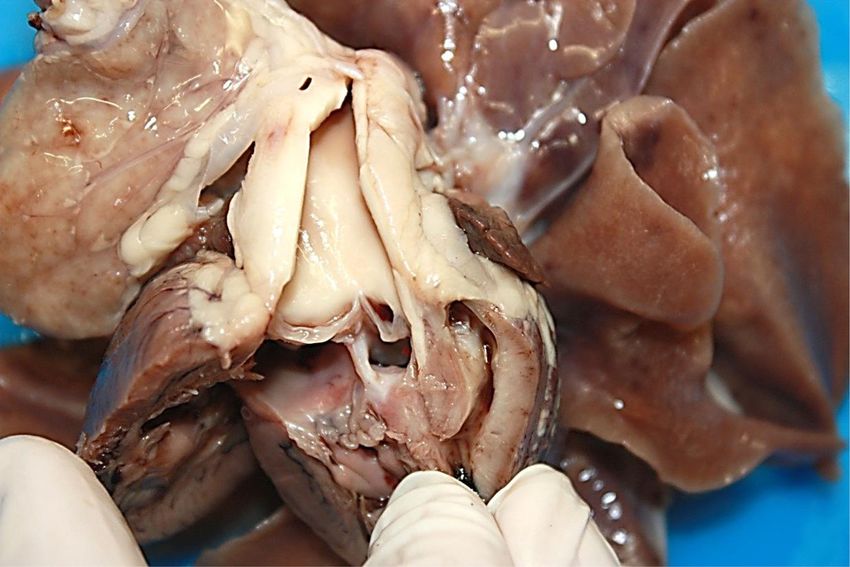
Pazzi et al. Acta Veterinaria Scandinavica 2014, 56:12                                                                                 Page 2 of 8
http://www.actavetscand.com/content/56/1/12

present. Initial management included oxygen supplemen-                     to 2.4 mm wide) with concomitant overriding aorta was
tation and 0.1 mg/kg of butorphanol (V-Tech Pharmacy,                      appreciable (Figure 3). A right-to-left ventricular shunt
Midrand, South Africa) intramuscularly, subsequently re-                   was detected on colour flow Doppler (Figure 4) with
ducing the patient’s respiratory distress and reducing the                 peak velocity up to 185 cm/s on spectral Doppler, with
severity of cyanosis. Serum biochemistry and electrolytes                  the majority of the shunted blood directed towards the
revealed no significant abnormalities, arterial blood gas                  left ventricular outflow tract and subaortic region. On
showed severe hypoxia - partial arterial pressure of oxy-                  the right parasternal short axis view, severe subvalvular PS
gen: 27.1 (normal range: 75–100 mmHg), mild acidosis                       characterised by marked narrowing of the right ventricular
pH: 7.341 (normal range: 7.350-7.450), low bicarbonate                     outflow tract was seen with post-stenotic peak velocity of
13.2 (normal range: 20–24 mmol/L) and low partial                          565 cm/s (Figure 5). Severe patient tachypnoea during the
arterial pressure of carbon dioxide (paCO2): 20.9 (normal                  echocardiographic examination resulted in marked cardiac
range: 32.0-45.0 mmHg).                                                    excursion and made it impossible to obtain an accurate
  A prominent main pulmonary arterial bulge was seen                       M-mode tracing. Nevertheless, with the overall findings of
superimposing over the aorta on the dorsoventral thor-                     high membranous VSD, overriding aorta, severe PS and
acic radiograph, corresponding with a soft tissue bulge                    right ventricular hypertrophy, a tentative diagnosis of TOF
at the cranial aspect of the base of the cardiac silhouette                with a right-to-left VSD shunt was made.
on the right lateral thoracic radiograph (Figure 1). There                   Due to the severity of the condition and the poor
was a mild but inconsistent increased interstitial lung                    prognosis the patient was euthanased and a necropsy
pattern seen in the cranial cupula and ventral aspect of                   conducted. The multiple echocardiographic findings were
the caudal lung lobes (seen only on lateral but not on                     confirmed during macroscopic examination and included
dorsoventral view). The overall radiological findings were                 an overriding aorta associated with a subaortic VSD,
suggestive of pulmonic stenosis with post-stenotic main                    concentric right ventricular hypertrophy and right atrial
pulmonary artery dilatation and therefore, echocardiog-                    dilatation, subpulmonary stenosis associated with localised
raphy was subsequently performed.                                          ventricular septal hypertrophy resulting in pulmonary
  On the right parasternal long axis echocardiography                      valve and trunk hypoplasia and aortic trunk dilatation. In
view, moderate thickening of the right ventricular free                    addition, an ASD, consistent with an ostium secundum
wall (two times the thickness of the left ventricular free                 was found (Figures 6, 7, 8, and 9). The caudal thor-
wall) and interventricular septum (1.5 times the thick-                    acic periaortic mediastinum exhibited multiple promin-
ness of the left ventricular free wall) was visible, indicat-              ent small tortuous blood filled vessels (veins) extending
ing moderate right ventricular hypertrophy (Figure 2).                     between the azygos - and costal veins and dorsocaudal
Marked enlargement of the right atrium was noted with                      pulmonary pleura (Figure 10). Prominent coronary veins
severe, turbulent high velocity trans-tricuspid regurgi-                   due to marked venous dilatation could also be detected
tation up to 559 cm/s detected on continuous wave                          macroscopically.
Doppler. On investigation of the left ventricular outflow                    No significant histopathological changes on routine
tract, a high membranous ventricular septal defect (up                     haematoxylin & eosin staining could be demonstrated in

                       A                                     B

                                                                             R

 Figure 1 Dorsoventral (A) and right lateral (B) thoracic radiographs. Prominent main pulmonary artery bulge seen superimposing over the
 descending aorta on dorsoventral view and corresponding to soft tissue opacity at the cranial aspect of the heart base on the orthogonal view.
 Arrow heads point at the bulge in the pulmonary artery.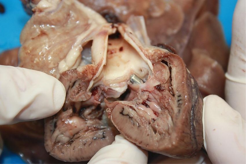
Pazzi et al. Acta Veterinaria Scandinavica 2014, 56:12                                                                                                    Page 3 of 8
http://www.actavetscand.com/content/56/1/12

                                                                                  RV                RA

                                                                             LV
                                                                                             LA

 Figure 2 Right parasternal long axis view of the heart with severe right atrial enlargement and moderate right ventricular hypertrophy.
 Note the thickening of the right ventricular free wall and interventricular septum. Right atrium (RA), right ventricle (RV), left atrium (LA), left ventricle (LV).

                                                                                        RV

                                                                                  IVS

                                                                        LV
                                                                                                   AO

 Figure 3 Right parasternal long axis left ventricular outflow tract view of the heart with ventricular septal defect and overriding aorta.
 A high membranous ventricular septal defect (arrow) is visible at the subaortic region. Right atrium (RA), right ventricle (RV), left atrium (LA), left
 ventricle (LV), interventricular septum (IVS).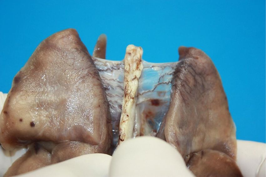
Pazzi et al. Acta Veterinaria Scandinavica 2014, 56:12                                                                                    Page 4 of 8
http://www.actavetscand.com/content/56/1/12

                                                                        RV

                                                                  IVS

                                                                LV
                                                                                           AO

 Figure 4 Right parasternal long axis left ventricular outflow tract view of the heart with colour flow Doppler with a reversed
 ventricular septal defect shunt. A right to left shunt characterised by blue-colour flow across the high membranous ventricular septal defect with
 concomitant overriding aorta. Right atrium (RA), right ventricle (RV), left atrium (LA), left ventricle (LV), interventricular septum (IVS).

myocardial fibres. Marked coronary vein dilatation histo-                    and terminal bronchiolar veins were generally signifi-
logically supported the macroscopic observation. The                         cantly distended. Histologically, the caudodorsal pul-
lungs showed generalised alveolar micro-atelectasis asso-                    monary pleural findings supported the macroscopic
ciated with pulmonary arterial collapse and hypoplasia                       observation of prominent venous dilatation in pleural
due to poor pulmonary arterial perfusion. The bronchial                      adventitia. Of other organs examined histologically, only

                                           RVOT

                                    AO

 Figure 5 Right parasternal short axis view of the heart base with right ventricular outflow tract and continuous wave Doppler showing
 severe subvalvular pulmonic stenosis. There is marked narrowing of the right ventricular outflow tract with post-stenotic peak velocity of
 564.5 cm/s. Right ventricular outflow tract (RVOT), aorta (AO).
Pazzi et al. Acta Veterinaria Scandinavica 2014, 56:12 Page 5 of 8
http://www.actavetscand.com/content/56/1/12
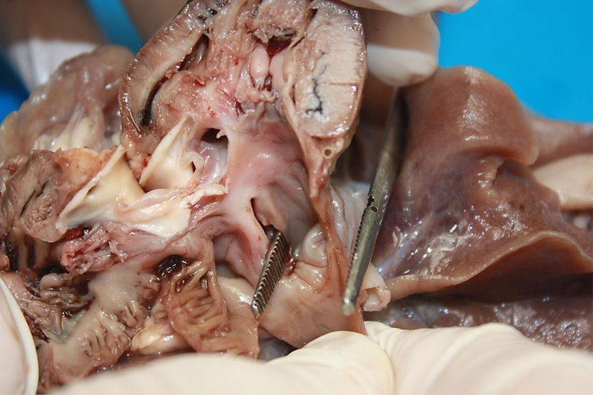
RV
RA
Figure 6 Sagittal section through the right ventricle (RV). There is an atrial septal defect (ostium secundum) between the right (RA) and left
atrium (arrow and forceps). The right atrium is also significantly dilated.
the liver showed significant change, manifesting as mod- the inbreeding of Keeshond dogs led to the suspicion of
erate global hepatic venous dilatation. a polygenetic threshold inheritance model for TOF
[24,25]. Specific genetic associations have not been eluci-
Discussion dated in dogs. Concurrent developmental abnormalities
Tetralogy of Fallot results from abnormal embryonic in addition to those causing TOF lead to ASD or PDA
development of the conotruncal septum, resulting in and resultant POF. TOF with ASD is a very rare condition
varying degrees of infundibular and valvular PS, pulmon- and has been reported in only 2 animal species as sporadic
ary artery hypoplasia, malalignment of the infundibular individual case reports [13,17]. The white colour of Bengal
septum, and a VSD [19]. Specific genetic associations Tigers is due to a recessive trait with selective inbreeding
with TOF in humans include alterations in JAG1 [20], in captivity often encouraging the expression of recessive
NKX2-5 [21], ZFPM2 [22] and VEGF [23] while in dogs traits, and although TOF has not been associated with
A
RV
Figure 7 Sagittal section through the right ventricle (RV). An overriding aorta (A) communicating with the right ventricle (RV) and left
ventricle through a subaortic ventricular septal defect (arrow and forceps). The right ventricular wall showed marked hypertrophy (double arrow).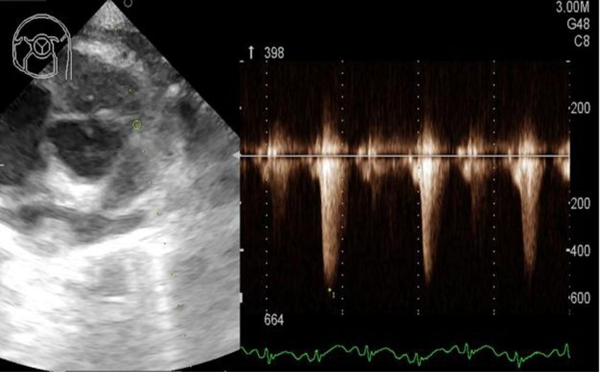
Pazzi et al. Acta Veterinaria Scandinavica 2014, 56:12                                                                                  Page 6 of 8
http://www.actavetscand.com/content/56/1/12

                                                                A
                                                                            PT

                                                           RA       VD

                                                                  RV

 Figure 8 Sagittal section through the right ventricle (RV). A significantly hypoplastic pulmonary trunk (PT) demonstrates markedly reduced
 pulmonary arterial blood flow (arrow). Note the compressive effect (stenosis) of a hypertrophic proximal interventricular septum (star) on the
 pulmonary trunk (PT). Overriding aorta (A). Ventricular septal defect (VD). Right atrium (RA).

Bengal or white Tigers to date, abnormalities of the visual                tachypnoea due to the hypoxaemia, causing CO2 to be
pathways have been associated with white Tigers [26].                      “blown-off” as well as only a mild right-to-left shunt seen
The clinical presentation of the cub with cyanosis and                     on Doppler echocardiography. A larger shunt fraction/
tachypnoea was supported by the arterial blood gas that                    pressure may have resulted in greater paCO2.
demonstrated severe hypoxaemia and concurrent mixed                          The radiological findings were supportive of PS with
acid–base disturbance (metabolic acidosis and respira-                     post-stenotic dilatation of the pulmonary artery but the
tory alkalosis) due to CO2 partial pressure lower than                     pulmonary pattern was not typical for cardiogenic pul-
would be expected for pure compensation for the meta-                      monary oedema. In contrast to previous reports in dogs
bolic acidosis. The metabolic acidosis was most likely                     [12-14], diffuse cardiomegaly was not visualised in this
secondary to anaerobic cellular metabolism due to severe                   Tiger. This may be due to the fact that the right-to-left
hypoxia, resulting in lactate accumulation. The lower-                     ventricular septal defect shunting blood was directed
than-expected paCO2 (considering the degree of periph-                     into the subaortic region, thus minimising the effect of
eral cyanosis) was most likely a result of the severe                      volume overload of the left heart while the moderate

                                                                     RV

                                                                      LV

 Figure 9 Cross section through the mid right & left ventricular region (LV). The right ventricle (RV) shows severe concentric hypertrophy
 due to pulmonary arterial stenosis. Note the marked ventricular septal hypertrophy (double arrow).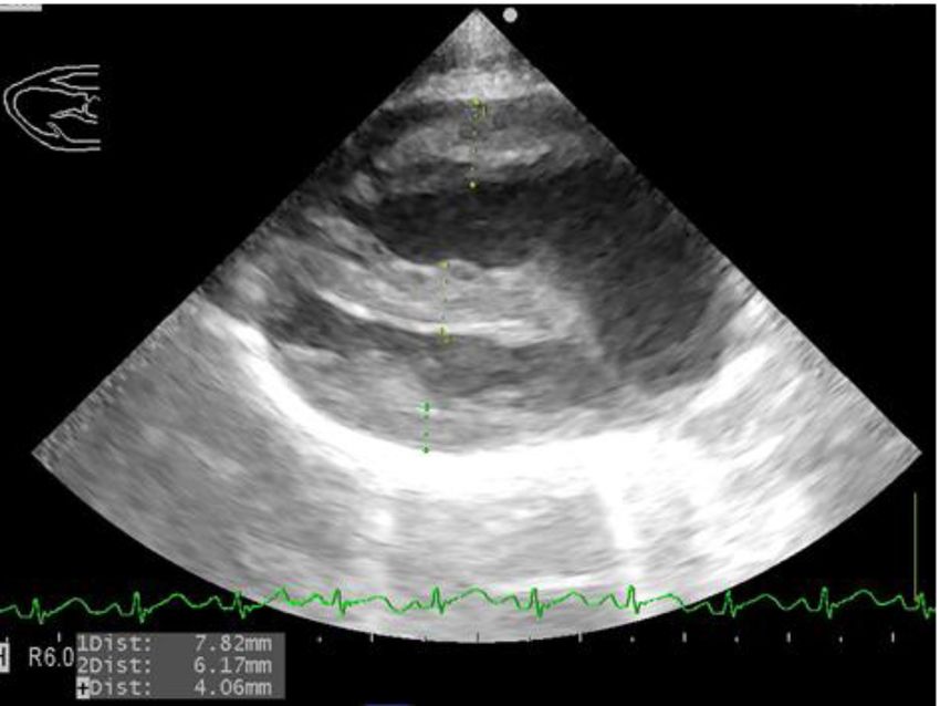
Pazzi et al. Acta Veterinaria Scandinavica 2014, 56:12                                                                                           Page 7 of 8
http://www.actavetscand.com/content/56/1/12

 Figure 10 Dorsal view of the lungs and bisecting thoracic aorta. There are numerous prominent small tortuous thin walled blood filled
 vessels (veins) between the caudal pulmonary pleurae and periaortic mediastinal connective tissue (arrows). They originate as fine hair-like vessels
 in the pulmonary pleura and anastomose to form larger vessels towards the aortic adventitia where they drain into the azygos and costal veins
 (not in picture).

concentric hypertrophy of the right ventricle was not                        of pulmonary truncal stenosis, resulting in diminishing
appreciable on radiographs.                                                  pulmonary arterial pressure. Diminished pulmonary ar-
   The echocardiographic findings in this case were                          terial pressure explains the pathological findings of pul-
typical for a TOF and surprisingly the ASD was only                          monary arteriolar collapse and hypoplasia associated
detected during necropsy. Failure to identify the ASD on                     with suspected increased flow resistance to the bronchial
echocardiography was most likely due to the small size                       arterial supply of the lung. This would result in most of
of the ASD while the absence of obvious shunting                             the bronchial arterial supply being shunted to the bron-
between the two chambers on colour flow Doppler was                          chial venous system (normally most of the bronchiolar
likely due to the equalisation of pressures between atria.                   arterial supply drains into the pulmonary arterial flow
The pulmonary outflow pressure in this cub was mildly                        via anastomosis), causing distension of bronchial and
increased compared to the previously reported value in                       pleural veins draining into the azygos and costal venous
the Korean Sapsaree dog also diagnosed with TOF and                          system. Coronary vein distension was most likely as a
ASD [13]. The ventricular right-to-left shunt velocity                       result of increased right atrial pressure subsequent to
measured in the Tiger cub may have included the left                         tricuspid valve insufficiency secondary to PS.
ventricular outflow tract due to the concomitant over-
riding aorta and could have resulted in a measured
shunt velocity that is not a true reflection. Interestingly                  Conclusions
the right-to-left shunt velocity was of lower velocity than                  This report documented the first clinical case of TOF
reported for the Korean Sapsaree dog [13], possibly re-                      with ASD (a subclass of POF) in a Bengal Tiger with
lated to the size of the VSD’s. The authors recommend if                     clinical and arterial blood gas signs of hypoxaemia,
the classic findings of a TOF are diagnosed, it is advised                   radiological and echocardiographic evidence of TOF and
to thoroughly exclude the possibility of a PDA or ASD                        necropsy findings consistent with TOF with ASD. The
to ensure the diagnosis of a POF is not missed.                              reported findings may assist in the antemortem diagnosis
   The necropsy findings were very similar to the previ-                     of Tetralogy or Pentalogy of Fallot in other species.
ously described adult Siberian Tiger [18], except no
endocardiosis of the mitral valve was present in this                        Abbreviations
Tiger cub. The other significant difference was the pres-                    ASD: Atrial septal defect; paC02: Arterial partial pressure of carbon dioxide;
                                                                             pa02: Arterial partial pressure of oxygen; PDA: Patent ductus arteriosis;
ence of locally extensive pleural venous distension in the                   POF: Pentalogy of Fallot; PS: Pulmonic stenosis; TOF: Tetralogy of Fallot;
caudal thoracic peri-aortic mediastinum covering the                         VSD: Ventricular septal defect.
area between the azygos vein and dorsocaudal pulmon-
ary pleura in this cub. The PS resulted in progressive                       Competing interests
right ventricular hypertrophy which increased the degree                     The authors declare that they have no competing interests.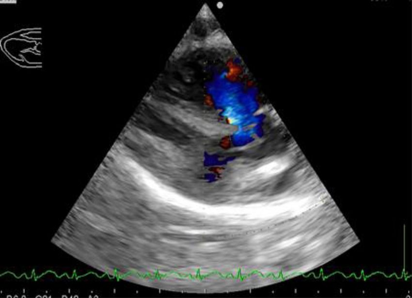
Pazzi et al. Acta Veterinaria Scandinavica 2014, 56:12                                                                                                     Page 8 of 8
http://www.actavetscand.com/content/56/1/12

Authors’ contributions                                                                20. Eldadah ZA, Hamosh A, Biery NJ, Montgomery RA, Duke M, Elkins R, Dietz
PP was the primary clinician on the case, collated all clinical and imaging               HC: Familial Tetralogy of Fallot caused by mutation in the jagged1 gene.
information and is the primary author of the paper. CKL carried out the                   Hum Mol Genet 2001, 10:163–169.
diagnostic imaging procedures and interpretation. JS performed the                    21. Goldmuntz E, Geiger E, Benson DW: NKX2.5 mutations in patients with
necropsy and histopathology examination and interpretation. All authors                   Tetralogy of Fallot. Circulation 2001, 104:2565–2568.
made intellectual contributions, reviewed and approved the final manuscript.          22. Pizzuti A, Sarkozy A, Newton AL, Conti E, Flex E, Digilio MC, Amati F,
                                                                                          Gianni D, Tandoi C, Marino B, Crossley M, Dallapiccola B: Mutations of
Author details                                                                            ZFPM2/FOG2 gene in sporadic cases of Tetralogy of Fallot. Hum Mutat
1
 Department of Companion Animal Clinical Studies, Faculty of Veterinary                   2003, 22:372–377.
Science, University of Pretoria, Private Bag X04, Onderstepoort 0110, South           23. Lambrechts D, Devriendt K, Driscoll DA, Goldmuntz E, Gewillig M, Vlietinck
Africa. 2Diagnostic Imaging Section, Department of Companion Animal                       R, Collen D, Carmeliet P: Low expression VEGF haplotype increases the
Clinical Studies, Faculty of Veterinary Science, University of Pretoria, Private          risk for Tetralogy of Fallot: a family based association study. J Med Genet
Bag X04, Onderstepoort 0110, South Africa. 3Current address: Department of                2005, 42:519–522.
Veterinary Clinical Sciences, College of Veterinary Medicine, Purdue                  24. Patterson DF: Genetic aspects of congenital heart disease in the dog.
University, West Lafayette, IN 47907-2026, USA. 4Section of Pathology,                    Gaines Dog Re Pro 1972, 4:7.
Department of Paraclinical Sciences, Faculty of Veterinary Science, University        25. Patterson DF, Pyle RL, Mierop L, Melbin J, Olson M: Hereditary defects of
of Pretoria, Private Bag X04, Onderstepoort 0110, South Africa.                           the conotruncal septum in Keeshond dogs: pathologic and genetic
                                                                                          studies. Am J Cardiol 1974, 34:187–205.
Received: 28 November 2013 Accepted: 27 February 2014                                 26. Guillery RW, Kaas JH: Genetic abnormality of the visual pathways in a
Published: 4 March 2014                                                                   “white” tiger. Science 1973, 180:1287–1289.

                                                                                       doi:10.1186/1751-0147-56-12
References
                                                                                       Cite this article as: Pazzi et al.: Tetralogy of Fallot and atrial septal defect
1. Tidholm A: Retrospective study of congenital heart defects in 151 dogs.             in a white Bengal Tiger cub (Panthera tigris tigris). Acta Veterinaria
    J Small Anim Pract 1997, 38:94–98.                                                 Scandinavica 2014 56:12.
2. Oliveira P, Domenech O, Silva J, Vannini S, Bussadori R, Bussadori C:
    Retrospective review of congenital heart disease in 976 dogs. J Vet Intern
    Med 2011, 25:477–483.
3. Bolton GR, Ettinger SJ, Liu SK: Tetralogy of Fallot in three cats. J Am Vet
    Med Assoc 1972, 160:1622–1631.
4. Kirby D, Gillick A: Polycythemia and Tetralogy of Fallot in a cat. Can Vet J
    1974, 15:114–119.
5. Fruganti A, Cerquetella M, Beribe F, Spaterna A, Tesei B: Clinic and
    ultrasonographic findings in a cat with Tetralogy of Fallot. Vet Res
    Commun 2004, 28:343–346.
6. Hall TL, Magdesian KG, Kittleson MD: Congenital cardiac defects in
    neonatal foals: 18 cases (1992–2007). J Vet Intern Med 2010, 24:206–212.
7. Mohamed T, Sato H, Kurosawa T, Oikawa S, Nakade T, Koiwa M: Tetralogy
    of Fallot in a calf: clinical, ultrasonographic, laboratory and postmortem
    findings. J Vet Med Sci 2004, 66:73–76.
8. Lacasta D, Ruiz S, Ramos JJ, Ferrer LM, Fernadez A, Gomez P: Tetralogy of
    Fallot in a three-month-old lamb: clinical, ultrasonographic and
    laboratory findings. Vet Rec 2011, 169:73.
9. Wenger S, Gull J, Glaus T, Blumer S, Wimmershoff J, Kranjc A, Steinmetz H,
    Hatt JM: Fallot’s Tetralogy in a European beaver (Castor fiber). J Zoo
    Wildlife Med 2010, 41:359–362.
10. Koie H, Abe Y, Sato T, Yamaoka A, Taira M, Nigi H: Tetralogy of Fallot in a
    Japanese macaque (Macaca fuscata). J Am Ass Lab Ani 2007, 46:66–67.
11. Agren E, Soderberg A, Morner T: Fallot’s Tetralogy in a European brown
    bear (Ursus arctos). J Wildl Dis 2005, 41:825–828.
12. McEntee K, Snaps F, Clercx C, Henroteaux M, Dondelinger R: Clinical
    vignette [Tetralogy of Fallot associated with a patent ductus arteriosus
    in a German Shepherd dog]. J Vet Intern Med 1998, 12:53–55.
13. InChul P, HyeSun L, JongTaek K, JoonSeok L, SeungGon L, ChangBaig H:
    Pentalogy of Fallot in a Korean Sapsaree dog. J Vet Med Sci 2007,
    69:73–76.
14. SeungKeun L, JinUng J, ChangBaig H: Pentalogy of Fallot with subaortic
    stenosis in a mixed dog. J Vet Clin 2009, 26:155–159.
15. Bayly WM, Reed SM, Leathers CW, Brown CM, Traub JL, Paradis MR, Palmer                  Submit your next manuscript to BioMed Central
    GH: Multiple congenital heart anomalies in five Arabian foals. J Am Vet                 and take full advantage of:
    Med Assoc 1982, 181:684–689.
16. Rahal C, Collatos C, Solano M, Bildfell R: Pentology of Fallot, renal
                                                                                           • Convenient online submission
    infarction and renal abscess in a mare. J Equine Vet Sci 1997, 17:604–607.
17. Pielmeier R, Engelke E, Legler M, Haist V, Hopster-Iversen C, Distl O:                 • Thorough peer review
    Congenital cardiac anomalies (Pentalogy of Fallot) in a two year old ram               • No space constraints or color figure charges
    with brachygnathia inferior [in German]. Berl Munch Tierarztl Wochenschr
                                                                                           • Immediate publication on acceptance
    2013, 126:256–263.
18. Scaglione FE, Tursi M, Chiappino L, Schroder C, Triberti O, Bollo E: Pentalogy         • Inclusion in PubMed, CAS, Scopus and Google Scholar
    of Fallot in a captive Siberian Tiger (Panthera tigris altaica). J Zoo Wildlife        • Research which is freely available for redistribution
    Med 2012, 43:931–933.
19. MacDonald KA: Congenital heart diseases of puppies and kittens.
    V Clin N Am - Small 2006, 36:503–531.                                                   Submit your manuscript at
                                                                                            www.biomedcentral.com/submit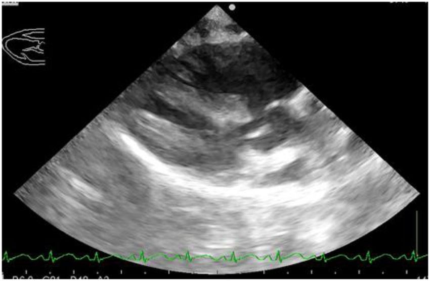
You can also read