The Dual Immunoregulatory function of Nlrp12 in T Cell-Mediated Immune Response: Lessons from Experimental Autoimmune Encephalomyelitis - MDPI
←
→
Page content transcription
If your browser does not render page correctly, please read the page content below
cells
Article
The Dual Immunoregulatory function of Nlrp12 in
T Cell-Mediated Immune Response: Lessons from
Experimental Autoimmune Encephalomyelitis
Marjan Gharagozloo, Shaimaa Mahmoud, Camille Simard, Tara M. Mahvelati               ID
                                                                                          ,
Abdelaziz Amrani and Denis Gris * ID
 Program of Immunology, Department of Pediatrics, CR-CHUS, Faculty of Medicine and Health Sciences,
 University of Sherbrooke, Sherbrooke, QC J1H 5N4, Canada; Marjan.Gharagozloo@usherbrooke.ca (M.G.);
 Shaimaa.Mahmoud@usherbrooke.ca (S.M.); Camille.Simard@usherbrooke.ca (C.S.);
 t.mahvelati@gmail.com (T.M.M.); Abdelaziz.Amrani@usherbrooke.ca (A.A.)
 * Correspondence: denis.gris@usherbrooke.ca; Tel.: +819-346-1110 (ext. 16632)

 Received: 3 August 2018; Accepted: 23 August 2018; Published: 27 August 2018

 Abstract: Although the etiology of multiple sclerosis (MS) remains enigmatic, the role of T cells is
 unquestionably central in this pathology. Immune cells respond to pathogens and danger signals via
 pattern-recognition receptors (PRR). Several reports implicate Nlrp12, an intracellular PRR, in the
 development of a mouse MS-like disease, called Experimental Autoimmune Encephalomyelitis (EAE).
 In this study, we used induced and spontaneous models of EAE, as well as in vitro T cell assays,
 to test the hypothesis that Nlrp12 inhibits Th1 response and prevents T-cell mediated autoimmunity.
 We found that Nlrp12 plays a protective role in induced EAE by reducing IFNγ/IL-4 ratio in lymph
 nodes, whereas it potentiates the development of spontaneous EAE (spEAE) in 2D2 T cell receptor
 (TCR) transgenic mice. Looking into the mechanism of Nlrp12 activity in T cell response, we found
 that it inhibits T cell proliferation and suppresses Th1 response by reducing IFNγ and IL-2 production.
 Following TCR activation, Nlrp12 inhibits Akt and NF-κB phosphorylation, while it has no effect
 on S6 phosphorylation in the mTOR pathway. In conclusion, we propose a model that can explain
 the dual immunoregulatory function of Nlrp12 in EAE. We also propose a model explaining the
 molecular mechanism of Nlrp12-dependent regulation of T cell response.

 Keywords: Nlrp12; CNS; inflammation; T cell; EAE; spontaneous EAE; TCR signaling; 2D2

1. Introduction
      Multiple sclerosis (MS) is a chronic autoimmune disease of the central nervous system (CNS),
where autoreactive immune responses are involved in demyelination and CNS damage. The etiology
and pathogenesis of the disease remain elusive. However, several lines of evidence demonstrate that
adaptive immune response plays a key role in the pathogenesis of MS and experimental autoimmune
encephalomyelitis (EAE), the mouse model of MS [1–3]. The major components of the adaptive
immunity, T cells, are initially activated by antigen presenting cells (APCs) in lymph nodes. Activated
T cells migrate into the CNS across the blood brain barrier (BBB) and reactivated again in perivascular
space, where CNS-resident cells including microglia and macrophages present myelin antigens to
T cells [4]. Thus, those activated CD4+ T cells orchestrate the functions of other adaptive immune
cells, such as CD8+ T cells and B cells, as well as innate immune cells in the CNS and periphery [5].
Depending on the composition of the cytokine milieu, naïve CD4+ T cells may differentiate into
different T helper (Th) subsets including Th1, Th2 and Th17 that produce signature cytokines such
as IFNγ, IL-4 and IL-17 respectively. The differentiation of naïve CD4+ T cells into Th1, Th2, or Th17

Cells 2018, 7, 119; doi:10.3390/cells7090119                                    www.mdpi.com/journal/cells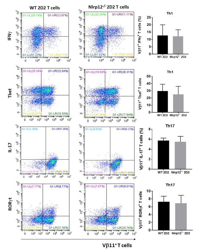
Cells 2018, 7, 119                                                                                 2 of 19

types are governed by transcription factors, known as Tbet, GATA3 and RORγt respectively [6].
Th subsets affect the CNS inflammation in different ways. Th1 and Th17 responses potentiate the CNS
inflammation, while Th2 response dampens inflammatory response and protects CNS damage [6].
These findings highlight the importance of T cell-mediated immunity in MS pathology.
     APCs including dendritic cells, macrophages and microglia are innate immune cells that trigger
T cell activation [7]. These cells create the first line of response by recognizing pathogens and/or
danger signals via pattern-recognition receptors (PRR) [8]. NOD-like receptors (NLRs) are intracellular
PRR that are mainly expressed by cells of hematopoietic origin and regulate both innate and adaptive
immune responses [9]. Recently, NLRs have gained more attention since 3 members of the family
including CIITA, Nlrc5 and Nlrp3 regulate transcription of molecules that shape adaptive immune
responses. CIITA [10] and Nlrc5 [11,12] show transcriptional activities for MHC II and MHC I
molecules respectively, while Nlrp3 acts as a Th2 transcription factor and promotes IL-4 production [13].
In addition, activation of NLRs often leads to the production and secretion of pro–inflammatory
cytokines such as IL-1β and IL-18 that in turn potentiate differentiation of Th1 and Th17 subsets [9,14].
These findings highlight the key role of NLR proteins in shaping T cell response and adaptive immunity.
     Not all NLRs are pro–inflammatory. Nlrp12 is a recently discovered member of NLRs that is shown
to be a negative regulator of both canonical and non-canonical nuclear factor-κB (NF-κB) signaling
pathways [15]. Previous studies showed that Nlrp12−/− mice are highly vulnerable to inflammatory
diseases such as experimental colitis and colorectal tumor development [16–19]. In the context of CNS
inflammation, the lack of Nlrp12 resulted in increased CNS inflammation and exacerbated course of
EAE [19]. Nlrp12−/− mice developed earlier and more severe form of EAE than wild-type (WT) mice.
This phenotype parallel with significant increases in the expression of pro-inflammatory genes in the
spinal cords of Nlrp12−/− mice relative to WT mice. Experiments using mouse primary microglia
cultures demonstrated that Nlrp12 significantly inhibits production of the inflammatory mediators
such as inducible nitric oxide synthase (iNOS), Tumor Necrosis Factor (TNF)α, IL-6 and nitric oxide
(NO) [19]. However, the ability of Nlrp12 to modulate T cell responses remains poorly defined.
     A recent article by Lukens et al. revealed that Nlrp12 is expressed not only by myeloid
cells but also by T cells. It negatively regulates NF-κB signaling, T cells proliferation and the
secretion of Th1/Th2/Th17 cytokines [20]. Non-surprisingly, Nlrp12 deficient mice developed
enhanced inflammatory symptoms in T-cell-mediated autoimmune diseases such as colitis and atopic
dermatitis [20]. However, in EAE model, lack of Nlrp12 promotes Th2 response and IL-4 secretion,
which results in a milder form of EAE with atypical symptoms, including ataxia and impaired balance
control [20]. Collectively, current findings and controversies indicate that the exact immunoregulatory
functions of Nlrp12 in T cell activation and T cell-mediated autoimmunity are poorly understood.
     In this study, we investigated the immunoregulatory role of Nlrp12 in T cell responses using
classical induced-EAE and spontaneous EAE (spEAE) models. We further characterized the role of
Nlrp12 in regulating T cell receptor (TCR) signaling pathways and IL-2 production.

2. Materials and methods

2.1. Mice
     All the protocols and procedures were approved by the University of Sherbrooke Animal Facility
and Use Committee (Protocols #280-15, 4 April 2017; #335-17B, 22 February 2018). Nlrp12 knock-out
(Nlrp12−/− ) mice on C57BL/6J background were kindly provided by Dr. Jenny P.Y. Ting (Chapel Hill,
NC, USA). Mice were backcrossed for at least 15 generation. The 2D2 transgenic mice expressing
a TCR specific for the myelin oligodendrocyte (MOG35–55 ) peptide were purchased from Jackson
Laboratory. Nlrp12−/− and WT mice were crossed with 2D2 mice to generate Nlrp12−/− 2D2 mice.
We genotyped all the animals for Nlrp12 and 2D2 (Supplementary protocol) and only those animals
that were Nlrp12−/− and 2D2+ were included in the study (Supplementary Figure S1). Moreover,
the expression of Vβ11 receptor was verified with flow cytometry. The mice were maintained under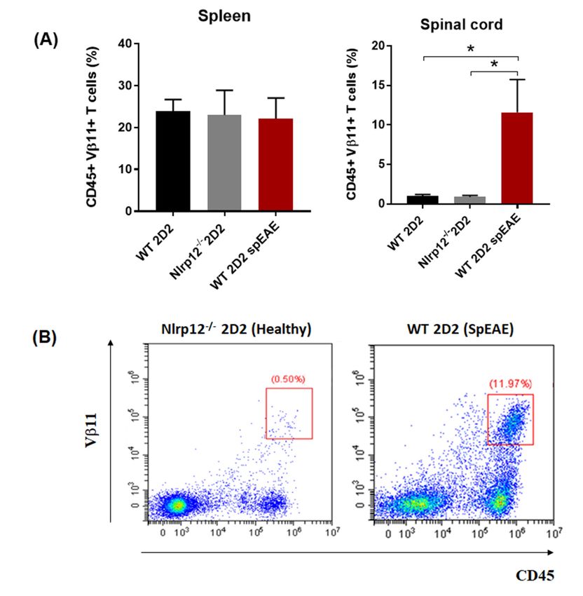
Cells 2018, 7, 119                                                                              3 of 19

specific pathogen-free conditions in the animal facility of the faculty of medicine, at the University
of Sherbrooke.

2.2. Induction of EAE and Tissue Collection
     EAE was induced in 8–10-week old WT or Nlrp12−/− female mice as previously described [19].
An emulsion mixture of MOG35−55 (Genemed Synthesis Inc., San Antonio, TX, USA), complete
Freund’s Adjuvant (CFA) (Sigma-Aldrich, St. Louis, MO, USA) and Mycobacterium tuberculosis H37
RA (Difco Laboratories, Detroit, MI, USA) was prepared and injected subcutaneously in the flank
with a total of 200 µg MOG35–55 and 500 µg Mycobacterium. Mice were also injected intraperitoneally
on days 0 and 2 with 200 ng Pertussis toxin (List Biological Laboratories Inc., Campbell, CA, USA).
After 3 weeks of immunization, mice were sacrificed, perfused with ice-cold phosphate-buffered saline
(PBS) (Wisent, St. Bruno, QC, Canada) and the CNS tissues were collected.

2.3. Intracellular Staining and Flow Cytometry
     CD4+ T cells were purified from lymph nodes and spleens using Mouse CD4+ T Cell Isolation
Kit (eBioscience, San Diego, CA, USA) and activated with plate-bound anti-CD3 (eBioscience,
clone:145-2C11, 1 µg/mL) and anti-CD28 (eBioscience, clone: 37.51, 2 µg/mL) antibodies for
indicated times. T cell proliferation was assessed by Ki67 intranuclear staining following fixation
and permeabilization in the Foxp3/Transcription Factor staining kit (eBioscience). For intracellular
staining of cytokines, the cells were stimulated with phorbol 12-myristate 13-acetate (PMA;
500 ng/mL, Sigma Chemical Co., St. Louis, MO, USA) and ionomycin (1 µg/mL, Calbiochem
Corp., La Jolla, CA, USA) for 5 h at 37 ◦ C in the presence of Brefeldin A (10 µg/mL, eBioscience).
Cells were stained with anti-CD4-FITC antibody (eBioscience), fixed, permeabilized and then stained
with anti-IFNγ-PE, anti-IL-4-PE, IL-17-PerCP-Cy5.5, Tbet-PE, or RORγt-PE antibody, as per the
manufacturer’s instructions (eBioscience). Sample acquisition was performed with Beckman Coulter
CytoFlex (Beckman Coulter, Brea, CA, USA) and data were analyzed using CytExpert 2 software
(Beckman Coulter, Brea, CA, USA). Plots were prepared using CytExpert 2 and FlowJo (San Carlos,
CA, USA) software.

2.4. Quantitative RT-PCR
    RNA was extracted from CD4+ T cells using TRIzol reagent (Life Technologies Inc., Burlington,
ON, USA) and cDNA was synthesized as previously described [19]. Reverse transcription PCR
(RT-PCR) was used to verify the expression of Nlrp12 in activated T cell using KiCqStart™ SYBR®
Green qPCR ReadyMix (Sigma Aldrich, St. Louis, MO, USA). Primers (IDT, Coralville, IA, USA)
sequences were as follows: Nlrp12F: 50 -CCT CTT TGA GCC AGA CGA AG-30 , Nlrp12R: 50 -GCC CAG
TCC AAC ATC ACT TT-30 , 18SF: 50 -CGG CTA CCA CAT CCA AGG AA-30 and 18SR: 50 -GCT GGA
ATT ACC GCG GCT-30 . The relative expression was calculated using the ∆∆CT method [21].

2.5. Cytokine Measurement
     IFNγ and IL-4 cytokines in the supernatant of activated CD4+ T cell culture and tissue lysates
were measured using ELISA kits as previously described [19]. Briefly, lymph node, spinal cord
and cerebellum tissues were homogenized in lysis buffer supplemented with protease inhibitors
(Cell Signaling Technology, Danvers, MA, USA) by rapid agitation using 3-mm stainless beads and a
TissueLyser II (Qiagen, Hilden, Germany) homogenizer for 2 min. The levels of IL-4 in tissue lysates
were determined using a high sensitivity IL-4 ELISA Kit (eBioscience, San Diego, CA, USA) according
to the manufacturer’s instruction. The amount of IFNγ was determined using IFNγ kit purchased
from PeproTech (Rocky Hill, NJ, USA).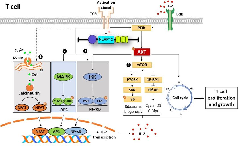
Cells 2018, 7, 119                                                                              4 of 19

2.6. Differentiation of 2D2 T Cells Toward Th1 or Th17 In Vitro
     Naïve CD4+ T cells were purified from the spleens and lymph nodes of Nlrp12−/− 2D2 and WT
2D2 mice using MagniSort Naïve CD4 T Cell Enrichment Kit (eBiosciences). Purified CD4+ T cells
were stimulated with MOG (50 µg/mL) in the presence of WT splenocytes at 1:1 ratio and Th1-,
Th2- or Th17- polarizing condition (Th1: IL-12 (10 ng/mL), anti-IL-4 (10 µg/mL), IL-2 (10 ng/mL); Th2:
IL-4 (10 µg/mL), hTGF-β1 (10 ng/mL), IL-2 (10 ng/mL) and Th17: anti-IL-12 (10 µg/mL), anti-IL-4
(10 µg/mL), anti-IFN-γ (10 µg/mL), mIL-6 (10 ng/mL), hTGF-β1 (10 ng/mL)). Recombinant cytokines
and antibodies were purchased from Biolegend (San Diego, CA, USA) and eBioscience (San Diego,
CA, USA), respectively. After 72 h of culture, cells were stained for MOG-TCR transgenic surface
marker, Vβ11 and Th1- or Th17- associated markers (intracellular cytokine and transcription factor).
The percentage of Th1 or Th17 cells were evaluated by Flow cytometry.

2.7. T Cell Activation and Signaling Pathways
     To analyze p65 phosphorylation upon CD3 cross-linking, CD4+ T cells were purified from
the lymph nodes of Nlrp12−/− and WT mice and incubated with 10 µg/mL anti-CD3 (eBioscience,
San Diego, CA, USA) for 30 min on ice, followed by washing and re-suspending the cells in serum-free
medium. T cells were incubated with 10 µg/mL cross-linking anti-mouse IgG antibody (R&D systems,
Minneapolis, MN, USA) for 20 min at 37 ◦ C. Akt phosphorylation was quantified after activation of
CD4+ T cells with PMA/Iono for 10 min at 37 ◦ C, while the phosphorylation of S6 was analyzed after
24 h activation of CD4+ T cells with plate-bound anti-CD3/CD28 antibodies. Following stimulation
in a defined period of time, T cells were washed and lysed in the lysis buffer plus proteinase
and phosphatase inhibitor (Cell Signaling Technology). Proteins were then separated by on 10%
SDS-polyacrylamide gels and transferred to nitrocellulose membrane. Transfers were blocked for 1h at
room temperature with 5% nonfat milk in TBS/0.1% Tween 20 (TBST) and then incubated overnight
at 4 ◦ C in the primary antibodies (Cell Signaling Technology) diluted in TBST. The membranes
were washed 3 times with TBST and incubated in horseradish peroxidase (HRP)-conjugated goat
anti-rabbit antibody (Cell Signaling Technology) diluted 1/1000 in TBST for 1 h at room temperature.
The immunoblots were developed with Lumigen ECL ultra reagent (Lumigen, Southfield, MI, USA),
imaged with ChemiDocTM (Bio-Rad, Hercules, CA, USA) and analyzed using Image Lab software
(Bio-Rad).

2.8. Phosphoflow Cytometry
     Phosphorylation of S6 ribosomal protein was confirmed by flow cytometry. Following 24 h
stimulation of CD4+ T cells with plate-bound anti-CD3/CD28, cells were immediately fixed and
permeabilized using Fix and Perm Kit (eBioscience). After washing with perm buffer (eBioscience) and
blocking with 5% Fetal Bovine Serum, cells were incubated with Phospho-S6 antibody (Cell Signaling
Technology) for 1h at room temperature, followed by washing with perm buffer and incubation
with Alexa Fluor® 555 (Cell Signaling Technology) conjugated antibody. Samples were acquired by
Beckman Coulter CytoFlex and data was analyzed using CytExpert 2 software (Beckman Coulter, Brea,
CA, USA).

2.9. SpEAE and Histological Analysis
     WT 2D2 or Nlrp12−/− 2D2 mice were monitored for the development of spEAE. Animals were
sacrificed after 4 months of monitoring or after the development of EAE at the peak score of 4.
The immunized mice were sacrificed as described in Section 2.2 and the spinal cords were removed
and fixed in 4% formaldehyde for 24 h. The spinal cord tissues were embedded in paraffin and cut
into 5-µm sections and stained with hematoxylin and eosin (H&E) stain and immunofluorescence for
astrocyte marker (GFAP), microglia marker (Iba1) and myelin basic protein (MBP). All slides were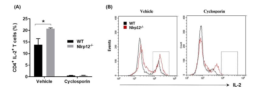
Cells 2018, 7, 119                                                                               5 of 19

scanned using a digital slide scanner NanoZoomer-XR C12000 (Hamamatsu Photonics, Hamamatsu,
Japan) and viewed using NDPview2 software (Hamamatsu Photonics, Hamamatsu, Japan).

2.10. Analysis of CNS-Infiltrating Mononuclear Cells by Flow Cytometry
     Dissected spinal cords were filtered through a 70 mm nylon sieve (BD Pharmingen, Becton
Dickinson, Franklin Lakes, NJ, USA) with a syringe plunger to get a uniform tissue homogenate
that was digested with collagenase D (2.5 mg/mL, Roche Diagnostics, Risch-Rotkreuz, Switzerland)
and DNase I (1 mg/mL, Sigma-Aldrich) with agitation at 37 ◦ C for 45 min. Mononuclear cells were
isolated by percoll (Sigma-Aldrich) centrifugation, in which 300 µL percoll was mixed with 1 mL cell
suspension overlaid with 700 µL Hank’s Balanced Salt Solution (HBSS). The samples were centrifuged
at 12,000× g for 15 min without break. Myelin and cell debris were aspirated and the mononuclear
pellet was re-suspended and washed with HBSS before staining for surface markers and analysis by
flow cytometry.

2.11. Statistical Analysis
     All statistical analyses were conducted using GraphPad Prism 7 software (GraphPad, San Diego,
CA, USA). Results were expressed as the mean ± standard deviation. Statistical differences between
WT and Nlrp12−/− samples were assessed by Student’s t-test. Level of Nlrp12 expression was assessed
by one-way ANOVA and cellular signaling by two-way ANOVA. The significance level was set at
p < 0.05.

3. Results

3.1. Nlrp12 Modulates Th1/Th2 Balance in EAE Mice
     Our previous study demonstrated that Nlrp12−/− mice develop earlier and more severe EAE
compared to WT mice (Supplementary Figure S2) [19]. Since Th1 cells play a crucial role in the
pathogenesis of EAE, we examined whether the imbalance of Th1/Th2 response might lead to
exacerbated EAE in Nlrp12−/− mice. We collected lymph nodes and the CNS tissues including spinal
cord and cerebellum from WT and Nlrp12−/− EAE mice at day 21 after MOG-CFA immunization and
measured the level of IFNγ and IL-4 as the designated cytokines for Th1 and Th2 cells respectively. As
shown in Figure 1, we found significantly higher levels of IFNγ in lymph node extracts from Nlrp12−/−
EAE mice, while IL-4 levels were significantly lower than WT EAE mice. In addition, the ratio of
IFNγ/IL-4 was higher in lymph node extracts of Nlrp12−/− EAE mice, suggesting a higher Th1/Th2
ratio in Nlrp12−/− EAE (Figure 1A). No difference was observed in the levels of IFNγ and IL-4 in
the extracts of CNS tissues (spinal cord and cerebellum) (Figures 1B and 1C), however, IFNγ/IL-4
ratio was significantly lower in spinal cords from Nlrp12−/− EAE mice compared to WT EAE mice
(Figure 1B). We then evaluated the production of cytokines in ex vivo activated CD4+ T cells.

3.2. Nlrp12 Inhibits the Production of IFNγ by CD4+ T Cells In Vitro
      To determine whether Nlrp12 could inhibit IFNγ production by T cells, we purified CD4+ T cells
from WT and Nlrp12−/− mice and stimulated them with anti-CD3/CD28 antibodies to activate T
cells with TCR and costimulatory signals simultaneously [22]. After 72 h, cell culture supernatants
were collected and the levels of IFNγ and IL-4 were measured by ELISA. As shown in Figure 2A,
activated CD4+ T cells from Nlrp12−/− mice secreted significantly higher levels of IFNγ and IL-4
compared to WT T cells. We then tested whether the increased levels of both Th1- and Th2-associated
cytokines were related to the increased proliferation of Nlrp12−/− T cells compared to WT T cells.
Ki67 is a nuclear protein that is expressed by the cells in all cell cycle phases except G0 phase [23].
Following activation by anti-CD3/CD28 antibody for 24 h, the CD4+ T cells were stained for Ki67
and analyzed by flow cytometry. Results showed significantly higher numbers of Nlrp12−/− T cells in
the cell cycle compared to WT T cells (Figure 2B). Flow cytometric analysis of intracellular cytokines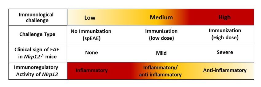
Cells 2018, 7, 119                                                                                       6 of 19

revealed that higher percentages of CD4+ T cells produced IFNγ in Nlrp12-/- activated T cells. However,
no significant difference was detected between Nlrp12−/− and WT T cells in the percentage of IL-4 or
IL-17 producing T cells after 3 days of activation with anti-CD3/CD28 antibodies in vitro (Figure 2C).
We then examined whether the exacerbated EAE in Nlrp12−/− mice was associated with the increased
     Cells 2018, 7, x FOR PEER REVIEW                                                                6 of 19
differentiation of myelin-specific T cells to Th1 or Th17 cells.

      Figure 1. Nlrp12 inhibits Th1 response in Experimental Autoimmune Encephalomyelitis (EAE).
           Figure 1. Nlrp12 inhibits Th1 response in Experimental Autoimmune Encephalomyelitis (EAE). Th1-
      Th1- and Th2- related cytokines in tissue lysates from EAE mice. Central nervous system (CNS)
           and Th2- related cytokines in tissue lysates from EAE mice. Central nervous system (CNS) tissues and
      tissues and lymph nodes were collected from EAE mice after 3 weeks of immunization with myelin
           lymph nodes were collected from EAE mice after 3 weeks of immunization with myelin
      oligodendrocyte/complete Freund’s adjuvant (MOG/CFA). The tissues were homogenized in lysis
           oligodendrocyte/complete Freund’s adjuvant (MOG/CFA). The tissues were homogenized in lysis
      buffer and cytokine levels were measured in tissue lysate by ELISA. (A) The levels of IFNγ, IL-4 and
           buffer and cytokine levels were measured in tissue lysate by ELISA. (A) The levels of IFNγ, IL-4 and
      IFNγ/IL-4 ratio in lymph node extracts from WT and Nlrp12−/− EAE     mice; (B) The levels of IFNγ, IL-4
           IFNγ/IL-4 ratio in lymph node extracts from WT and Nlrp12−/− EAE mice; (B) The levels of IFNγ, IL-4
      and their ratios in the spinal cord and (C) cerebellum of EAE mice, n = 5–6 per group, * p < 0.05.
           and their ratios in the spinal cord and (C) cerebellum of EAE mice, n = 5–6 per group, * p < 0.05.

      3.2. Nlrp12 Inhibits the Production of IFNγ by CD4+ T Cells In Vitro
           To determine whether Nlrp12 could inhibit IFNγ production by T cells, we purified CD4+ T cells
      from WT and Nlrp12−/− mice and stimulated them with anti-CD3/CD28 antibodies to activate T cells
      with TCR and costimulatory signals simultaneously [22]. After 72 h, cell culture supernatants were
      collected and the levels of IFNγ and IL-4 were measured by ELISA. As shown in Figure 2A, activated
      CD4+ T cells from Nlrp12−/− mice secreted significantly higher levels of IFNγ and IL-4 compared to WT
      T cells. We then tested whether the increased levels of both Th1- and Th2-associated cytokines were
      related to the increased proliferation of Nlrp12−/− T cells compared to WT T cells. Ki67 is a nuclear
      protein that is expressed by the cells in all cell cycle phases except G0 phase [23]. Following activation
      by anti-CD3/CD28 antibody for 24 h, the CD4+ T cells were stained for Ki67 and analyzed by flow
      cytometry. Results showed significantly higher numbers of Nlrp12−/− T cells in the cell cycle compared
      to WT T cells (Figure 2B). Flow cytometric analysis of intracellular cytokines revealed that higher
Cells 2018, 7, x FOR PEER REVIEW 7 of 19
T cells after 3 days of activation with anti-CD3/CD28 antibodies in vitro (Figure 2C). We then
examined whether the exacerbated EAE in Nlrp12−/− mice was associated with the increased
Cells 2018, 7, 119 7 of 19
differentiation of myelin-specific T cells to Th1 or Th17 cells.
Figure
Figure 2. Nlrp12
2. Nlrp12 inhibits
inhibits T cell
T cell proliferation
proliferation and and cytokine
cytokine productionbybyCD4
production CD4+ +TTcells.
cells. (A)
(A) Purified
Purified
CD4+CD4 T cells from WT andNlrp12
Nlrp12−/−mice micewere stimulated with anti-CD3/CD28 for 72 for
h. Cell culture
+ −/−
T cells from WT and were stimulated with anti-CD3/CD28 72 h. Cell
supernatants were collected and the levels of IFNγ and IL-4 were measured
culture supernatants were collected and the levels of IFNγ and IL-4 were measured by ELISA, n =by ELISA, n = 4; (B)
4;
Purified CD4++ T cells from WT and Nlrp12−/− − /mice
− were stimulated with anti-CD3/CD28 for 24 h and
(B) Purified CD4 T cells from WT and Nlrp12 mice were stimulated with anti-CD3/CD28 for 24 h
the proliferation of CD4+ T cells was evaluated using Ki67 staining and flow cytometry; (C) CD4+ T
and the proliferation of CD4+ T cells was evaluated using Ki67 staining and flow cytometry; (C) CD4+
cells were activated with anti-CD3/CD28 for 48 h and the expression of intracellular cytokines was
T cells were activated with anti-CD3/CD28 for 48 h and the expression of intracellular cytokines was
measured using flow cytometry. Representative flow cytometric plots show the expression of IFNγ,
measured using flow cytometry. Representative flow cytometric plots show the expression of IFNγ,
IL-4 and IL-17 in CD4
+
+ T cells, n = 4; * p < 0.05.
IL-4 and IL-17 in CD4 T cells, n = 4; * p < 0.05.
3.3. Nlrp12 Has No Effect on T Cell Differentiation toward Th1 and Th17
We evaluated whether Nlrp12 would favor differentiation of naïve T cells towards inflammatory
Th subsets. We purified naïve CD4+ T cells from lymph nodes and spleens of Nlrp12−/− 2D2 and WT
2D2 mice and cultured with MOG-pulsed WT splenocytes in the presence or absence of Th1 or Th17
polarizing cytokines. After 72 h of incubation, cells were harvested and stained for CD4 surface marker3.3. Nlrp12 Has No Effect on T Cell Differentiation toward Th1 and Th17
We evaluated whether Nlrp12 would favor differentiation of naïve T cells towards inflammatory
Th subsets. We purified naïve CD4+ T cells from lymph nodes and spleens of Nlrp12−/− 2D2 and WT
2D2
Cells mice
2018, and cultured with MOG-pulsed WT splenocytes in the presence or absence of Th1 or Th17
7, 119 8 of 19
polarizing cytokines. After 72 h of incubation, cells were harvested and stained for CD4 surface
marker and Th1 (IFNγ and Tbet) or Th17 (IL-17 and RORγt) intracellular markers. Flow cytometry
andresults show that
Th1 (IFNγ andNlrp12
Tbet) did not affect
or Th17 (IL-17theand
differentiation of CD4+ T cells
RORγt) intracellular toward
markers. Th1 cytometry
Flow or Th17 (Figure
results
3). that Nlrp12 did not affect the differentiation of CD4+ T cells toward Th1 or Th17 (Figure 3).
show
Figure
Figure 3. 3. Nlrp12does
Nlrp12 doesnot
notaffect
affectthe
thedifferentiation
differentiationofofnaïve CD4++ T
naïveCD4 T cells
cells to
to Th1
Th1 or
or Th17.
Th17. Naïve CD4++ T
Naïve CD4
T cells
cells fromfrom
WT andWTNlrp12 −
and Nlrp12
/ − micemice
−/−
werewere purified
purified and stimulated
and stimulated with MOG-splenocytes
with MOG-splenocytes in the in the
presence
of presence
Th1 or Th17 of Th1 or Th17 polarizing
polarizing cytokines for cytokines
3 days.for 3 days.
After After 5 h stimulation
5 h stimulation with PMA/ionomycin,
with PMA/ionomycin, cells were
cells were
stained stained
for Vβ11 forTh1-
and Vβ11or and Th1-associated
Th17- or Th17- associated cytokine/transcription
cytokine/transcription factor
factor and and analyzed
analyzed by flow
by flow cytometry, n = 3; Representative flow cytometric plots show the expression
cytometry, n = 3; Representative flow cytometric plots show the expression of Th1- and Th17-relatedof Th1- and Th17-
relatedin
markers markers
Vβ11+in cells. T cells.
T Vβ11
+
3.4. Nlrp12 Expression Is Increased in Activated T Cells
To test whether TCR activation modulated the expression of Nlrp12, we activated CD4+ T cells
with either anti-CD3 or anti-CD3/CD28 antibodies for 24 h and measured the expression of Nlrp12
gene by qPCR. As shown in Figure 4A, the mRNA expression of Nlrp12 was significantly increased in
T cells following activation by anti-CD3 or anti-CD3/CD28 antibodies. The level of Nlrp12 expression
remains high even after 48 h stimulation of T cells by anti-CD3/CD28 (Figure 4B). Since we observed a
change in Nlrp12 expression soon after T cell activation, we asked the question whether Nlrp12 could
modify early TCR signaling events before commitment of T cells to a certain Th subset.withTo test anti-CD3
whether TCR activation modulated thefor
expression
24 h andofmeasured
Nlrp12, we
theactivated CD4 + T cells
either or anti-CD3/CD28 antibodies expression of Nlrp12
with
gene either
by qPCR. anti-CD3 or anti-CD3/CD28
As shown antibodies
in Figure 4A, the mRNA for 24 h andofmeasured
expression Nlrp12 wasthesignificantly
expression of Nlrp12
increased
gene by qPCR. As shown in Figure 4A, the mRNA expression of Nlrp12 was significantly
in T cells following activation by anti-CD3 or anti-CD3/CD28 antibodies. The level of Nlrp12 increased
in T cells following
expression activation
remains high by 48
even after anti-CD3 or anti-CD3/CD28
h stimulation antibodies. The(Figure
of T cells by anti-CD3/CD28 level of
4B).Nlrp12
Since
expression
Cellswe observed
2018, remains high even after 48 h stimulation of T cells by anti-CD3/CD28 (Figure 4B).
7, 119 a change in Nlrp12 expression soon after T cell activation, we asked the question whether Since
9 of 19
we observed
Nlrp12 couldamodify
changeearly
in Nlrp12
TCRexpression soon after
signaling events T cell
before activation,of
commitment weTasked the
cells to a question
certain Thwhether
subset.
Nlrp12 could modify early TCR signaling events before commitment of T cells to a certain Th subset.
Figure
Figure 4. 4. T cell
T cell activationincreases
activation increasesNlrp12
Nlrp12 mRNA
mRNA expression.
expression. (A)
(A)Increased
Increasedexpression
expression ofof
Nlrp12
Nlrp12
Figure
mRNA 4.inTCD4
cell
+ + activation
T cells increases
activated Nlrp12
with mRNA
anti-CD3 or expression. (A) antibodies
anti-CD3/CD28 Increased expression
for 24 h. of Nlrp12
The higher
mRNA in CD4 T cells activated with anti-CD3 or anti-CD3/CD28 antibodies for 24 h. The higher
mRNA in CD4
expression was
+ T cells activated with anti-CD3 or anti-CD3/CD28 antibodies for 24 h. The higher
found
expression was found inin T Tcells
cellsactivated
activatedwith
withanti-CD3/CD28
anti-CD3/CD28 antibody
antibodycompared
comparedtotoT T cell treatment
cell treatment
expression
with wasor
anti-CD3 found
PBS; in T The
(B) cellsexpression
activated with
of anti-CD3/CD28
Nlrp12 is antibody
increased in compared
activated CD4 tocells
+ T
+ T cell treatment
after 2424
and
with anti-CD3 or PBS; (B) The expression of Nlrp12 is increased in activated CD4+ T cells after and
with
48 h anti-CD3
activation orwith
PBS;anti(α)-CD3/CD28
(B) The expressionantibody;
of Nlrp12results
is increased
are in activated
presented CD4 toT the
relative cellsexpression
after 24 andof of
48 h activation with anti(α)-CD3/CD28 antibody; results are presented relative to the expression
48 h activation
Nlrp12 with anti(α)-CD3/CD28 0.05. antibody; results are presented relative to the expression of
Nlrp12 in in inactive
inactive cells;
cells; n n= =4,4,* *ppFigure 5. Nlrp12 inhibits phosphorylation of Akt and p65 in CD4+ T cells. The cells were activated
either with PMA/ionomycin for 10 min or with cross-linking anti-CD3 antibody (anti-CD3/IgG) for 20
min at 37 °C. (A) Samples were analyzed by SDS-PAGE and immunoblotting for phospho-Akt (S473)
and phospho-p65. Total Akt and p65 expression in the same lysates were used to normalize the
Cells 2018, 7, 119
expression of phosphorylated molecule between samples; (B) The ratio of phosphorylated to total 10 of 19
molecules were compared between WT and Nlrp12−/− cells, n = 3, * p < 0.05.
3.6. Nlrp12 Inhibits IL-2 Synthesis but Does Not Modify Ca2+ 2+ /Calmodulin-Dependent T Cell Activation
3.6. Nlrp12 Inhibits IL-2 Synthesis but Does Not Modify Ca /Calmodulin-Dependent T Cell Activation
Since
Since thethe
phosphorylation
phosphorylation of of
NF-κB
NF-κB promotes
promotes IL-2 synthesis,
IL-2 synthesis,wewe
tested
testedwhether
whether Nlrp12
Nlrp12 inhibit IL-2
inhibit
production in MOG-specific transgenic CD4 + T cells. CD4+ T cells from Nlrp12−/− 2D2 and WT 2D2
IL-2 production in MOG-specific transgenic CD4+ T cells. CD4+ T cells from Nlrp12−/− 2D2 and WT 2D2
mice were activated withwith
MOG-pulsed splenocytes and IL-2 + T cells was quantified
mice were activated MOG-pulsed splenocytes andexpression by CD4by
IL-2 expression CD4+ T cells was
using Nlrp12 2+
quantified using flow cytometry. Furthermore, to test whether Nlrp12 modifies Ca2+/calmodulin- T
flow cytometry. Furthermore, to test whether modifies Ca /calmodulin-dependent
celldependent
activation, we activation,
incubatedwe theincubated
T cells with 2+
T cell the Tand
cellsCawith/calmodulin inhibitor
and Ca2+/calmodulin (cyclosporin)
inhibitor for 24 h
(cyclosporin)
and + + 6, Nlrp12
forquantified the percentage
24 h and quantified of CD4 IL-2
the percentage of CD4T cells
+ IL-2 by
+ flowbycytometry.
T cells As shown
flow cytometry. in Figure
As shown in Figure 6,
inhibits
Nlrp12IL-2 production
inhibits by MOG-activated
IL-2 production CD4+ CD4
by MOG-activated T cells, however,
+ T cells, it does
however, notnot
it does interfere with
interfere withthe
the immunosuppressive
immunosuppressive activities
activities of cyclosporine,
of cyclosporine, sincecyclosporin
since cyclosporin inhibits
inhibits IL-2
IL-2production
productioninin both
both
Nlrp12 − /
Nlrp12 and−
−/− and WT T cells.
cells.
Figure Nlrp12
6. 6.
Figure Nlrp12 inhibits IL-2
inhibits IL-2synthesis
synthesisbybyactivated
activatedTTcells.
cells.(A)
(A)CD4
+
CD4+ T cells from
from WT WT and Nlrp12−/−−/−
and Nlrp12
mice were
mice activated
were activated with
withanti-CD3/CD28
anti-CD3/CD28 for 24 hh in inthe
thepresence
presenceofofcyclosporin
cyclosporin ororitsits vehicle
vehicle (DMSO)
(DMSO)
forfor
24 24
h and
h andintracellular
intracellularexpression
expressionofofIL-2
IL-2 were quantifiedusing
were quantified usingflow
flowcytometry;
cytometry;(B) (B) Representative
Representative
flow
flow cytometric
cytometric histogramsshowing
histograms showingthe population of IL-2++TTcells
thepopulation cellsininvehicle
vehicleororcyclosporin-treated
cyclosporin-treated
group,
group, n =n 3,
= 3,
* p* p<Cells 2018, 7, 119 11 of 19
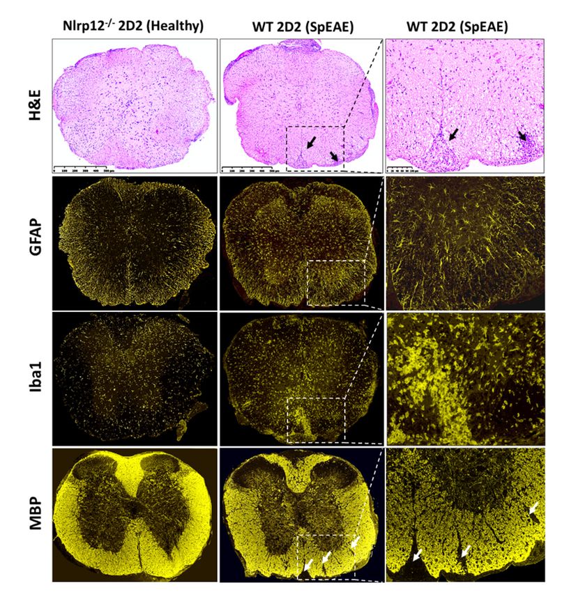
3.8. Nlrp12−/− 2D2 Mice Are Resistant to the Development of spEAE
Given the anti-inflammatory role of Nlrp12 in EAE [19,20], we investigated whether Nlrp12−/−
2D2 mice would develop spEAE. As expected, 6% of WT 2D2 mice developed EAE spontaneously
(Table 1). However, surprisingly, none of Nlrp12−/− 2D2 mice developed spEAE (Table 1). Pathological
examination of WT 2D2 spEAE mice revealed marked inflammation associated with infiltration of
mononuclear cells to the spinal cord and increased expression of microglia and astrocyte markers (Iba1
and GFAP respectively) (Figure 8). The inflammation was associated with demyelination, as shown
in by immunofluorescence staining of myelin basic protein (MBP) (Figure 8). Using flow cytometry,
we found a similar percentage of myelin-specific T cells (Vβ11+ ) in the spleens of Nlrp12−/− 2D2 mice
and WT 2D2 mice (Figure 9). However, a high percentage of leukocytes (CD45high ) including Vβ11+ T
Cells 2018, 7, x FOR PEER REVIEW 12 of 19
cells infiltrated to the spinal cord of WT 2D2 spEAE compared to healthy mice (Figure 9).
Figure 8. SpEAE in WT 2D2 mice is associated with spinal cord inflammation and demyelination.
Figure 8. SpEAE in WT 2D2 mice is associated with spinal cord inflammation and demyelination.
Representative histopathological examination of the spinal cords from healthy Nlrp12−/− 2D2 mice and
Representative histopathological examination of the spinal cords from healthy Nlrp12−/− 2D2 mice and
spEAE WT 2D2 mice. H&E staining reveals an extensive mononuclear cell infiltration to the spinal
spEAE WT 2D2 mice. H&E staining reveals an extensive mononuclear cell infiltration to the spinal cord
cord of spEAE mice (shown by black arrows). Immunofluorescent staining of spinal cords from WT
of spEAE mice (shown by black arrows). Immunofluorescent staining of spinal cords from WT 2D2
2D2 spEAE shows an increased expression of GFAP (astrocyte marker) and Iba1
spEAE shows an increased expression of GFAP (astrocyte marker) and Iba1 (macrophage/microglia
(macrophage/microglia marker) mice compared to Nlrp12−/− 2D2 healthy mice. Focal demyelinating
marker) mice compared to Nlrp12−/− 2D2 healthy mice. Focal demyelinating lesions (shown by white
lesions are
arrows) (shown by white
observed arrows)
in spEAE arecords
spinal observed
usinginMBP
spEAE spinalSimilar
staining. cords histopathological
using MBP staining. Similar
features to
histopathological
healthy Nlrp12 − / − features to healthy Nlrp12 −/− 2D2 mice were found in healthy WT 2D2 mice (images
2D2 mice were found in healthy WT 2D2 mice (images not shown).
not shown).Cells 2018, 7, 119 12 of 19
Cells 2018, 7, x FOR PEER REVIEW 13 of 19
Myelin-specificTTcells
Figure9.9.Myelin-specific
Figure cellsinfiltrate
infiltrateto
tothe
thespinal
spinalcord
cordof
ofspEAE
spEAEmice.
mice.(A)
(A)The
Thepercentage
percentageofofTT
cells expressing myelin-specific transgenic TCR (Vβ11 + ) were comparable in the spleen of healthy and
cells expressing myelin-specific transgenic TCR (Vβ11 ) were comparable in the spleen of healthy and
+
spEAEmice,
mice,however,
however,thethe percentage + T cells were increased in the spinal cord of spEAE
spEAE percentage of of Vβ11
Vβ11 + T cells were increased in the spinal cord of spEAE mice
mice compared Nlrp12 −/− healthy mice (n = 6), * p < 0.05; (B) Representative flow
compared to WTtohealthy
WT healthy
and and
Nlrp12 −/− healthy mice (n = 6), * p < 0.05; (B) Representative flow
cytometryplots
plotsofofCD45
CD45 + Vβ11 + T cells in the spinal cord of Nlrp12 −/− 2D2 healthy and WT 2D2
cytometry + Vβ11 + T cells in the spinal cord of Nlrp12−/− 2D2 healthy and WT 2D2 spEAE
spEAE
mice. mice.
Table 1. Nlrp12 does not prevent the development of spEAE. None of Nlrp12 −/−
2D2 mice developed
4. Discussion
spEAE, while 6 WT 2D2 mice out of 101 developed spEAE, manifested with ascending paralysis.
Early
Nlrp12reports
−/− 2D2showed themonitored
mice were expression
forand anti-inflammatory
16 weeks functionEAE
and no sign of classical of Nlrp12 in innate immune
was observed.
cells of myeloid origin such as DC and macrophages [18,26]. However, a very recent report by Lukens
et al. revealedGenotype
the expression Total of Nlrp12
(n) inSpEAE
T cells(%)[20]. The
Age current
of Onset study
(Weeks) aimedEAEto investigate
Score the
immunoregulatory WT 2D2function of Nlrp12
101 in T cell-mediated
6 immune response
10.1 ± 4.7 in EAE. Our
3.6 ± 0.5 results
suggest thatNlrp12
Nlrp12
−/−plays
2D2 pivotal30 role in Th1/Th2 0 balance by inhibiting- Th1 peripheral - responses in
the favor of Th2. We demonstrated that in - lymph nodes of Nlrp12 mice, Th1 to Th2 ratio is−/−
increased compared to WT mice. This shift, in part, can be explained by significant increases in the
4. Discussion
production of IFNγ by Nlrp12−/− T cells. Interestingly, Nlrp12 does not play a role in the differentiation
Early
of naïve reports
T cells but showed
upregulatesthe expression and anti-inflammatory
IL-2 production and proliferation function Nlrp12
of CD4+ Tofcells. Theineffect
innateofimmune
Nlrp12
cells of myeloid origin such as DC and macrophages [18,26]. However, a very
is associated with its increased expression and inhibition of major molecular pathways including recent report by Lukens
Akt
et al.
and revealed
NF-κB T cells. of Nlrp12 in T cells [20]. The current study aimed to investigate the
the expression
in activated
immunoregulatory
Previously, wefunction
published of Nlrp12 in T cell-mediated
that Nlrp12 −/− mice develop immune
more response
severe form in EAE. Ourthan
of EAE results
WTsuggest
mice,
that Nlrp12 plays pivotal role in Th1/Th2 balance by inhibiting Th1 peripheral
which is associated with exacerbated spinal cord inflammation and increased activation of microgliaresponses in the favor of
Th2. We demonstrated that into - lymph nodes of Nlrp12 −/− mice, Th1 to Th2 ratio is increased compared
in Nlrp12 −/− mice compared WT mice [19]. In the present study, we found an increased Th1
to WT mice. This shift, in part, can be explained
dominant response in lymph nodes of Nlrp12 EAE mice, suggesting
−/− by significant increases in Nlrp12
that the production
suppressesof IFNγ
Th1
− /
by Nlrp12in the − T cells. Interestingly, Nlrp12we does
activation periphery. Interestingly, didnotnotplay
find aany
rolechange
in the differentiation
in the levels of of naïve
IFNγ andT IL-4
cells
butthe
upregulates + T cells. The effect of Nlrp12 is associated
in CNS fromIL-2 production
Nlrp12 −/− EAE miceand proliferation
compared toofWT CD4EAE mice. However, the IFNγ/IL-4 ratio
significantly decreased in the spinal cord of Nlrp12−/− EAE mice compared to WT EAE mice, which is
consistent with Lukens et al. observation of enhanced Th2 response and increased IL-4 production inCells 2018, 7, 119 13 of 19
with its increased expression and inhibition of major molecular pathways including Akt and NF-κB in
activated T cells.
Previously, we published that Nlrp12−/− mice develop more severe form of EAE than WT mice,
which is associated with exacerbated spinal cord inflammation and increased activation of microglia in
Nlrp12−/− mice compared to WT mice [19]. In the present study, we found an increased Th1 dominant
response in lymph nodes of Nlrp12−/− EAE mice, suggesting that Nlrp12 suppresses Th1 activation
in the periphery. Interestingly, we did not find any change in the levels of IFNγ and IL-4 in the CNS
from Nlrp12−/− EAE mice compared to WT EAE mice. However, the IFNγ/IL-4 ratio significantly
decreased in the spinal cord of Nlrp12−/− EAE mice compared to WT EAE mice, which is consistent
with Lukens et al. observation of enhanced Th2 response and increased IL-4 production in the CNS
of Nlrp12−/− EAE mice [20]. In contrast to Lukens’ study where Nlrp12−/− mice developed atypical
EAE signs, we found severe classical EAE signs in Nlrp12−/− mice compared to WT mice [19]. Several
plausible explanations of these discrepancies were proposed in our recent review [9]. One possibility
might be related to the difference in MOG-adjuvant immunization protocols between various labs.
In our report, WT and Nlrp12−/− animals were immunized with a total dose of 200 µg MOG [19],
which is two-fold higher than the immunization dose used by Lukens et al. [20]. Interestingly, a recent
study showed that immunization with low or high MOG concentration can modify the patterns of
inflammatory cytokines [27]. Their study demonstrates that anti-inflammatory cytokines such as IL-10
and TGFβ significantly increase in the CNS of EAE animal immunized with 100 µg MOG compared to
300 µg MOG immunization. Therefore, it is possible that the severe EAE signs in Nlrp12−/− mice in
our study were driven by lower levels of IL-10 and TGFβ anti-inflammatory cytokines or by higher
levels of other inflammatory cytokines such as Granulocyte-macrophage colony-stimulating factor
(GM-CSF) in the CNS [28,29]. Another possible explanation for observing different EAE profiles is the
difference in the environmental conditions and different knockout strategies. It was shown that some
C57BL/6 colonies have acquired a missense mutation in the Nlrp12 gene that can affect neutrophil
responses [30]. In another study, genetic ablation of Nlrp12 was found to cause significant changes in
microbiota [17]. Nevertheless, these variabilities highlight the complex immunoregulatory nature of
Nlrp12 that warrants further investigation.
Given the important role of T cells in the pathogenesis of EAE, we further investigated whether
Nlrp12 controls T cell proliferation and activation in a T cell-intrinsic manner. We found a significant
increase of IFNγ and IL-4 levels in the supernatant of Nlrp12−/− compared to WT T cells. However,
when we measured the levels of intracellular cytokines by flow cytometry, higher percentage of
CD4+ IFNγ+ T cells were found in Nlrp12−/− group, while the percentages of CD4+ IL-4+ T cells
or CD4+ IL-17+ T cells did not change between both groups. We addressed this discrepancy with
Ki67 staining and our flow cytometry results revealed that activated Nlrp12-/- T cells proliferate
significantly more than WT T cells, which explains why we found increased production of both IFNγ
and IL-4 in the supernatant of activated Nlrp12−/− T cells. Consistent with our findings, Lukens et al.,
observed the higher expression of activation markers, enhanced proliferation and elevated secretion
of Th1/Th2/Th17 cytokines by Nlrp12−/− T cells compared to WT T cells in vitro [20]. Collectively,
these results show that Nlrp12 inhibits the activation of inflammatory T cell subsets including Th1
and Th17.
In the presence of polarizing cytokines, we found no difference between Nlrp12−/− or WT T cells
in the expression of Th1- or Th17- associated molecules, suggesting that Nlrp12 does not affect the
differentiation of naïve T cells to Th1 or Th17. In a recent study by Cai et al., purified CD4+ T cells from
Nlrp12−/− or WT mice were activated with anti-CD3 and polarizing cytokines for 6 days. They showed
that the differentiation of Nlrp12−/− T cells to Th1 or Th17 cells were significantly lower than WT T
cells. However, no difference was found between WT and Nlrp12−/− T cells in Th2 differentiation [31].
Taken together, it appears that Nlrp12−/− T cells respond differently to the environmental stimuli,
depending on the type of activating signals, incubation period and polarizing conditions.Cells 2018, 7, 119 14 of 19
Cells 2018, 7, x FOR PEER REVIEW 15 of 19
Our results, in agreement with Lukens’ study [20], showed that Nlrp12 inhibits T cell activation
and
(Figure 10). TheseWe
proliferation. found
results arethat
in the expression
agreement Nlrp12 was
withofprevious significantly
studies that show increased in T cells
Nlrp12 suppresses
following
canonical activation by anti-CD3
and non-canonical NF-κBorpathways
anti-CD3/CD28 antibodies. The increased level of Nlrp12
[16,20,32,33].
expression
The NF-κB signaling cascade interacts with severalTparallel
remained high even after 48 h stimulation of cells, suggesting
pathways that Nlrp12the
including modulates
signaling
Tcascades
cell signaling pathways upon TCR stimulation.
initiated by phosphatidylinositol 3-kinase (PI3K) and Akt. We found a significant increase
Since multiple
of Akt(Ser signaling pathways
473) phosphorylation in Nlrp12 are−/−involved in T cell activation
T cells compared and proliferation,
to WT T cells. These results we are
hypothesized that signaling pathways were inhibited by Nlrp12
supported by a study that demonstrates Nlrp12 negatively regulates Akt signaling in activated T cells.
pathwayIt isin
well-established 2+ /calmodulin-dependent phosphatase),
affected tumor that TCRinactivation
tissues triggers
colitis model [16].calcineurin (a CaAkt
Interestingly, acts upstream of NF-κB, where the
MAPK and of
activation NF-κB signaling
PI3K/Akt pathways,
signaling pathwayleading to the
leads activation of the nuclear
to phosphorylation factor of activated
and degradation T cell
of IκB protein,
(NFAT),
resulting in nuclear translocation and transcriptional activation of NF-κB [34]. Taken together, we
AP1 and NF-κB transcription factors that initiate IL-2 transcription [22]. In this regard, our
evaluated the phosphorylation of p65 (NF-κB subunit) and Akt in activated CD4 + T cells. Western blot
results demonstrate that Nlrp12 inhibits Akt signaling pathway, which, subsequently, affects
results demonstrated a significant −/− T cells
Nlrp12the
downstream pathways includingincrease in the phosphorylation
NF-κB pathway. of p65 in activated
Moreover, phosphorylated Akt blocks activity
compared to WT T cells,
of two G1-checkpoint highlighting
inhibitors, p21 and thep27,
inhibitory
and promotes Nlrp12
effect ofcell cycleon NF-κB signaling
progression pathway
[35]. Collectively,
(Figure 10). These
our results results
suggest that are in agreement
Nlrp12 controls Twith cell previous studies
proliferation andthat show Nlrp12
cytokine suppresses
production canonical
by inhibiting the
and non-canonical NF-κB pathways [16,20,32,33].
activation of Akt, that, in turn, affects several downstream effectors (Figure 10).
Figure10.
Figure 10. Possible
Possible mechanism
mechanismofofaction actionofofNlrp12
Nlrp12in regulating
in regulatingearly TCRTCR
early signaling pathways,
signaling T cell
pathways,
Tactivation and proliferation.
cell activation Activation
and proliferation. signals signals
Activation from TCR activates
from multiplemultiple
TCR activates downstream signaling
downstream
pathwayspathways
signaling in T cells,inpresented as (1) NFAT,
T cells, presented as (1) (2) NF-κB,
NFAT, (3) MAPK
(2) NF-κB, and (4)and
(3) MAPK mTOR pathways.
(4) mTOR These
pathways.
pathways
These lead to
pathways nuclear
lead translocation
to nuclear of NFAT,
translocation NF-κB
of NFAT, and and
NF-κB AP1AP1transcription factors
transcription and and
factors IL-2
transcription.
IL-2 IL-2IL-2
transcription. binds to to
binds its its
receptor
receptor ononthe
thecell
cell surface
surface and mTOR pathway.
and initiates mTOR pathway.TCR TCR
activating
activatingsignals
signalsinduce
inducethe expressionofofNlrp12
theexpression Nlrp12that
thatconsequently
consequentlyinhibits
inhibitsNF-κB
NF-κBandandMAPKMAPK
signaling
signalingpathways,
pathways,which
whichsuppress
suppressIL-2 IL-2production.
production.Upstream
Upstreamof ofNF-κB pathway,Nlrp12
NF-κBpathway, Nlrp12inhibits
inhibits
Akt
Aktphosphorylation
phosphorylationthat thatcontrols
controlsNF-κBNF-κBsignaling
signalingon ononeoneside
sideand
andmTOR
mTORactivity
activityand
andcell
cellcycle
cycle
progression
progressionon onthe
theother
otherside.
side.Taken together,Nlrp12
Takentogether, Nlrp12has hasaabroad
broadrange
rangeofofregulatory
regulatoryactivity
activitythat
that
controls hyper-proliferation and activation of T cells. The pathways shown in dashed
controls hyper-proliferation and activation of T cells. The pathways shown in dashed lines are not lines are not
affected
affectedbybyNlrp12
Nlrp12including
includingNFATNFATsignaling
signalingandandmTOR/S6
mTOR/S6phosphorylation.
phosphorylation.
OneNF-κB
The of the signaling
signaling cascade
pathways downstream
interacts of Akt parallel
with several phosphorylation
pathwaysisincluding
mTOR pathway, which
the signaling
induces protein synthesis and cell growth by regulating ribosomal p70S6 kinase 1 (S6K1)
cascades initiated by phosphatidylinositol 3-kinase (PI3K) and Akt. We found a significant increase and
ofeukaryotic translation
Akt(Ser 473) factor 4E-binding
phosphorylation protein
in Nlrp12 −/− T 1cells
(4EBP1). S6K1 phosphorylates
compared to WT T cells. and activates
These results S6,
area
supported by a study that demonstrates Nlrp12 negatively regulates Akt signaling pathwayand
ribosomal subunit involved in initiating protein synthesis machinery. Using western blotting in
flow cytometry, we found no difference between Nlrp12−/− T cells and WT T cells in the level of S6
phosphorylation. Therefore, it is possible that Nlrp12 regulates protein synthesis and cell growth viaCells 2018, 7, 119 15 of 19
affected tumor tissues in colitis model [16]. Interestingly, Akt acts upstream of NF-κB, where
the activation of PI3K/Akt signaling pathway leads to phosphorylation and degradation of IκB
protein, resulting in nuclear translocation and transcriptional activation of NF-κB [34]. Taken together,
our results demonstrate that Nlrp12 inhibits Akt signaling pathway, which, subsequently, affects
downstream pathways including NF-κB pathway. Moreover, phosphorylated Akt blocks the activity
of two G1-checkpoint inhibitors, p21 and p27, and promotes cell cycle progression [35]. Collectively,
our results suggest that Nlrp12 controls T cell proliferation and cytokine production by inhibiting the
activation of Akt, that, in turn, affects several downstream effectors (Figure 10).
One of the signaling pathways downstream of Akt phosphorylation is mTOR pathway,
which induces protein synthesis and cell growth by regulating ribosomal p70S6 kinase 1 (S6K1)
and eukaryotic translation factor 4E-binding protein 1 (4EBP1). S6K1 phosphorylates and activates
S6, a ribosomal subunit involved in initiating protein synthesis machinery. Using western blotting
and flow cytometry, we found no difference between Nlrp12−/− T cells and WT T cells in the level of
S6 phosphorylation. Therefore, it is possible that Nlrp12 regulates protein synthesis and cell growth
via modulating the activity of 4EBP1 (Figure 10). Further investigation is warranted to uncover the
regulatory mechanism of Nlrp12 on mTOR signaling pathway.
The results of this study and previous publications suggest that Nlrp12 inhibits NF-κB and MAPK
signaling pathways in activated T cells [20,36]. The transcription of IL-2 gene is regulated by NF-κB,
MAPK and Ca2+ /calmodulin-dependent pathways [37,38]. Accordingly, we hypothesized that Nlrp12
inhibits IL-2 production by activated CD4+ T cells. Flow cytometry data revealed a higher percentage
of CD4+ IL-2+ T cells in activated Nlrp12−/− CD4+ cells compared to WT CD4+ T cells, suggesting
that Nlrp12 suppresses IL-2 production by activated T cells. Incubation with cyclosporin inhibited
IL-2 production by both Nlrp12−/− and WT T cells, indicating that lack of Nlrp12 does not affect
Ca2+ /calmodulin signaling pathway in T cells.
Our in vitro findings, together with results obtained from in vivo EAE model, support the idea
that Nlrp12 inhibits T cell responses. In 2D2 mice, due to the presence of many MOG-reactive T
cells, about 4–14% of mice developed spEAE [39,40]. In 2D2 mice, the percentage of Vβ11+ T cells
in the spleen were the same between healthy and spEAE animals, showing that our affected and
non-affected animals had similar percentage of MOG-reactive T cells. However, a high percentage of
MOG-reactive T cells infiltrated to the spinal cord of WT 2D2 spEAE mice, which confirms the presence
of autoreactive T cells in inflamed spinal cord. The healthy Nlrp12−/− 2D2 animals have only a few
Vβ11+ T cells in the spinal cord and do not contain pathology. The fact that none of Nlrp12−/− 2D2 mice
develop disease suggests that Nlrp12 does not inhibit the development of spEAE and even can serve
as contributing factor in the pathology of EAE. Due to slow rate of breeding of Nlrp12−/− 2D2 mice
(unpublished observation), these conclusions are based on the observation of thirty Nlrp12−/− 2D2 mice.
Interestingly, previous studies report that in induced EAE, Nlrp12 can prevent [19] or promote [20] CNS
inflammation. To explain the observed controversy, we propose a dual immunoregulatory function for
Nlrp12, in which Nlrp12 can act as an inflammatory or anti-inflammatory molecule, depending on the
type and severity of immunological challenge. The hypothetical model of Nlrp12 immunoregulation is
shown in Figure 11.
The bifunctional nature of Nlrp12 has been previously reported in several
studies [16,19,20,31,36,41,42]. Early in vitro studies suggest that Nlrp12 is an inflammatory
NLR that interacts with ASC to form inflammasome [43]. Recent reports also support this idea
and show that Nlrp12 activates inflammasome in Yersinia Pestis and Plasmodium infections [42,44].
Moreover, several behavioral outcomes are similar between ASC−/− and Nlrp12−/− mice [45]. On the
other hand, there are studies that classify Nlrp12 as an anti-inflammatory molecule and the inhibitor
of NF-κB signaling pathway [16,18,32,46]. In this regard, Nlrp12−/− mice were shown to be highly
susceptible to inflammatory diseases of intestine such as experimental colitis and colon cancer [16,18].
Taken together, these findings support the dual immunoregulatory nature of Nlrp12, that may vary in
a cell-specific or stimulus-specific manner.You can also read