The Dutch Working Party on Antibiotic Policy recommendations for the diagnosis and management of COVID-19 associated pulmonary aspergillosis
←
→
Page content transcription
If your browser does not render page correctly, please read the page content below
The Dutch Working Party on Antibiotic Policy (SWAB)
recommendations for the diagnosis and management of COVID-19
associated pulmonary aspergillosis

This evidence-based document is a supplement to the SWAB guidelines for the Management of
Invasive Fungal Infections, revised version December 2017.

Committee:

N.M.A. Blijlevens,1 R.J.M. Brüggemann,2 J.J.W.M. Janssen,3 D.W. de Lange,4 J.F. Meis,5 A.M.L. Oude
Lashof,6 M.H.E. Reijers,7 B.J.A. Rijnders,8 J.A. Schouten,9 F.L. van de Veerdonk,10 T.S. van der Werf,11
T.F.W. Wolfs,12 A.R.H. van Zanten,13 and P.E. Verweij (chair).14 The committee was supported by J.B.
Buil14 for literature review and writing and editing of the draft documents.

1
 Department of Hematology, Radboud University Medical Center, Nijmegen; 2Department of Clinical
Pharmacy, Radboud University Medical Center, Nijmegen; 3Department of Hematology, Amsterdam
University Medical Center, location VUmc, Amsterdam; 4Department of Intensive Care, University
Medical Center Utrecht, Utrecht; 5Department of Medical Microbiology and Infectious Disease,
Canisius Wilhelmina Hospital, Nijmegen; 6Department of Infectious Diseases, Maastricht University
Medical Center, Maastricht; 7Department of Pulmonology, Radboud University Medical Center,
Nijmegen; 8Department of Medical Microbiology & Infectious Diseases, Erasmus Medical Center,
Rotterdam; 9Department of Intensive Care, Radboud University Medical Center, Nijmegen;
10
  Department of Internal Medicine, Radboud University Medical Center, Nijmegen; 11Department of
Internal Medicine, Division of Infectious Diseases, University Medical Center Groningen, Groningen;
12
  Wilhelmina Children's Hospital, University Medical Center Utrecht, Utrecht; 13Department of
Intensive Care Medicine, Gelderse Vallei Hospital, Ede; 14Department of Medical Microbiology,
Radboud University Medical Center, Nijmegen.

©2021 SWAB
www.swab.nl
                                                                                                      1
Contents
Introduction and methodology ............................................................................................................... 3
Key questions .......................................................................................................................................... 6
1. What is the case definition of COVID-19 associated pulmonary aspergillosis? .............................. 6
2. What is the optimal approach towards diagnosing or refuting CAPA in patients with COVID-19? .... 7
3. What is the reported incidence of Aspergillus pneumonia in patients with COVID-19? .................. 10
4. What are the host- / risk factors that are associated with COVID-19 associated pulmonary
aspergillosis (CAPA)? ............................................................................................................................. 12
5. What is the optimal antifungal choice for patients with proven or high likelihood of COVID-19 and
suspected Aspergillus pneumonia? ....................................................................................................... 14
6. How should invasive Aspergillus tracheobronchitis be managed in CAPA patients? ................... 16
7. What is the role of immunomodulating agents in the management of CAPA in ICU patients? ... 17
Final considerations............................................................................................................................... 18
Recommendations ................................................................................................................................ 20
Flow chart .............................................................................................................................................. 21
Practical guidance for antifungal drug administration and drug target concentrations ...................... 23
References ............................................................................................................................................. 24
2Introduction and methodology
General introduction
Soon after the start of the COVID-19 pandemic, reports of suspected invasive pulmonary aspergillosis
(IPA) complicating COVID-19 appeared. Although initial reports from China were not specific regarding
the frequency and identification of fungal pathogens causing secondary infection in COVID-19 patients,
reports from Europe indicated that Aspergillus was frequently cultured in airway samples from COVID-
19 patients admitted to the ICU. IPA secondary to influenza was recognized as a clinical entity over the
last years in patients admitted to the intensive care unit (ICU) with respiratory failure. Cases of
influenza associated pulmonary aspergillosis (IAPA) were observed in 19% of influenza patients in the
ICU in the Netherlands and Belgium.1 The mortality rate was 51% in a retrospective multicenter cohort
study compared to 28% in influenza patients without IAPA.1 Characteristics of IAPA are listed in Table
1.
Table 1. Characteristics of influenza associated pulmonary aspergillosis (IAPA).
Characteristic Ref.
1-3
IAPA occurs in 16% - 23% of patients with influenza in the ICU
1
IAPA develops in 32% of influenza patients with EORTC/MSGERC host factor compared to
14% in EORTC/MSGERC negative influenza patients in the ICU.
1-4
Between 30% and 78% of IAPA patients have no underlying EORTC/MSGERC host factor
1-3,5
IAPA occurs within 48 hrs of ICU admission in the majority of patients (early IAPA, co-
infection) but is less frequently observed at a later point as well (late IAPA, secondary
infection)
3,4
Between 30% and 56% of IAPA patients present with invasive Aspergillus tracheobronchitis
1
IAPA occurs in patients with influenza A as well as influenza B infection
1
Influenza is an independent risk factor for IAPA
1,3
Use of corticosteroids before ICU admission was independently associated with IAPA.
Other risk factors include higher APACHE II score and male sex
1-4
Serum galactomannan is positive in 57% to 78% of IAPA patients
4
Serum Beta-D-glucan is positive in 82% of IAPA patients
1-3
BAL galactomannan is positive in 88% - 100% of IAPA patients
1-4
BAL Aspergillus culture is positive in 63%-89% of IAPA patients and 100% of patients with
invasive tracheobronchitis
2
Azole resistance has been reported in IAPA with a frequency up to 29%
1-4
ICU mortality of IAPA is 51%-61% compared to 28% in influenza patients without IAPA
4
ICU mortality was 90% in influenza patients with invasive Aspergillus tracheobronchitis
compared to 44% in influenza patients with other forms of IAPA.
3Based on the emerging risk of IAPA and the high frequency in the Netherlands, a recommendation was
given in the SWAB guidelines for the Management of Invasive Fungal Infections (revised version
December 2017):6
Recommendation 17 ICU patients with confirmed influenza should undergo sampling for serum
galactomannan. It is recommended that ICU patients with confirmed influenza and
radiologic abnormalities on chest X-ray should undergo bronchoscopy and BAL for
galactomannan and culture.
In case of tracheobronchitis, a positive serum galactomannan or a positive BAL
galactomannan (index ≥0.8), patients should be treated with combination
azole+echinocandin or azole+L-AmB therapy. Monotherapy with L-AmB is considered
as a second choice in these patients.
If cultures reveal no Aspergillus growth, galactomannan-positive BAL material should
be tested by PCR for the presence of Cyp51 mutations.
ICU patients with influenza and negative aspergillus serum and bronchoscopy/BAL
screening, and non-ICU influenza patients should undergo (repeat) serum and
bronchoscopy/BAL diagnostics if new respiratory complications or clinical worsening
occur, or if sputum/tracheal Aspergillus colonization cultures are positive.
The increasing number of reports on Aspergillus superinfections in critically ill COVID-19 patients as
well as the frequent detection of Aspergillus species or galactomannan (GM) in airway samples from
critically ill COVID-19 patients, resulted in uncertainty about its clinical relevance as well as the best
diagnostic and treatment strategies of these patients. Below we will use the term COVID-19 associated
pulmonary aspergillosis (CAPA) when we refer to patients considered to have a tissue invasive infection
with Aspergillus.
The current evidence was reviewed based on the following seven key questions:
Table 2. Key questions
1. What is the case definition of COVID-19 associated pulmonary aspergillosis?
2. What is the preferred approach towards diagnosing or refuting CAPA in patients with COVID-
19?
3. What is the risk of Aspergillus pneumonia in patients with COVID-19?
4. Are there host/risk factors that are associated with CAPA?
5. What is the treatment of choice for patients with CAPA?
6. How should invasive Aspergillus tracheobronchitis be managed in CAPA patients?
7. What is the role of immunomodulating agents in the management of CAPA in ICU patients?
Conflicts of interest policy and funding
The Guidelines Committee would like to thank all individuals and societies who contributed to the
development of these guidelines. Members of the preparatory committee reported the following
potential conflicts of interest: NMAB…..; RJMB has received unrestricted grants, speaker fees or
consultancy fees from Amplyx, Astellas, F2G, Gilead, Munidipharma, MSD and Pfizer. All contracts were
with Radboudumc and all invoices were paid to Radboudumc; JJWMJ has no conflicts of interest; DWdL
has no conflicts of interest; JFM received grants or speaker fees from F2G, Gilead and United Medical;
AMLOL no conflicts of interest; MHER has no conflicts of interest; BJAR has received research grants
4from Gilead Sciences, outside the submitted work; JAS has no conflicts of interest; FLvdV has no
conflicts of interest; TSvdW has no conflicts of interest; TFWW has no conflicts of interest; ARHvZ has
no conflicts of interest; PEV has received unrestricted grants, speaker fees or consultancy fees from
MSD, Pfizer, F2G, Gilead Sciences, Munidipharma and Thermofisher. All contracts were with
Radboudumc and all invoices were paid to Radboudumc.
Methodology
The current addendum was based on seven key questions considering population, intervention,
comparison, and outcomes (PICO) relevant for the Dutch clinical setting (Table 2). For each key
question we developed short evidence summaries after searching PubMed and other sources
considered relevant. The evidence was subsequently assessed using the Grading of Recommendations
Assessment, Development, and Evaluation (GRADE) system as described in the SWAB sepsis guideline
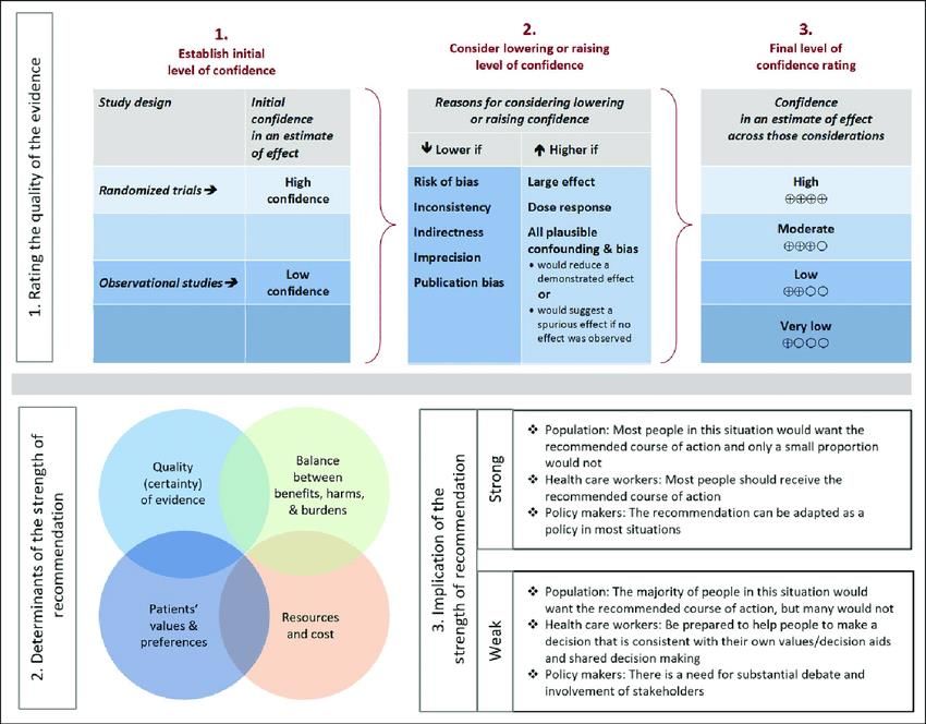
(Figure 1).7,8
Figure 1. Overview of GRADE methodology. Approach and implications to rating the quality of evidence
and strength of recommendations using the Grading of Recommendations Assessment, Development
and Evaluation (GRADE) methodology.
The PubMed search strategy included ((coronavirus and 2020) or COVID-19 or SARS-CoV-2) and
(Aspergillus* or aspergillosis* or CAPA) and included publications until December 15th 2020. When
available, in-press publications not yet available on PubMed were included as well. Case-reports were
excluded. In addition, unpublished cohort data were used from a multicenter CAPA registry study
5conducted in Belgium and the Netherlands. Quality of evidence for clinically relevant outcomes was
graded from high to very low. A multidisciplinary committee formulated recommendations after
structured discussions as strong or weak. The committee anticipated on limited high-quality evidence
due to the recent emergence of SARS-CoV-2. When evidence could not be obtained, recommendations
were provided on the basis of opinions and experiences with other viral pneumonias, notably influenza
(good practice statements, GPS). Based on this process, we formulated 13 recommendations on the
management of patients with a proven COVID-19 and Aspergillus colonization or a suspected or proven
CAPA (see recommendations below).
Key questions
1. What is the case definition of COVID-19 associated pulmonary
aspergillosis?
Evidence summary
A rapidly increasing number of papers on CAPA in COVID-19 patients admitted to the ICU are being
reported in the literature. One problem is the lack of a consensus CAPA case definition, and various
definitions have been used to classify CAPA. The invasive fungal infection case definition of the
European Organization for Research and Treatment of Cancer (EORTC) / Mycosis Study Group
Education and Research Consortium (MSGERC) is rarely applicable because it only applies to patients
with a specific host factors, which are typically absent in patients suspected of having CAPA and due
to the fact that the ICU-setting was excluded in the definition document.9 Several studies have used
the algorithm that was proposed by Blot et al. to distinguish between IPA and Aspergillus colonization
in the ICU.10 As the classification is based on a positive culture, sometimes revised definitions were
used which include the biomarker GM to classify patients in addition to those with a positive culture.
Other papers have used the criteria which were used by Schauwvlieghe et al.1 to classify patients with
IAPA. Recently, an expert group proposed a case definition to classify patients with IAPA. This case
definition has also been used to classify patients with CAPA.11 Finally, a consensus CAPA case definition
was published by the European Confederation for Medical Mycology (ECMM) and the International
Society for Human and Animal Mycology (ISHAM),12 categorizing patients as proven, probable and
possible CAPA. This document has only been published very recently and has not yet been used to
classify CAPA patients. Furthermore, as described below, as long as the literature regarding autopsy
and biopsy proven cases of CAPA remains very limited, the sensitivity and specificity of these
definitions remains uncertain. As proven IPA relies on the demonstration of invasive growth of
Aspergillus hyphae, there is little variation between definitions regarding this category. Variation in
the probable category is notably relating to host factors, clinical factors (including radiologic imaging)
and mycological criteria.12
Conclusions
A consensus CAPA case definition has been published which aimed to improve standardization and
comparability of clinical studies. Due to the recent publication of the case definition and lack of proven
CAPA cases, the sensitivity and specificity of these consensus definitions remains unclear.
62. What is the optimal approach towards diagnosing or refuting CAPA in patients
with COVID-19?
Evidence summary
Mycology
Demonstration of tissue invasive growth of septate hyphae and identification of Aspergillus through
PCR or culture is considered the gold standard of proven IPA. This would require obtaining tissue
through an invasive procedure or at autopsy. However, particularly during the first months of the
pandemic, invasive diagnostic procedures have been discouraged due to the assumed risk of health
care associated infection via aerosolization. This may explain the limited number of biopsy or autopsy
proven CAPA cases (six) so far (Table 3). However, another possible explanation of the limited number
of proven CAPA cases, despite the frequent documentation of Aspergillus species by culture, PCR or
antigen testing in airway samples from critically ill COVID-19 patients may be that tissue invasion is
indeed less frequent .
Table 3. Reported proven CAPA cases.
Sex / Underlying condition Procedure Respiratory GM/ culture Highest Ref.
age serum GM
13
M, 38 Obesity, Biopsy during BAL: >2.8 / A. fumigatus 0.3
hypercholesterolemia bronchoscopy
13
M, 62 Diabetes Biopsy during BAL: 2 / A. fumigatus 0.2
bronchoscopy
13
M, 73 Obesity, diabetes, Biopsy during BAL: >2.8 / A. fumigatus 0.1
hypertension, bronchoscopy
hypercholesterolemia
13
M, 77 Diabetes, chronic Biopsy during BAL: 2.79 / A. fumigatus 0.1
kidney disease, bronchoscopy
hypertension,
pemphigus foliaceus
14
M, 71 Hypertension, Autopsy, molecular BAL: not performed 4.3
diabetes, chronic ID A. penicillioides
kidney disease
15
M, 73 Obesity, diabetes, Autopsy Bronchial aspirate: A. positive
hypertension, atrial fumigatus
fibrillation
As invasive aspergillosis commonly presents as pulmonary infection, bronchoscopy with bronchial
alveolar lavage (BAL) has become the most important tool to diagnose IPA. BAL samples have been
validated for microscopy (using optical brighteners such as Blankophor P or calcofluor white),
Aspergillus culture, detection of GM and Aspergillus DNA for species identification and detection of
azole resistance markers (AsperGenius). Detection of Aspergillus antigen may take place through ELISA
test (Platelia Aspergillus) or through lateral flow device (LFD) point-of-care tests that allow rapid
7detection of Aspergillus antigen (IMMY and OLM Diagnostics). Alternatively beta-D-glucan (BDG),
which is a panfungal marker, may be detected in serum of patients with IPA.
An important issue in performing diagnostic procedures in COVID-19 patients has been the risk of
aerosolization associated with bronchoscopy and BAL. Although bronchoscopy has generally been
discouraged in COVID-19 patients, evaluation for co-infection is considered an indication to perform
this procedure,16 provided that adequate preventive measures are taken to protect health care
workers.17 In addition, exclusion of co-infection is considered relevant before starting corticosteroid
treatment in COVID-19 patients with secondary clinical (respiratory) worsening that is attributed to
pulmonary fibrosis or to organizing (non-infectious) pneumonia (also called Cryptogenic Organizing
pneumonia, COP). Due to restricted availability of bronchoscopy, alternative specimens and
procedures have been used, including testing of sputum, bronchial (BA)/ tracheal aspirates (TA) and
non-bronchoscopic bronchial lavage (NBL).12 Important drawbacks of these specimens include
sampling of the upper respiratory tract rather than lower respiratory tract, lack of validation of
Aspergillus biomarkers for these specimens, and inability to visualize the airways, which is critical to
diagnose invasive Aspergillus tracheobronchitis. However, during the second wave availability of BAL
has increased and the procedure is now safely used in most centers. An overview of the performance
of Aspergillus diagnostics in reported case series is shown in Table 4.
Table 4. Overview of performance of diagnostic tests in CAPA
Country # of CAPA BAL (#positive / Aspergillus species TA/BA (#positive / Serum (#positive / Ref.
cases #performed) #performed) #performed)
France 9 Culture 5 / 7 A. fumigatus (7) Culture 2 / 2 GM 1 / 9 18
GM 2 / 7 GM - BDG 4 / 8
PCR 3 / 7 PCR 2/ 2
Germany 5 Culture 1 / 3 Culture 2 / 3 GM 2 / 5 19
A. fumigatus (4)
GM 3 / 3 GM ND BDG -
PCR 3 / 3 PCR 1 / 2
NL 6 Culture 2 / 3 A. fumigatus (5) Culture 3 / 3 GM 0 / 3 20
GM 3 / 3 GM - BDG -
PCR - PCR -
Belgium 6 Culture 5 / 6 A. fumigatus (5), A. Culture - GM 1 / 5 13
flavus (1)
GM 5 / 6 GM - BDG -
PCR - PCR -
Italy 30 Culture 19 / 30 A. fumigatus (16), A. Culture - GM 1 / 30 21
niger (3), A. flavus (1)
GM 30 / 30 GM - BDG -
PCR 20 /30 PCR -
UK 19 Only NBL performed; A. fumigatus (9), A. Denominator not Denominator not 22
Denominator not versicolor (1) reported reported
reported
8Belgium 4 Culture 4 / 4* Not specified GM - 23
GM 4 / 4 BDG -
PCR 2 /2
Switzerland 3 Culture - A. fumigatus (3) Culture 3 / 3 GM 1 / ? 24
GM - GM - BDG 1 / ?
PCR - PCR 1 / ?
France 19 Culture 7 / 9 A. fumigatus (14), A. Culture 9 / 10 GM 1 / 12 25
calidoustus (1), A. niger
GM 7 / 9 GM - BDG -
(1)
PCR - PCR -
Pakistan 5 Not specified A. fumigatus (1), A. Not specified GM 0 / 5 26
flavus (4), A. niger (1)
BDG 1 / 5
USA 4 Not specified A. fumigatus (4) Not specified GM 1 / 3 27
BDG -
France 7 Culture not specified / 5 A. fumigatus (5) Not specified GM 1 / 7 33
GM 3 / 5 BDG 2 / 7
PCR 2 / 5
*BAL and BA were not distinguished.
Overall, BAL GM was positive in 57 of 64 (89%) of CAPA patients, while BAL culture was positive in 43
of 59 (73%). It should be noted that positive GM was considered entry criterion in the study of
Bartoletti et al, which included 30 patients.21 Aspergillus PCR in BAL was positive in 30 of 47 (64%) CAPA
patients. Serum GM was positive in 7 of 71 (10%) patients.21 The performance of LFD tests has been
studied in ICU-patients,28 and one study compared the performance of the Sona Aspergillus
Galactomannan Lateral Flow Assay (IMMY) with that of the Platelia Aspergillus (Biorad) in TA obtained
from CAPA patients.29 However, as bronchoscopy was not performed in this study, a reliable
classification of CAPA patients was not achieved.
Imaging
The typical appearance of COVID-19 includes peripheral, bilateral, ground-glass opacities with or
without consolidation or visible intralobular lines (i.e. crazy paving) in early stages; multifocal ground
glass opacities of rounded morphology with or without consolidation or crazy paving at peak stage;
reverse halo sign as well as other findings of organizing pneumonia at late stages are observed as
well.30 Many signs of COVID-19 pneumonia can mimic CAPA, and vice versa, and lesions suggestive of
CAPA may be hidden. Radiological findings that were previously shown to be sufficiently specific to
diagnose IPA in immunocompromised patients are the halo sign, air-crescent sign, cavitating lung
lesions and well-defined intrapulmonary nodule(s). In ICU patients with influenza cavitating lung
lesions and well-described nodule(s) are also considered useful. Whether or not, any of these criteria
can help in distinguishing Aspergillus colonization from infection in COVID-19 patients is as yet
uncertain. Indeed, an intrinsic part of severe COVID-19 is intravascular thrombosis due to
9endotheliopathy, which can result in infarction and cavitating lesions as well as the halo sign.12
Therefore, the role of imaging as a reliable criterion for diagnosing CAPA is probably limited.
Importantly, CT may contribute to diagnose other reasons for respiratory deterioration. Nevertheless,
for critically ill COVID-19 patients new nodules with cavitation or halo sign or consolidations have been
recommended to trigger a diagnostic work-up for CAPA.31 Autopsy data of a sufficient number of
patients with these radiological findings present in the days preceding death are needed in order to
improve our understanding of the radiology of CAPA.
Conclusions
Very few biopsy or autopsy proven cases of CAPA have been reported. Therefore, definite
conclusions on the diagnostic characteristics of a single diagnostic test or combinations of tests
cannot be made (very low quality of evidence).
The performance of TA or NBL for diagnosing CAPA remains unclear. These specimens are not
validated for the use of GM, PCR and BDG detection and do not allow diagnosis of invasive
Aspergillus tracheobronchitis (very low quality of evidence).
BAL GM may be the most reliable test to diagnose CAPA but a positive BAL GM test should not
be considered definite proof of CAPA (low quality of evidence).
Serum GM was positive in very few patients (Belgium AspICU10 6 / 34 (21%) 8 (2 – 16) 6/6 4/-/2 13
Italy Modified IAPA11 30 / 108 (28%) 4 (2 – 8) days * (study used 30 / 30 0 / 30 / - 21
screening protocol)
AspICU10 19 / 108 (18%) 8 (0 – 35)† 19 / 19 0 / - / 19
UK AspICU10 8 / 135 (6%) 7/8 0/-/8 22
IAPA1 20 / 135 (15%) 15 / 20 0 / - / 20
Own definition 19 / 135 (14%) 14 / 19 0 / - / 19
Belgium Modified AspICU10 4 / 131 (3%) 4 4/4 0/-/4 23
Switzerland Modified IAPA11 3 / 80 (4%)¥ 6 (3-8) 3/3 0/1/2 24
France Modified AspICU10 19 / 106 (18%) 11 (2-23) 18 / 19 0 / - / 19 25
France EORTC/MSGERC (if 7 / 145 (5%) 10 (median) 27 / 27 0/0/7 33
immunocompromised)9
and IAPA2
*Includes cohort of Alanio et al.18
¥
80 mechanical ventilated patients of a total of 118 patients admitted to the ICU.
†
of 16 patients with multiple Aspergillus cultures positive.
∞
Time to positivity: Antinori et al. culture sample taken at day 4 positive.15 Ghelfenstein-Ferreira et al.
culture positive with A. fumigatus of sample taken at day 6 after ICU admission.34 Meijer et al.
recovered A. fumigatus from a tracheal aspirate culture at ICU admission.35 Mitaka et al. found that
the 6 patients were mechanically ventilated for a mean of 6.8 days (range 1-14 days) before Aspergillus
isolation. The two patients described by Helleberg had growth of A. fumigatus in respiratory samples
1 and 5 days after starting mechanical ventilation.36
Overall, 10 CAPA case series in the ICU reported 120 CAPA cases in 1,155 COVID-19 patients (10%,
range between 3% and 33%). Only in four cases CAPA was proven, while the majority had a probable
or putative diagnosis. One study from the Netherlands reported 6 CAPA cases in a cohort of 31 COVID-
19 patients admitted to the ICU, of which three cases could be classified as probable CAPA.16 Yet, a
subsequent report from the same hospital reported on the results of postmortem pathology findings
in these patients. The premortem diagnosis of CAPA could not be histologically confirmed. This
however, does not exclude CAPA because no autopsy was performed but rather blinded percutaneous
lung biopsies were evaluated.37 A recent, yet unpublished cohort of 520 COVID-19 patients admitted
to the ICU in centers in Belgium and the Netherlands during the first wave, showed that 41 patients
(8%) could be classified as proven or probable CAPA according to the new consensus definitions.38
The cohort studies show that the vast majority of ICU patients that were diagnosed with CAPA were
mechanically ventilated, although this may be explained by the fact that diagnostic procedures like
BAL are rarely performed in non-ventilated patients with COVID-19. Furthermore, the majority of
patients developed CAPA on average between day 4 and 11 after ICU admission. The study of Bartoletti
which involved systematic bronchoscopy on day 0 and 7 of ICU admission indicated that 14 of 108
(13%) patients were BAL GM positive (index >1) at ICU admission.21
11Conclusions
Observational studies on CAPA in COVID-19 patients reported frequencies between 3% and
33% in the ICU, using variable case definitions (very low quality of evidence).
CAPA is almost exclusively reported in mechanically ventilated patients and can be diagnosed
early as well as late after ICU admission (low quality of evidence).
4. What are the host- / risk factors that are associated with COVID-19 associated
pulmonary aspergillosis (CAPA)?
Evidence summary
Case series published to date show that only a minority of patients have traditional EORTC/MSGERC
host factors (Table 6). Three patients (2%) were reported with a hematological malignancy, two (1,5%)
with other malignancies and five (4%) with solid organ transplantation. One study identified the
presence of an EORTC/MSGERC host factor and solid organ transplantation as significant risk for
invasive fungal infection. 33
Table 6. Underlying diseases and identified risk factors for CAPA in published case series.
Country # of CAPA EORTC/MSGERC host Other underlying diseases Identified risk factors Ref.
cases factor (# pts)
France 9 Myeloma + steroids Hypertension, obesity, N.A. 18
(1), steroids (1) diabetes, ischemic heart
disease, asthma
France* 21 Solid organ transplant Hypertension, diabetes, Cumulative corticosteroid 32
(1), myeloma (1) obesity, coronary disease, dose ≥100 mg higher in CAPA
asthma (OR, 3.7; IC95% 1.0 -9.7)
Germany 5 Not present Hypertension, obesity, N.A. 19
hypercholesterolemia,
diabetes, COPD, emphysema
NL 6 Not present COPD, asthma, cardiomyopathy N.A. 20
Belgium 6 AML (1) Hypertension, obesity, N.A. 13
hypercholesterolemia,
diabetes, HIV
Italy 30 Malignancies (2), solid Hypertension, obesity, Chronic steroid therapy was 21
significantly more frequent in
organ transplant, hypercholesterolemia,
CAPA compared to non-CAPA
chronic steroid diabetes, coronary disease, (p=0.02)
treatment cerebrovascular disease,
chronic kidney disease, COPD
12UK 19 Hematological Vasculitis, essential Association between multiple 22
positive Aspergillus/BDG
malignancy (1), hydrocortisone
positive test and the use of
corticosteroid therapy thrombocytopenia, diabetes, high dose corticosteroids
(p=0.007) and chronic
(15) – dose and chronic respiratory illness, solid
respiratory condition (p=0.05)
duration not specified cancer, autoimmune disease,
obesity, hypertension,
Alzheimer disease, chronic
kidney disease,
Belgium 4 Not present Obesity, chronic kidney disease N.A. 23
Switzerland 3 Not present Hypertension, obesity, N.A. 24
diabetes, pulmonary fibrosis,
asthma
France 19 Not present Hypertension, diabetes, N.A. 25
malignancy, COPD, asthma,
tuberculosis, cardiopathy,
ABPA, schizophrenia, glaucoma,
HIV
France 7 Kidney Hypertension, obesity, Preexisting host factor 33
(p=0.03), solid organ
transplantation (2), diabetes, tabagism,
transplant (p=0.004), long
liver transplantation dyslipidemia term (>3weeks) corticosteroid
therapy (any dose)(p=0.01)
(1), steroids (1)
*Includes cohort of Alanio et al.18
Three cohort studies have identified risk factors for CAPA. Chronic steroid treatment (at dosages higher
than or equivalent to prednisone 16 mg/day for at least 15 days) was found to be significantly more
frequent in patients with CAPA compared to those without CAPA.21 The use of high dose
corticosteroids (dose not defined) and the presence of chronic lung disease were associated with
multiple positive Aspergillus tests.22 A third cohort found that corticosteroids administered at any dose
for > 3 weeks was a risk factor for invasive fungal infection.33 A forth study did not find that high dose
corticosteroids was associated with CAPA risk (11.5% versus 28.6%; p=0.08), but observed cumulative
dose ≥100 mg to be higher among CAPA patients.32
All but few of the reports on CAPA come from a setting where corticosteroid therapy was not yet the
standard of care but rather the exception. Since the publication of the Recovery trial, corticosteroid
therapy has become the standard of care for all patients admitted with severe COVID-19. 39 Therefore,
the data regarding the impact as well as the magnitude of the impact of corticosteroid use on the
incidence of CAPA should be considered preliminary. In particular, the question remains if a certain
cumulative dose is required and to what extent a 10 day regimen of dexamethasone that was used in
the recovery trial and has become the standard of care in the Netherlands poses a significant risk for
CAPA.
13Conclusions
Most CAPA patients lack EORTC/MSGERC host factors (low quality of evidence).
Multiple cohort studies show that corticosteroid therapy is associated with increased risk for
CAPA (low quality of evidence).
Host factors that have been implicated to increase the risk for CAPA include, any
EORTC/MSGERC host factor, solid organ transplant or chronic respiratory disease (very low
quality evidence).
5. What is the optimal antifungal choice for patients with proven or high
likelihood of COVID-19 and suspected Aspergillus pneumonia?
Evidence summary
Despite the difficulty in distinguishing between Aspergillus colonization and invasive infection, several
studies have shown excess mortality in Aspergillus positive COVID-19 patients in the ICU (Table 7). In
the study of Bartoletti et al, of the 30 CAPA patients, 16 received antifungal therapy of which 13
received voriconazole.21 Fourteen patients did not receive antifungal therapy due to post mortem
diagnosis (7 patients) or due to clinical decision (7 patients). Survival of patients treated with
voriconazole was 54% (7 of 13), and for those not receiving voriconazole 41% (7 of 17)(p=.39).21 A
relationship between initial BAL GM index and 30-day survival was noted. The odds of death within 30
days of ICU admission increased 1.41-fold (1.10–1.81; P = .007) for each point increase in the initial
BAL GM index. When adjusted for age, need for renal replacement therapy, and SOFA score at ICU
admission, the initial BAL GM index was still independently associated with increased odds of death
within 30 days of ICU admission (OR, 1.44; 95% CI, 1.08–1.94; P = .014).21 In the study of White et al.,
mortality rates ranged from 46.7% (95% CI, 24.8–69.9) in CAPA patients receiving appropriate
antifungal therapy to 100% (95% CI, 51.1–100) in patients not receiving appropriate antifungal
therapy.22 In the cohort from Belgium and the Netherlands, 20 of 41 (48.8%) CAPA patients died
compared to 135 of 476 (28.2%) of COVID-19 without CAPA (pUK 19 58% (day 77?) 38% 22 NL CAPA12 41 48.8% 28.2% (p
Pharmacokinetic drug-drug interactions will play an important role. Triazole drugs are mostly
perpetrator drugs but can be victim drugs as well. In the Netherlands drug-drug interactions are
monitored by electronic prescribing systems with direct feedback to the clinician. Nevertheless, these
systems might not identify drug interactions with new or experimental drugs. A highly recommended
and reliable source for drug interactions can be found at https://www.covid19-druginteractions.org/.
This data source setup by the University of Liverpool together with the Radboudumc provides the latest
insights into relevant drug interactions including the azole drugs.
Next to pharmacokinetic interactions, pharmacodynamic interactions (drug with similar toxicity
profiles) may be relevant. These interactions include interactions between (lipid) formulations of
amphotericin B and nephrotoxic drugs, potassium losing agents and posaconazole, and many more.
Please consult your hospital pharmacist for management of these interactions.
(See Practical guidance for antifungal drug administration and drug target concentrations)
Conclusions
There is currently not enough evidence to draw any definitive conclusion on the optimal
antifungal treatment strategy for patients with proven or high likelihood of COVID-19 and
suspected Aspergillus co-infection.
For antifungal therapy of patients with CAPA the SWAB 2017 recommendations for treatment
of invasive aspergillosis should be followed.
Concomitant administration of dexamethasone and posaconazole may reduce posaconazole
plasma concentrations (very low quality of evidence).
Many variables impact on triazole drug exposure in critically ill patients, supporting the use of
TDM (very low quality of evidence).
6. How should invasive Aspergillus tracheobronchitis be managed in CAPA
patients?
Evidence summary
Invasive Aspergillus tracheobronchitis was found to be a frequent and highly lethal manifestation of
IAPA, which may be due in part to epithelial erosion of trachea and bronchi due to the influenza virus.
Autopsy studies indicate that focal white patches may be present in the trachea and large bronchi of
92% of COVID-19 patients.43 Histology shows mucosal ulceration with mixed inflammatory cell
infiltration, including neutrophils and fibrin. This is likely to be due to viral tropism as the epithelium
of the conducting airways was shown to support the replication of SARS-CoV.44 Local epithelial damage
may provide a port of entry for Aspergillus to cause invasive airway disease. Pseudomembranous
plaques or ulcers were visible in 6 of 30 (20%) patients with CAPA,21 and bronchial ulcers reported in
two of 8 Aspergillus positive COVID-19 patients, but the patients in the latter study were not classified
according to published definitions.45 In the Dutch-Belgian cohort of 41 CAPA cases, 4 (10%) proven
invasive Aspergillus tracheobronchitis cases were registered.38 These data indicate that the frequency
of invasive Aspergillus tracheobronchitis in CAPA is probably lower than observed in IAPA. However,
the diagnosis of invasive Aspergillus tracheobronchitis is made through visualization of plaques in the
16airways, and since the use of bronchoscopy has been restricted, tracheobronchitis cases may be
underreported.
The mortality of invasive Aspergillus tracheobronchitis is unknown in CAPA, but was reported to be as
high as 90% in IAPA patients.4 There are no data on mortality associated with invasive Aspergillus
tracheobronchitis in CAPA. Systemic antifungal therapy alone might not be sufficient to effectively
treat this disease manifestation due to intraluminal growth of the fungus. Inhaled (liposomal)
amphotericin B has been recommended in these cases as adjunctive therapy by the IDSA.46 To date
only one IAPA patient with invasive Aspergillus tracheobronchitis was reported to be treated with
nebulized liposomal amphotericin B in addition to systemic antifungal therapy.47
Conclusions
Invasive Aspergillus tracheobronchitis is occasionally observed in patients with COVID-19 (very
low quality of evidence).
Invasive Aspergillus tracheobronchitis in CAPA is diagnosed through visual inspection and
mucosal biopsy of suspected lesions (very low quality of evidence).
Systemic antifungal treatment of invasive Aspergillus tracheobronchitis in critically ill COVID-
19 patients is indicated, but there is little evidence to support additional nebulized liposomal
amphotericin B therapy (very low quality of evidence).
7. What is the role of immunomodulating agents in the management of CAPA
in ICU patients?
Evidence summary
Corticosteroids in influenza and other coronavirus respiratory syndromes have shown no benefit or
possible harm.48,49 Early consensus was against corticosteroids in COVID-19.50 During the pandemic the
RECOVERY trial, a meta-analysis of steroid trials by the WHO, and the REMAP-CAP trial have changed
practice by showing benefit of corticosteroids in COVID-19 patients in the ICU.39,51,52 RECOVERY
reported that in over 6,000 patients the administration of 6 mg dexamethasone for ten days was
associated with significantly reduced 28-day mortality.39 This result was most pronounced among
patients requiring mechanical ventilation (rate ratio = 0.65, 95% CI = 0.48 – 0.88, p = 0.0003) and
changed clinical practice immediately. The question arose whether there would be additive effects of
other immunomodulatory drugs on top of corticosteroids. Recently, REMAP-CAP showed that in an
ICU population blocking the IL-6 pathway with tocilizumab or sarilumab could further reduce mortality
and organ free support days in the ICU when started within 24 hours of admission to the ICU.53 Median
organ support-free days were 10 (interquartile range [IQR] -1, 16), 11 (IQR 0, 16) and 0 (IQR -1, 15) for
tocilizumab, sarilumab and control, respectively. Hospital mortality was 28% (98/350) for tocilizumab,
22.2% (10/45) for sarilumab and 35.8% (142/397) for control. Again similar to corticosteroids there
were no reports of increased adverse events, including secondary infections during treatment.39,48,51,53
Immune-modulation has thus become a cornerstone of treatment of COVID-19 in the ICU. And current
practice will include combinations of corticosteroids and blocking IL-6 in critically ill patients. Although
there is no evidence for increased frequency of IPA in this population with immunotherapy compared
to no immunotherapy, the difficulty of diagnosing CAPA as outlined here and the fact that registrations
17of fungal infection complications are not optimal do not allow us to know whether the incidence of
CAPA is the same, lower or higher in this COVID-19 population receiving immunotherapy. The risk
factors identified for CAPA thus far do include corticosteroids and the population with the highest
CAPA incidence was a study where over 70% of patients had received tocilizumab.21 However the
argument not to start immunomodulatory therapy in the ICU because of risk of CAPA is not valid given
the reported beneficial effects in the ICU population.
When CAPA is diagnosed we do not have data which supports that stopping dexamethasone or other
immune modulatory agents would be beneficial in addition to antifungal treatment. In light of the
beneficial effects demonstrated in RCTs we therefore advise to continue immunomodulatory
treatment. More data is needed on the incidence and significance of CAPA in COVID-19 patients in the
ICU with immunomodulatory treatment.
Conclusions.
There is insufficient data on the incidence and significance of CAPA in COVID-19 patients in the
ICU with immunomodulatory treatment (very low level of evidence).
There is no evidence to stop immunomodulatory therapy once CAPA is diagnosed (very low
level of evidence).
Final considerations
Most publications to date have involved patient cohorts during the first corona wave and the studies
were hampered by the lack of a consensus case definition and the reluctance to perform bronchoscopy
and invasive procedures, including autopsy. These factors have contributed to areas of uncertainty
regarding the pathophysiology, diagnosis and management of CAPA. Upper respiratory tract
specimens, such as TA and sputum, have been used to identify CAPA patients, but detection of
Aspergillus in these specimens may represent respiratory tract colonization rather than invasive
infection. Furthermore, sputum and TA are not validated for the detection of GM or Aspergillus DNA.
The poor performance of serum biomarkers for the diagnosis of CAPA, underscores the need to obtain
lower respiratory tract material to be able to diagnose CAPA. Increasing experience with bronchoscopy
in critically ill CAPA patients, has shown that this procedure can be performed safely provided
adequate infection prevention measures are taken to prevent exposure of health care workers to
infectious aerosols. As bronchoscopy also allows visual inspection of the airways and thus enables the
diagnosis of invasive Aspergillus tracheobronchitis, bronchoscopy and BAL remain the main diagnostic
procedure to diagnose CAPA. In addition to diagnosing CAPA and other respiratory infections, BAL may
also be useful to exclude CAPA in patients that require corticosteroid therapy, for instance for the
treatment of COP or the prevention of pulmonary fibrosis.
Positive TA Aspergillus culture or any unexplained respiratory deterioration in critically ill COVID-19
patients are considered triggers to perform a bronchoscopy and BAL (flow chart). Antifungal therapy
should be started as soon as possible. BAL GM results may be awaited if available the same day, but if
not, antifungal therapy should be started pre-emptively while awaiting Aspergillus test results. In this
setting, positive Aspergillus LFD tests may help to decide to promptly start antifungal therapy, but
there are currently no test validation data in COVID-19 patients. Nevertheless, the performance of
Aspergillus LFD tests in BAL in another ICU patient population was good.28 The Aspergillus LFD test
may be especially useful in treatment centers where regular GM tests are not routinely performed and
samples must sent to other laboratories, extending the time to diagnosis.
18The use of dexamethasone for the standard treatment of COVID-19 might increase the risk for the
development of CAPA, but an increased CAPA frequency during the second corona wave has not yet
been reported in the literature. The need to administer corticosteroids for the treatment of COVID-19
and the associated risk for CAPA, present a dilemma in the management of critically ill COVID-19
patients. Although the decision to continue corticosteroids in critically ill patients who develop CAPA
needs to be assessed on an individual patient basis, we believe that dexamethasone therapy should
be continued for the timeframe as suggested by the SWAB guideline 54 if possible. This also applies to
patients who are treated with high dose corticosteroid therapy for pulmonary fibroproliferation in the
course of ICU stay and develop CAPA. Discontinuation or tapering of corticosteroids could be
considered in patients that do not respond to antifungal therapy or with underlying EORTC/MSGERC
host factors, although this is not supported by scientific data.
There are currently no CAPA cases reported in children, which may indicate that the risk to develop
CAPA in this population is low. Other secondary fungal infections have been reported in critically ill
COVID-19 patients, including candidemia, fusariosis, scedosporiosis and mucormycosis, and should be
considered in critically ill COVID-19 patients with positive cultures.
The COVID-19 field is rapidly evolving and new COVID-19 treatment regimens such as the use of
dexamethasone and/or immunotherapy may alter the risk for CAPA. Furthermore, autopsy studies are
likely to increase our understanding of CAPA and help to validate case definition and diagnostic tests.
As these developments are likely to have impact on our recommendations, we aim to update the
recommendations when deemed necessary.
19Recommendations
Recommendation Strength Quality of
evidence
1. A CAPA diagnostic work up is recommended in mechanically Strong Low
ventilated COVID-19 patients with unexplained respiratory
deterioration or a positive Aspergillus culture from the
respiratory tract.
2. We recommend maximum efforts to perform a bronchoscopy Strong Low
for inspection of the airways and bronchoalveolar lavage (BAL)
to diagnose CAPA in patients with proven or high likelihood
COVID-19 in the ICU.
3. Screening of critically ill COVID-19 patients for serum GM or Strong Low
BDG is not recommended.
4. There is no recommendation against or in favor of using lateral Weak Very low
flow devices based assays for diagnosing CAPA.
5. Patients with visible plaques in trachea and bronchi should Strong Low
undergo mucosal biopsy or brush to diagnose invasive
Aspergillus tracheobronchitis.
6. Detection of Aspergillus in sputum and tracheal aspirate is Strong Low
considered insufficient evidence to support CAPA diagnosis,
but warrants further diagnostics through bronchoscopy and
BAL.
7. Standard CT imaging is not recommended to refute or diagnose Weak Very low
CAPA.
8. Antifungal therapy is indicated in patients with proven or Strong Low
probable CAPA.
9. We recommend to follow the SWAB Management of Invasive Strong Low
Fungal Infections 2017 guideline on antifungal therapy of
CAPA.
10. We recommend not to stop concomitant dexamethasone or Weak Very low
corticosteroid therapy in CAPA patients that require antifungal
therapy.
11. We recommend to consider pre-emptive therapy for CAPA in Weak Very low
patients in who(m) a BAL has been performed and BAL GM
results are pending.
12. In patients with a negative BAL GM, discontinuation of pre- Weak Very low
emptive antifungal therapy is recommended.
13. Therapeutic drug monitoring (TDM) is recommended in Strong Low
critically ill CAPA patients receiving triazole therapy.
20Flow chart
Proposed clinical guidance for the management of CAPA
21(@)
This does not mean that a lung CTand serum GM testing should be standard of care for all ICU patients with
COVID-19. Instead, the flow diagram is meant to be used when a CT is done during routine patient care and
shows cavitating or well-described nodular lung lesions or a serum GM is measured for a suspected CAPAand
turns outpositive.
(*)
SOC = Standard of care. The SOCof COVID-19 is likely to change in the future but for now it includes
thromboembolic prophylaxis, therapy with dexamethasone, exclusion of pulmonary embolism with CT.
Other causes of clinical respiratory deterioration may also need to be have been excluded: pneumothorax,
atelectasis, progressive pulmonary fibrosis.
($)
If there is growth of Aspergillus, phenotypic resistance testing can be used e.g. with VIPcheck on site or at a
mycology reference laboratory. In culture negative but GM positive BALsamples, the CYP51A Aspergillus PCRcan
be used to exclude the presence of the 2 most frequent resistance mutations that confer azole resistance in
the Netherlands (TR34/TR46pattern).
(#)
Formally, only when septate hyphae size 2.5 to 4.5 µm in diameter are seen ANDthe presence of Aspergillus
DNA is documented as well, the infection is classified as proven CAPA. However, the presence of hyphae
compatible with Aspergillus suffices to start antifungal therapy.
(†)
Serum GM is generally negative, but increases the probability of CAPAif positive in combination with
positive BALGM.
It is recommended to start antifungal therapy as early as possible. If BALtest results are available the same
day these can be awaited before antifungal therapy is started. If not, immediately available it is
recommended to consider starting antifungal therapy pre-emptively while awaiting test results. Rapid LFD
tests may be used to decide whether or not to start antifungal therapy, but due to lack of data the
committee does not recommend against or in favor of this strategy. If the patient shows clinical signs of
aspergillus tracheobronchitis, we recommend to repeat bronchoscopy once per week to evaluate the effect
of antifungaltherapy.
22Practical guidance for antifungal drug administration and
drug target concentrations
1. In the setting of gastro-intestinal dysfunction, intravenous voriconazole is preferred over oral
voriconazole. The voriconazole solvent sulpha-butyl-ether-cyclodextrin (SBECD) is considered
only to accumulate in the setting of poor renal function, but is not nephrotoxic itself.
2. We recommend against the use of posaconazole oral suspension.
3. Posaconazole intravenous administration should be done over a central venous catheter due
to the very low pH of the intravenous solution.
4. Posaconazole tablets cannot be crushed and thus not be used for administration over the
nasogastric tube.
5. Isavuconazole capsules can be opened and given over the nasogastric tube (expert opinion).
The “ syringe method” should be used as the drug has a very bad taste.
6. Lipid formulations of amphotericin B (typically Ambisome) is recommended over amphotericin
B deoxycholate.
7. Special patient populations such as obese patients require attention with regards to risk of
underdosing for posaconazole, isavuconazole and all echinocandins and risk of overdosing for
voriconazole and liposomal amphotericin B.
8. Hepatic function should be monitored for all triazoles. Renal function should be monitored for
liposomal amphotericin B. Electrolyte disturbances, specifically hypokalemia is frequently
observed when using liposomal amphotericin B but also with posaconazole.
9. In all circumstances, trough concentrations should be assessed between day 3-5 after start of
triazole therapy unless Model Informed Precision Dosing is used to derive the area-under-the-
concentration time curve. However, in the absence of good population pharmacokinetic
models, it is relatively unlikely that such a technique will be deployed.
Triazole drug target concentrations. An overview of target concentrations is presented in Table 8.
The target concentrations are derived for the setting of azole-susceptible Aspergillus infection.
Table 8. Target plasma concentrations for triazoles.
Drug Lower target Upper target
Voriconazole > 1.5 - 2 mg/L < 4-6 mg/L
Posaconazole >1 mg/L 3.75 mg/L * limited evidence
to support higher
concentrations
Isavuconazole >2 mg/L 4 mg/L # marginal evidence to
support higher concentrations
*The upper threshold for posaconazole is not well defined. For the oral suspension, the product
leaflet recommends an upper limit of 3.75 mg/L for posaconazole. In clinical studies this threshold
appeared safe. Recently new data has emerged on the use of intentional (in the setting of
intermediate susceptible species) as well as occasional high concentrations of posaconazole
supporting safety at higher exposures. 55
Target concentrations for isavuconazole have not yet been clearly established. Currently, the
exposure attained in patients responding to therapy is pursued which amounts to 2-4 mg/L. From
the phase III studies, it was observed that toxicity occurred at an area under the concentration
time curve of 233 mg*h/L.56 This equals a trough concentration of about 9 mg/L. Therapeutic drug
monitoring of liposomal amphotericin B is not warranted.
23References
1. Schauwvlieghe A, Rijnders BJA, Philips N, et al. Invasive aspergillosis in patients admitted to
the intensive care unit with severe influenza: a retrospective cohort study. The Lancet
Respiratory medicine. 2018;6(10):782-792.
2. van de Veerdonk FL, Kolwijck E, Lestrade PP, et al. Influenza-associated aspergillosis in
critically ill patients. American journal of respiratory and critical care medicine.
2017;196(4):524-527.
3. Wauters J, Baar I, Meersseman P, et al. Invasive pulmonary aspergillosis is a frequent
complication of critically ill H1N1 patients: a retrospective study. Intensive Care Med.
2012;38(11):1761-1768.
4. Nyga R, Maizel J, Nseir S, et al. Invasive tracheobronchial aspergillosis in critically ill patients
with severe influenza. A Clinical Trial. American journal of respiratory and critical care
medicine. 2020;202(5):708-716.
5. Vanderbeke L. Unpublished. In:2021.
6. Kullberg BJ, Blijlevens NM, Janssen JJWM, et al. SWAB guidelines for the management of
invasive fungal infections. www.swab.nl/richtlijnen. Published 2017. Accessed 14 dec.,
2017.
7. Guyatt GH, Oxman AD, Vist GE, et al. GRADE: an emerging consensus on rating quality of
evidence and strength of recommendations. BMJ (Clinical research ed). 2008;336(7650):924-
926.
8. Sieswerda E, Bax HI, Hoogerwerf JJ, et al. The Dutch Working Party on Antibiotic Policy
(SWAB) guideline for empirical antibacterial therapy of sepsis in adults. The Dutch Working
Party on Antibiotic Policy (SWAB). 2020.
9. Donnelly JP, Chen SC, Kauffman CA, et al. Revision and update of the consensus definitions of
invasive fungal disease from the European Organization for Research and Treatment of
Cancer and the Mycoses Study Group Education and Research Consortium. Clinical infectious
diseases : an official publication of the Infectious Diseases Society of America.
2020;71(6):1367-1376.
10. Blot SI, Taccone FS, Van den Abeele AM, et al. A clinical algorithm to diagnose invasive
pulmonary aspergillosis in critically ill patients. American journal of respiratory and critical
care medicine. 2012;186(1):56-64.
11. Verweij PE, Rijnders BJA, Bruggemann RJM, et al. Review of influenza-associated pulmonary
aspergillosis in ICU patients and proposal for a case definition: an expert opinion. Intensive
Care Med. 2020;46(8):1524-1535.
12. Koehler P, Bassetti M, Chakrabarti A, et al. Defining and managing COVID-19-associated
pulmonary aspergillosis: the 2020 ECMM/ISHAM consensus criteria for research and clinical
guidance. The Lancet Infectious diseases. 2020.
13. Rutsaert L, Steinfort N, Van Hunsel T, et al. COVID-19-associated invasive pulmonary
aspergillosis. Annals of intensive care. 2020;10(1):71.
14. Santana MF, Pivoto G, Alexandre MAA, et al. Confirmed invasive pulmonary aspergillosis and
COVID-19: the value of postmortem findings to support antemortem management. Rev Soc
Bras Med Trop. 2020;53:e20200401.
15. Antinori S, Rech R, Galimberti L, et al. Invasive pulmonary aspergillosis complicating SARS-
CoV-2 pneumonia: A diagnostic challenge. Travel Med Infect Dis. 2020;38:101752.
16. Pritchett MA, Oberg CL, Belanger A, et al. Society for Advanced Bronchoscopy Consensus
Statement and Guidelines for bronchoscopy and airway management amid the COVID-19
pandemic. J Thorac Dis. 2020;12(5):1781-1798.
17. Koehler P, Cornely OA, Kochanek M. Bronchoscopy safety precautions for diagnosing COVID-
19 associated pulmonary aspergillosis-A simulation study. Mycoses. 2021;64(1):55-59.
2418. Alanio A, Delliere S, Fodil S, Bretagne S, Megarbane B. Prevalence of putative invasive
pulmonary aspergillosis in critically ill patients with COVID-19. The Lancet Respiratory
medicine. 2020;8(6):e48-e49.
19. Koehler P, Cornely OA, Bottiger BW, et al. COVID-19 associated pulmonary aspergillosis.
Mycoses. 2020;63(6):528-534.
20. van Arkel ALE, Rijpstra TA, Belderbos HNA, van Wijngaarden P, Verweij PE, Bentvelsen RG.
COVID-19-associated pulmonary aspergillosis. American journal of respiratory and critical
care medicine. 2020;202(1):132-135.
21. Bartoletti M, Pascale R, Cricca M, et al. Epidemiology of invasive pulmonary aspergillosis
among COVID-19 intubated patients: a prospective study. Clinical infectious diseases : an
official publication of the Infectious Diseases Society of America. 2020.
22. White PL, Dhillon R, Cordey A, et al. A national strategy to diagnose COVID-19 associated
invasive fungal disease in the ICU. Clinical infectious diseases : an official publication of the
Infectious Diseases Society of America. 2020.
23. Sarrazyn C, Dhaese S, Demey B, Vandecasteele S, Reynders M, Van Praet JT. Incidence, risk
factors, timing and outcome of influenza versus Covid-19 associated putative invasive
aspergillosis. Infection control and hospital epidemiology. 2020:1-7.
24. Lamoth F, Glampedakis E, Boillat-Blanco N, Oddo M, Pagani JL. Incidence of invasive
pulmonary aspergillosis among critically ill COVID-19 patients. Clinical microbiology and
infection : the official publication of the European Society of Clinical Microbiology and
Infectious Diseases. 2020;26(12):1706-1708.
25. Dupont D, Menotti J, Turc J, et al. Pulmonary aspergillosis in critically ill patients with
Coronavirus Disease 2019 (COVID-19). Medical mycology. 2021;59(1):110-114.
26. Nasir N, Farooqi J, Mahmood SF, Jabeen K. COVID-19-associated pulmonary aspergillosis
(CAPA) in patients admitted with severe COVID-19 pneumonia: An observational study from
Pakistan. Mycoses. 2020;63(8):766-770.
27. Mitaka H, Perlman DC, Javaid W, Salomon N. Putative invasive pulmonary aspergillosis in
critically ill patients with COVID-19: An observational study from New York City. Mycoses.
2020;63(12):1368-1372.
28. Mercier T, Dunbar A, Veldhuizen V, et al. Point of care aspergillus testing in intensive care
patients. Critical care (London, England). 2020;24(1):642.
29. Roman-Montes ACM, Martinez-Gamboa A, Diaz-Lomeli P, et al. Accuracy of galactomannan
testing on tracheal aspirates in COVID-19-associated pulmonary aspergillosis. Mycoses. 2020.
30. Lang M, Som A, Mendoza DP, et al. Detection of unsuspected Coronavirus Disease 2019 cases
by computed tomography and retrospective implementation of the Radiological Society of
North America/Society of Thoracic Radiology/American College of Radiology consensus
guidelines. J Thorac Imaging. 2020.
31. Armstrong-James D, Youngs J, Bicanic T, et al. Confronting and mitigating the risk of COVID-
19 associated pulmonary aspergillosis. The European respiratory journal. 2020;56(4).
32. Delliere S, Dudoignon E, Fodil S, et al. Risk factors associated with COVID-19-associated
pulmonary aspergillosis in ICU patients: a French multicentric retrospective cohort. Clinical
microbiology and infection : the official publication of the European Society of Clinical
Microbiology and Infectious Diseases. 2020.
33. Fekkar A, Lampros A, Mayaux J, et al. Occurrence of invasive pulmonary fungal infections in
severe COVID-19 patients admitted to the ICU. American journal of respiratory and critical
care medicine. 2020;0(ja):null.
34. Ghelfenstein-Ferreira T, Saade A, Alanio A, et al. Recovery of a triazole-resistant Aspergillus
fumigatus in respiratory specimen of COVID-19 patient in ICU - A case report. Medical
mycology case reports. 2020.
25You can also read