The Great War of Today: Modifications of CAR-T Cells to Effectively Combat Malignancies - MDPI
←
→
Page content transcription
If your browser does not render page correctly, please read the page content below
cancers
Review
The Great War of Today: Modifications of CAR-T
Cells to Effectively Combat Malignancies
Andriy Zhylko, Magdalena Winiarska and Agnieszka Graczyk-Jarzynka *
 Department of Immunology, Medical University of Warsaw, 02-097 Warsaw, Poland;
 zhylko.andrey@gmail.com (A.Z.); magdalena.winiarska@wum.edu.pl (M.W.)
 * Correspondence: agnieszka.graczyk-jarzynka@wum.edu.pl

 Received: 18 June 2020; Accepted: 21 July 2020; Published: 24 July 2020

 Abstract: Immunotherapy of cancer had its early beginnings in the times when the elements of the
 immune system were still poorly characterized. However, with the progress in molecular biology, it has
 become feasible to re-engineer T cells in order to eradicate tumour cells. The use of synthetic chimeric
 antigen receptors (CARs) helped to re-target and simultaneously unleash the cytotoxic potential of
 T cells. CAR-T therapy proved to be remarkably effective in cases of haematological malignancies,
 often refractory and relapsed. The success of this approach yielded two Food and Drug Administration
 (FDA) approvals for the first “living drug” modalities. However, CAR-T therapy is not without flaws.
 Apart from the side effects associated with the treatment, it became apparent that CAR introduction
 alters T cell biology and the possible therapeutic outcomes. Additionally, it was shown that CAR-T
 approaches in solid tumours do not recapitulate the success in the haemato-oncology. Therefore,
 in this review, we aim to discuss the recent concerns of CAR-T therapy for both haematological and
 solid tumours. We also summarise the general strategies that are implemented to enhance the efficacy
 and safety of the CAR-T regimens in blood and solid malignancies.

 Keywords: chimeric antigen receptor; T cells; immunotherapy

1. Introduction
      After so many years of searching for a cancer cure, it is hard to believe that the most potent
weapon is within our bodies. Employing and redirecting T cell cytotoxicity against haematological
malignancies with the help of advanced molecular engineering has recently proven to be a “magic
bullet”. Chimeric antigen receptors (CARs) allow us to control the army of potent killers among
the immune cells. Modified with CARs, T cells (CAR-T) demonstrated hardly ever seen anticancer
efficacy [1–3] and CAR-T cells are now dignified as a revolution in immunotherapy. But is it a revolution
or the consequence of a long-lasting process of evolution?
      The modern history of cancer immunotherapy starts in Germany where the pioneers of
immunotherapy were performing their first steps. The first trials employing the immune system against
neoplasm were performed by Fehleisen and Busch, who reported tumour regressions in patients
intentionally infected with erysipelas [4,5]. Later, American surgeon William Coley, titled the father
of immunotherapy, started treating inoperable sarcoma patients with heat-inactivated bacteria [6].
Some of his patients benefited from this extraordinary treatment method [7]. However, the cause
of tumour regressions remained unknown at this time. For the time being, this fledgling and
unpredictable immunotherapy had to give way to the successful development of radiotherapy and
chemotherapy regimens.
      Immunotherapy began to resurface as adoptive cell therapy (ACT) as soon as the immune cell
subpopulations and their functions were characterised. The first approach of ACT was shown by
Rosenberg et al. who proved that it is feasible to harvest tumour infiltrating lymphocytes (TILs) from

Cancers 2020, 12, 2030; doi:10.3390/cancers12082030                          www.mdpi.com/journal/cancers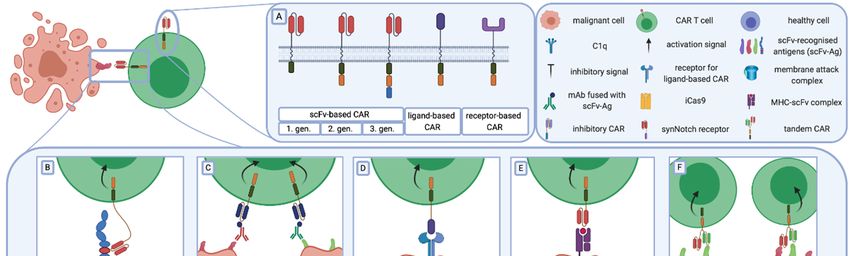
Cancers 2020, 12, 2030                                                                               2 of 29

resected melanomas, expand them ex vivo in the presence of IL-2 and reintroduce them into patients
with metastatic melanoma with a remarkable objective regression rate [8]. Unfortunately, TILs are
relatively rarely detectable in tumours. Therefore, novel solutions, achieved to a great extent with the
help of genetic engineering, were necessary to retarget T cell cytotoxicity. These approaches allowed
researchers to either to redesign the specificity of the native T-cell receptors (TCRs) or to develop
artificial CAR receptors [9]. Now, the development of CARs has become the apogee of ACT. However,
the development of CARs has come a long way since the first two studies appeared at the end of
the 1980s [10,11]. These studies described the construction of a TCR fused with variable antibody
fragments that enabled T cell activation in a major histocompatibility complex (MHC)-independent
manner. It took almost 15 years from the first description [12] to the approval of CAR-T cells as
a therapeutic modality for the treatment of relapsed B-cell acute lymphoblastic leukaemia (B-ALL)
in children.
      Today, CAR-T cell therapy is a fundamental option for patients with CD19-positive haematological
malignancies. Two types of CD19-targeting CARs were approved by the Food and Drug Administration
(FDA) for treating relapsed and refractory cases. In August 2017, tisagenlecleucel (KymriahTM , Novartis,
Morris Plains, NJ, USA) was approved to treat B-ALL in children and young adults. In May 2018,
it was approved as a treatment for diffused large B-cell lymphoma (DLBCL) [13]. The second CAR,
axicabtagene ciloleucel (YescartaTM , Kite, El Segundo, CA, USA) was approved by the FDA in October
2017 for the therapy of DLBCL [14]. The long-term observation of patients treated with CD19 CAR
allows us to pinpoint the drawbacks of CAR-T regimens and helps to reveal important clues of cancer
tumour escape from CAR-T surveillance. It becomes clear that the composition of the CAR-T cell
infusion should be optimised to increase CAR safety and persistence [15–17]. Furthermore, it is
apparent that there is no simple translation of CAR-T cell success in haematology to solid tumours.
Indeed, CAR-T treatments of solid tumours are faced with the harsh conditions of the tumour milieu
or poor trafficking that disrupts effective anti-tumour activity. In this review, we aim to discuss the
general strategies that are currently being tested to improve CAR therapy safety and efficacy. We will
also elaborate on the current urgent issues in the case of haematological CAR-T treatments and the
greatest barriers in studies testing CAR-T cells in solid tumours.
      Promising clinical outcomes, along with approval by the FDA, significantly increased the
enthusiasm around the ACT that contributed to the spread of CAR-T therapy around the world.
As CARs become more widely applied, it is necessary to be aware of their properties, their effects on T
cell functionality, and their limitations and unique side effects.

2. Chimeric Antigen Receptor Structure
     The chimeric antigen receptor (CAR) is a synthetic transmembrane receptor that is designed to
activate effector cells, such as T cells or natural killer (NK) cells, in response to the recognition of
surface antigen. A typical CAR construct consists of four parts differing in their function: (i) an antigen
recognising domain, (ii) a hinge, (iii) a transmembrane domain and (iv) intracellular activation domains.
Despite the simplicity of the modular CAR structure, it is worth noting that even small changes in its
sequence could affect the biologic functions of CAR-T cells, their efficacy and persistence [18].

2.1. Antigen Recognising Domain
     The extracellular antigen recognition domain determines the specificity of the CAR. This domain
is usually composed of variable fragments of the light and heavy chain of an antibody, joined together
with a peptide linker to form a single-chain variable fragment (scFv) [19]. scFv-based CARs are the
most thoroughly studied “classical” antigen recognition domain of CARs. Up to now, both of the
clinically approved CARs contain the same scFv, based on the FMC63 antibody [20], that enables
them to recognise the epitope within the extracellular part of transmembrane glycoprotein—CD19,
the marker of normal and neoplastic B cells [21]. Binding to cognate antigen in an antibody-like manner
allows antigen recognition and activation of effector cells in an MHC-independent way.
Cancers 2020, 12, 2030                                                                             3 of 29

     Other molecules capable of specific antigen binding are also tested as extracellular domains of
CAR. It was shown that ankyrin repeat proteins (DARPins) [22], a natural cytotoxic receptor NKp30 [23],
membrane-tethered IL-13 [24], proliferation-inducing ligand (APRIL) [25], Fms-like tyrosine kinase
3 ligand (FLT3L) [26], granulocyte-macrophage colony-stimulating factor (GM-CSF) [27] and
programmed death-ligand 1 (PD-L1) [28] can be successfully applied as target recognition domains
and have shown some encouraging results in preclinical or even clinical settings [29].

2.2. A Hinge and a Transmembrane Domain
      Effective CAR-T cell activation can also be regulated by hinge length [30]. The hinge is an
extracellular part of a CAR that links the recognition and transmembrane domains. Usually derived
from CD8α, CD28, IgG1 or IgG4, the hinge shows up as a multifaceted player in CAR structure
responsible for functions such as antigen access, signalling and persistence of modified T cells. It was
shown that appropriate adjustments in the hinge length should be introduced depending on the
epitope spatial location. Epitopes located adjacent to the target cell membrane require longer and more
flexible hinges than epitopes that are more easily accessible [31].
      The transmembrane (TM) domain is an integral part of the CAR that connects the extracellular and
intracellular portions and anchors the receptor in the cell membrane. Usually derived from inducible
co-stimulator (ICOS), CD8α, CD3ζ or CD28 proteins, the transmembrane part plays a significant role in
CAR function and stability. CAR constructs containing a CD3ζ transmembrane domain were shown to
enhance cytokine production upon antigen stimulation by creating heterodimers with the endogenous
T-cell receptor (TCR) [32]. Later, the replacement of the CD3ζ transmembrane domain with CD8α or
CD28 has improved surface CAR expression, leading to increased anticancer efficacy [23]. A recent
study on the third generation of CAR-T cells (described further) failed to confirm the critical influence
of the TM domain on surface CAR expression, as was revealed during the investigation of the first and
second-generation CAR constructs. In this study, however, the modulation of CAR-T cell’s anticancer
efficacy and persistence by different TM domains was described [33].

2.3. Intracellular Activation Domains
      Along with the recognition domain, activation domains are the most vigorously investigated parts
of the CAR structure. The first described chimeric receptors, now classified as first-generation CARs,
contained only a single activation domain—CD3ζ, derived from the endogenous T cell receptor [12]
(Figure 1A). First-generation CAR-T cells, by binding to a target antigen, acquire the first activation
signal (the one that T cells receive upon engagement of TCR to the peptide–MHC complex) [34].
The main obstacle for this CAR generation is the insufficient persistence and activation of CAR-T
cells [35].
      Physiologically, T cells require the first (provided by TCR) and second signal (provided by
costimulatory molecules) to achieve optimal activation [36]. The costimulatory activation signal,
upon CAR binding to antigen, is provided by the incorporation of the second activation domain
adjacent to CD3ζ. In comparison to the first-generation, CAR-T cells modified with the second CAR
generation are characterised by enhanced IL-2 secretion and proliferation capacity upon repeatable
interaction with a cognate antigen [37]. CD28 and 4-1BB (CD137) are the most frequently used
coactivation domains [38]. Both of the currently approved FDA CAR-T cell therapies are based on
second-generation CARs—Tisagenlecleucel (Kymriah/Novartis) on a construct containing 4-1BB [13],
and axicabtagene ciloleucel (Yescarta/Kite Pharma) on a construct containing the CD28 [14] coactivation
domain. Despite differences within CAR constructs, both therapeutics have shown impressive
clinical outcomes.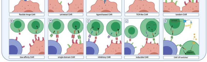
Cancers 2020, 12, 2030 4 of 29
Cancers 2020, 12, x 4 of 29
FigureFigure
1. The 1.CAR
The structure
CAR structure and general
and general strategies
strategies to enhance
to enhance CAR-TCAR-T cell therapy.
cell therapy. (A)generations
(A) Three Three
generations
of chimeric of chimeric
antigen receptorsantigen receptors
are based onarethebased on the composition
composition of the intracellular
of the intracellular part. First-
part. First-generation
CARsgeneration
contain one CARs contain one
stimulation stimulation
domain, whereasdomain,
secondwhereas second
and third and third generations
generations have one orhavetwoone or
additional
two additional costimulatory domains, respectively. Besides scFv, other molecules (ligands or
costimulatory domains, respectively. Besides scFv, other molecules (ligands or receptors) are used as
receptors) are used as antigen recognition domains. (B–F) Strategies to increase CAR-T cell efficacy
antigen recognition domains. (B–F) Strategies to increase CAR-T cell efficacy and selectivity against
and selectivity against tumour cells. (B) Epitopes, located adjacent to the cell membrane, require a
tumour cells. (B) Epitopes, located adjacent to the cell membrane, require a longer and more flexible
longer and more flexible hinge than those easily accessible for CARs. (C) In universal CARs, by
hingetargeting
than those easily accessible for CARs. (C) In universal CARs, by targeting an epitope fused with
an epitope fused with an antibody, specificity relies mostly on antibody selectivity. (D) The
an antibody,
use of a ligand insteadrelies
specificity mostly
of scFv on antibody
as an antigen selectivity.
recognition domain(D)could The use ofthe
increase a ligand instead
specificity of scFv
of CAR-
as anTantigen recognition domain could increase the specificity of CAR-T cells.
cells. (E) TCR-like CAR-T cells target intracellular cancer-specific antigens that are presented by (E) TCR-like CAR-T
cells MHC.
target(F)intracellular
Tandem CARs cancer-specific
are designed toantigens
target morethat areone
than presented by MHC. (F) Tandem
antigen simultaneously. CARs are
They recognise
designed to targetantigens
two different more thanand one antigen
provide simultaneously.
effective Theycells
lysis of malignant recognise
expressingtwoonedifferent
or bothantigens
cognate and
antigens.
provide (G–K)
effective Strategies
lysis to decrease
of malignant on-target off-tumour
cells expressing one or both toxicity and antigens.
cognate the probability
(G–K) of Strategies
healthy to
cells on-target
decrease lysis. (G) Decreased
off-tumour affinity toward
toxicity and cognate antigen enables
the probability CAR-T
of healthy cells
cells to distinguish
lysis. (G) Decreasedhealthy
affinity
towardcells with a antigen
cognate low expression
enableslevel
CAR-Tof TAAs
cells from malignant healthy
to distinguish cells with a high
cells withlevel
a lowof TAAs. (H) The
expression level of
division of the full activation signal to two independent CARs with different antigen specificities
TAAs from malignant cells with a high level of TAAs. (H) The division of the full activation signal
restrict the CAR-T cell’s cytotoxicity to malignant cells that express both targeted antigens while
to two independent CARs with different antigen specificities restrict the CAR-T cell’s cytotoxicity to
sparing healthy cells with only one antigen on their surface. (I) Additional co-modification of CAR-T
malignant cells that express both targeted antigens while sparing healthy cells with only one antigen on
cells with iCAR enables specific recognition of non-malignant cells and inhibition of toxicity against
their them.
surface.
(J) (I)
TheAdditional
expression co-modification
of the “effector” of CARCAR-T cells with
is regulated by iCAR enablesreceptor
the synNotch specific redirected
recognition of
non-malignant
against anothercells TAA.
and inhibition of toxicity against
(K) CAR “off-switches” them.
are based on (J)
theThe expression of
co-modification of the
CAR-T“effector” CAR is
cells with
regulated by the synNotch
antibody-recognised receptor
molecules or redirected
with iCas9. against another
This enables TAA.
selective (K) CAR
CAR-T “off-switches”
cell clearance are based
while CAR-
on the co-modification
related of CAR-T
life-threatening cells with
side effects occur.antibody-recognised moleculesantigen
Abbreviations: CAR—chimeric or withreceptor,
iCas9. This enables
scFv—
single-chain
selective CAR-Tvariable fragment,
cell clearance mAb—monoclonal
while antibody, Ag—antigen,
CAR-related life-threatening iCas9—inducible
side effects caspase
occur. Abbreviations:
9, TCR—T-cell
CAR—chimeric receptor,
antigen MHC—major
receptor, histocompatibility
scFv—single-chain complex, mAb—monoclonal
variable fragment, TAA—tumour-associated antibody,
antigen, iCAR—inhibitory CAR, synNotch receptor—synthetic Notch
Ag—antigen, iCas9—inducible caspase 9, TCR—T-cell receptor, MHC—major histocompatibility receptor.
complex, TAA—tumour-associated antigen, iCAR—inhibitory CAR, synNotch receptor—synthetic
Physiologically, T cells require the first (provided by TCR) and second signal (provided by
Notch receptor.
costimulatory molecules) to achieve optimal activation [36]. The costimulatory activation signal,
upon CAR binding to antigen, is provided by the incorporation of the second activation domain
To combine benefits from different coactivation domains, the third generation of CARs,
adjacent to CD3ζ. In comparison to the first-generation, CAR-T cells modified with the second CAR
which consists of two coactivation domains, was developed [39]. T cells modified with the third
generation are characterised by enhanced IL-2 secretion and proliferation capacity upon repeatable
CAR generation have shown enhanced anti-tumour activity and increased persistence [33]. However,
interaction with a cognate antigen [37]. CD28 and 4-1BB (CD137) are the most frequently used
the combinatory effect of two costimulatory domains within the single CAR construct does not appear
to be a simple consequence of the costimulatory domain properties. For instance, the proximity of
the costimulatory domain to the intracellular part of the cell membrane may determine the dominant
domain and establish functional characteristics of CAR-T cells. Intriguingly, some studies report no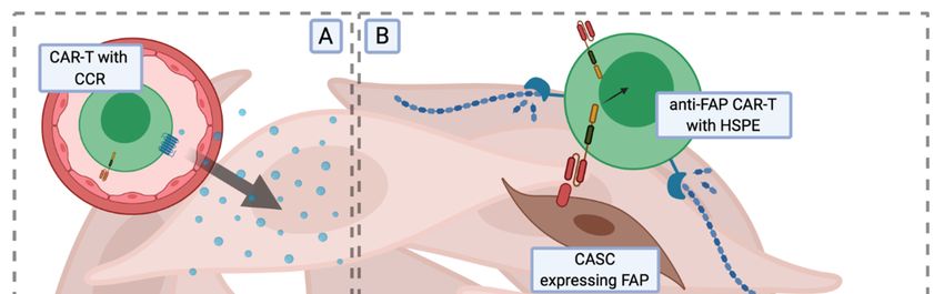
Cancers 2020, 12, 2030                                                                                5 of 29

superiority [40] of the third CAR generation and claim even worse performance than second-generation
counterparts [41,42]. A deeper understanding of coactivation domain interactions within a single CAR
structure is needed to create potent third-generation CARs.
      The third activation signal [43], which non-modified T cells usually gain through soluble factors
and typically emerges after the first two signals, has often been neglected in the context of CAR-T
activation. However, the enhanced anti-tumour efficacy of T cells redirected for universal cytokine
killing (TRUCKs), also called fourth-generation CARs (described in the further part of this review),
and studies reporting the essential role of cytokines for full CAR-T cells activation [44] open a new
path for further investigations aiming to increase CAR efficiency.

3. CAR T-Cell Biology
      Pharmacokinetics and pharmacodynamics are critical properties of newly discovered compounds
that contribute immensely to their further fate and could make or break the drug. However, in the case
of CAR-T cells, the level of complicacy is even higher due to their biological features.
      The basic studies on CAR-T cell biology are complicated due to many factors contributing to their
properties. The phenotype of lymphocytes, the expression level of the CAR on the cell surface and even
the locus of CAR DNA integration [45] can all affect CAR-T functionality. Moreover, the variations
in CAR structure alter the cytophysiology of modified cells and should be taken into account while
composing CAR-T cell infusions for patients. Despite this, several studies appeared and lifted the
curtain of secrecy surrounding CAR-T cell biology.
      To determine the cytotoxic potential of CAR-T cells, a model of T cells co-expressing the TCR
and CAR with different cognate antigen specificity was created and its cytolytic ability through each
of these receptors was investigated [46]. The transduction of T cells with a CAR does not influence
TCR-mediated activation or proliferation. Thus, CAR-T cells remain capable of lysing multiple
tumour cells after TCR or CAR-mediated engagement to a cognate antigen. Perforin and granzyme B
expression, required for CAR-mediated toxicity, remained at the same level after the cell modification.
The MHC-independent manner of T cell stimulation through a CAR enables activation of both CD4+
and CD8+ lymphocytes and provides effective serial killing [47]. In addition to perforin/granzyme
B-mediated toxicity, upregulation of Fas ligand (FasL) expression on CAR-T cells upon activation was
shown to enhance their anticancer efficacy against bystander antigen-negative tumour cells [48].
      Further studies on a mechanism of CAR-T cell interaction with a target cell revealed
differences within immune synapses formed after antigen recognition by TCR and CAR [49].
Following TCR-mediated antigen recognition within the MHC complex, T cells form a well-studied
“bull0 s eye-like” immune synapse (IS) [50–52]. Highly organised and stable TCR ISs contain three
concentric areas, known as supramolecular activation clusters (SMACs). The central SMAC, with the
TCR–MHC and Lck clusters, and peripheral SMAC, with lymphocyte function-associated antigen-1
(LFA-1) adhesion molecules responsible for IS stabilisation [53], are surrounded by the distal SMAC,
where actin accumulation takes place. The study by Davenport et al. [49] revealed that the IS
formed after antigen recognition by CAR differs significantly from the classical one mediated by TCR.
The CAR-mediated binding to the antigen leads to the formation of a disorganised synapse with no
concentric rings and randomly distributed microclusters of LFA-1, Lck and CAR–antigen complexes.
The disorganised structure of CAR ISs does however have its benefits, including faster activation and
deactivation of proximal and distal signals, faster lytic granule delivery to the synaptic cleft and shorter
duration of the IS. These benefits of the CAR IS result in quicker CAR-T cell detachment from dying
cells and movement towards another malignant cell.
      Future studies are needed to investigate the influence of other coactivation domains [54] and
different scFv affinities on CAR-T cell biology. This long and bumpy road lies in front of those who
would dare to study the biology of the new human-made CAR-T cells.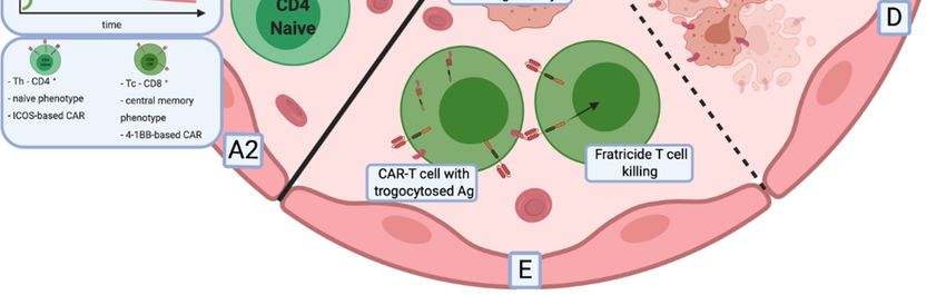
Cancers 2020, 12, 2030                                                                               6 of 29

4. Side Effects of CAR-T
     Currently, CAR-T cell therapy brings hope to cancer patients and challenges to scientists and
physicians. The remarkably rapid response to CAR-T infusions and durable remission rate observed in
patients with relapsed and refractory cancer comes inseparably with unique acute toxicity. The main
challenges that remain an obstacle are cytokine-release syndrome, which can be accompanied or
followed by neurotoxicity and on-target off-tumour toxicity.

4.1. Cytokine-Release Syndrome
      Cytokine-release syndrome (CRS) [55] is the most common adverse effect of CAR-T therapy and
is triggered by the recognition of cognate antigen followed by the activation of T cells and bystander
immune cells. Among these activated immune cells, macrophages were shown to play a crucial role
in the pathophysiology of CRS [56,57]. The excessive release of pro-inflammatory cytokines such
as IL-1, IL-6, IL-15 and interferon gamma (IFN-γ) [58], cause generalised immune activation which
clinically manifests as high fever, hypotension, hypoxia and even multiorgan dysfunction, which can
be fatal [59–61]. Severe CRS more commonly affects patients with bulky disease [62–64]. The onset
of CRS typically occurs within the first week after the CAR infusion and can be managed by the
use of tocilizumab (anti-IL-6R antibody) [65]. The usage of tocilizumab in the management of CRS
does not seem to interfere with the anticancer efficacy of CAR-T. This observation contributed to the
beginning of an ongoing clinical trial that aims to evaluate the impact of tocilizumab prophylaxis
on CAR-T efficacy (NCT02906371, https://clinicaltrials.gov/ct2/show/NCT02906371). For tocilizumab
non-responders and those with high-grade CRS, corticosteroids are indicated [65], despite reported
interference with CAR-T efficiency [64].
      Rarely, another systemic hyperinflammatory disorder may follow the CAR-T cells infusion [65].
With similar clinical manifestation and laboratory findings to CRS, hemophagocytic lymphohistiocytosis
(HLH) could also appear and requires additional therapies like etoposide or cytarabine. HLH is
associated with a higher mortality rate [66,67].

4.2. Immune Effector Cell-Associated Neurotoxicity Syndrome (ICANS)
      Immune effector cell-associated neurotoxicity syndrome (ICANS) is the second most frequently
seen undesirable result of CAR-T cell activation. In mild cases, patients usually experience diminished
attention, language disturbance and impaired handwriting. However, in severe cases, seizures,
motor weakness, increased intracranial pressure, papilledema, and cerebral oedema can also occur.
Despite the hair-raising clinical picture of a patient with ICANS, signs are generally fully reversible
with proper management [55]. The pathophysiology of ICANS is not fully known. There are two
main hypotheses of ICANS occurrence—cytokine diffusion to the brain [68,69] and trafficking of T
cells into the central nervous system [68,70]. Neurotoxicity appearing simultaneously with CRS is
typically low grade and of a shorter duration, while neurotoxicity appearing after CRS (more common)
is usually more severe (grade ≥ 3) and protracted [65]. Similar to CRS, ICAN management depends on
the toxicity grade, with a more significant role of corticosteroids and restriction of tocilizumab usage
for patients with grade ≥ 1 ICANS with concurrent CRS.

4.3. Cross-Reactivity of CAR-T Cells (On-Target Off-Tumour Toxicity)
     The increasing number of targeted antigens tested for future CAR-T cell therapies brings into
consideration the safety of the overall treatment. The main issue today is not whether we can redirect
T cell cytotoxicity, but whether we can control it. The results of antigen recognition on malignant and
healthy cells are also observed for the FDA approved CD19 CAR-T cell therapies. B-cell aplasia with
concomitant hypogammaglobulinemia is an example of on-target off-tumour cytotoxicity of CD19
redirected modified-T cells [71–73]. After clearance of cancerous cells, persistent CAR-T cells not only
provide surveillance, but also contribute to prolonged B cell aplasia long after cancer cell disappearance.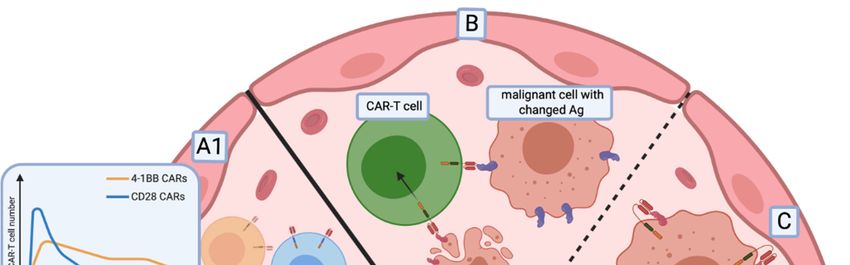
Cancers 2020, 12, 2030                                                                                 7 of 29

Thankfully, loss of B cells is a relatively low price to pay for a cancer cure and can be managed with
intravenous immunoglobulin infusions [74]. However, targeting more abundant molecules might lead
to profound on-target off-tumour cytotoxicity, fatal in fact, as was observed during the anti-human
epidermal growth factor receptor 2 (HER2)-CAR-T clinical trial [75].

5. General Strategies to Enhance CAR Therapy
      After the first remarkable success of CAR-T cells in the field of haemato-oncology, further spreading
of this therapeutic modality faced many roadblocks, one of which was the lack of tumour-specific
antigens (TSAs) [76–78]. Due to the shortage of TSAs, most of the clinically investigated CARs are
redirected against tumour-associated antigens (TAAs) [79]. To diminish off-target effects, the specific
construction of CAR plasmids is indispensable. Much effort is also put into CAR construct optimisation
for safety and efficacy reasons. By optimising the length of the hinge and TM domain, Ying et al. [80]
increased the safety (no neurotoxicity or CRS greater than grade 1) of CAR-T cells while maintaining the
anticancer efficacy. Following antigen recognition, CAR-T cells were shown to secrete lower amounts
of inflammatory cytokines and were less susceptible to activation-induced cell death (AICD) when the
hinge and a transmembrane domain were derived from CD8α in comparison to CD28 [81].
      In the case of CARs built with a long spacer consisting of IgG1 or IgG4, certain susceptibility to
AICD was observed as a result of constant fragment (Fc) antibody region recognition by FcR-bearing
cells and off-target CAR-T cell activation. The mutation or elimination of FcR-recognised regions were
crucial for proper CAR-T cell activation and persistence [82–84].

5.1. CARs Improvements to Target Malignant Cells
      Selecting the specific antigen as a target for CAR-T cells is a crucial step in developing an effective
and safe weapon against cancer. However, there are very few antigens that are expressed selectively
on malignant cells. Among factors contributing to this phenomenon are: (i) the limited spectrum
of antigens exhibited extracellularly, (ii) the risk of potential “off-tumour” cytotoxicity in case of
more ubiquitously expressed antigens, and (iii) the relatively small database covering cancer-specific
modification of antigens, such as glycosylation [85] or splicing-dependent antigen changes [86].
      Modifications in the hinge region [87], TCR-like CARs or tandem CARs are among many proposed
strategies to enhance malignant cell targeting. The importance of a cancer-specific modification along
with the hinge length [31] was elegantly pinpointed in a study comparing CARs targeting the MUC1
glycoprotein. Due to the underglycosylation of MUC1 in malignant cells, some of the cryptic epitopes
remain uncovered and can be targeted using specific antibodies. It was shown that by choosing scFv
targeting these unmasked epitopes and by adjusting hinge length with an IgD linker, certain selectivity
of CAR toward malignant cells can be acquired [87] (Figure 1B).
      Another strategy covers the idea of creating a “universal” CAR that would be usable in
different malignancies and would possibly overcome the antigen specificity issue (Figure 1C). In this
approach, redirecting CAR cytotoxicity relies on labelled antibodies or small fluorescein-based
adapters that recognise various tumour-associated epitopes. CAR-T cells are designed to recognise
the tag or fluorescein and, therefore, their cytotoxicity is limited only to cells coated with the
antibodies/adapters [88–92]. This approach enables not only the use of the same CAR in various
malignancies, but also the targeting of many tumour antigens simultaneously. Moreover, this strategy
increases the probability of overcoming intratumoural and intertumoural heterogeneity [93].
      In some cases, ligands are the preferred antigen recognition domain in comparison to scFvs due to
their lower affinity profile and better malignant cell discrimination [24–27] (Figure 1D). It was shown
to be feasible to discriminate PD-1high from PD-1low targets by substituting the anti-PD-1 scFv with the
PD-L1 extracellular domain [28].
      Antigen binding that is unrestricted to MHC presentation, the main advantage of CARs, has serious
drawbacks; it restricts the pool of targeted antigens to those presented on the cell surface. To overcome
this issue, TCR-like antibodies were recently created [94] and integrated as an antigen recognition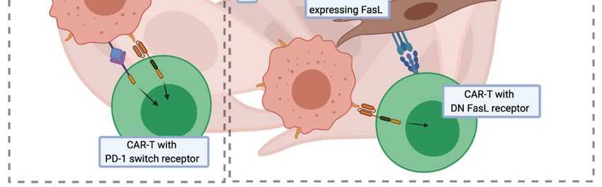
Cancers 2020, 12, 2030                                                                               8 of 29

domain of CAR to form the TCR-like CAR [95–97]. With this strategy, CARs are capable of recognising
intracellular antigens presented by MHC complexes on the surface of a cell (Figure 1E). Similarly, it was
shown that fusing the extracellular part of the TCR with intracellular CAR domains can transmit the
MHC-restricted recognition of antigen and trigger effector cell cytotoxicity. This strategy additionally
widens the pool of cells that can be used in CAR-based approaches, as shown for the NK cell-derived
cell line NK-92 [98].
      The heterogeneous expression pattern of the target antigen and antigen-negative relapses in long
term follow-ups, caused by the selection of malignant cells resistant to CAR-T, revealed the unmet
need for targeting more than one antigen at the same time. Targeting multiple TAAs simultaneously
significantly improves the discrimination of cancerous cells [99] and decreases the risk of CAR-T-resistant
cancer relapse [100]. For instance, two or more [101] different CARs could be expressed within the same T
cell or alternatively, separately transduced T cells could be injected sequentially or simultaneously [102].
Furthermore, “tandem CARs” were invented to link two recognition domains encoded by two
scFvs in a single construct [103] (Figure 1F). Indeed, tandem CARs showed enhanced anti-tumour
efficacy in comparison to T cells expressing two individual CARs [104]. This result paved the way
for several ongoing clinical trials (NCT03241940, https://clinicaltrials.gov/ct2/show/NCT03241940;
NCT03233854, https://clinicaltrials.gov/ct2/show/NCT03233854; NCT03448393, https://clinicaltrials.
gov/ct2/show/NCT03448393) for tandem CARs.

5.2. Strategies that Enable CARs to Discriminate between Normal and Malignant Cells
      Several methods were proposed to distinguish the variation of TAA antigen expression present on
both malignant and healthy tissues [105–108]. They rely, among others, on the modulation of CAR-T cell
affinity toward the antigen, dividing the full activation signal into two distinct CARs, the replacement
of the coactivation domains to inhibitory ones to create iCARs, or inducible expression systems.
      Apart from specific antigen recognition, CAR-T cell activation also depends on the affinity of the
antigen recognition domain towards the antigen. It was shown that scFv differing in their affinity could
regulate to a great extent and set a threshold for antigen recognition. The proliferation and cytokine
production of CAR-T cells following antigen binding could be enhanced by increasing the affinity to
the epitope [30,109]. Unnecessarily high affinity may, however, lead to exhaustion or AICD [110].
      The possibility of regulating the antigen recognition threshold allows researchers to redirect the
cytotoxicity of CAR-T cells towards cells with a high expression level of the target antigen while sparing
the ones with low expression [111]. A tumour-associated antigen is usually expressed at a significantly
higher level on tumour cells compared to non-malignant tissues. By decreasing the affinity of scFv,
it becomes possible to eradicate cancerous cells and spare bystander untransformed cells with low
expression of the target antigen [111–113] (Figure 1G).
      Simultaneous targeting of multiple antigens is currently used not only to enhance anticancer
efficacy, but also to increase the safety of CAR-T cell therapy. Indeed, it seems feasible to modify a
single T cell with two CARs differing in the antigen specificity, where the first CAR contains only the
CD3ζ activation domain and another contains only the CD28 domain [114–116] (Figure 1H). In this
setting, the cytotoxicity mediated by CAR-T cells is restricted to target cells that have both cognate
antigens. However, restricting toxicity against neoplastic cells with both target antigens opens an
escape window for malignancy to evade CAR-T cells by losing one of the target epitopes [114].
      As an alternative strategy, it was proposed to co-transduce a single T cell with a second CAR that
has inhibitory properties (iCAR) (Figure 1I). In this approach, cytotoxicity against cells expressing the
antigen recognised by the iCAR, expressed on healthy tissues, is repressed by transmitting inhibitory
signals derived from the intracellular domains of checkpoint proteins, such as cytotoxic lymphocyte
protein 4 (CTLA-4) or program cell death 1 (PD-1) [117].
      Another strategy to limit on-target off-tumour toxicity is to express an “effector CAR” responsible
for T cell activation only in defined circumstances (Figure 1J). The synthetic Notch (synNotch) system
has been recently created, in which the synNotch receptor redirected against the first TAA induces the
Cancers 2020, 12, 2030 9 of 29
expression of the “effector CAR” specific against the other TAA [118,119]. Alternatively, by using the
hypoxia-inducible CAR system, Kosti et al. recently employed the pan-ErbB antigen as a target for
CAR-T cells. Despite widespread expression of the target antigen, there was no off-tumour toxicity
observed along with the potent anticancer efficacy within the tumour site [120]. CAR expression can
also be controlled by the administration of small molecules. A tetracycline regulation system was used
to modulate the expression of a CD19 CAR. While showing minimal background leakage without
doxycycline (the activator of the system), this system showed similar efficacy to CD19 CAR-T cells
in terms of cytokine production and proliferation rate upon engagement to cognate antigen in the
presence of doxycycline [121].
As described above, some life-threatening side effects may appear following CAR-T cell infusion.
To address this problem, “safe switches” for CAR-T cells were created (Figure 1K). The co-modification
of CAR-T cells with surface antigens that could be targeted with therapeutics, like rituximab and
cetuximab, enables the selective clearance of modified T cells [122,123]. However, CAR-related
toxicity could have fulminant manifestation and may require rapid CAR-T cell ablation, for which
antibody-mediated clearance would be too slow. A far more potent and clinically applicable strategy is
the co-modification of CAR-T cells with inducible caspase 9 (iCasp9) which, in the presence of a small
inert molecule, dimerises and induces rapid apoptosis of T cells [124,125].
A different and ambitious strategy to overcome on-target off-tumour toxicity was recently
described. Targeting CD33, the molecule expressed on healthy and neoplastic myeloid cells [126],
to fight the acute myeloid leukaemia (AML) may result in off-tumour toxicity and destruction of healthy
myeloid cells. Kim et al. [127] “made” CD33 specific for AML by depleting this molecule from normal
haematopoietic stem and progenitor cells (HSPC) prior to autologous HSPC transplantation. After the
effective engraftment of CD33-deficient HSPCs, healthy myeloid cells were resistant to CD33-CAR-T
cells, enabling sufficient AML clearance without myelotoxicity.
In addition to these general issues aiming to increase CAR-T therapy safety and efficacy,
some further problems, described below, were revealed in a long-term follow-up of patients receiving
the approved CAR-T cell treatments.
6. Remaining Issues in Haemato-Oncology
Long-term observation of patients in complete response after CAR-T cell therapy revealed a
depressingly high rate of disease recurrence [128]. The majority of relapses are thought to be caused by
either one or all of the following reasons:
6.1. Insufficient CAR-T Cell Persistence and Proliferation
Adequate proliferation rate and sufficient persistence of CAR-modified T cells were shown to
correlate with the durability of remission [129]. Selecting the optimal subpopulation of lymphocyte
ratio [130] along with the structural CAR modifications [131–133] that aim to increase the viability of
CAR-T cells showed encouraging results in preclinical and clinical settings [15,16,134].
Understanding the impact of costimulatory domains on a CAR-T cell phenotype opens the
possibility of modifying the CAR structure to enhance the persistence of T cells [135]. Indeed,
the influence of domains became evident when comparing phenotypic and metabolic changes occurring
in CAR-T cells [135]. T cells that contained a CD28 domain in their CARs acquired an effector memory
phenotype with brisk proliferation and glucose metabolism, which led to faster cancer clearance
at the cost of limited persistence. In contrast, 4-1BB-based CAR-T cells showed slower anticancer
activity with T cells differentiating predominantly into central memory cells with enhanced endurance,
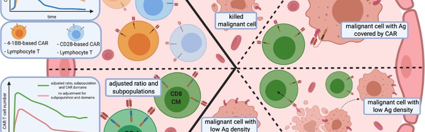
oxidative metabolism and increased mitochondrial biogenesis (Figure 2A1).Cancers 2020, 12, x 10 of 29
phenotype with brisk proliferation and glucose metabolism, which led to faster cancer clearance at
the cost of limited persistence. In contrast, 4-1BB-based CAR-T cells showed slower anticancer
activity with T cells differentiating predominantly into central memory cells with enhanced
Cancers 2020, 12, 2030 10 of 29
endurance, oxidative metabolism and increased mitochondrial biogenesis (Figure 2A1).
Figure 2. 2. Remaining issues of CAR-T cell cell therapies
therapies inin haemato-oncology.
haemato-oncology. Relapses in a long-term
long-term
observation after CAR-T
observation after CAR-T cell infusion have two primary sources:
infusion have two primary sources: CAR-T cells persistence
persistence (A1,A2) and
the escape of malignant cells from immune surveillance (B–E). (A1) The persistence and proliferation
rate ofof CAR-T
CAR-T cells
cells depends
depends on on the
the co-stimulation
co-stimulation domains.
domains. Additionally, (A2) subpopulation
subpopulation
composition of the CAR-T cell infusion, as well as the the phenotype
phenotype ofof lymphocytes,
lymphocytes, influence the
persistence of
proliferation and persistence of modified
modified effector
effector cells.
cells. (B–E) Mechanisms of immune escape of
neoplastic cells from CAR-T cells. (B) (B) Targeted antigen changes as a result of mutation mutation or splicing
splicing
alteration. (C)
(C) Unintentional
Unintentional modification
modification of of neoplastic
neoplastic BB cells with a CAR that masks the targeted
targeted
epitope, rendering it undetectable
undetectable by CAR-TCAR-T cells.
cells. (D) Infusion of CAR-T cells triggers the selection
selection
of malignant cells with low antigen antigen density
density that
that are
are resistant
resistant to
to effector
effector cells.
cells. (E) The decrease of of
antigen surface level results from CAR-T cell-mediated trogocytosis,
from CAR-T cell-mediated trogocytosis, with subsequent
subsequent induction of
fratricide T cell
cell killing
killing and
and CAR-T
CAR-T cells
cells with
with ananexhausted
exhaustedphenotype.
phenotype. Abbreviations:
Abbreviations: Th—helper T
cell, Tc—cytotoxic
Tc—cytotoxic TT cell,
cell, ICOS-based
ICOS-basedCAR—inducible
CAR—inducibleco-stimulator-based
co-stimulator-basedCAR.CAR.
Clonal
Clonal expansion
expansionofofCAR-T
CAR-T cells hashas
cells alsoalso
beenbeen
reported as a result
reported as a of the lentiviral
result vector-mediated
of the lentiviral vector-
unintended insertion of the CAR gene into the TET2 gene. This observation was described
mediated unintended insertion of the CAR gene into the TET2 gene. This observation was described in a patient
with a mutation
in a patient within the secondinTET2
a mutation allele [136].
the second TET2 allele [136].
Moreover,
Moreover,the thepersistence
persistenceand
andactivity
activityofofdifferent subpopulations
different subpopulations of of
lymphocytes
lymphocytes seem to rely
seem on
to rely
different coactivation domains. Cytotoxic (CD8+) CAR-T cell persistence was shown
on different coactivation domains. Cytotoxic (CD8+) CAR-T cell persistence was shown to depend on to depend on
4-1BB
4-1BB signalling,
signalling, while
while helper
helper (CD4+) CAR-T cells
(CD4+) CAR-T cells require
require ICOS
ICOS signalling. The redirection
signalling. The redirection of
of TT cells
cells
with CAR molecules adjusted for subpopulations led to enhanced persistence and anticancer
with CAR molecules adjusted for subpopulations led to enhanced persistence and anticancer efficacy efficacy
of
of CAR-T
CAR-T cells
cells in
in mouse
mouse models
models [33]
[33] (Figure
(Figure 2A2).
2A2).
Preclinical investigations revealed that CAR-modified T cells with less differentiated phenotypes,
like naïve or central memory, have higher anticancer efficacy [130]. By reducing the duration of ex
vivo expansion of CAR-T cells, Ghassemi et al. showed enhanced anti-tumour efficacy of the modified
T cells, which was caused by the less differentiated phenotype and enhanced effector functions inCancers 2020, 12, 2030 11 of 29
a murine xenograft model of ALL [137]. Additionally, the subpopulation composition of CAR-T
cells emerged as a way to impact therapy outcome [17]. The first CAR-T cell therapy with a defined
CD4/CD8 ratio [15,16] appeared to be applicable even in patients with severe leukopenia and is
currently under the FDA approval process.
However, without potent T cells with high proliferation potential, even the perfect chimeric antigen
receptor performs weakly. Preclinical experiments are often based on healthy donors0 T cells and do not
take into count changes occurring during tumourigenesis. Studies indicate that during tumourigenesis,
T cells acquire an exhaustion phenotype [138], characterised by a decreased proliferation capacity [139],
and this change seems to be irreversible in the advanced stages of cancer. Exhausted central memory T
cells have a distinct transcriptional status compared to healthy ones [140,141]. This knowledge should
stimulate further studies on using healthy donor cells as a base for off-the-shelf therapeutics.
6.2. Relapse of Antigen-Negative Disease
The data collected during clinical trials demonstrate that CD19 antigen loss is responsible for
the majority of relapses in B-ALL patients following CD19 CAR-T therapy. CD19 antigen loss was
also shown to occur in NHL patients [142]. Two main mechanisms accountable for antigen loss were
recently described: antigen escape and lineage switch [143].
The recurrence of phenotypically identical disease with the lack of cognate epitope characterises
antigen escape (Figure 2B). There are several splice variants of CD19 described in B-ALL. Some variants
lack the epitope recognised by CAR-T cells in the extracellular portion of the antigen and others lack
the transmembrane region, causing the loss of CD19 surface expression [144]. CD19 splice variants in
tumour cells can already be detected in patients before the CAR-T infusion [145]. CAR-T cells simply
stimulate the selection of malignant cell variants resistant to therapy. However, other mechanisms
of antigen escape were also reported. Braig et al. have shown that post-transcriptional alteration of
CD81, a protein that regulates CD19 maturation and trafficking, leads to the loss of CD19 expression
and relapse of disease [146]. On the other hand, the lineage switch mechanism depends on changes of
a cancerous cell from a lymphoid to myeloid phenotype in response to the therapy [147]. The main
approach to overcome these obstacles is described above and relies on the simultaneous targeting of
multiple epitopes.
The most alarming issue with the lack of recognition of CD19 antigen by CAR-T cells is the
semi-controllable introduction of CAR genes [148]. Unintentional transduction of a single neoplastic B
cell during the production process of CAR-T led to the relapse of leukaemia with the epitope masked
by the CAR on the surface of malignant cells [149] (Figure 2C). This finding illustrates the need for
further improvement of manufacturing technologies to clean out engineered T cells from residual
tumour cells.
6.3. Low Antigen Density
Low antigen density is most commonly associated with solid tumours, where the antigen
expression level is very heterogeneous. Less frequently, a low antigen expression pattern is described
as a problem in haematological malignancies. It was, however, portrayed by the clinical trial of CD22
CAR-T cells where some of the patients, after achieving a complete response, relapsed with malignant
cells expressing low levels of CD22. At the same time, CD22 CAR-T cells were still detectable in their
blood [150] (Figure 2D). This data is in agreement with previous studies reporting incomplete CAR-T
cell activation after contacting the cells expressing an insufficient level of target antigen [151–153].
Trogocytosis is another mechanism responsible for low surface antigen level on malignant B
cells [154]. CAR-T cells, after binding to a cognate antigen, may extract the antigen from a cancerous cell,
causing a reversible decline of surface antigen expression level and increased resistance of malignant
cells to CAR-T. At the same time, T cells with trogocytosed antigen acquire not only an exhausted
phenotype, but also undergo fratricide T-cell killing caused by CD19 surface expression (Figure 2E).Cancers 2020, 12, 2030 12 of 29
7. Driving CARs through Solid Tumour Roadblocks
The development of CARs has undisputedly revolutionised the field of cancer therapy. However,
the stunning efficacy of CAR-T cell therapy in haematological diseases has not, up until now,
translated to the efficient treatment of the solid tumours, which account for approximately 90% of total
cancer-caused deaths [155]. Complex cell interactions, along with the specific microenvironment within
the solid mass, create a challenging labyrinth with no way-out [156]. The most significant challenges
for CAR-T therapy include: (i) the lack of TSA, as described above, (ii) poor or limited trafficking of
CAR-T cells to the tumour mass, and (iii) the immunosuppressive tumour microenvironment (TME).
7.1. Trafficking and Infiltration
Inappropriate trafficking seems to be the most potent barrier for CAR-T cells to infiltrate the
tumour site. To overcome this obstacle, the direct infusion of CAR-T cells to solid masses was applied
for tumours originating from different tissues [29,157–160]. Studying an orthotopic model that mimics
human pleural malignancy, Adusumilli et al. showed that intrapleurally-administered CAR-T cells
outperform intravenously infused cells in tumour eradication efficacy and also promote the elimination
of extrapleural tumours [161]. These encouraging results contributed to the development of the
ongoing clinical trial of mesothelin-targeted CAR-T cells administrated intrapleurally (NCT02414269,
https://clinicaltrials.gov/ct2/show/NCT02414269). In another approach, the co-modification of CAR-T
cells with a chemokine receptor that guides the CAR-T cells to the tumour site was proposed.
Preclinical studies of CAR-T cells with an increased expression level of a chemokine receptor
showed enhanced anti-tumour efficiency, as a result of improved migration and infiltration capacity.
Forced expression of CC-chemokine receptor 4 (CCR4) on CD30-CAR-T cells enhanced the migration
and anti-tumour activity in a mouse xenograft Hodgkin lymphoma model [162]. In another study,
CXCR2-expressing anti-glypican-3 (GPC3)-CAR-T cells showed improved migration and activity in a
xenograft hepatocellular carcinoma (HCC) tumour model [163]. Increased infiltration and anti-tumour
efficacy was also shown in xenograft models of neuroblastoma and mesothelioma by co-expressing
CCR2b with CARs directed against GD2 [164] and mesothelin antigens [165] (Figure 3A).
Once a T cell reaches the destination point (the tumour site), the next challenge is to penetrate
the tumour extracellular matrix (ECM), which is composed of various fibrous proteins, glycoproteins,
and proteoglycans. By developing CAR-T cells against fibroblast activation protein (FAP; a proteinase
involved in extracellular matrix remodelling, expressed abundantly in epithelial cancers [166]),
Wang et al. [167] demonstrated that clearance of cancer-associated stromal cells (CASCs) by FAP CAR-T
cells inhibits tumour growth and increases tumour infiltration by host immune cells. Another study by
Caruana et al. [168] showed that CAR-T cells lose the expression of heparanase (HSPE), the enzyme
responsible for ECM degradation, as a result of the ex vivo expansion process. Engineered CAR-T
cells with an increased expression of HSPE showed enhanced infiltration and anti-tumour efficacy
(Figure 3B).Cancers 2020, 12, 2030 13 of 29
Cancers 2020, 12, x 13 of 29
Figure 3. Examples of
3. Examples of preclinical
preclinical concepts
concepts supporting
supporting CAR-T
CAR-T cell anti-tumour
anti-tumour activity in solid
tumours. The The effective
effective eradication
eradication of of solid
solid tumours
tumours by by CAR-T
CAR-T cellscells is jeopardised by impaired
trafficking and infiltration into the tumour mass (A,B), competition competition with inhibitory signals (C) and
reduced
reduced survival
survivalwithin
within thethe
TME.
TME.(A) (A)
CAR-TCAR-Tcells additionally co-modified
cells additionally with a chemokine
co-modified receptor
with a chemokine
(CCR) migrate towards and infiltrate solid masses in response to the chemokine
receptor (CCR) migrate towards and infiltrate solid masses in response to the chemokine gradient. gradient. (B) Forced
expression of heparanase (HSPE) and redirection of CAR-T cells against
(B) Forced expression of heparanase (HSPE) and redirection of CAR-T cells against fibroblast fibroblast activation protein
(FAP) further
activation enhances
protein (FAP)the further
infiltration of solidthe
enhances masses. Blocking
infiltration of inhibitory
solid masses.molecules, suchinhibitory
Blocking as PD-L1,
by CAR-T cells
molecules, suchsecreting
as PD-L1, antibodies
by CAR-T (C, top)
cells or modifiedantibodies
secreting with switch (C,receptors (C, bottom)
top) or modified providing
with switch
activation
receptors (C, bottom) providing activation signals leads to increased anticancer efficacy ofwith
signals leads to increased anticancer efficacy of the therapy. (D) Preconditional therapy the
cyclophosphamide and fludarabine
therapy. (D) Preconditional therapy(Cy/Flu) decreases the expression
with cyclophosphamide level of IDO
and fludarabine that inhibits
(Cy/Flu) decreases T cell
the
functions.
expression(E) Modification
level of IDO thatofinhibits
CAR-T T cells
cellwith dominant-negative
functions. (E) Modification(DN)ofreceptors for FasL
CAR-T cells withleads to the
dominant-
resistance of CAR-T
negative (DN) cellsfor
receptors to FasL
proapoptotic signals
leads to the present
resistance ofin the TME.
CAR-T cells(F) Armoured CARs
to proapoptotic (TRUCKs)
signals present
engineered to secrete proinflammatory cytokines have enhanced anticancer
in the TME. (F) Armoured CARs (TRUCKs) engineered to secrete proinflammatory cytokines have efficacy and increase the
activation of tumour infiltrating lymphocytes (TILs). Abbreviations: TME—tumour
enhanced anticancer efficacy and increase the activation of tumour infiltrating lymphocytes (TILs). microenvironment,
PD-L1—programmed
Abbreviations: TME—tumour death-ligand 1, IDO—indoleamine
microenvironment, 2,3 dioxygenase, TRUCK—T
PD-L1—programmed cells redirected
death-ligand 1, IDO—
for universal cytokine killing, CASC—cancer-associated stromal
indoleamine 2,3 dioxygenase, TRUCK—T cells redirected for universal cytokine killing,cell, PD-1—program cell CASC—
death 1,
FasL—Fas ligand. stromal cell, PD-1—program cell death 1, FasL—Fas ligand.
cancer-associated
7.2. Inhibitory Signalling
7.2. Inhibitory Signalling
After successfully trafficking to the tumour site, CAR-T cells are still challenged with other
After successfully trafficking to the tumour site, CAR-T cells are still challenged with other
obstacles such as the inhibitory signals present in the TME [169]. Among many, immune checkpoints
obstacles such as the inhibitory signals present in the TME [169]. Among many, immune checkpoints
play a crucial role in cancer immune evasion. Checkpoint molecules, such as PD-1 and CTLA-4,
play a crucial role in cancer immune evasion. Checkpoint molecules, such as PD-1 and CTLA-4, upon
upon ligation with their ligands, PD-L1/PD-L2 and CD80/CD86, respectively, provide inhibitory signals
ligation with their ligands, PD-L1/PD-L2 and CD80/CD86, respectively, provide inhibitory signals to
T cells. Blocking the checkpoint molecules with immune checkpoint inhibitors (ICIs) appeared
recently as a game-changing therapy that contributed to the revolution of immunotherapy [170]. ICIsCancers 2020, 12, 2030 14 of 29
to T cells. Blocking the checkpoint molecules with immune checkpoint inhibitors (ICIs) appeared
recently as a game-changing therapy that contributed to the revolution of immunotherapy [170].
ICIs emerge to have a striking effect in patients, especially those with melanoma and non-small cell
lung cancer. The anticancer effect of ICIs is caused mainly by the unleashing of a host anticancer
immune response. As CAR-T cell therapy is based on T cells, it is vulnerable to immune checkpoint
blockade [169]. Several groups studied the combination of ICIs and CAR-T cells and reported enhanced
anticancer efficacy [171,172]. However, ICIs may not fully block immune checkpoint activity due to
the capture of antibodies by tumour-associated macrophages. Therefore, another strategy based on
PD-1/PD-L1 axis disruption by PD-1 gene knockout in CAR-T cells has been recently proposed and is
currently being investigated in clinical settings [173].
Instead of gene disruption, the PD-1/PD-L1 axis could also be blocked by additional CAR-T cell
modification with either a dominant-negative PD-1 receptor [174] that lacks an intracellular inhibitory
domain, or a switch PD-1 receptor [175] with the inhibitory domain changed to the CD28 coactivation
domain. Other studies describe CAR-T cells secreting anti-PD-L1 antibodies [176] or PD-1 binding
scFv [177,178]. These modifications were shown to reduce PD-1-mediated CAR-T cell exhaustion and
to improve their anticancer efficacy (Figure 3C).
7.3. Tumour Microenvironment (TME)
Decreased nutrient availability accompanied by tumour metabolites and immunosuppressive
cytokine abundance creates a fearfully perfect immunocompromised TME niche. This niche can
additionally be hypoxic, prone to oxidative stress, with enzymatic systems overrode by tumour
malignant signalling.
As a tumour grows, it starts to build up its vascular network, which supplies the enlarging tumour
mass with nutrients and oxygen. Tumour endothelial cells (EC) were shown to contribute to the
immune evasion of the solid tumours. In addition to the downregulation of adherent molecules [179],
EC can induce the apoptosis of immune cells by expressing FAS ligand [180]. Several strategies were
developed to destroy neoplastic vessels by targeting vascular endothelial growth factor receptor-1
(VEGFR-1) [181], VEGFR-2 [182–184], prostate-specific membrane antigen (PSMA) [185] or tumour
endothelial marker 8, also known as anthrax toxin receptor 1 (TEM8/ANTXR1) [186] molecules to starve
the tumour and enhance tumour infiltration. Due to abnormal vascularisation and rapid malignant cell
proliferation, some parts of the tumour mass are exposed to hypoxic conditions. While the influence
of hypoxia on CAR-T cell function remains mostly unexplored, it is known that hypoxic conditions
increase adenosine production within the TME [187]. In fact, adenosine (acting mainly through the
adenosine A2a receptor (A2aR)), can accumulate in concentrations [188] capable of inhibiting both
native [189,190] and CAR-modified T cells. The activation of CAR-T cells through either the TCR
or CAR leads to a further increase of A2aR expression [191]. Targeting A2aR with either genetic or
pharmacological blockade [192], which is now thoroughly investigated as an anticancer drug [193],
was shown to enhance CAR-T cell efficacy, especially in combination with PD-1 inhibitors [191].
Another pathway that leads to the inhibition of T cell activation and proliferation relies on
prostaglandin E2 (PGE2) signalling, which activates protein kinase A (PKA) in a cyclic AMP
(cAMP)-dependent manner [194,195]. Newick et al. showed that it is feasible to modify CAR-T
cells with a small regulatory subunit l anchoring disruptor (RIAD) peptide to block the association
of PKA and ezrin A-kinase anchoring protein (AKAP), which is responsible for tethering PKA to
membrane lipid rafts [196], and, in turn, to increase the function of CAR-T cells and improve their
trafficking into the tumour site [197].
Nutrient deprivation is another crucial feature of the TME which protects solid masses from
the immune response. Decreased concentrations of arginine in serum and locally within the TME
was reported in diverse types of malignancies and was shown to inhibit the expansion of both
non-modified T cells and CAR-T cells [198–200]. The insertion of argininosuccinate synthase (ASS) and
ornithine transcarbamylase (OTC) enzymes, responsible for arginine synthesis, increased CAR-T cellYou can also read