THE JOURNEY OF THE OOCYTE - Rener UPSkill Health Series bioclinic naturals
←
→
Page content transcription
If your browser does not render page correctly, please read the page content below
THE JOURNEY OF THE
      OOCYTE
    Rener UPSkill Health Series
         bioclinic naturals

           www.naturalhealthfertility.com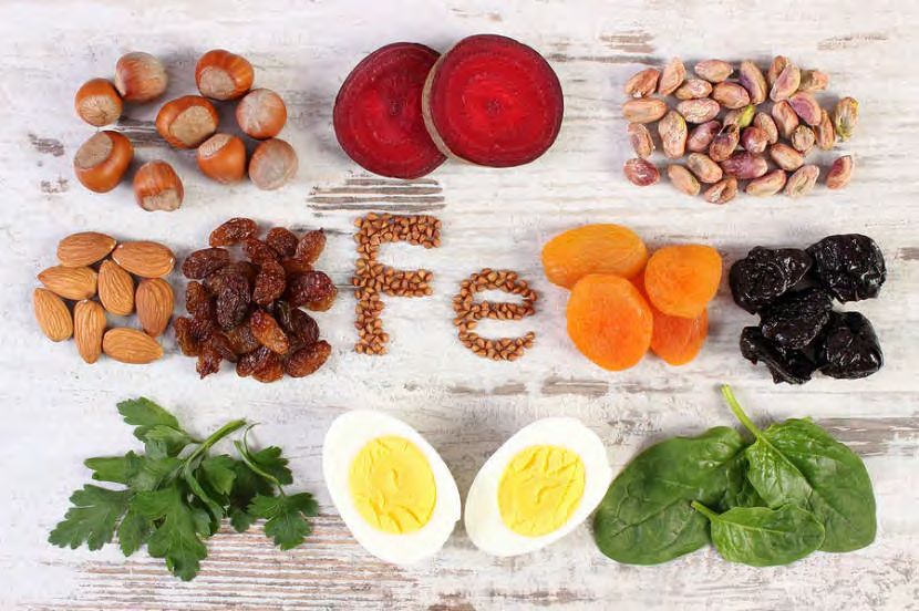
THE OOCYTE
                                                                                                                        Mature human oocyte
                                                                                                                        contains more
                                                                                                                        mitochondria and
                                                                                                                        mtDNA than other cell
                                                                                                                        types
                                                                                                                        Mitochondria - key
                                                                                                                        factors mediating
                                                                                                                        reproductive
                                                                                                                        competence
Cecchino, G., Seli, E., Alves da Motta, E., & García-Velasco, J. (2018). The role of mitochondrial activity in female fertility and assisted reproductive technologies: overview and current insights. Reproductive
Biomedicine Online, 36(6), 686–697. https://doi.org/10.1016/j.rbmo.2018.02.007

© Leah Hechtman 2020                                                                    www.naturalhealthfertility.com                                      2
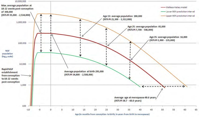
OVARIAN BIOLOGY
20/40 gestation: 6-7 million
Ovarian atresia onwards (independent of ovulation)
At birth: 700,000 follicles
At puberty: 400,000 follicles
Monthly ovulation: 20-30 follicles per cycle

 © Leah Hechtman 2020   www.naturalhealthfertility.com   3
THE HUMAN OOCYTE

© Leah Hechtman 2020   www.naturalhealthfertility.com   4
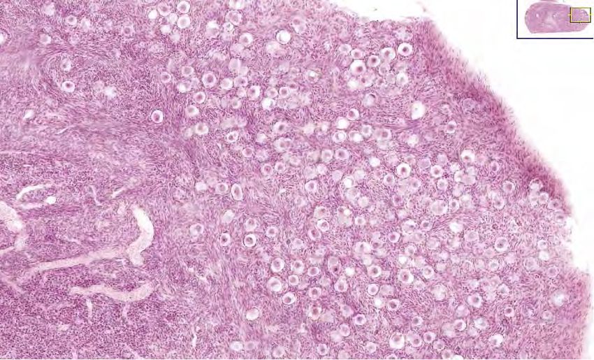
HISTOLOGY OF INFANT OVARIAN
          TISSUE

© Leah Hechtman 2020   www.naturalhealthfertility.com   5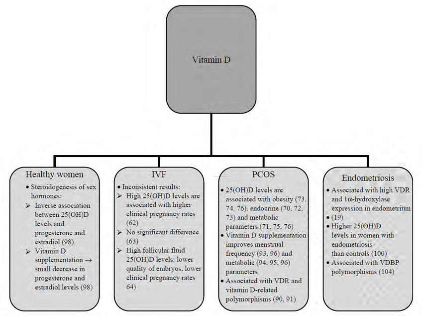
OVARIAN BIOLOGY
     Constant oocyte atresia
     Early menopause or POI/POF can occur in any
     woman
      • a decrease in the initial primordial follicle number
      • an increase in apoptosis or follicle destruction
      • a failure of the follicle to respond to
        gonadotrophin stimulation

                                                             6
© Leah Hechtman 2020        www.naturalhealthfertility.com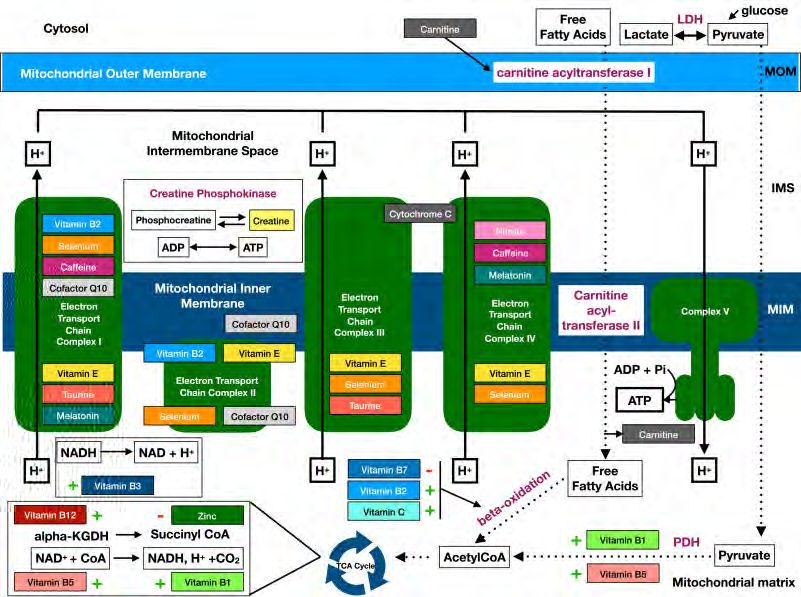
NORMAL
                                          FOLLICULOGENESIS
                                                                                                                                                1. Primordial follicles
                                                                                                                                                2. Primary follicles
                                                                                                                                                3. Preantral follicles
                                                                                                                                                4. Antral follicles
                                                                                                                                                5. Ovulation

Sheikhansari, Golshan, Leili Aghebati-Maleki, Mohammad Nouri, Farhad Jadidi-Niaragh, and Mehdi Yousefi. 2018. ‘Current Approaches for the Treatment of Premature Ovarian Failure with Stem Cell Therapy’. Biomedicine & Pharmacotherapy 102 (June): 254–62.
https://doi.org/10.1016/j.biopha.2018.03.056.
Chan, K. A., M. W. Tsoulis, and D. M. Sloboda. 2015. ‘Early-Life Nutritional Effects on the Female Reproductive System’. Journal of Endocrinology 224 (2): R45–62. https://doi.org/10.1530/JOE-14-0469.

                                                                                                                                                                                                   7
© Leah Hechtman 2020                                                                                              www.naturalhealthfertility.com
OVARIAN FOLLICLE
                      CLASSIFICATION
                                Alternate                                           Size          Size
       Class                  nomenclature     Type              No. of cells   (diameter)    ultrasound
Primordial follicle               Small        1, 2, 3                     25      1000     > 6000µm     18 – 28mm
follicle

       © Leah Hechtman 2020                   www.naturalhealthfertility.com       8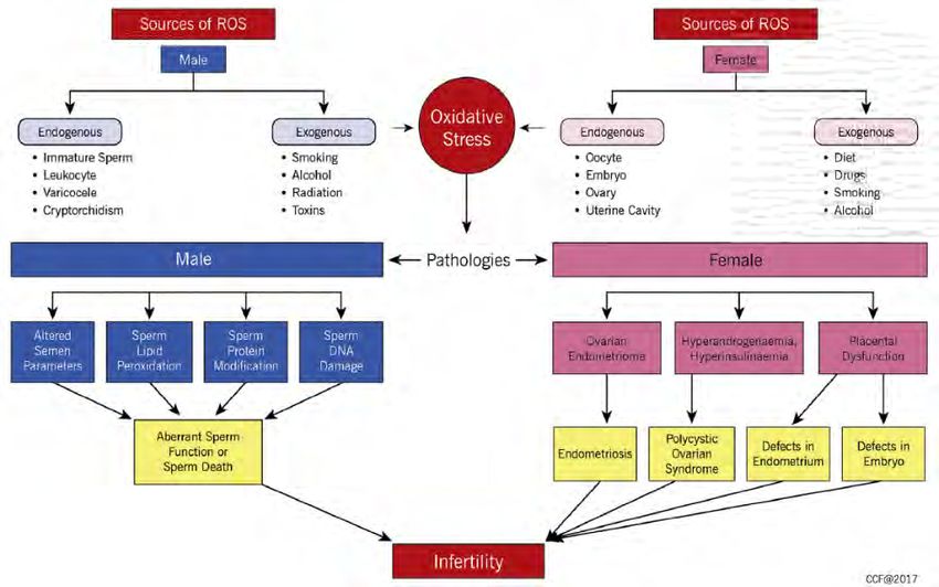
THE HUMAN OOCYTE

© Leah Hechtman 2020   www.naturalhealthfertility.com   9
TRIGENERATIONAL IMPACT

Chan, K. A., M. W. Tsoulis, and D. M. Sloboda. 2015. ‘Early-Life Nutritional Effects on the Female Reproductive System’. Journal of Endocrinology 224 (2): R45–62.
https://doi.org/10.1530/JOE-14-0469.

© Leah Hechtman 2020                                                    www.naturalhealthfertility.com
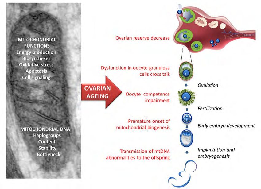
OVARIAN AGEING

         www.naturalhealthfertility.com
FEMALE FERTILITY
Ovarian ageing – decrease in quantity and
quality of oocytes
Aged oocytes – reduced amounts of
mitochondria
Labarta, E., Santos, M. J. de los, Escribá, M. J., Pellicer, A., & Herraiz, S. (2019). Mitochondria as a tool for oocyte rejuvenation. Fertility and Sterility, 111(2), 219–
226. https://doi.org/10.1016/j.fertnstert.2018.10.036
© Leah Hechtman 2020 www.naturalhealthfertility.comMITOCHONDRIAL HEALTH
Mitochondria represent the primary source of ATP
production within oocytes and are critical for normal
oocyte maturation
Embryogenesis is an energy-demanding process,
and oocyte-derived mitochondria are required to
support blastocyst formation
Ovarian ageing is also associated with increased
accumulation of mitochondrial DNA mutations which
are likely to affect mitochondrial biogenesis and
impact oocyte quality
Dumollard, R., Duchen, M., & Carroll, J. (2007). The role of mitochondrial function in the oocyte and embryo. Current Topics in Developmental Biology, 77, 21–49.
https://doi.org/10.1016/S0070-2153(06)77002-8
May-Panloup, P., Boucret, L., Chao de la Barca, J.-M., Desquiret-Dumas, V., Ferré-L’Hotellier, V., Morinière, C., Descamps, P., Procaccio, V., & Reynier, P. (2016). Ovarian ageing: The
role of mitochondria in oocytes and follicles. Human Reproduction Update, 22(6), 725–743. https://doi.org/10.1093/humupd/dmw028
© Leah Hechtman 2020 www.naturalhealthfertility.com 13MITOCHONDRIAL HEALTH
May-Panloup, P., Boucret, L., Chao de la Barca, J.-M., Desquiret-Dumas, V., Ferré-L’Hotellier, V., Morinière, C., Descamps, P., Procaccio, V., & Reynier, P. (2016). Ovarian ageing: The role of mitochondria in
oocytes and follicles. Human Reproduction Update, 22(6), 725–743. https://doi.org/10.1093/humupd/dmw028
© Leah Hechtman 2020 www.naturalhealthfertility.com 14MITOCHONDRIAL HEALTH DNA methylation in oocytes is established during growth Global DNA methylation is low in early oogenesis and peaks as oocytes reach full size DNA methylation is necessary to establish imprinted gene expression The epigenome of the oocyte is dramatically remodelled during oogenesis One sheep study • Subtle, long-term programming effects associated with modest reductions in B vitamin and methionine status around the time of conception • Key components of the methionine cycle within the ovarian follicle were altered, including the ratio of SAM to SAH, which is associated with the extent of DNA methylation © Leah Hechtman 2020 www.naturalhealthfertility.com 15
MITOCHONDRIAL HEALTH
Reik, W. (2001). Epigenetic Reprogramming in Mammalian Development. Science,
293(5532), 1089–1093. https://doi.org/10.1126/science.1063443
Tomizawa, S.-I., Nowacka-Woszuk, J., & Kelsey, G. (2012). DNA methylation establishment
during oocyte growth: Mechanisms and significance. The International Journal of
Developmental Biology, 56(10–12), 867–875. https://doi.org/10.1387/ijdb.120152gk
Tian, X., & Diaz, F. J. (2013). Acute dietary zinc deficiency before conception compromises
oocyte epigenetic programming and disrupts embryonic development. Developmental
Biology, 376(1), 51–61. https://doi.org/10.1016/j.ydbio.2013.01.015
Sinclair, K. D., Allegrucci, C., Singh, R., Gardner, D. S., Sebastian, S., Bispham, J.,
Thurston, A., Huntley, J. F., Rees, W. D., Maloney, C. A., Lea, R. G., Craigon, J., McEvoy, T.
G., & Young, L. E. (2007). DNA methylation, insulin resistance, and blood pressure in
offspring determined by maternal periconceptional B vitamin and methionine status.
Proceedings of the National Academy of Sciences of the United States of America,
104(49), 19351–19356. https://doi.org/10.1073/pnas.0707258104

  © Leah Hechtman 2020              www.naturalhealthfertility.com
SOURCES OF OXIDATIVE
                  STRESS

Roychoudhury, S., Agarwal, A., Virk, G., & Cho, C.-L. (2017). Potential role of green tea catechins in the management of oxidative stress-associated infertility.
Reproductive Biomedicine Online, 34(5), 487–498. https://doi.org/10.1016/j.rbmo.2017.02.006

          © Leah Hechtman 2020                                      www.naturalhealthfertility.com
NUTRITION AND
MITOCHONDRIAL HEALTH
Increase respiratory chain flux (e.g. CoQ10,
riboflavin)
Serve as antioxidants (e.g. CoQ10, ALA, vitamin
C and E), and/or act as cofactors (e.g. riboflavin,
thiamine)
Function as mitochondrial substrates (e.g. L-
carnitine)
Hirano, M., Emmanuele, V., & Quinzii, C. M. (2018). Emerging therapies for mitochondrial diseases. Essays in Biochemistry, 62(3), 467–481.
https://doi.org/10.1042/EBC20170114
© Leah Hechtman 2020 www.naturalhealthfertility.comMITOCHONDRIAL DYSFUNCTION
AND BIOACTIVE FOOD
Mafra, D., Gidlund, E.-K., Borges, N. A., Magliano, D. C., Lindholm, B., Stenvinkel, P., & von Walden, F. (2018). Bioactive food and exercise in chronic kidney
disease: Targeting the mitochondria. European Journal of Clinical Investigation, 48(11), e13020. https://doi.org/10.1111/eci.13020
© Leah Hechtman 2020 www.naturalhealthfertility.comOVERVIEW OF RELEVANT
NUTRIENTS IN BIOENERGETIC
MITOCHONDRIAL PROCESSES
Wesselink, E., Koekkoek, W. a. C., Grefte, S., Witkamp, R. F., & van Zanten, A. R. H. (2018). Feeding mitochondria: Potential role of nutritional components to
improve critical illness convalescence. Clinical Nutrition (Edinburgh, Scotland). https://doi.org/10.1016/j.clnu.2018.08.032
© Leah Hechtman 2020
www.naturalhealthfertility.comANTIOXIDANTS
Mitochondria-targeted antioxidants have shown
great potential because they cross the
mitochondrial phospholipid bilayer and eliminate
ROS at the heart of the source
Some conflicting evidence – more research
needed
Oyewole, A. O., & Birch-Machin, M. A. (2015). Mitochondria-targeted antioxidants. FASEB Journal: Official Publication of the Federation of American Societies for
Experimental Biology, 29(12), 4766–4771. https://doi.org/10.1096/fj.15-275404
© Leah Hechtman 2020 www.naturalhealthfertility.comCOENZYME Q10 © Leah Hechtman 2020 www.naturalhealthfertility.com 22
COENZYME Q10 (COQ10)
        Highest concentration in the mitochondria
        Decrease in mitochondrial activity associated with
        CoQ10 deficiency affects the granulosa cells’
        capacity to generate ATP
        Supplementation delayed age-mediated oocyte loss
        CoQ10 production slows with ageing, making the
        body less effective at protecting the eggs from
        oxidative damage
Ben-Meir, A., Burstein, E., Borrego-Alvarez, A., Chong, J., Wong, E., Yavorska, T., … Jurisicova, A. (2015). Coenzyme Q10 restores oocyte mitochondrial function and fertility during reproductive aging. Aging
Cell, 14(5), 887–895. https://doi.org/10.1111/acel.12368
Ben-Meir, A., Yahalomi, S., Moshe, B., Shufaro, Y., Reubinoff, B., & Saada, A. (2015). Coenzyme Q-dependent mitochondrial respiratory chain activity in granulosa cells is reduced with aging. Fertility and
Sterility, 104(3), 724–727. https://doi.org/10.1016/j.fertnstert.2015.05.023
Özcan, P., Fıçıcıoğlu, C., Kizilkale, O., Yesiladali, M., Tok, O. E., Ozkan, F., & Esrefoglu, M. (2016). Can Coenzyme Q10 supplementation protect the ovarian reserve against oxidative damage? Journal of Assisted
Reproduction and Genetics, 33(9), 1223–1230. https://doi.org/10.1007/s10815-016-0751-z
Hernández-Camacho, J. D., Bernier, M., López-Lluch, G., & Navas, P. (2018). Coenzyme Q10 Supplementation in Aging and Disease. Frontiers in Physiology, 9, 44. https://doi.org/10.3389/fphys.2018.00044
Xu, Y., Nisenblat, V., Lu, C., Li, R., Qiao, J., Zhen, X., & Wang, S. (2018). Pretreatment with coenzyme Q10 improves ovarian response and embryo quality in low-prognosis young women with decreased ovarian
reserve: a randomized controlled trial. Reproductive Biology and Endocrinology : RB&E, 16. https://doi.org/10.1186/s12958-018-0343-

             © Leah Hechtman 2020                                                      www.naturalhealthfertility.com
COENZYME Q10 (COQ10)
Lowered aneuploidy rate
Delayed ovarian reserve depletion
Restored oocyte mitochondrial gene expression
Improved mitochondrial activity and distribution
Lowered ROS levels in oocytes
Increased mitochondrial mass and polarization
Increased ATP levels in oocytes
Cecchino, G., Seli, E., Alves da Motta, E., & García-Velasco, J. (2018). The role of mitochondrial activity in female fertility and assisted reproductive technologies: overview and
current insights. Reproductive Biomedicine Online, 36(6), 686–697. https://doi.org/10.1016/j.rbmo.2018.02.007
© Leah Hechtman 2020 www.naturalhealthfertility.comCOENZYME Q10 (COQ10) Teran, E., Hernández, I., Tana, L., Teran, S., Galaviz-Hernandez, C., Sosa-Macías, M., … Calle, A. (2018). Mitochondria and Coenzyme Q10 in the Pathogenesis of Preeclampsia. Frontiers in Physiology, 9, 1561. https://doi.org/10.3389/fphys.2018.01561 © Leah Hechtman 2019 www.naturalhealthfertility.com 25
COQ10: TRANSLATE INTO
               PRACTICE
     Form and delivery
     Dose
     Frequency
     Duration of treatment
     Response prediction and outcome assessment

© Leah Hechtman 2020   www.naturalhealthfertility.com   26
IRON © Leah Hechtman 2020 www.naturalhealthfertility.com 27
IRON
       Greater intake (dietary or supplemental) =
       increased fertility due to ovarian utilization
       One study – 18555 women
         • Supplemented – significantly lower risk of ovulatory
           infertility
         • Non-haem iron preferred over haem-derived

Buhling, K. J., & Grajecki, D. (2013). The effect of micronutrient supplements on female fertility. Current Opinion in Obstetrics & Gynecology, 25(3), 173–180.
https://doi.org/10.1097/GCO.0b013e3283609138
Chavarro, J. E., Rich-Edwards, J. W., Rosner, B. A., & Willett, W. C. (2006). Iron intake and risk of ovulatory infertility. Obstetrics and Gynecology, 108(5), 1145–
1152. https://doi.org/10.1097/01.AOG.0000238333.37423.ab

© Leah Hechtman 2020                                                 www.naturalhealthfertility.com                       28
IRON
Peripheral tissues for synthesizing ATP and
protein – lack of ATP = ↓ cells’ ability to synthsize
DNA and mRNA
• cofactor in the expression and activation of various
metabolic enzymes involved in glycolysis
• TCA cycle
• Electron chain transfer
• Pentose phosphate pathways
© Leah Hechtman 2020 www.naturalhealthfertility.com 29Barrientos, T., Laothamatas, I., Koves, T. R., Soderblom, E. J., Bryan, M., Moseley, M. A.,
Muoio, D. M., & Andrews, N. C. (2015). Metabolic Catastrophe in Mice Lacking Transferrin
Receptor in Muscle. EBioMedicine, 2(11), 1705–1717.
https://doi.org/10.1016/j.ebiom.2015.09.041
Li, Y. Q., Cao, X. X., Bai, B., Zhang, J. N., Wang, M. Q., & Zhang, Y. H. (2014). Severe iron
deficiency is associated with a reduced conception rate in female rats. Gynecologic and
Obstetric Investigation, 77(1), 19–23. https://doi.org/10.1159/000355112
Dhur, A., Galan, P., & Hercberg, S. (1989). Effects of different degrees of iron deficiency on
cytochrome P450 complex and pentose phosphate pathway dehydrogenases in the rat.
The Journal of Nutrition, 119(1), 40–47. https://doi.org/10.1093/jn/119.1.40
Chitambar, C. R., & Narasimhan, J. (1991). Targeting iron-dependent DNA synthesis with
gallium and transferrin-gallium. Pathobiology: Journal of Immunopathology, Molecular and
Cellular Biology, 59(1), 3–10. https://doi.org/10.1159/000163609
© Leah Hechtman 2020 www.naturalhealthfertility.com 30IRON
Recent study
• Low iron = low oestrous cycle = impaired follicle
development and fertility
• Lowered iron in serum, liver, ovaries
• Lowered ATP in ovaries and mRNA expressions of follicle
development markers (Fshr, Cyp19a1 and Ccnd2 mRNA)
• Lowered oestrogen production
• Failed follicle development and fertility
Tonai, S., Kawabata, A., Nakanishi, T., Lee, J. Y., Okamoto, A., Shimada, M., & Yamashita, Y. (2020). Iron deficiency induces female infertile in order to failure of
follicular development in mice. The Journal of Reproduction and Development. https://doi.org/10.1262/jrd.2020-074
© Leah Hechtman 2020 www.naturalhealthfertility.com 31IRON OVERLOAD
Excess iron leads to reduced production of LH and FSH
from the anterior pituitary, suggesting impaired oocyte
maturation and low ovarian reserve
In patients with beta-thalassemia, multiple blood
transfusions and increased gastrointestinal iron
absorption leads to iron overload in the body and
infertility
Haemochromatosis -> leads to subfertility
Thalassemia = low AMH and low AFC = harm to ovarian
reserve
© Leah Hechtman 2020 www.naturalhealthfertility.com 32IRON OVERLOAD
Singer, S. T., Vichinsky, E. P., Gildengorin, G., van Disseldorp, J., Rosen, M., & Cedars, M. I. (2011). Reproductive
capacity in iron overloaded women with thalassemia major. Blood, 118(10), 2878–2881.
https://doi.org/10.1182/blood-2011-06-360271
Mishra, A. K., & Tiwari, A. (2013). Iron Overload in Beta Thalassaemia Major and Intermedia Patients.Mædica,
8(4), 328–332.
Tweed, M. J., & Roland, J. M. (1998). Haemochromatosis as an endocrine cause of subfertility.BMJ : British
Medical Journal, 316(7135), 915–916.
Chang, H.-H., Chen, M.-J., Lu, M.-Y., Chern, J. P. S., Lu, C.-Y., Yang, Y.-L., Jou, S.-T., Lin, D.-T., Yang, Y.-S., &
Lin, K.-H. (2011). Iron overload is associated with low anti-müllerian hormone in women with transfusion-
dependent β-thalassaemia. BJOG: An International Journal of Obstetrics and Gynaecology, 118(7), 825–831.
https://doi.org/10.1111/j.1471-0528.2011.02927.x
Roussou, P., Tsagarakis, N. J., Kountouras, D., Livadas, S., & Diamanti-Kandarakis, E. (2013). Beta-Thalassemia
Major and Female Fertility: The Role of Iron and Iron-Induced Oxidative Stress. Anemia, 2013.
https://doi.org/10.1155/2013/617204
Uysal, A., Alkan, G., Kurtoğlu, A., Erol, O., & Kurtoğlu, E. (2017). Diminished ovarian reserve in women with
transfusion-dependent beta-thalassemia major: Is iron gonadotoxic? European Journal of Obstetrics, Gynecology,
and Reproductive Biology, 216, 69–73. https://doi.org/10.1016/j.ejogrb.2017.06.038
Mensi, L., Borroni, R., Reschini, M., Cassinerio, E., Vegetti, W., Baldini, M., Cappellini, M. D., & Somigliana, E.
(2019). Oocyte quality in women with thalassaemia major: Insights from IVF cycles. European Journal of
Obstetrics & Gynecology and Reproductive Biology: X, 3, 100048. https://doi.org/10.1016/j.eurox.2019.100048
© Leah Hechtman 2020 www.naturalhealthfertility.com 33IRON AND ENDOMETRIOSIS Defrère, S., Lousse, J. C., González-Ramos, R., Colette, S., Donnez, J., & Van Langendonckt, A. (2008). Potential involvement of iron in the pathogenesis of peritoneal endometriosis. Molecular Human Reproduction, 14(7), 377–385. https://doi.org/10.1093/molehr/gan033 © Leah Hechtman 2020 www.naturalhealthfertility.com 34
IRON THROUGH THE
PREGNANCY
Milman, N. (2011). Iron in pregnancy: How do we secure an appropriate iron status in the mother and child? Annals of Nutrition & Metabolism, 59(1), 50–54.
https://doi.org/10.1159/000332129
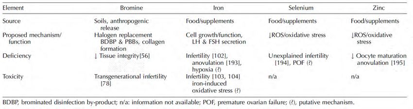
© Leah Hechtman 2020 www.naturalhealthfertility.com 35TRACE ELEMENTS AND
PROPOSED EFFECT ON
OVARIAN FUNCTION
© Leah Hechtman 2020 www.naturalhealthfertility.com 36IRON: TRANSLATE INTO
PRACTICE
Form and delivery
Dose
Frequency
Duration of treatment
Response prediction and outcome assessment
© Leah Hechtman 2020 www.naturalhealthfertility.com 37VITAMIN D © Leah Hechtman 2020 www.naturalhealthfertility.com 38
EFFECT OF VITAMIN D ON
                 TISSUES AND FERTILITY

Lerchbaum, E., & Obermayer-Pietsch, B. (2012). Vitamin D and fertility: A systematic review. European Journal of Endocrinology, 166(5), 765–778.
https://doi.org/10.1530/EJE-11-0984

          © Leah Hechtman 2018                                   www.naturalhealthfertility.com                     39
VITAMIN D
Even before pregnancy, vitamin D initiates
and/or sustains actions to facilitate fertilization
and implantation
Hypovitaminosis D is known to lead to
subfertility, infertility and pathological alteration
of critical reproductive tissues, such as the
endometrium
© Leah Hechtman 2020 www.naturalhealthfertility.com 40VITAMIN D
Blomberg Jensen, M. (2014). Vitamin D and male reproduction. Nature Reviews. Endocrinology, 10(3), 175–186.
https://doi.org/10.1038/nrendo.2013.262
Dabrowski, F., Grzechocinska, B., & Wielgos, M. (2015). The Role of Vitamin D in Reproductive Health—A Trojan
Horse or the Golden Fleece? Nutrients, 7(6), 4139–4153. https://doi.org/10.3390/nu7064139
Luk, J., Torrealday, S., Neal Perry, G., & Pal, L. (2012). Relevance of vitamin D in reproduction. Human
Reproduction (Oxford, England), 27(10), 3015–3027. https://doi.org/10.1093/humrep/des248
Refaat, B., Ahmad, J., Idris, S., Kamfar, F. F., Ashshi, A. M., Batwa, S. A., & Malibary, F. A. (2017).
Characterisation of vitamin D-related molecules and calcium-sensing receptor in human Fallopian tube during the
menstrual cycle and in ectopic pregnancy. Cell and Tissue Research, 368(1), 201–213.
https://doi.org/10.1007/s00441-016-2519-2
Shin, J. S., Choi, M. Y., Longtine, M. S., & Nelson, D. M. (2010). Vitamin D Effects on Pregnancy and the
Placenta. Placenta, 31(12), 1027–1034. https://doi.org/10.1016/j.placenta.2010.08.015
Anagnostis, P., Karras, S., & Goulis, D. G. (2013). Vitamin D in human reproduction: A narrative review: Vitamin D
and reproduction. International Journal of Clinical Practice, 67(3), 225–235. https://doi.org/10.1111/ijcp.12031
Blomberg Jensen, M., Gerner Lawaetz, J., Andersson, A.-M., Petersen, J. H., Nordkap, L., Bang, A. K., Ekbom, P.,
Joensen, U. N., Prætorius, L., Lundstrøm, P., Boujida, V. H., Lanske, B., Juul, A., & Jørgensen, N. (2016). Vitamin
D deficiency and low ionized calcium are linked with semen quality and sex steroid levels in infertile men. Human
Reproduction (Oxford, England), 31(8), 1875–1885. https://doi.org/10.1093/humrep/dew152
© Leah Hechtman 2020 www.naturalhealthfertility.com 41VITAMIN D RECEPTOR (VDR)
Interest in the reproductive functions of vitamin D
surfaced following the discovery of the vitamin D
receptor (VDR) and the metabolizing enzyme 1α-
hydroxylase in the decidua, placenta, ovary,
endometrium and pituitary gland
VDR is expressed in ovarian granulosa cells and
fallopian epithelial cells and this expression
increases during pregnancy
Dabrowski, F., Grzechocinska, B., & Wielgos, M. (2015). The Role of Vitamin D in Reproductive Health—A Trojan Horse or the Golden Fleece? Nutrients, 7(6),
4139–4153. https://doi.org/10.3390/nu7064139
Mousa, A., Abell, S., Scragg, R., & de Courten, B. (2016). Vitamin D in Reproductive Health and Pregnancy. Seminars in Reproductive Medicine, 34(2), e1-13.
https://doi.org/10.1055/s-0036-1583529
© Leah Hechtman 2020 www.naturalhealthfertility.com 42VITAMIN D: HORMONE
REGULATION
Placental steroidogenesis
Decidualization of the endometrium through
different signalling pathways of the VDR
Aleyasin, A., Hosseini, M. A., Mahdavi, A., Safdarian, L., Fallahi, P., Mohajeri, M. R., Abbasi, M., & Esfahani, F. (2011). Predictive value of the level of vitamin D in
follicular fluid on the outcome of assisted reproductive technology. European Journal of Obstetrics, Gynecology, and Reproductive Biology, 159(1), 132–137.
https://doi.org/10.1016/j.ejogrb.2011.07.006
© Leah Hechtman 2020 www.naturalhealthfertility.com 43VITAMIN D: IMPACT TO OVARIAN
TISSUE
Cholecalciferol stimulates
• Progesterone production by 13%
• Oestradiol production by 9%
• Oestrone production by 21%
Parikh, G., Varadinova, M., Suwandhi, P., Araki, T., Rosenwaks, Z., Poretsky, L., & Seto-Young, D. (2010). Vitamin D regulates steroidogenesis and insulin-like
growth factor binding protein-1 (IGFBP-1) production in human ovarian cells. Hormone and Metabolic Research = Hormon- Und Stoffwechselforschung =
Hormones Et Metabolisme, 42(10), 754–757. https://doi.org/10.1055/s-0030-1262837
© Leah Hechtman 2020 www.naturalhealthfertility.com 44VITAMIN D: AMH PATTERNING
Vitamin D changes AMH production patterns in
ovarian granulosa cells and alters FSH
sensitivity
• Ovarian follicle development
• Ovarian reserve preservation
• PCOS women: Abnormal AMH levels normalized with
Vitamin D
Merhi, Z. (2014). Advanced glycation end products and their relevance in female reproduction. Human Reproduction, 29(1), 135–145.
https://doi.org/10.1093/humrep/det383
Dabrowski, F., Grzechocinska, B., & Wielgos, M. (2015). The Role of Vitamin D in Reproductive Health—A Trojan Horse or the Golden Fleece? Nutrients, 7(6),
4139–4153. https://doi.org/10.3390/nu7064139
© Leah Hechtman 2020 www.naturalhealthfertility.com 45PROPOSED ASSOCIATIONS OF
VITAMIN D STATUS WITH
FEMALE REPRODUCTION
Lerchbaum, E., & Obermayer-Pietsch, B. (2012). Vitamin D and fertility: A systematic review. European Journal of Endocrinology, 166(5), 765–778.
https://doi.org/10.1530/EJE-11-0984
© Leah Hechtman 2020 www.naturalhealthfertility.com 46VITAMIN D: TRANSLATE INTO
PRACTICE
Form and delivery
Dose
Frequency
Duration of treatment
Response prediction and outcome assessment
© Leah Hechtman 2020 www.naturalhealthfertility.com 47Thank you © Leah Hechtman 2020 www.naturalhealthfertility.com 48
Q&A

© Leah Hechtman 2020   www.naturalhealthfertility.com   49
You can also read