The Mechanosensory Structure of the Hair Cell Requires Clarin-1, a Protein Encoded by Usher Syndrome III Causative Gene
←
→
Page content transcription
If your browser does not render page correctly, please read the page content below
The Journal of Neuroscience, July 11, 2012 • 32(28):9485–9498 • 9485

Cellular/Molecular

The Mechanosensory Structure of the Hair Cell Requires
Clarin-1, a Protein Encoded by Usher Syndrome III
Causative Gene
Ruishuang Geng,1 Sami Melki,1 Daniel H.-C. Chen,1 Guilian Tian,2 David N. Furness,3 Tomoko Oshima-Takago,4
Jakob Neef,4 Tobias Moser,4 Charles Askew,5 Geoff Horwitz,5 Jeffrey R. Holt,6 Yoshikazu Imanishi,2
and Kumar N. Alagramam1
1Otolaryngology Head and Neck Surgery, University Hospitals Case Medical Center and 2Pharmacology, Case Western Reserve University, Cleveland, Ohio
44106, 3Institute for Science and Technology in Medicine, School of Life Sciences, Keele University, Staffordshire, ST5 5BG, United Kingdom, 4Department
of Otolaryngology and Center for Molecular Physiology of the Brain, University of Goettingen, D-37073 Goettingen, Germany, 5Department of
Neuroscience, University of Virginia School of Medicine, Charlottesville, Virginia 22908, and 6Department of Otolaryngology, Children’s Hospital Boston
and Harvard Medical School, Boston, Massachusetts 02115

Mutation in the clarin-1 gene (Clrn1) results in loss of hearing and vision in humans (Usher syndrome III), but the role of clarin-1 in the sensory
hair cells is unknown. Clarin-1 is predicted to be a four transmembrane domain protein similar to members of the tetraspanin family. Mice
carrying null mutation in the clarin-1 gene (Clrn1⫺/⫺) show loss of hair cell function and a possible defect in ribbon synapse. We investigated the
role of clarin-1 using various in vitro and in vivo approaches. We show by immunohistochemistry and patch-clamp recordings of Ca 2⫹ currents
and membrane capacitance from inner hair cells that clarin-1 is not essential for formation or function of ribbon synapse. However,
reduced cochlear microphonic potentials, FM1-43 [N-(3-triethylammoniumpropyl)-4-(4-(dibutylamino)styryl) pyridinium dibromide]
loading, and transduction currents pointed to diminished cochlear hair bundle function in Clrn1⫺/⫺ mice. Electron microscopy of
cochlear hair cells revealed loss of some tall stereocilia and gaps in the v-shaped bundle, although tip links and staircase arrangement of
stereocilia were not primarily affected by Clrn1⫺/⫺ mutation. Human clarin-1 protein expressed in transfected mouse cochlear hair cells
localized to the bundle; however, the pathogenic variant p.N48K failed to localize to the bundle. The mouse model generated to study the
in vivo consequence of p.N48K in clarin-1 (Clrn1N48K) supports our in vitro and Clrn1⫺/⫺ mouse data and the conclusion that CLRN1 is an
essential hair bundle protein. Furthermore, the ear phenotype in the Clrn1N48K mouse suggests that it is a valuable model for ear disease
in CLRN1N48K, the most prevalent Usher syndrome III mutation in North America.

Introduction                                                                                                                      understanding hair cell biology and hearing loss linked to bundle
Hearing and balance rely on hair cells, specialized epithelial cells                                                              pathology. Mouse mutants have played an important role; many
in the inner ear that develop a set of stereocilia (hair bundle) at                                                               of the deaf mutants have defects in the sensory hair bundle and
their apical surface. The hair bundle detects vibration-induced                                                                   provide in vivo models for investigating gene functions in the hair
movements in the ear and transduces these stimuli into electrical                                                                 bundle (Leibovici et al., 2008).
signals. Investigating gene function in the bundle is critical to                                                                     Usher syndrome (USH), an autosomal recessive disorder, ac-
                                                                                                                                  counts for ⬃50% of the cases of combined inherited blindness
Received Jan. 21, 2012; revised April 26, 2012; accepted May 8, 2012.
                                                                                                                                  and deafness (Saihan et al., 2009). USH type III (USH3) is caused
    Author contributions: R.G., G.T., T.M., J.R.H., Y.I., and K.N.A. designed research; R.G., S.M., D.H.-C.C., G.T., D.N.F.,      by mutation in the clarin-1 gene (CLRN1) and is characterized by
T.O.-T., J.N., T.M., C.A., G.H., and Y.I. performed research; R.G., S.M., D.N.F., J.N., T.M., J.R.H., Y.I., and K.N.A. analyzed   progressive hearing loss and variable balance impairment (Joen-
data; R.G. and K.N.A. wrote the paper.                                                                                            suu et al., 2001; Ness et al., 2003). The primary structure of the
    This work was supported by Usher III Initiative (Y.I. and K.N.A.), Elden foundation (Y.I.), National Institutes of
Health Grants R01-DC010816 (K.N.A.) and R01-DC05439 (J.R.H.), and Deafness Research UK funds (D.F.), and a grant
                                                                                                                                  clarin-1 protein (CLRN1) predicts four transmembrane domains
from the German Research Foundation (T.M.) via the Collaborative Research Center 889, and the state of Lower                      similar to a large family of membrane proteins, including tetras-
Saxony (through a “Neurosenses” PhD fellowship to T.O.-T.). We are grateful to Richard Lee and Corrine Thai for their             panins and claudins (Adato et al., 2002). Based on in vitro and
help in the process of generating N48K knock-in mice and Chie Iioka for her help in generating the CLRN1–YFP
                                                                                                                                  biochemical assays, Tian et al. (2009) posited a possible role for
construct. The monoclonal antibody ␣5 developed by D. M. Fambrough was obtained from the Developmental
Studies Hybridoma Bank developed under the auspices of the National Institute of Child Health and Human Devel-                    CLRN1 in the regulation and homeostasis of actin filaments. We
opment and maintained by The University of Iowa, Department of Biological Sciences (Iowa City, IA).                               reported the first animal model for ear disease in USH3: a mouse
    The authors declare no competing financial interests.                                                                         carrying a null allele of Clrn1 (Clrn1⫺/⫺) (Geng et al., 2009).
    Correspondence should be addressed to Dr. Kumar Alagramam, Otolaryngology Head and Neck Surgery, Univer-
                                                                                                                                  Clrn1⫺/⫺ mice showed early-onset profound hearing loss and
sity Hospitals Case Medical Center, Case Western Reserve University, Cleveland, OH 44106. E-mail: kna3@case.edu.
    DOI:10.1523/JNEUROSCI.0311-12.2012                                                                                            variable balance impairment. Elevated auditory-evoked brains-
Copyright © 2012 the authors 0270-6474/12/329485-14$15.00/0                                                                       tem response (ABR) and vestibular evoked potential thresholds
9486 • J. Neurosci., July 11, 2012 • 32(28):9485–9498 Geng et al. • Clarin-1 Is an Essential Bundle Protein
in Clrn1⫺/⫺ mice were associated with reduced amplitudes and Cochlear microphonics and CAP. Cochlear microphonic (CM) and
delayed latencies of the compound action potential (CAP) of CAP recordings were conducted as a variant of ABR described previ-
cochlear and vestibular ganglion neurons. The cochlear hair bun- ously. With this setup, we stimulated the ear with a 100 s rectangular
dle structure was disrupted in Clrn1⫺/⫺ mice at a young age pure tone stimulus at a firing rate of 20 per second. Stimuli were pre-
sented through a rubber tube from a Tucker Davis Technology speaker
[postnatal day 2 (P2) to P10] without concomitant loss of gan-
directly to the studied ear. Then, 1024 sweeps were recorded and aver-
glion cells. We speculated that a hair bundle defect caused the
aged in the condensation and rarefaction phases separately. The two
elevated threshold, and the observed delay in peak latency is a resulting waves were then added to extract the CAP (Henry and Chole,
secondary consequence of the hair bundle defect in Clrn1⫺/⫺ 1979). The CM was visualized in the condensation and rarefaction trac-
mice. Alternatively, the mutant phenotype could be attributable ings. The CM amplitude was measured as the difference between the
to a defect in hair cell-to-afferent neuron communication (a rib- maximum peak of a given polarity and the maximum peak of the oppo-
bon synapse defect). These findings led us to two mutually non- site polarity (Santarelli et al., 2006). The maximum intensity of 110 dB
exclusive hypotheses: (1) the primary role of CLRN1 is in SPL was then used and decreased by 10 dB steps until the threshold was
maintenance of the structural integrity of the hair bundle, and/or reached. The threshold was assessed as the intensity reproduced a clearly
(2) CLRN1 is essential for hair cell ribbon synapse formation or identifiable CAP. The CM was measured from peak to trough at ⬃1 ms
function. To test these hypotheses, we performed a series of ex- after recording started for all the mice. The stimulus used to measure the
CM is the condensation stimulus. Data were collected in Excel (Mi-
periments focusing on the hair bundle and ribbon synapses of the
crosoft) and analyzed using a Mann–Whitney U test for nonparametric
Clrn1⫺/⫺ mice. Furthermore, we explored the consequence of a data in the MedCalc program. A one-tailed p value ⬍0.05 was considered
missense mutation CLRN1N48K (p.N48K) in the context of the significant. This test was conducted at all three frequencies tested, namely
hair cells in vitro and in vivo. Here we report, for the first time, the 2, 4, and 8 kHz. For technical reasons linked to the setup of the Tucker Davis
generation of the Clrn1N48K (knock-in) mouse model for ear phe- Technologies software/hardware, we could not record CM responses at fre-
notypes in CLRN1N48K patients. Our investigation has defined quencies ⬎8 kHz. Clrn1⫺/⫺ mice were tested at ages P18 –P19 before the
the role of clarin-1 in sensory hair cells and has uncovered a onset of profound hearing loss (P21–P25) (Clrn1⫹/⫺, n ⫽ 8; Clrn1⫺/⫺, n ⫽
pathogenic mechanism for the most prevalent North American 9). Therefore, we chose a younger time point (P18 –P19), when some hear-
USH3 mutation (Adato et al., 2002; Fields et al., 2002). ing function was retained.
The results are shown as a scatter plot, as well as a box and whisker plot.
The scatter plot shows the spread of raw data. In the box and whisker
Materials and Methods plot, the central box represents the values from the 25th to 75th percen-
The genes and proteins used in this study. The following are the genes used: tiles. The middle line is the median. A line extends from the minimum to
Clrn1⫺/⫺, homozygous knock-out allele; Clrn1⫹/⫺, heterozygous knock- the maximum value, excluding “outside” and “far-out” values, which are
out allele; Clrn1⫹/N48K, heterozygous N48K knock-in allele; Clrn1N48K/N48K, displayed outside the error bars. An outside value is defined as a value
homozygous N48K knock-in allele; CLRN1, human clarin-1 gene; hCLRN1, that is smaller than the lower quartile minus 1.5 times the interquar-
human clarin-1 protein; Clrn1, mouse clarin-1 gene; mCLRN1, mouse tile range, or larger than the upper quartile plus 1.5 times the interquar-
clarin-1 protein; clarin-1, a general reference to the gene and protein in more tile range. A far-out value is defined as a value that is smaller than the
than one species. lower quartile minus 3 times the interquartile range, or larger than the
Mice. The use of mice at Case Western Reserve University (CWRU) upper quartile plus 3 times the interquartile range. The error bars within
was approved by the Institutional Animal Care and Use Committee (pro- the whiskers are the 95% confidence interval of the mean.
tocol 2010-0074). Unless stated otherwise, all mice used in this study Cochlear culture preparation. Cochlear organ cultures were prepared
were derived from homozygous-by-heterozygous mating, and mice of from mutant and control mice at 0 –3 d of age as described previously
either sex were used. Mice heterozygous for the clarin-1 mutation were (Russell and Richardson, 1987) and maintained on Cell-Tak-coated glass
indistinguishable from their wild-type siblings and, therefore, used as the coverslips in Maximow side assemblies at 37°C in a medium containing
control for all experiments. 90% MEM, 10% horse serum, 10 mM HEPES, pH 7.2, and 10 mg/ml
ABRs. ABR recording was conducted as described previously (Geng et ampicillin for 24 h before use.
al., 2009). Briefly, mice aged P18 and older were anesthetized with a FM1-43 labeling. FM1-43 [N-(3-triethylammoniumpropyl)-4-(4-
dilute intraperitoneal injection of ketamine, xylazine, and acepromazine (dibutylamino)styryl) pyridinium dibromide] dye loading experiments
at doses of 40, 5, and 1 mg/kg, respectively. Their body temperature was were performed as described previously (Gale et al., 2001; Alagramam et
maintained at 37–38°C by placing them on a homeothermic heating pad al., 2011). In brief, coverslips with adherent cochlear cultures were
(Harvard Apparatus), and they were placed in a soundproof chamber washed once with HEPES-buffered (10 mM, pH 7.2) HBSS (HBHBSS),
during testing. ABR testing was performed using a SmartEP system from dipped for 10 s in HBHBSS containing 3 mM FM1-43, and washed im-
Intelligent Hearing Systems. Platinum subdermal needle electrodes were mediately three times in a large volume of HBHBSS (10 s for each wash).
inserted at the vertex and ventrolaterally to the right ear and left ear. To The coverslips were then placed on a glass slide and viewed with an
test hearing function, mutant mice were presented with 16 kHz pure tone upright microscope equipped with epifluorescence optics and FITC fil-
stimuli at a stimulus intensity starting at 100 decibel sound pressure level ters (488 nm excitation, 520 nm emission) using a 40⫻ dry objective.
(dB SPL) and decrementing in 10 dB steps; this sequence was repeated in Images were captured at fixed time points after a dye application using a
5 dB steps until the lowest intensity that evoked a reproducible ABR 12-bit cooled CCD camera (Retiga Exi Aqua Blue; Q-Imaging). FM1-43
waveform (peaks I–IV) was detected. The stimulus was presented for 100 levels were quantified for Clrn1⫹/⫺, Clrn1⫺/⫺, cadherin 23 heterozygous
ms duration and for at least 500 sweeps to both the left and right ear (one (Cdh23⫹/⫺) or cadherin 23 homozygous (Cdh23⫺/⫺) hair cells. We fo-
at a time) through high-frequency transducers (a closed system). For cused on the basal turn for quantification because hair cells are more
each mouse, ABR thresholds from the left and right were averaged and mature at the base compared with the apex in neonates. Images were
used for statistical analysis. Number of mice tested were as follows: imported into Adobe Photoshop CS3 (Adobe Systems), and gray levels
Clrn1⫺/⫺, n ⫽ 12; Clrn1N48K/N48K, n ⫽ 25; and Clrn1 ⫹/N48K, n ⫽ 5. were measured in 24 hair cells from each explant using a 400 pixel (20 ⫻
Statistical method used to analyze ABR data. A one-way ANOVA was 20) region of interest. Data were collected from nine Clrn1⫹/⫺ (n ⫽ 9), 10
used to determine whether the difference in hearing thresholds observed Clrn1⫺/⫺ (n ⫽ 10), three Cdh23⫹/⫺ (n ⫽ 3), and three Cdh23⫺/⫺ (n ⫽ 3)
among the three groups of mice was significant. Briefly, the data were explants. Differential interference contrast (DIC) images were captured
arranged in columns using Excel (Microsoft), and the statistics were to map location of hair cells. Data was exported to Excel (Microsoft) and
conducted with Prism (GraphPad Software). A Bonferroni’s multiple analyzed using the software PAST. Because the sample sizes within each
comparison test was then used to determine which groups were signifi- group were small and unlikely to have a normal distribution, a Mann–
cantly different to account for the overall total difference. Whitney U test was used to compare the two groups. Grayscale levels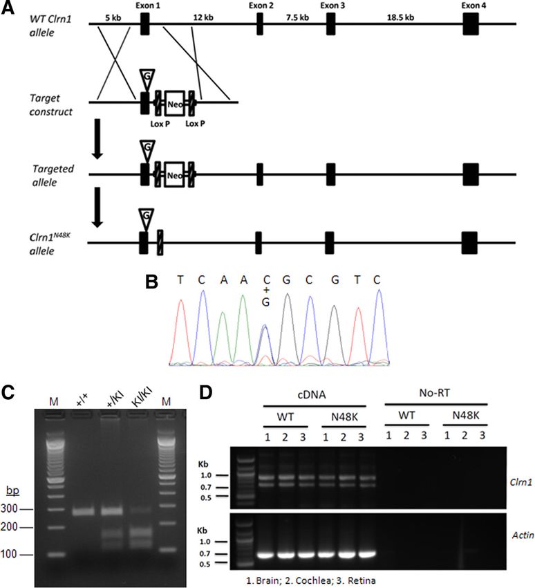
Geng et al. • Clarin-1 Is an Essential Bundle Protein J. Neurosci., July 11, 2012 • 32(28):9485–9498 • 9487 from Clrn1⫹/⫺ and Clrn1⫺/⫺ explants were plotted as a box and whisker low-pass eight-pole Bessel filter (Krohn-Hite) to eliminate residual plot as well as a scatter plot. The Cdh23⫺/⫺ explants showed no uptake, probe resonance. Hair bundle deflections were continuously monitored and it was not plotted. via video microscopy during recording to ensure that the probe and Tissue preparation for electrophysiology. Cochleae and utricles were ex- stereocilia bundle moved in unison. Pictures taken of the probe at static cised from wild-type and Clrn1⫺/⫺ mouse pups at P1–P8 in accordance voltage steps were used to calibrate the stimulus displacement relative to with protocols approved by the Animal Care and Use Committee of the the probe rest position to 2 m in both the positive and negative direc- University of Virginia (Protocol 3123). Briefly, pups were killed by rapid tion (spatial resolution of ⬃4 nm). Video images (temporal resolution of decapitation, and the temporal bone was removed and bathed in MEM ⬃30 ms) of the probe were recorded to confirm on-axis motion. The supplemented with 10 mM HEPES and 0.05 mg/ml ampicillin, pH 7.4. 10 –90% rise time of the probe was ⬃20 s. Utricle sensory epithelia were carefully removed, treated briefly with Expression constructs. Constructs designed to express human CLRN1 or subtilisin to facilitate removal of the otoconia, and mounted as described CLRN1 N48K mutant protein, each fused to an HA epitope sequence, were previously (Holt et al., 1997). Organs of Corti were gently isolated, and reported previously (Tian et al., 2009). The CMV–GFP–Myo15a construct tectorial membranes were mechanically removed. The tissue was then was obtained from Dr. Thomas Friedman (National Institute on Deafness divided in half turns (base and apex) and pinned flat beneath a pair of and Other Communication Disorders, Bethesda, MD). The CMV–YFP thin glass fibers adhered to a round glass coverslip. The hair cells were plasmid vector (mVenus–N1) and the CMV–mouse Prestin–YFP expres- visualized from the apical surface using an upright Axioskop FS micro- sion construct were obtained from Dr. Jian Zuo (St. Jude Children’s Re- scope (Carl Zeiss) equipped with a 63⫻ water-immersion objective with search Hospital, Memphis, TN) (Wu et al., 2007). Constructs designed to differential interference contrast (DIC) optics. Images were acquired express human CLRN1–YFP or CLRN1 N48K–YFP were generated as fol- with a C2400 CCD camera and Argus image processor (Hamamatsu). lows. Full-length human CLRN1 coding sequence (National Center for Bio- Hair cell electrophysiology. The sensory tissue was bathed in standard technology Information Reference Sequence NM_174878) was cloned from artificial perilymph solution containing the following (in mM): 144 NaCl, human retina RNA (Clontech) with primers 5-gtttctcatcatgccaagccaacagaag- 0.7 NaH2PO4, 5.8 KCl, 1.3 CaCl2, 0.9 MgCl2, 5.6 D-glucose, and 10 3 and 5-gtgaccaaagcaagtctactcccttgta-3, and cloned into a mammalian ex- HEPES, NaOH-adjusted to pH 7.4 (320 mOsm/kg). Vitamins (1:50; cat- pression vector containing a CMV promoter and a C-terminal YFP coding alog #11120) and amino acids (1:100; catalog #11130) were added from sequence. An infusion cloning protocol kit from Invitrogen was used to concentrates (Invitrogen). Recording pipettes (3–5 M⍀) were pulled recombine cDNA into the expression vector (a schematic diagram of the from R6 capillary glass (King Precision Glass) and filled with an intracel- constructs is shown in Results.) lular solution containing the following (in mM): 135 KCl, 5 EGTA-KOH, Gene gun transfection. Inner-ear sensory epithelium cultures were pre- 5 HEPES, 2.5 K2ATP, 2.5 MgCl2, and 0.1 CaCl2, pH 7.4. Currents were pared from the organ of Corti of P1–P4 C57BL/6 as described previously recorded under whole-cell voltage clamp (holding potential, ⫺64 mV) at (Belyantseva et al., 2003). Cultures were then transfected using a Helios room temperature using an Axopatch 200B (Molecular Devices), filtered gene gun (Bio-Rad). Gold particles (1.0 m; Bio-Rad) were coated with at 1–10 kHz with a low-pass Bessel filter, digitized at ⱖ20 kHz with a plasmid DNA at a ratio of 2 g plasmid DNA to 1 mg of gold particles and 12-bit acquisition board (Digidata 1322) and pClamp 8.2 (Molecular precipitated onto the inner wall of Tefzel tubing, which was cut into Devices), and stored on disk for offline analysis using OriginPro 7.1 individual cartridges containing ⬃1 g of plasmid DNA. Samples were (OriginLab). Data are presented as means ⫾ SE unless otherwise noted. bombarded with the gold particles from one cartridge per culture, using Patch-clamp analysis of inner hair cell presynaptic function. For this, we 110 psi of helium. After an additional 18 –24 h in culture, samples were performed perforated patch-clamp recordings in apical coils of freshly fixed in 4% paraformaldehyde, stained with phalloidin-conjugated Alexa dissected organs of Corti (Moser and Beutner, 2000) from Clrn1⫺/⫺ mice Fluor 546, and observed using a Leica confocal microscope. For each and age-matched wild-type mice (C57BL/6N) at P14 –P20. The pipette transfection, three to six transfected hair cells were observed. solution contained 130 mM Cs-gluconate, 10 mM TEA-Cl, 10 mM 4-AP Heterologous expression and detection of hCLRN1 or hCLRN1N48K pro- (Merck), 1 mM MgCl2, 10 mM HEPES, pH adjusted with HCl to 7.17 tein in human embryonic kidney cells. Human embryonic kidney 293 (osmolarity of ⬃290 mOsm), and 300 g/ml amphotericin B (Calbio- (HEK293) cell lines stably expressing either hCLRN1 or hCLRN1 N48K chem). The extracellular solution contained (in mM): 104 NaCl, 35 TEA- were stained by the immunofluorescence labeling method as described Cl, 2.8 KCl, 10 CaCl2, 1 MgCl2, 10 HEPES, 1 Cs-gluconate, 5 4-AP, 11.1 previously (Tian et al., 2009). Hemagglutinin (HA) and FLAG epitope D-glucose, pH adjusted with NaOH to 7.2 (osmolarity of ⬃300 mOsm). sequences were fused to the C-terminal tail of CLRN1 and CLRN1 N48K EPC-9 amplifier controlled by Pulse software (HEKA) was used for mea- to facilitate the detection of protein expression and localization. The surements. All voltages were corrected for liquid-junction potentials. stable cell lines were treated with proteasome inhibitor bortezomib (15 Currents were sampled at 20 kHz and low-pass filtered at 2 kHz. Cells nM) for 16 h and then fixed with 4% paraformaldehyde in PBS. The fixed that displayed a holding current exceeding ⫺50 pA were discarded from cells were costained with antibodies against HA tag (Covance) and an analysis. Ca 2⫹ currents were further isolated using a P/n protocol. Series endoplasmic reticulum (ER) marker calreticulin (Sigma-Aldrich) or a resistance (RS) was ⬍30 M⍀. Patch-clamp data were analyzed with Igor plasma membrane marker Na/K ATPase (␣5; Developmental Studies (Wavemetrics). Hybridoma Bank at University of Iowa, Iowa City, IA) to visualize the Mechanical stimulation. Vestibular type II hair cells were mechanically localization of expressed proteins. Cells were then labeled with anti- stimulated by coupling the kinocilium to a stimulus pipette filled with mouse IgG conjugated with Alexa Fluor 488 and anti-rabbit IgG conju- extracellular solution using negative pressure (Holt et al., 2002). A piezo- gated with Cy3. Samples were visualized by a Leica SP2 confocal electric device with a 10 –90% rise time of 0.6 ms was used to drive microscope. movement of the stimulus pipette and sensory hair bundle unit. Voltage Generation of Clrn1N48K knock-in mice. The transgenic mouse was gen- steps were used to calibrate the motion of the stimulus probe ⫾2 m erated in collaboration with inGenious Targeting Laboratory. Briefly, a relative to its rest position. Movement of the hair bundle and stimulus bacterial artificial chromosome (BAC) clone harboring Clrn1 gene was probe was monitored visually, and video images of the probe were re- isolated from RP23 mouse genomic BAC library. The genomic region corded to confirm the absence of off-axis motion and to calibrate the containing promoter, exon 1, and intron 1 regions of Clrn1 were sub- probe motion (spatial resolution of ⬃4 nm). Coupling quality of the cloned from the BAC clone. A QuikChange site-directed in vitro mu- stimulus pipette and conformity of hair bundle movement were moni- tagenesis kit (Stratagene) was used to introduce a single nucleotide tored before, during, and after the recording using video microscopy. change in exon 1 that would result in the translation of lysine (K) in place Cochlear outer hair bundles were stimulated using an angled stiff glass of asparagine (N) at position 48 in the amino acid sequence of CLRN1 probe with a fire-polished tip ⬃3–5 m in diameter as described previ- (referred to as “N48K” mutation). A cassette containing the neomycin ously (Stauffer and Holt, 2007). Briefly, the probe was mounted on a resistance gene (Neo) flanked by loxP sites was cloned into the long one-dimensional PICMA chip piezo actuator (Physik Instruments) homologous arm downstream of exon 1 of the targeting construct using driven by a 400 mA ENV400 amplifier (Piezosystem). Voltage steps were the recombineering technology (Copeland et al., 2001). The targeting used to evoke bundle deflections with a stimulus filtered at 20 kHz by a construct was subsequently electroporated into C57BL/6J and 129/Sv

9488 • J. Neurosci., July 11, 2012 • 32(28):9485–9498                                                                    Geng et al. • Clarin-1 Is an Essential Bundle Protein

hybrid embryonic stem (ES) cells, and recombinant clones were selected
using G418. Recombined ES cells were microinjected into BALB/c blas-
tocysts and implanted into pseudo-pregnant mothers. Chimeric progeny
were obtained, and highly chimeric animals were subsequently mated
with C57BL/6J mice to identify germ-line transmission of the Clrn1 de-
letion. Founder animals were identified, and heterozygous progeny were
delivered to CWRU. Clrn1⫹/N48K mice were crossed with B6.C-
Tg(CMV– cre)1Cgn/J mice to delete the Neo cassette. Clrn1N48K/N48K
mice were derived by mating Clrn1⫹/N48K mice. The new allele described
in this study was maintained by crossing it to the C57BL/6J (B6) strain,
which was used in all parts of this study.
   Scanning electron microscopy. Cochleae from Clrn1⫹/⫺, Clrn1⫺/⫺,
Clrn1⫹/N48K, or Clrn1N48K/N48K mice at P3 were fixed by intra-
labyrinthine perfusion with 2.5% glutaraldehyde in a 0.1 M sodium caco-
dylate buffer containing 2 mM CaCl2, pH 7.4, and then immersed in the
fixative for 2 h. They were stored in 0.25% glutaraldehyde diluted with a
buffer at 4°C until additional processing, subsequently washed in the
buffer, dissected to reveal the organ of Corti, and postfixed with
cacodylate-buffered 1% OsO4 for 1 h. After washing in the cacodylate
buffer, samples were impregnated with osmium using the osmium-
thiocarbohydride (OTOTO) method (for details, see Furness and
Hackney, 1986). After OTOTO, they were dehydrated through a series of
increasing ethanol concentrations up to 100% ethanol dried over a mo-
lecular sieve and critical point dried from liquid CO2 using a Polaron
critical point dryer. Cochleae were affixed to specimen stubs using silver
conducting paint (Agar Scientific) and examined in a Hitachi S4500 field
emission scanning electron microscope operated at 5 kV. For each geno-
type, three cochlear specimens were examined by scanning the electron
microscopy.
   Immunofluorescence analysis of inner hair cell ribbon synapse. Immu-
nostaining was performed as described previously (Khimich et al., 2005;
Meyer et al., 2009). Briefly, the freshly dissected organ of Corti (apical
turn) was fixed with 2% paraformaldehyde for 15 min at room temper-
ature [for staining with antibody to glutamate receptor subunits 2 and 3
(anti-GluR2/3) or anti-CtBP2/RIBEYE]. The following antibodies were
used: mouse IgG1 anti-CtBP2 (also recognizing the ribbon protein RIB-
EYE; 1:150; BD Biosciences), rabbit anti-GluR2/3 (1:200; Millipore Bio-
science Research Reagents), and secondary Alexa Fluor 488- and Alexa
Fluor 594-labeled antibodies (1:200; Invitrogen). Samples were visual-
ized by Leica SP2 confocal microscope.

Results
Loss of clarin-1 disrupts hair bundle function
The CM potential was significantly diminished in Clrn1⫺/⫺ mice.
Clrn1⫺/⫺ mice show bundle morphology defects in cochlear hair
cells and profound hearing loss by P21 (Geng et al., 2009). We
hypothesized that hair bundle defect is the primary reason for loss
of hair cell function and significantly elevated ABR thresholds in
Clrn1⫺/⫺ mice. However, ABR recordings reflect the electrical
responses of both the cochlear ganglion neurons and the nuclei of
the central auditory pathway to sound stimulation, making it                 Figure 1. Cochlear microphonics. CM was recorded from Clrn1⫺/⫺ or Clrn1⫹/⫺ mice (P18 –
difficult to evaluate loss of cochlear function in isolation in the          P19) at 2, 4, and 8 kHz. The graph shows the spread of the data at each frequency tested for
mutant model. In contrast, CM responses can be used to evaluate              Clrn1⫺/⫺ (n ⫽ 9) and Clrn1⫹/⫺ (n ⫽ 8) mice. The results show that, on average, the receptor
cochlear function (Santarelli et al., 2006; Cheatham et al., 2011).          (hair cell) potential was lower in the Clrn1⫺/⫺ mice compared with the controls. Amplitude
CM response is an electrical potential arising from hair cell de-            reduction in Clrn1⫺/⫺ mice was statistically significant at 2 kHz (p ⫽ 0.01) and 4 kHz (p ⫽
polarization, and it reflects hair cell mechanoelectrical transduc-          0.04) but not 8 kHz (p ⫽ 0.15).
tion (MET). CM was recorded from the round window. CM and
ABR recording techniques depend on the presentation and trans-               0.04), and 8 kHz ( p ⫽ 0.15) (Fig. 1). Although reduction in CM
mission of sound stimuli through the ear, and mice acquire that              amplitude did not reach statistical significance at 8 kHz, it was
capability at ⬃2 weeks after birth; therefore, these recordings              clearly reduced in the mutant, with most values at or below the
cannot be performed in neonates.                                             median value of the control. Although CM were not recorded at
    We recorded CM responses from Clrn1⫺/⫺ mice at P18, be-                  higher frequencies because of technical reasons (explained in
fore the onset of hair cell degeneration, which is reported to be at         Materials and Methods), we believe that CM amplitudes will be
approximately P21 (Geng et al., 2009). The amplitude of the                  attenuated at all frequencies in the Clrn1⫺/⫺ cochlea. This con-
signal from the Clrn1⫺/⫺ mice at P18 was lower compared with                 clusion is supported by the fact that Clrn1⫺/⫺ mutants showed
age-matched Clrn1⫹/⫺ mice at 2 kHz ( p ⫽ 0.01), 4 kHz ( p ⫽                  hearing loss at 8, 16, and 32 kHz, and hair bundle defects were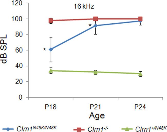
Geng et al. • Clarin-1 Is an Essential Bundle Protein J. Neurosci., July 11, 2012 • 32(28):9485–9498 • 9489
Figure 3. MET currents in Clrn1 ⫹/⫹ and Clrn1⫺/⫺ hair cells. A–D, Representative families
of MET currents evoked by rapid hair bundle deflections that ranged in amplitude from ⫺0.5 to
2 m. Currents were recorded in voltage-clamp mode (Vhold ⫽ ⫺64 mV) from the indicated
organs and genotypes between P1 and P6. The scale bars in B and D also apply to A and C,
respectively. E, Mean maximal transduction currents measured from 73 hair cells as the differ-
ence between the smallest currents evoked by negative bundle deflection and the largest cur-
rents evoked by positive deflections. The number of cells for each of the four groups is indicated
above. F, Mean maximal transduction currents plotted for different cochlear regions and ages.
The number of cells for each group is indicated above.
Figure 2. FM1-43 uptake in Clrn1⫺/⫺ and Cdh23⫺/⫺ cochlear hair cells in culture at P3.
Uptake in Clrn1⫺/⫺ (B) is diminished compared with the control (A). Aⴕ and Bⴕ are DIC images open at rest in nonstimulated hair bundles (Gale et al., 2001;
of A and B showing the location of the hair cells. Hair cells from Cdh23v2J/v2J mice hair cells fail to Meyers et al., 2003). Cochlear cultures from Clrn1⫹/⫺ and
load with FM1-43 (C, D). Cⴕ and Dⴕ are DIC images of C and D showing the location of the hair Clrn1⫺/⫺ mice were examined for their ability to load with
cells. When there is complete loss of hair cell function, no dye uptake is observed (no back- FM1-43 in response to a brief, 10 s exposure to the dye. Hair cells
ground). E, Quantitative analysis of FM1-43 dye loading in the basal coils of the Clrn1⫹/⫺ from Clrn1⫺/⫺ mice showed dye loading in the apical and basal
controls (n ⫽ 8) and Clrn1⫺/⫺ mutants (n ⫽ 9). The number of hair cells included to determine turns of the cochlea, although it was clearly reduced in both turns
the mean grayscale levels per tissue is 24. The reduction in dye loading was significant ( p ⫽ compared with the controls (images from midbasal section
0.0205). The error bars represent SD. shown in Fig. 2A–D). Quantitative analysis of the basal turn of the
Clrn1⫺/⫺ mice indicated a 30 – 40% reduction in dye loading
noted in apical, midbasal, and basal turns of Clrn1⫺/⫺ cochlea
relative to that observed in the controls (Fig. 2 E), and the differ-
from 2- to 3-week-old mice (Geng et al., 2009). Together, these
ence between the two groups was statistically significant ( p ⫽
data support our hypothesis that hair bundle defect is the cause of
0.0205). The reduced uptake could be attributable to loss of tip
hair cell dysfunction in Clrn1⫺/⫺ mice, suggesting a possible role
links or MET channels, namely mutation in Clrn1 directly affects
for CLRN1 in maintaining bundle integrity. Loss of CLRN1 could
the formation of the tip-link complex or MET channels. Alterna-
lead to disintegration of the bundle over time, but this process
tively, reduced FM1-43 loading could be a consequence of bundle
may not be uniform along cochlear turn (especially early on),
disruption, leading to a reduced number of tip links or functional
leading to the observed variation in the CM output noted above.
MET channels open at rest in mutant hair cells. If mutation in
However, it could also be argued that a reduced CM response
Clrn1 directly affects the formation of the tip-link complex or
could be caused by abnormalities of the tectorial membrane,
MET channels, then hair cells carrying Clrn1⫺/⫺ (a null) mutation
MET channel, or other factors needed for proper hair cell depo-
would be expected to show no dye loading, but that is not the case.
larization. To test the bundle defect hypothesis directly, we ex-
However, it is possible that FM1-43 enters hair cells through routes
amined FM1-43 dye loading in nonstimulated hair bundles and
independent of the MET channels (during the 10 s exposure) and
recorded transduction currents after mechanical stimulation of
hence the observed loading. To address these issues and as an impor-
the bundles.
tant reference control, cochlear cultures prepared from mice carry-
Hair cells of Clrn1 ⫺/⫺ mice show reduced FM1-43 loading ing a presumptive null allele of tip-link protein cadherin 23,
FM1-43 is an amphipathic styryl dye that is known to rapidly Cdh23v2J/v2J (Di Palma et al., 2001), were examined for their ability to
accumulate in sensory hair cells via the MET channels that are load FM1-43 under the same condition used to test the cochlear9490 • J. Neurosci., July 11, 2012 • 32(28):9485–9498 Geng et al. • Clarin-1 Is an Essential Bundle Protein
culture from Clrn1⫺/⫺ mice. No dye loading
was observed in Cdh23v2J/v2J culture (Fig.
2C,D), indicating that dye loading occurs
exclusively through MET channels in our
experimental condition. If CLRN1 was di-
rectly involved in the development of tip
links or in the formation of the MET chan-
nel, then we would expect no dye loading in
the Clrn1 knock-out background similar to
that observed with Cdh23v2J/v2J cultures.
However, these results (Fig. 2A,B,E) suggest
that reduced FM1-43 loading in Clrn1⫺/⫺
mice is more likely attributable to compro-
mised MET channel function as a result of
loss of hair bundle integrity.
Transduction currents of hair cells of
Clrn1⫺/⫺ mice had significantly reduced
amplitudes and sensitivity
MET currents were recorded from 39
Clrn1 ⫹/⫹ hair cells and 34 Clrn1⫺/⫺ hair
cells. Clrn1⫺/⫺ hair bundles with mor-
phologies most similar to wild type were
selected for analysis. Representative trans-
duction currents are shown in Figure 3 for
both cochlear and vestibular hair cells. In
both cell types, the deletion of Clrn1 re-
⫹/⫹
sulted in a reduction of the maximal Figure 4. I( X) relationships from P1–P6 hair cells excised from Clrn1 and Clrn1⫺/⫺ mice. Peak current is plotted as a
transduction current that could be evoked function of the stimulus amplitude for utricle (A) and cochlea (B) cells, respectively. The data points were fitted with second-order
by stiff probe deflection of the hair bun- Boltzmann equations. The data shown in A and B were normalized ⫺/⫺ to the maximal current and replotted to illustrate the broader
relationships in hair cells from the utricle (C) and cochlea (D) of Clrn1 mice. To quantify the sensitivity of the cells, we measured
dle. On average, the maximal transduc-
the 10 –90% operating range for each of the 73 cells. The mean operating ranges are plotted on the bar graphs for utricle (E) and
tion current was reduced by 32% in the cochlea (F ) hair cells. The number of cells in each group is indicated on the graphs.
cochlear outer hair cells (OHCs) and by
44% in utricular type II hair cells (Fig. 3A– effects are likely the consequence of disrupted hair bundle
E). To identify possible developmental contributions of Clarin-1, stereocilia.
hair cell transduction was recorded during the first postnatal
week, at time points when mouse transduction development has
CLRN1 is a membrane protein that localizes to hair
been well characterized (Lelli et al., 2009). When we subdivided
bundle stereocilia
the pooled cochlear data according to developmental age and
Several attempts were made to detect endogenous mouse
cochlear region, we found similar reductions in transduction cur-
CLRN1 by immunolabeling, but we have not achieved specific
rent amplitude over all ages and regions examined (Fig. 3F ),
labeling yet. We raised antibodies against two peptides from
suggesting that the effect of Clrn1 deletion is not tonotopically or
the extracellular loops, one peptide from cytoplasmic tail and
developmentally regulated.
⫺/⫺ full-length mCLRN1 protein. Although these antibodies rec-
Hair cells of Clrn1 mice were significantly less sensitive
ognize mCLRN1 expressed in cell lines, their specificity could
than those of Clrn1 ⫹/⫹ cells. To investigate sensitivity, we exam-
⫹/⫹ not be confirmed in mouse inner ear tissue (data not shown).
ined the stimulus–response [I( X)] relationships of Clrn1 and
⫺/⫺ All antibodies mentioned in this paragraph were diligently
Clrn1 cells (Fig. 4). The I( X) data were extracted from those
tested in two different laboratories (those of K.N.A. and Y.I.),
presented in Figure 3 and are plotted as the peak transduction
and we observed the same labeling pattern in cochlear tissue
current as a function of the hair bundle deflection. Figure 4, A and from Clrn1⫹/⫹ and Clrn1⫺/⫺ mice, although Clrn1 mRNA is not
B, shows representative I( X) curves from vestibular cells as well as detected in the cochlea of Clrn1⫺/⫺ mice (Geng et al., 2009). This
cochlear cells. The raw data were fit with a second-order Boltz- is different from Cosgrove and colleagues, who reported CLRN1
mann equation. The I( X) curves revealed both reduced current expression in the inner ear and retina using a rabbit CLRN1
amplitudes in the Clrn1⫺/⫺ cells and reduced sensitivity, which is antiserum they generated (Zallocchi et al., 2009, 2012). We sus-
evident as the broader I( X) relationship, with a shallower slope. pect that the expression level of CLRN1 in the mouse ear is low,
To highlight this finding, we normalized the I( X) relations in making it difficult to detect the endogenous protein. To circum-
Figure 4, C and D. To quantify the change in sensitivity, we mea- vent this problem, we took the alternate approach described be-
sured the displacement required to activate 10 –90% of the re- low, and all evidence presented here suggests that we were
sponse. The mean 10 –90% operating ranges are presented for successful in determining the CLRN1 localization in hair cells.
⫹/⫹
both utricle type II hair cells and cochlear OHCs from Clrn1
⫺/⫺
and Clrn1 cells in Figure 4, E and F. Hair cells excised from CLRN1 localizes to the hair bundle
Clrn1⫺/⫺ mice had significantly broader I( X) relationships. In Data from the cochlear and hair cell physiology experiments led
summary, vestibular and cochlear hair cells of Clrn1⫺/⫺ mice had to the hypothesis that CLRN1 is a necessary hair bundle protein
significantly reduced amplitudes and reduced sensitivity. Both and, therefore, should be expressed in the hair bundle. TargetingGeng et al. • Clarin-1 Is an Essential Bundle Protein J. Neurosci., July 11, 2012 • 32(28):9485–9498 • 9491
in CLRN1 is replaced by lysine. Bionfor-
matic analysis shows that asparagine at
position 48 is a conserved amino acid in
clarin-1 across species and a conserved
glycosylation site (Adato et al., 2002). It is
known that lack of N-glycosylation could
lead to abnormal posttranslational pro-
cessing and localization of membrane
proteins (Smith et al., 2011). Based on this
information and the results presented
previously (Figs. 1–5A), we hypothesized
that hCLRN1 N48K protein will not localize
to the hair bundle. Here, we show that
hCLRN1 carrying the pathogenic mutation
N48K fails to reach the hair bundle and in-
stead was retained in the soma of the hair
cell (Fig. 5B). All cells transfected with the
hCLRN1 N48K–YFP construct showed local-
ization of the fusion protein to the soma but
not to the hair bundle (n ⫽ 24 hair cells).
These data support our hypothesis that
hCLRN1 N48K fails to localize to the hair
bundle and, for the first time in hair cells,
reveals a possible mechanism underlying a
common pathogenic mutation in CLRN1.
To negate the concern that the ob-
served localization of hCLRN1 to the hair
bundle is an artifact of the CMV-driven
overexpression and/or YFP fusion, we
performed the following control experi-
ments. The mouse organ of Corti was
transfected with mouse Prestin–YFP or
GFP–Myo15a cDNA construct. Prestin–
YFP localized to the hair cell plasma mem-
brane, not to the hair bundle (Fig. 5C),
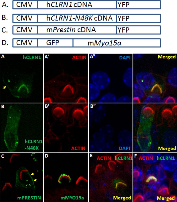
Figure 5. CLRN1 localizes to the hair bundle. P3 wild-type mouse cochlear epithelia explants transfected with a specific and GFP–Myo15a accumulated at the tips
construct (using gene gun) and neighboring nontransfected hair cells after 1 d in culture and ⬃20 h after transfection. Expression of the stereocilia, as reported previously
is driven by the CMV promoter in all cases, and detection of the fluorescent protein marks transfected cells; the tissue was (Belyantseva et al., 2000, 2003), and not
counterstained with phalloidin-conjugated Alexa Fluor 546 (red) and DAPI (blue). A, CLRN1–YFP (green); Aⴕ and Aⴖ are phalloidin anywhere else in the hair cell (Fig. 5D).
and DAPI counterstained images of A. CLRN1–YFP predominantly localized to the bundle with a small amount of the fusion protein Results from these control experiments
localizing to the plasma membrane in the soma (arrow). B, CLRN1 N48K–YFP (green); Bⴕ and Bⴖ are phalloidin and DAPI counter-
indicate that CMV-driven expression and
stained images of B. C, Merged image showing Prestin–YFP (green)-expressing hair cell and phalloidin-labeled bundles on
transfected and nontransfected cells. Arrows point to membrane localization of Prestin–YFP. D, GFP–Myo15a (green)-expressing
YFP fusion do not interfere with native lo-
hair cell and phalloidin-labeled bundles on transfected and nontransfected cells. GFP–Myo15a appears at the tip of the stereocilia calization of the expressed protein in hair
as expected. E, F, YFP/phalloidin merged images from two separate (from A) rounds of transfection with CLRN1–YFP construct to cells, nor is hair bundle localization an arti-
show reproducibility of results shown in A. fact of overexpression of a protein fused to
YFP. The lack of bundle targeting of overex-
pressed hCLRN1 N48K further supports the
of hCLRN1 to the stereocilia in wild-type hair cells was demon- notion that the bundle localization of the hCLRN1 protein is not an
strated by transfection of mouse organ of Corti explants using artifact of overexpression.
gene gun transfection protocol. hCLRN1–YFP accumulated in
the stereocilia of hair cells that were transfected with a hCLRN1– Heterologous expression confirms membrane localization of
YFP cDNA construct (Fig. 5A). The tissue was counterstained hCLRN1 and mislocalization of the hCLRN1 N48K protein
with phalloidin–Alexa Fluor 546. Phalloidin is a high-affinity fil- Clarin-1 is predicted to be a membrane protein (Adato et al., 2002).
amentous actin (F-actin) probe. Fluorescently labeled phalloidin Gene gun transfection showed that hCLRN1 localizes to the hair
highlights F-actin-rich structures (stereocilia) within the hair bundle (Fig. 5), but the resolution of those images was not sufficient
bundle. Merged images showed localization of hCLRN1–YFP to to discern membrane localization. To examine the subcellular local-
the bundle (Fig. 5 A, A⬘,merged image). In some of the transfected ization of wild-type hCLRN1 and hCLRN1 N48K mutant, we gener-
hair cells (n ⫽ 4), faint fluorescent labeling was observed in ated two HEK293 cell lines stably expressing either hCLRN1 or
plasma membrane (Fig. 5A, arrow). All cells transfected with the hCLRN1 N48K fused with HA tag for detection. It has been reported
hCLRN1–YFP construct showed hair bundle localization of the that N48 is the only N-glycosylation site in CLRN1, and N48K mu-
fusion protein (n ⫽ 27 hair cells). tation led to the mutant protein lacking glycosylation (Tian et al.,
Next, we focused on one of the common USH3 mutations in 2009). Under normal conditions, the expression level of
North America, namely p.N48K. Here, asparagine at position 48 hCLRN1 N48K is very low because of active degradation of this pro-9492 • J. Neurosci., July 11, 2012 • 32(28):9485–9498 Geng et al. • Clarin-1 Is an Essential Bundle Protein
tein in HEK293 cells (Tian et al., 2009). We
treated the two stable cell lines with bort-
ezomib to enhance the protein level and to
facilitate our detection of immunofluores-
cence. The cells were costained with anti-
bodies against the HA tag and a plasma
membrane marker Na/K ATPase (Fig. 6A).
Wild-type hCLRN1 localized to the plasma
membrane (Fig. 6A, top row) with a small
amount observed in the ER. The cells
were also costained with antibodies
against the HA tag and calreticulin, a
major Ca 2⫹ binding (storage) chaper-
one in the ER (Fig. 6 B). Unlike wild-
type hCLRN1, the hCLRN1 N48K mutant
failed to localize to the plasma membrane
(Fig. 6 A, bottom row) and mainly colo-
calized with calreticulin (Fig. 6 B, bottom
row), suggesting that most of the mutated
proteins were retained in the ER. Results
described above show that N48K muta-
tion in hCLRN1 alters normal plasma
membrane localization of hCLRN1 and
support an idea that hCLRN1 is localized
to the plasma membrane compartment of
the hair bundle.
Generation of Clrn1 N48K knock-in mice
Results described thus far led to the hypoth-
esis that CLRN1 is an essential hair bundle
protein and that loss of hearing associated
with the CLRN1N48K mutation in USH3 is
attributable to defective intracellular traf-
ficking of the CLRN1 N48K protein to the
hair bundle. To study the effect of N48K
mutation in vivo, we generated a transgenic
N48K
mouse carrying missense mutation in the Figure 6. Localization of CLRN1 and its CLRN1 mutant heterologously expressed inN48K HEK293 cells. The HEK cell lines stably
first coding exon (exon 1) of the endoge- expressing either wild-type human CLRN1 fused to HA tag (top rows of A and B) or CLRN1 fused to HA tag (bottom rows of A
nous (mouse) Clrn1 gene. The amino acid and B) were treated with bortezomib (15 nM) for 16 h before fixed by 4% paraformaldehyde. A, The cells were stained by
antibodies against HA tag (green) and Na/K ATPase (red), which localizes to the plasma membrane. CLRN1 localizes to the
sequence of mouse and human clarin-1 are plasma membrane, whereas CLRN1 N48K fails to reach the plasma membrane. B, The cells were stained by antibodies against HA
highly conserved (⬎85%), including the tag (green) and calreticulin (red). The majority of CLRN1 N48K is observed in the ER and the cytoplasm. Scale bars, 20 m.
conserved glycosylation site at position 48
(Adato et al., 2002), making the mouse an tral auditory pathway to sound stimulation. Clrn1⫹/N48K mice
ideal in vivo model to generate the N48K knock-in mutation. The showed wild-type thresholds at all time points tested, confirming
knock-in transgenic mouse was produced by a homologous recom- the recessive nature of the phenotype associated with clarin-1
bination of engineered construct carrying the missense mutation; mutation and, at the same time, arguing against dominant-
the wild-type exon 1 was replaced with exon 1 carrying N48K mu- negative effects by a single Clrn1N48K allele. Clrn1N48K/N48K mice
N48K
tation (Fig. 7A). The knock-in allele was designated Clrn1 . Nor- displayed early-onset hearing loss that deteriorated to profound
mal Mendelian segregation of the Clrn1N48K allele (wild-type hearing loss after weaning. In contrast to Clrn1⫺/⫺ mice, how-
⫹/⫹ ⫹/N48K N48K/
Clrn1 , heterozygous Clrn1 , and homozygous Clrn1 ever, the Clrn1N48K/N48K mice displayed thresholds indicative of
N48K) was observed. DNA sequence analysis of cDNA generated
hearing loss but not profound hearing loss at P18 ( p ⬍ 0.0001) or
from Clrn1⫹/N48K mouse tissue confirmed mutation at the target site P21 ( p ⬍ 0.0001). ABR thresholds of Clrn1N48K/N48K mice were
(Fig. 7B,C), and RT-PCR analysis confirmed that the expression of quite variable at P18: although some mice showed threshold close
mouse Clrn1 mRNA and the sizes of splicing variants were indistin- to 40 dB SPL, others were in the 70 – 80 dB SPL range. The hearing
⫹/⫹
guishable from those observed in Clrn1 mice (Fig. 7D). These function deteriorated to near deafness shortly after weaning age
experiments confirm that we have successfully generated a knock-in ( p ⬍ 0.0001) (Fig. 8). Although ABR recordings can be per-
allele carrying the missense mutation in the mouse. formed in mice 2 weeks or older, it is not (yet) technically feasible
Clrn1 N48K/N48K mice show early onset hearing loss that progresses to record transduction currents from cochlear hair cells at this
to deafness around weaning age age. We believe the ABR data to be a good reflection of the func-
To assess hearing function in Clrn1N48K/N48K mice, we recorded tional status of the cochlea in the Clrn1N48K/N48K mice. It is known
ABR from Clrn1N48K/N48K, Clrn1⫹/N48K, and Clrn1⫺/⫺ mice at that USH3 patients with CLRN1N48K mutation develop hearing
P18, P21, and P24. ABR recordings reflect the electrical responses loss with variable onset and severity (Sadeghi et al., 2005; Herrera
of both the cochlear ganglion neurons and the nuclei of the cen- et al., 2008). Here we report that N48K mutation in the mouseGeng et al. • Clarin-1 Is an Essential Bundle Protein J. Neurosci., July 11, 2012 • 32(28):9485–9498 • 9493
in Fig. 9). Of the 15 OHCs in each mutant
panel, ⬃40% from the Clrn1N48K/N48K
mice showed mild bundle disturbance
compared with ⬃20% in the Clrn1⫺/⫺
specimen; conversely, the organ of Corti
from the Clrn1⫺/⫺ mouse showed a
greater number of moderately (50%) to
severely (20%) disturbed hair bundles
compared with that of the Clrn1N48/K/N48K
mouse (30% moderate, 20% severe). Spe-
cifically, the disruption includes splits in
the bundle and loss of some of the tall ste-
reocilia. Some of the hair cells from the
Clrn1⫺/⫺ mice showed more severe dis-
ruption of the hair bundle arrangement
compared with hair cells from Clrn1N48K/
N48K mice (Fig. 10 B, E). Hair cells from
both mutants bear kinocilia, and the bun-
dle orientation (vertex of the “v” facing
the lateral edge) is generally not affected.
Even in disrupted hair bundles, the
graded heights of the stereocilia and tent-
ing of the shorter stereocilia toward the
taller stereocilia were apparent in both mu-
tants (Fig. 10A,D,E). Tip links were identi-
fied in hair bundles examined from both
mutants, with some exceptions. Overall, the
profile of the hair bundle phenotype was the
same in both mutants, although the degree
of disruption in the bundle was more severe
in Clrn1⫺/⫺ mice compared with Clrn1N48K/
⫺/⫺
N48K mice. Results from the Clrn1 mice
show that Clrn1 is not required for the for-
mation of tip links, development of graded
Figure 7. Generation of Clrn1N48K knock-in mouse. A, Targeting map for Clrn1 N48K knock-in mice. Top, Mouse Clrn1 consists of heights of the stereocilia, or polarized orien-
four exons. Second row, The targeting vector contained 5.7 kb of 5⬘ and 2.2 kb of 3⬘ homologous sequence with C-to-G transversion tation of the hair bundle. Rather, clear dis-
at the codon 48. Third row, As a result of homologous recombination, codon 48 in exon 1 was changed to AAG, and the loxP flanked
ruption in hair bundle structure in the
Neo gene was inserted after exon 1. Bottom row, Neo was removed from targeted locus by cre–loxP recombination. B, DNA
sequence analysis confirms mutation at the target nucleotide in exon 1: both wild-type nucleotide C and mutant nucleotide G were
Clrn1⫺/⫺ mutants associated with loss of
observed in a cDNA sample from heterozygous knock-in mice (⫹/KI). C, The missense mutation introduced a BsaHI restriction site, stereocilia and a milder phenotype of simi-
N48K/N48K
which was used to distinguish the wild-type (⫹/⫹), heterozygous (⫹/KI), and homozygous (KI/KI) mutants. D, Mouse Clrn1 lar profile in Clrn1 mice suggests
mRNA is expressed in brain, cochlea, and retina; the upper band (⬃850 bp) and the lower band (⬃750 bp) seen in the upper half that mCLRN1 may be required to maintain
is typical Clrn1 mRNA and alternative splicing expression pattern. Results show that Clrn1 mRNA expression is not affected by the the structure of the bundle after it is formed.
missense mutation. WT, Wild type. However, it is likely that morphogenesis and
maintenance of bundle stereocilia share
common mechanisms to some degree, and,
Clrn1 gene also causes hearing loss with variable levels of severity
N48K/N48K therefore, it is difficult to exclude CLRN1 from either process at this
between mutants. However, all Clrn1 mice lose all hear-
time. Although the mechanism by which mCLRN1 exerts its func-
ing during the young-adult stage (P20 –P30). The hearing loss
N48K/N48K tion in the bundle stereocilia remains to be elucidated, data pre-
phenotype observed in Clrn1 mutants, combined with
sented here clearly demonstrate that CLRN1 is a protein essential for
data reported above (Figs. 1– 6), suggest an important role for
either maintenance or morphogenesis of hair bundles but is not
CLRN1 in the hair bundle.
essential for establishing planer cellular polarity.
Mutation in Clrn1 disrupts hair bundle morphology, not tip links
Next, scanning electron microscopy was used to examine and Clrn1 is not essential for hair cell ribbon synapse
compare the impact of null and missense mutation in Clrn1 on development or function
hair bundle development and structure. Cochlear hair bundles Previous reports suggested a possible role for clarin-1 in the sen-
were studied by field emission scanning electron microscopy in sory synapses based on limited amino acid sequence homology to
the Clrn1⫺/⫺ and Clrn1N48K/N48K mouse at P3–P4. Specifically, cerebellar synapse protein stargazin (Adato et al., 2002). Also,
we looked at the overall organization of the hair bundle, bundle Geng et al. (2009) reported a delay in peak latencies in ABRs and
orientation, staircase arrangement, and tip links. The Clrn1⫺/⫺ vestibular evoked potential recordings. In the case of the ABR,
and Clrn1N48K/N48K both showed disruption of the hair bundle peaks I–IV represent the response time (of the electrical re-
integrity compared with the control (Clrn1⫹/⫺) (Fig. 9). The sponses) of both the cochlear ganglion neurons (or hair cell to
bundle disturbance in these two alleles of Clrn1 was categorized afferent neurons) (peak I) and various nuclei of the central audi-
into mild, moderate, and severe disturbance (marked by asterisks tory pathway (peaks II–IV) to sound stimulation. Reevaluation of9494 • J. Neurosci., July 11, 2012 • 32(28):9485–9498 Geng et al. • Clarin-1 Is an Essential Bundle Protein
Figure 8. Assessment of hearing in Clrn1N48/K/N48K knock-in compared with the Clrn1⫺/⫺
mice over time. The plot shows that mean ABR thresholds of Clrn1N48K/N48K mice (n ⫽ 25) are
significantly elevated compared with the thresholds of Clrn1⫹/N48K mice (n ⫽ 6), which dis-
played wild-type thresholds at all time points tested. The plot also shows that ABR thresholds of
Clrn1N48K/N48K mice are significantly lower compared with Clrn1⫺/⫺ mice (n ⫽ 12) at P18 and
P21; the difference is not significant at P24 because mice from both groups display profound
hearing loss at P24. *p ⬍ 0.0001.
Clrn1⫺/⫺ mice since the 2009 study confirmed delay in the peak
latencies, but closer examination showed that a delay in the initial
response or peak I could be associated with the mutant pheno-
type; whereas delays in the timing of peaks II–IV were secondary
to the delay in peak I since interpeak latency (IPL) value was the
same in Clrn1⫹/⫹ and Clrn1⫺/⫺ mice (Table 1). A delay in initial
response suggested a hair cell-to-afferent communication delay
or ribbon synapse defect. We used two approaches to determine
whether Clrn1 is essential for hair cell presynaptic function
and/or development.
Inner hair cell ribbon synapse develops normally in
Clrn1 ⫺/⫺ mice
We explored whether null mutation in Clrn1⫺/⫺ affects develop-
ment of the inner hair cell (IHC) ribbon synapse in Clrn1⫺/⫺ Figure 9. Scanning electron microscopy of the organ of Corti showing regions of ⬃15 OHCs
mice using presynaptic and postsynaptic marker antibodies. The each from the Clrn1⫺/⫺, Clrn1N48K/N48K, and Clrn1⫹/⫺ mice. Mild (*), moderate (**), and
afferent ribbon synapses of P15 Clrn1⫹/⫺ and Clrn1⫺/⫺ mice severe (***) bundle disruption can be seen in both the Clrn1⫺/⫺ and Clrn1N48/K/N48K panels
were examined by immunofluorescence microscopy, using both compared with the Clrn1⫹/⫺ panel. However, the organ of Corti from the Clrn1⫺/⫺ mouse
an antibody directed against the nuclear protein CtBP2, which showed a greater number of moderately to severely disturbed hair bundles compared with that
also detects RIBEYE, a major structural component of the ribbon of the Clrn1N48/K/N48K mouse. Scale bar, 5 m.
(Schmitz et al., 2000), and an antibody against GluR2/3, associ-
ated with the dendrites of afferent neurons. Intact ribbon syn-
apses were defined by the presence of juxtaposed RIBEYE and amplitude nor the voltage dependence of CaV1.3-mediated Ca 2⫹
GluR2/3 fluorescent spots (Khimich et al., 2005). Whole mounts influx was altered. We did not find any obvious changes in exo-
of organs of Corti from P15 Clrn1⫹/⫺ and Clrn1⫺/⫺ mice double cytosis. This argues against an essential function for clarin-1 in
immunolabeled for CtBP2 and GluR2/3 were analyzed by confo- hair cell presynaptic function.
cal microscopy. Quantitative analysis showed that the IHCs of
Discussion
Clrn1⫺/⫺ had ⬃90% the number of ribbon synapses observed in
Here, we show that (1) CLRN1 is a hair bundle protein needed
the Clrn1⫹/⫺ specimen at P15 (Fig. 11 A, B; Table 2). The ob-
for development or maintenance of the normal shape of the
served reduction, ⬃10% compared with control, is not consid-
bundle, and (2) the pathogenic mechanism underlying
ered significant and is probably secondary to the deteriorating
CLRN1N48K mutation in USH3 is a likely failure to express the
condition of the hair cells in the Clrn1⫺/⫺ background. Our re-
mutated protein in the bundle. These findings have been con-
sults show that Clrn1 is not required for the development or
firmed using a novel mouse model carrying a Clrn1N48K
maintenance of ribbon synapse, and the observed phenotype,
knock-in mutation to investigate the consequence of the mis-
namely delayed initial response in evoked potentials, is secondary
sense mutation N48K in mCLRN1 in vivo.
to the compromised hair bundle structure/function.
Our previous report showed that mice homozygous for the
Clrn1 is not essential in hair cell presynaptic function targeted deletion (or knock-out) of first coding exon in Clrn1
Next, we explored the physiological impact of null mutation in displayed inner ear dysfunction and loss of Clrn1 mRNA expres-
Clrn1 on exocytic membrane capacitance changes (⌬Cm) in re- sion in the inner ear (Geng et al., 2009); hence, the knock-out
sponse to Ca 2⫹ influx (ICa) evoked by step depolarizations in allele is considered a null allele of Clrn1 and is represented as
perforated-patch recordings from IHCs at P14 –P20. As illus- Clrn1⫺/⫺. To investigate the specific role of clarin-1 in hair cells,
trated for representative (Fig. 12 A) and averaged (Fig. 12 B, C) we started with an in-depth analysis of hair cell function in a
data, Clrn1⫺/⫺ IHCs showed normal ICa and ⌬Cm. Neither the Clrn1⫺/⫺ mice. Measures of hair cell function (CM, FM1-43 up-You can also read