The Mosquito Immune System and the Life of Dengue Virus: What We Know and Do Not Know - MDPI
←
→
Page content transcription
If your browser does not render page correctly, please read the page content below
pathogens
Review
The Mosquito Immune System and the Life of
Dengue Virus: What We Know and Do Not Know
Debica Mukherjee , Sandeepan Das, Feroza Begum, Sweety Mal and Upasana Ray *
 CSIR-Indian Institute of Chemical Biology, 4, Raja S.C. Mullick Road, Jadavpur,
 Kolkata 700032, West Bengal, India; debicamukherjee@csiriicb.res.in (D.M.); sandeepan@csiriicb.res.in (S.D.);
 ferozabegum@csiriicb.res.in (F.B.); sweetymal1994@gmail.com (S.M.)
 * Correspondence: upasana.ray@iicb.res.in or ray.upasana@gmail.com; Tel.: +91-968-1878-333

 Received: 4 April 2019; Accepted: 30 May 2019; Published: 13 June 2019

 Abstract: Flaviviruses are largely transmitted to humans by their arthropod vectors such as mosquitoes
 or ticks. The dengue virus (DENV) is one of the members of the family Flaviviridae and is the causative
 agent of dengue fever. In the mosquito vector, DENV enters through viremic blood meal and
 replicates in the mid-gut. Newly formed virion particles circulate to various mosquito organs and
 get transmitted to the next host in subsequent bites. Aedes aegypti and Aedes albopictus have intricate
 immune control to allow DENV production at a sub-pathogenic level. In the mosquito, antimicrobial
 peptides (AMP) and RNA inference (RNAi) are the two main antiviral strategies used against DENV.
 Apart from innate immunity, mosquito resident microbes play a significant role in modulating DENV
 replication. In this review, we discuss different immune mechanisms and preventive strategies that
 act against DENV in two of its vectors: Aedes aegypti and Aedes albopictus.

 Keywords: Aedes; immunity; dengue; RNAi; gut-microbiome; AMP

1. Introduction
      Vector-borne diseases such as yellow fever, dengue fever, etc. are known for their chronic effects
on human health. These diseases are spread by vectors such as mosquitoes and other arthropods.
Mosquitoes are known to be an important vector of virus transmission. Geographically, tropical and
subtropical areas are the most suitable regions for optimal growth of mosquitoes and thus mosquito
borne diseases pose great risk the world over.
      The dengue virus (DENV) is a single-stranded, positive-sense RNA virus belonging to the
Flaviviridae and is transmitted to humans and other primates by Aedes mosquitoes, importantly
Aedes aegypti (Ae. aegypti) and Aedes albopictus (Ae. albopictus). In humans, DENV infection causes
dengue fever (DF) and in severe cases, dengue hemorrhagic fever (DHF) or dengue shock syndrome
(DSS). In the transmission cycle, upon injection into the human body, the virus gains entry into the
host’s cells, hijacks the host’s cell machinery to replicate, and escapes the host’s immune strategies to
elicit pathology symptoms [1]. Conversely, in the mosquitoes, the virus replicates, but it is under strong
immune control so that the vector’s life span is not compromised. DENV can inhibit mosquito immunity
to an extent that allows it to multiply to a non-pathogenic level [2]. Thus, a fine immunological balance
exists in the mosquito so that the virus can persist and multiply.
      A female mosquito needs to feed on vertebrate blood before egg laying for egg maturation.
The virus enters the mosquito’s system via viremic blood and infects the midgut epithelial cells.
It replicates in the midgut cells and then gets released into the hemocoel and disseminates to other
tissues via hemolymph, ultimately reaching the salivary glands to infect fresh hosts in subsequent bites.

Pathogens 2019, 8, 77; doi:10.3390/pathogens8020077                             www.mdpi.com/journal/pathogens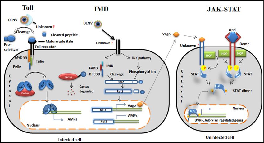
Pathogens 2019, 8, 77                                                                                                2 of 16

Pathogens 2018, 7, x FOR PEER REVIEW                                                                                2 of 16

     In the mosquitoes, innate immunity and a resident microbiome work together against DENV.
     In the mosquitoes, innate immunity and a resident microbiome work together against DENV.
This review summarizes the molecular mechanism of immune function in the mosquito system against
This review summarizes the molecular mechanism of immune function in the mosquito system
DENV pathogenesis (Figure 1).
against DENV pathogenesis (Figure 1).

      Figure 1. Mosquito immune strategies against the dengue virus (DENV). JAK-STAT = Janus
      Figure  1. Mosquito
      kinase-signal       immune
                    transducer  andstrategies
                                    activator against  the dengue
                                               of transcription;    virus (DENV).
                                                                 AaSR-C            JAK-STAT
                                                                          = Ae. aegypti homolog= Janus  kinase-
                                                                                                  of scavenger
      signal transducer and activator of transcription; AaSR-C   = Ae. aegypti
      receptor-C; AsMCR = Ae. aegypti macroglobulin complement related factor. homolog  of scavenger  receptor-
      C; AsMCR = Ae. aegypti macroglobulin complement related factor.
2. Innate Immunity
Innate Immunity
     The mosquito’s innate immunity includes three main strategies:
     The mosquito’s innate immunity includes three main strategies:
1.    Antiviral signaling pathways: Toll-like pathway, immune deficiency (IMD), and Janus kinase-signal
1. Antiviral
      transducersignaling   pathways:
                    and activator       Toll-like pathway,
                                    of transcription     (JAK-STAT)immune      deficiency (IMD), and Janus kinase-
                                                                          pathway.
2. signal    transducer proteins:
      Complement-like      and activator
                                       Theseof transcription     (JAK-STAT) pathway.
                                                 include Thioester-containing             proteins (TEP); A. aegypti
2. Complement-like        proteins: These
      macroglobulin complement                  include
                                          related    factorThioester-containing
                                                              (AaMCR) and A. aegypti     proteins   (TEP);of A.
                                                                                               homolog             aegypti
                                                                                                               scavenger
     macroglobulin      complement
      receptor-C (AaSR-C).               related    factor   (AaMCR)        and  A.   aegypti  homolog     of  scavenger
3. receptor-C
      Small RNAs:  (AaSR-C).
                      These include small interfering RNA (siRNA), micro RNA (miRNA) (derived from
3. Small    RNAs:    These  include small RNA
      the virus), and PIWI-interacting        interfering   RNA (siRNA), micro RNA (miRNA) (derived from
                                                     (piRNA).
     the virus), and PIWI-interacting RNA (piRNA).
3. Antiviral Signaling Pathways
Antiviral Signaling Pathways
     Toll-like pathway: In Ae. Aegypti, dorsal ortholog Rel1 and Relish ortholog Rel2 act as Toll and
IMD Toll-like
      pathway pathway:
                 components, In Ae.  Aegypti, dorsal
                                 respectively,          ortholog
                                                  and induce         Rel1 andofRelish
                                                                 expression              orthologpeptides
                                                                                   antimicrobial   Rel2 act as   Toll and
                                                                                                              (AMPs)    [3].
IMD
It waspathway
        observed components,
                   that ten days respectively,
                                    post infectionandwith
                                                       induce
                                                            DENV,expression    of antimicrobial
                                                                       the oxidative              peptides (AMPs)
                                                                                       damage preventative      enzymes[3].
It
arewas observed that
    suppressed,         ten days
                   but Toll        post infection
                             and JAK-STAT            with DENV,
                                                 pathway     effectorsthealong
                                                                           oxidative
                                                                                with damage
                                                                                       pathogen preventative
                                                                                                  recognitionenzymes
                                                                                                                 receptor
are suppressed,
(PRR)  expressionbut areToll and JAK-STAT
                         up-regulated    [3]. Thepathway     effectors
                                                    Toll pathway      is aalong with anti-dengue
                                                                           powerful    pathogen recognition      receptor
                                                                                                     defense system     for
(PRR)   expression
Ae. aegypti, as wellareas up-regulated
                          against multiple [3].DENV
                                                The Toll    pathway
                                                        serotypes    [4].is a powerful anti-dengue defense system
for Ae. aegypti,
     Toll        as well
          receptor         as against
                      activation       multiple
                                  requires         DENV serotypes
                                              virus-derived      ligand [4].
                                                                           binding to unknown extracellular PRRs
     Toll receptor
and proteolytic       activation
                  cleavage         requires virus-derived
                              of pro-spaetzle     to activate theligand    binding
                                                                     cytokine        to unknown
                                                                                spaetzle,  which isextracellular
                                                                                                      the ligand forPRRs
                                                                                                                       the
and  proteolytic cleavage
transmembrane      receptor ofofthe
                                 pro-spaetzle
                                     Toll pathway to activate   the cytokine
                                                      [5]. Spaetzle    activatesspaetzle,  which isby
                                                                                  the Toll receptor   thecross-linking
                                                                                                          ligand for the of
transmembrane      receptor
receptor ectodomains          of the Toll
                          followed         pathway
                                      by the   relay of[5].
                                                         theSpaetzle     activates
                                                              signal through        the Tollproteins
                                                                                 adaptor     receptor   by cross-linking
                                                                                                     (Figure   2).
of receptor ectodomains followed by the relay of the signal through adaptor proteins (Figure 2).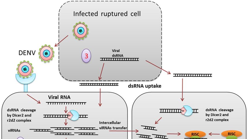
Pathogens 2019, 8, 77 3 of 16
Pathogens 2018, 7, x FOR PEER REVIEW 3 of 16
Immune deficiency (IMD) pathway: The immune deficiency pathway operates by virus-receptor
binding followed
Immune by recruitment
deficiency of adaptor
(IMD) pathway: Theproteins.
immune In Drosopila,
deficiency the IMD
pathway signaling
operates pathway
by virus-
activates
receptoran NF-kβfollowed
binding transcription factor, Relish,
by recruitment of whereas in mosquitoes,
adaptor proteins. Relish ortholog
In Drosopila, the IMD Rel2 acts as a
signaling
transcription factor. The IMD pathway in mosquitos gets activated when a virus
pathway activates an NF-kβ transcription factor, Relish, whereas in mosquitoes, Relish ortholog Rel2 binds to an unknown
receptor
acts as a[6], which recruits
transcription factor.various
The IMD adaptor
pathway proteins. Henceforth,
in mosquitos the pathway
gets activated when ahas two
virus segments
binds to an as
described
unknowninreceptor
Figure [6],
2, one of which
which recruits activates
various the Janusproteins.
adaptor kinase (JNK) signaling
Henceforth, that phosphorylates
the pathway has two
segments
Rel2 as other
[7,8], the described in Figure
part recruits IMD,2, one
FADDof which activatesdeath
(fas-associated the Janus kinase
domain), and(JNK)
DREDD signaling
(deaththat
related
phosphorylates Rel2 [7,8], the other part recruits IMD, FADD (fas-associated
ced-3/Nedd2-like protein) proteins to cleave the phosphorylated Rel2 [8]. Cleaved Rel2 eventually death domain), and
DREDD (death
transcribe relatedAMP
IMD related ced-3/Nedd2-like
genes (Diptericinprotein)andproteins
Cecropin) to cleave the phosphorylated
and a signaling Rel2 [8].
molecule called Vago,
Cleaved
which Rel2 eventually
is known to have an transcribe
antiviralIMD rolerelated
againstAMP Westgenes
Nile (Diptericin and Cecropin)
virus and Drosophila and [9,10].
C virus a signaling
Vago is
molecule
secreted fromcalled Vago, which
the infected is known
cell and acts as atoligand
have for
an the
antiviral role pathway
JAK-STAT against Westin theNile virus andcells
neighboring
Drosophila
(Figure 2). C virus [9,10]. Vago is secreted from the infected cell and acts as a ligand for the JAK-
STAT pathway in the neighboring cells (Figure 2).
Figure2.2.Evolutionarily
Figure Evolutionarily conserved signalingpathways.
conserved signaling pathways.Toll Toll pathway:
pathway: TheThe virus
virus is recognized
is recognized by anby an
unknown cytoplasmic receptor. Cytokine pro-spleatzle is cleaved to active cytokine
unknown cytoplasmic receptor. Cytokine pro-spleatzle is cleaved to active cytokine spleatzle and spleatzle and binds
tobinds
the Toll receptor. Adaptor proteins are recruited to the Toll receptor. A negative
to the Toll receptor. Adaptor proteins are recruited to the Toll receptor. A negative regulator of regulator of cactus
degrades and a free
cactus degrades andRel1 dimer
a free Rel1translocates to the nucleus.
dimer translocates to the The Rel1 The
nucleus. dimerRel1acts as a transcription
dimer acts as a
factor for Toll regulated
transcription genes
factor for Toll and produces
regulated genes and antimicrobial peptides (AMPs)
produces antimicrobial (cecropin
peptides (AMPs)and defensin).
(cecropin
IMD
and pathway:
defensin).TheIMDvirus bindsThe
pathway: to avirus
transmembrane receptor of the receptor
binds to a transmembrane cell and splits
of the the
cellpathway
and splitsinto
thetwo
segments.
pathway One into segment activates
two segments. One thesegment
JNK pathway andthe
activates phosphorylates
JNK pathwayRel2. and Another segmentRel2.
phosphorylates recruits
IMD and other
Another segment adaptor proteins
recruits IMD and {Immune deficiency
other adaptor (IMD),
proteins fas-associated
{Immune deficiencydeath domain
(IMD), (FADD) and
fas-associated
death
deathrelated
domainced-3/Nedd2-like
(FADD) and death protein
related(DREDD)} to cleave
ced-3/Nedd2-like the C-terminal
protein (DREDD)}phosphorylated domain of
to cleave the C-terminal
phosphorylated
Rel2.11. Cleaved domain of Rel2.11.toCleaved
Rel2 translocates Rel2 and
the nucleus translocates
transcribesto the nucleus
AMPs and and transcribes
a secretory AMPs
protein, Vago.
and a secretory
JAK/STAT pathway:protein, Vago. JAK/STAT
This pathway is activatedpathway:
eitherThis pathway is activated
by cytokine-like secretoryeither
proteinby from
cytokine-like
an infected
secretory
cell or by theprotein from an
conserved infected ligand
JAK-STAT cell or Upd.
by theVago
conserved JAK-STAT
is secreted ligand Upd.
from nearby Vago
infected is secreted
cells, binds to an
from nearby
unknown infected
receptor, andcells, bindshopscotch
recruits to an unknown(HOP) receptor, and recruits
kinase. Similarly, Dome hopscotch
receptors (HOP)
bindkinase.
Upd and
Similarly, Dome receptors bind Upd and receptor phosphorylation occurs
receptor phosphorylation occurs through HOP kinase. The phosphorylated receptor is a docking through HOP kinase. The site
phosphorylated receptor is a docking site for STAT. STAT phosphorylation
for STAT. STAT phosphorylation leads to dimerization. STAT dimer translocates to the nucleus and leads to dimerization.
STAT dimer
transcribes translocates to the nucleus
JAK/STAT-regulated genes andand dengue
transcribes JAK/STAT-regulated
virus genes and dengue virus
restriction factor (DVRF).
restriction factor (DVRF).
DENV infection significantly up-regulates the expression of cecropin-like AMPs [11]. Another
study DENV
revealedinfection significantly
that activation up-regulates
of the the by
IMD pathway expression of the
inhibiting cecropin-like AMPs [11].
negative regulator Another
(Casper) have
study revealed that activation of the IMD pathway by inhibiting the negative regulator (Casper) have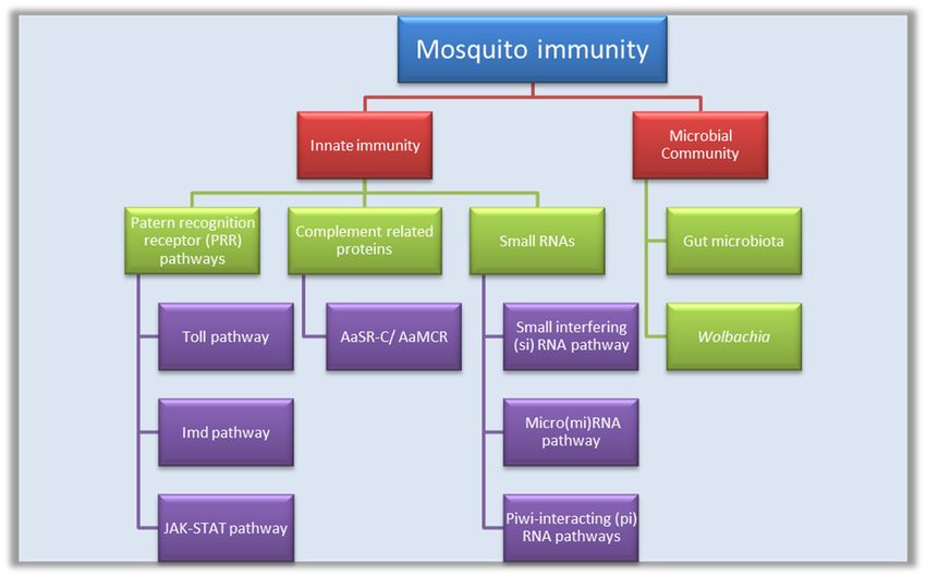
Pathogens 2019, 8, 77                                                                               4 of 16

no effect on DENV in the midgut of a susceptible Ae. Aegypti strain [3,12] whereas in a refractory
Ae. Aegypti strain, blocking IMD pathway results in an increase in viral replication [12].
     In Drosophila, IMD is linked to a protein complex (TAK1/TAB2) via a polyubiquitin chain.
The TAK1/TAB2 complex leads to the activation of one segment of the IMD pathway [13]. However, in
mosquitoes, the Ubiquitine variant (Ub3881) lacks one of the essential lysine residues. Hence, Ub3881
possibly has other residues that target the DENV envelope protein for degradation and down-regulate
the production of infectious virus particle [14,15].
     The JAK-STAT pathway is an essential defense pathway for anti-dengue immunity in
invertebrates [11,16]. In Ae. aegypti, the JAK-STAT pathway is activated by different ligands. Unpaired
(Upd) is the common ligand that binds to its receptor Dome. Receptor dimerization leads to activation
of receptor-associated JAK and downstream signaling (Figure 2).
     According to Souza-Neto et al., mosquitoes become more susceptible to DENV infection if the
receptor Dome or JAK homolog HOP is suppressed by RNA inference (RNAi). On the other hand,
DENV resistance increases if the negative regulator of the JAK-STAT pathway, protein inhibitor of
activated STAT (PIAS) blocks the signaling [16]. The effector genes of DENV are named dengue virus
restriction factors (DVRFs). DVRF 1 is the transmembrane receptor of the pathway and DVRF 2
recognizes the virus by the antifreeze and allergen domains. The allergen domain has been reported in
Anopheles gambiae pattern recognition receptor (PRR) MDL1 immune gene. MDL1 immune gene is
known to have anti-plasmodium activity. Hence, DVRF2 could be a PRR and be involved in DENV
recognition [16].
     Although these main signaling pathways restrict viral propagation to non-pathogenic levels,
DENV multiplies and accumulates in salivary glands, making the vector a competent virus transmitter.

4. Organ-Specific Antiviral Strategies
      Mid-gut: The mid-gut (Figure 3A) is the initial tissue that comes into contact with the
virus-containing blood meal. The first line of defense in the mid-gut are physical barriers such as the
mid-gut infection barrier (MIB) and mid-gut escape barriers [17–20]. The mid-gut infection barrier may
form due to the lack of entry receptors on the epithelial cells [17] or pathogen compartmentalization
by the peritrophic matrix [18]. After successful entry of the virus inside midgut cells, uncoating,
replication, and new virus particle assembly occurs. If the newly formed virions are not able to cross the
basal lamina of the epithelial cells to spread in the hemolymph or are unable to infect secondary organs,
the prevention of these events are referred to as midgut escape barriers (MEB) [19]. In Ae. aegypti,
the innate immune signaling pathways become active during DENV infection. Exogenous siRNA
pathways work against viral infection in the midgut (Figure 3B) [21]. Additionally, the gut microbiome
plays an important role (discussed later).
      Hemolymph: From the mid-gut, DENV is released into the hemocoel, then disseminated to other
organs. In hemolymph, hemocytes allow virus replication, but innate immunity limits the distribution.
Host nitric oxide (NO) inhibits DENV replication in hemocytes. NO production increases during
infection and exogenous NO reduces the DENV replication level (Figure 3C) [22].
      Complement related proteins: Another defense mechanism in the hemolymph, which controls
flaviviral infection, comprises the complement-related proteins. The mosquito genome encodes a
thioester-containing protein (TEP) that recognizes flaviviruses and leads to the expression of an
antimicrobial peptide [23]. Ae. aegypti macroglobulin complement related factor (AaMCR) belongs
to the insect TEP family, which recognizes viral particles. The Ae. aegypti homolog of the Scavenger
receptor C (AaSR-C) mediates the binding of AaMCR to the DENV particles [24]. Thus, when expression
of AMPs in the hemocytes is up-regulated, AMP diffuses to the hemolymph and plays an anti-dengue
role (Figure 3C).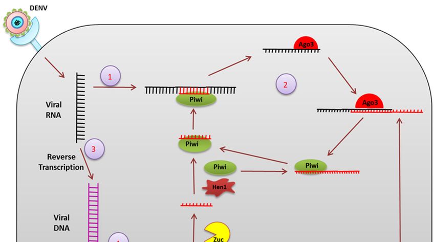
Pathogens 2019, 8, 77 5 of 16
Pathogens 2018, 7, x FOR PEER REVIEW 5 of 16
Figure3. 3.Organ-specific
Figure Organ-specificmosquito
mosquitoantiviral
antiviral strategies.(A):
strategies. A: In
Inthe
theAe.
Ae.aegypti
aegyptisystem,
system,DENV
DENVenters
enters
throughblood
through bloodmeal
mealand
andreplicates
replicatesinside
insidethe
themid-gut.
mid-gut.FromFromthethemid-gut,
mid-gut,DENVDENVisisreleased
releasedtotothe
the
hemolymph,salivary
hemolymph, salivaryglands,
glands,andand brain.
brain. B: Inside
(B): Inside the
themid-gut
mid-gutepithelial
epithelialcells,
cells,(1)
(1)RNA
RNAinterference
interference
limits
limits viral
viral replication
replication andand
(2)(2) immune
immune signaling
signaling pathways
pathways generate
generate AMPs AMPs
that that
blockblock the virus
the virus insideinside
the
the and
cells cellsalso
anddiffuse
also diffuse outhemolymph.
out to the to the hemolymph.
(C): In theC:hemocytes,
In the hemocytes, (3) complement-like
(3) complement-like factor
factor AaMCR
and its scavenger
AaMCR and itsreceptor AaSR-C
scavenger interactAaSR-C
receptor with theinteract
signalingwith
AMPthe production.
signaling(4) Phenoloxidase
AMP production.(PO) (4)
encapsulates
Phenoloxidase DENV(PO)in encapsulates
the hemolymph. DENV(D):inInthe
thehemolymph.
neurons, AaHig binds
D: In the to the envelop
neurons, AaHigprotein
bindsoftothe
the
DENV
envelop as well as the
protein cell DENV
of the membrane to block
as well as theendocytosis.
cell membrane (E): In
to the salivary
block glands E:
endocytosis. (5)In
extracellular
the salivary
cecropin-like peptides inhibit the virus and (6) intracellular cystatin and ankyrin
glands (5) extracellular cecropin-like peptides inhibit the virus and (6) intracellular cystatin and repeat-containing
protein
ankyrin (ARP) limit virus production.
repeat-containing protein (ARP) limit virus production.
Salivary
Complementglandsrelated Salivary glands
(SGs): proteins: Another contain
defense themechanism
major poolinofthe thehemolymph,
virus before which
transmission.
controls
Active replication
flaviviral of the
infection, DENV occurs
comprises leading to a high virus
the complement-related titer. In
proteins. The themosquito
SGs, the virus
genome again has to a
encodes
pass through salivary gland infection and escape barriers (SGIB and SGEB),
thioester-containing protein (TEP) that recognizes flaviviruses and leads to the expression of an which are the ultimate
restriction points
antimicrobial before [23].
peptide virusAe.
transmission. The SGIB may
aegypti macroglobulin form due related
complement to the lowerfactoramount
(AaMCR) of virus
belongstiterto
inthe
theinsect
hemolymph [25] or the presence of the basal lamina of the SG cells, which
TEP family, which recognizes viral particles. The Ae. aegypti homolog of the Scavenger hide cellular entry
receptors
receptor [26]. The SGEBmediates
C (AaSR-C) may result thebecause
bindingofof incomplete
AaMCR to apoptosis
the DENV of theparticles
SG acinar cells,
[24]. which
Thus, is
when
required
expressionfor the virus in
of AMPs to the
release via saliva
hemocytes [27]. The DENV
is up-regulated, AMP induces multiple
diffuses immune effectors
to the hemolymph in thean
and plays
salivary glands. In Ae. Aegypti,
anti-dengue role (Figure 3C). Toll and IMD pathways are activated and this gives rise to a putative
antibacterial
Salivary peptide,
glandscecropin-like
(SGs): Salivary peptide
glands (CEC-like
contain thepeptide)
major(Figure
pool of3E),the which showstransmission.
virus before anti-DENV
activity
Active[11]. Other immune
replication regulatory
of the DENV occursgenes thatto
leading area upregulated in a In
high virus titer. DENV infection
the SGs, include
the virus thehas
again Tollto
pathway receptor and adaptor proteins (Toll5A and MYD88). However, a peptide
pass through salivary gland infection and escape barriers (SGIB and SGEB), which are the ultimate from the Defensin
family is markedly
restriction downregulated
points before [11].
virus transmission. The SGIB may form due to the lower amount of virus
In the SGs, complement-like factors
titer in the hemolymph [25] or the presence of the AaMCR andbasal
AaSR-C
laminahaveof athe
roleSGascells,
antivirals
whichthat hide mediate
cellular
virus recognition and AMP production [24]. Putative cystatin (CS) gene expression
entry receptors [26]. The SGEB may result because of incomplete apoptosis of the SG acinar and ankyrin repeat
cells,
containing protein (ARP)
which is required for thegene expression
virus to release alsoviaincrease
saliva in theThe
[27]. DENV DENV infection along
induces with 12
multiple other
immune
effectors in the salivary glands. In Ae. Aegypti, Toll and IMD pathways are activated and this gives
rise to a putative antibacterial peptide, cecropin-like peptide (CEC-like peptide) (Figure 3E), which
shows anti-DENV activity [11]. Other immune regulatory genes that are upregulated in a DENV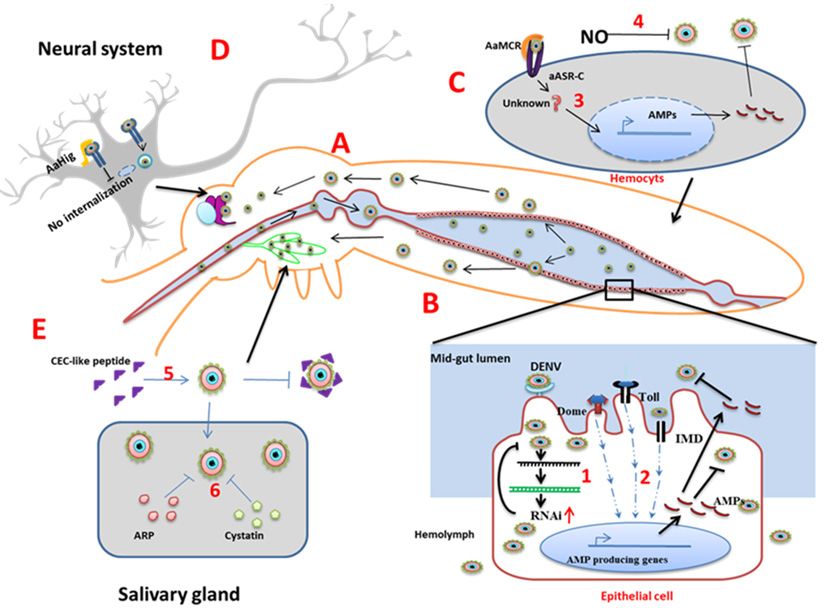
Pathogens 2019, 8, 77                                                                              6 of 16

immune modulator genes (Figure 3E) [28]. Silencing of these genes results in an increased viral load,
which indicates their role in antiviral immunity.
      Neuronal system: The neuron-specific resistance mechanism of mosquitoes against viral infection
is largely underexplored. In mosquito brains, an immune factor named Ae. aegypti homolog of
Hikaru Genki (AaHig) is expressed ubiquitously [29]. The Aahig protein contains the conserved
immunoglobulin domain and complement control protein domain (CCP) (Sushi domain) [29].
The AaHig protein specifically localizes at membranes of the neural cells and its CCP domain
interacts with the surface envelope proteins of the flaviviruses. The binding of AaHig to the virus
envelope directly blocks viral entry (endocytosis) (Figure 3D).
      Antimicrobial peptides/proteins: Antimicrobial peptides/proteins (AMPs) are the main
components of the humoral immune response and are synthesized in fat bodies, hemocytes, and
epithelial cells in response to DENV infection [24]. AMPs are secreted into hemolymph, distributed
to different organs, and display antimicrobial activity against a broad spectrum of microorganisms.
In response to microbial invasion, multiple signaling pathways get activated such as the IMD, Toll,
and JAK-STAT pathways, with the JAK-STAT pathway being the most important in the case of viral
infection [16]. These signaling events lead to production of AMPs that are the important immune
effectors. AMPs have also been shown to be important for maintaining mosquito gut immunity [3].
Seventeen AMPs have been discovered in A. aegypti and are classified into five independent groups:
cysteine-rich defensins, alpha helical peptides cercopins, cysteine-rich peptides gambicins, glycine-rich
peptides attacins, and diptericins. Defensins are the predominant AMPs of the Aedes mosquitoes, the
production of which is induced in response to infection by bacteria, filarial worms, and viruses [30].
Defensins are cysteine-rich in nature [31] and have been shown to function by attaching to cell
membranes, leading to pore formation, which causes efflux of various essential nutrients [32]. DENV-2
infection-induced defensin expression in C6/36 cells was enhanced with increased viral multiplicity of
infection (MOI). Defensins A, C, and D were found to increase due to DENV infection [24]. Cecropins
are small proteins which inhibit proline uptake, causing leaky membranes. Cecropins have several
isoforms and many of them were found to increase during DENV infection. They can lyse cellular
membranes in the case of bacteria and are also capable of inhibiting proline uptake and inducing
membrane leakage [33,34]. A 59 amino-acid peptide (AAEL000598) ceropin isolated from the salivary
glands of Ae. aegypti showed antiviral activity against both the DENV and Chikungunya viruse [7].
Cercopin P1 has been shown to inhibit release of viral particles [35]. Gambicin is another AMP found
in mosquitoes and other insects, the mature form of which is a 61 residue peptide having eight cysteins
connected with four disulfide bridges. Gambicin expression increases by more than two-fold during a
DENV2 infection of Ae. aegypti [24]. Attacin is a 20 kD protein that acts by inhibiting biosynthesis of
the outer membrane proteins in gram-negative bacteria [24]. Attacin mRNA levels increase due to
DENV infection in mosquitoes. The same study reported a slight increase of yet another AMP called
‘diptericin’ during DENV2 infection in Ae. Aegypti.
      Although the expressions of defensin, cecropin, and gambicin increase during viral infection in
mosquitoes and other insects, their mechanism of action is still unclear and needs further research. In
contrast, the other two AMPs, namely attacin and diptericin, have been shown to exert an antiviral
response in Drosophila by controlling viral RNA synthesis during Sindbis virus (SINV) infection, and
knocking down these genes increases the viral load in Drosophila [36]. Although the importance of AMPs
in mosquito immunity is now an established fact, technical difficulties in the isolation of hemocytes
(AMP production site) and the unavailability of suitable cell lines have made the characterization of the
molecular mechanisms of AMP function challenging. The importance of AMPs as immune effectors
and antiviral agents warrants detailed investigation of their molecular mechanisms of action.

5. Small RNA Mediated Immunity
     siRNA: Small interfering RNA (siRNA) is one of the important components of the RNAi mechanism
(Figure 4). Gaines et al. showed that infection of C6/36 cells with a double subgenomic Sindbis (dsSIN)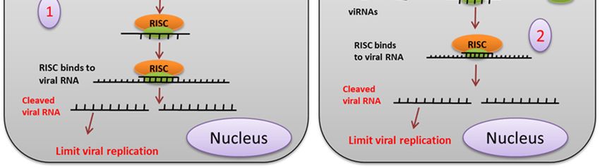
as immune effectors and antiviral agents warrants detailed investigation of their molecular
mechanisms of action.

Small RNA Mediated Immunity
Pathogens 2019, 8,Small
      siRNA:      77  interfering RNA (siRNA) is one of the important components of the RNAi      7 of 16
mechanism (Figure 4). Gaines et al. showed that infection of C6/36 cells with a double subgenomic
Sindbis (dsSIN) virus-carrying precursor of the membrane (prM) coding region of the DENV2,
virus-carrying precursor of the membrane (prM) coding region of the DENV2, provides resistance
provides resistance against DENV2 but not DENV3 and DENV4 [37]. Northern blot and
against DENV2 but not DENV3 and DENV4 [37]. Northern blot and immunofluorescence confirmed
immunofluorescence confirmed the presence of sense and antisense prM RNA and prM proteins,
the presence of sense and antisense prM RNA and prM proteins, respectively, in the infected cells.
respectively, in the infected cells. RNAi as an innate antiviral immunity mechanism against the
RNAi as an innate antiviral immunity mechanism against the DENV was confirmed when expression
DENV was confirmed when expression of untranslatable prM in C6/36 cells showed resistance
of untranslatable prM in C6/36 cells showed resistance against DENV2. Expressing dsRNA derived
against DENV2. Expressing dsRNA derived from the genome of the DENV in C6/36 cells restricts
from the genome of the DENV in C6/36 cells restricts DENV2 replication [38]. Intrathoracic injection of
DENV2 replication [38]. Intrathoracic injection of a dsSIN virus containing the prM of DENV2 in Ae.
a dsSIN virus containing the prM of DENV2 in Ae. aegypti restricted DENV2 RNA accumulation in the
aegypti restricted DENV2 RNA accumulation in the head tissue, salivary glands, and midgut [39].
head tissue, salivary glands, and midgut [39]. DENV2 serotype-specific vsiRNAs are generated upon
DENV2 serotype-specific vsiRNAs are generated upon infection in Ae. Aegypti, and knockdown of
infection in Ae. Aegypti, and knockdown of the siRNA pathway components dcr2, Ago2, and r2d2
the siRNA pathway components dcr2, Ago2, and r2d2 leads to an increase in viral replication and
leads to an increase in viral replication and shortening of the extrinsic incubation period [21].
shortening of the extrinsic incubation period [21].

     Figure 4. siRNA mediated pathway. (1) The exogenous siRNA pathway is activated when the
     Figure 4. siRNA mediated pathway. (1) The exogenous siRNA pathway is activated when the virus-
     virus-derived dsRNA is recognized and cleaved by the Dcr2 and r2d2 complex into about 19-21 bp
     derived dsRNA is recognized and cleaved by the Dcr2 and r2d2 complex into about 19-21 bp long
     long siRNA. siRNAs duplexes are loaded onto the RISC (RNA induced silencing complex), which
     siRNA. siRNAs duplexes are loaded onto the RISC (RNA induced silencing complex), which
     degrades the passenger strand (2). The RNAi signal or the siRNA from the virus-infected cell enters
     degrades
     the        the passenger
         neighboring           strand
                       uninfected       (2). The aRNAi
                                  cell through           signal or
                                                   gap junction   orthe  siRNA from
                                                                      cytoplasmic     the(3).
                                                                                  bridge  virus-infected
                                                                                              Viral dsRNAcell enters
                                                                                                           released
     the neighboring
     from  the infecteduninfected   cellisthrough
                         ruptured cell      taken upa bygapthejunction   or cytoplasmic
                                                                cells, which             bridge (3).
                                                                              provides adaptive       Viral dsRNA
                                                                                                  immunity    by the
     released from
     siRNA pathway.  the infected ruptured    cell is taken  up  by  the cells, which provides  adaptive immunity
     by the siRNA pathway.
     It was found that DCR2 and AGO2 transcript levels were significantly increased in the midgut
with DENV infected blood meal in the early days of infection, but the levels were equalized in later
stages [40]. Direct transfection of synthetic siRNA against the membrane glycoprotein precursor gene
of DENV1 in C6/36 cells reduced the viral load and increased cell survival rate [41,42].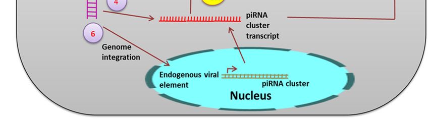
Pathogens 2019, 8, 77 8 of 16
Previously, it was thought that the siRNA mediated RNAi against the virus by a local and
cell-autonomous phenomenon, but recent studies have shown that the RNAi signal can pass from one
cell to another. In insects, the virus-derived siRNA can pass from an infected cell to a non-infected cell
via gap junctions or cytoplasmic bridges, thereby transferring the RNAi response to neighboring cells.
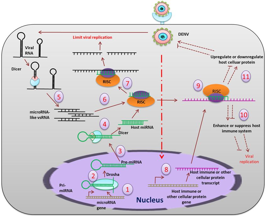
miRNA: In the mosquito, miRNAs can interact with RNA viruses either directly or indirectly
(Figure 5), where the direct interaction involves binding of the miRNA to the viral RNA genome,
and the indirect interaction introduces a virus-mediated change in the host transcriptome [43]. In a
time-dependent miRNA expression profile study in Ae. aegypti, it was shown that at 2–4 days
post-exposure (dpe), five miRNAs were modulated, but this increased to twenty-three at 9 dpe. In silico
analyses revealed 464 gene targets when miRNA bound to the 30 untranslated region (UTR). These
genes include those expressing proteins involved in transport, transcriptional regulation, mitochondrial
function, chromatin modification, and signal transduction processes that help in viral replication and
dissemination. A number of endogenous miRNAs are induced after blood feeding in Ae. aegypti
(e.g., aae-miR-375). The targets of aae-miR-375 include two immune-related genes, cactus and REL1.
Cactus is upregulated by aae-miR-375, whereas REL1 is downregulated. The aae-miR-375 also enhances
DENV2 replication during infection [44]. In Ae. albopictus, sixty-six differentially expressed miRNAs
were identified during DENV infection. Among these, miR-34-5p, which was upregulated, targets
the Toll pathway signaling protein (REL-1), and the peptidoglycan recognition protein LE and AMP
defensin D. miR87, which were down-regulated, target the Toll pathway [45].
During persistent viral infection, the miRNA expression profile changes [46]. Some of miRNAs
that are upregulated are miR-927, miR-87, miR-210, miR-2a3p, miR-190, and miR-970 whereas the
miRNAs that are downregulated include miR-252, miR-263a-3p, miR-92b, miR-10-5p, miR-9a-5p,
miR-9a-1, miR-124, miR-286a, and miR-286b [46]. Studies of the targets for these miRNAs revealed
target proteins to be those involved in ubiquitination, innate immune response, oxidative stress
response, cytoskeletal maintenance, fatty acid biosynthesis, intracellular protein transport, exocytosis,
autophagy, and pH regulation [46]. Modulation of protein expression by miRNAs helps in maintaining
an equilibrium between viral replication and host antiviral response during persistent infection.
MicroRNAs like viral small (vs) RNAs are produced from the viral genome. Twenty-three
microRNA-like vsRNAs were identified and were found to originate from the 50 UTR and 30 UTR of
the DENV2 in the argonaute 2 (AGO2) dependent pathway [47]. Among those, DENV-vsRNA-5
was found to inhibit DENV2 replication by targeting the non-structural protein-1 (NS1). Recently,
researchers have been trying to develop transgenic mosquitoes that are resistant to DENV infection
and transmission. A genetically engineered Ae. aegypti was developed, in which artificial antiviral
miRNA genes were introduced under the polyubiquitin promoter targeting the DENV3 non-structural
proteins NS2B, NS3, and NS5. This reduced the DENV3 transmission rate effectively [48].
piRNA: piRNAs are 24–30 nucleotides long, small RNA formed from the intergenic region termed
a piRNA cluster and are processed from the single-stranded piRNA precursor (pre-piRNA) via a
dicer-independent mechanism [49] (Figure 6).
In Drosophila melanogaster, pre-piRNAs are trimmed at both the 50 and 30 ends by mitochondria-
associated nuclease Zucchini (Zuc) [50] and by an unknown 30 -50 exonuclease [51]. The piRNAs
are loaded onto PIWI proteins before 30 trimming and are 20 -O-methylated at the 30 end by
a methyltransferase, DmHen1/Pimet, forming mature PIWI-piRNA complexes [52]. Aub- and
PIWI-bound primary piRNAs are antisense and uridine (1U) biased at the 50 end. AGO3 binds
to these piRNAs to form the piRNA-induced silencing complexes (piRISCs) which are transported
to the nucleus to cleave complementary target transcripts. This process generates a new secondary
sense piRNA that pairs with the antisense piRNA precisely by 10 nts at the 50 end. This secondary
piRNA specifically contains adenosine at position 10 (10A◦ ) and again undergoes the same process of
20 -O-methylation at the 30 end followed by AGO3 binding to generate Aub-bound piRNAs. This piRNA
amplification loop is referred to as the “ping-pong” cycle [53,54].Ae. aegypti (e.g., aae-miR-375). The targets of aae-miR-375 include two immune-related genes, cactus
and REL1. Cactus is upregulated by aae-miR-375, whereas REL1 is downregulated. The aae-miR-375
also enhances DENV2 replication during infection [44]. In Ae. albopictus, sixty-six differentially
expressed miRNAs were identified during DENV infection. Among these, miR-34-5p, which was
upregulated,
Pathogens 2019, 8,targets
77 the Toll pathway signaling protein (REL-1), and the peptidoglycan recognition
9 of 16
protein LE and AMP defensin D. miR87, which were down-regulated, target the Toll pathway [45].
Figure 5.5. The
ThemiRNA
miRNA pathway
pathway mediated
mediated inhibition
inhibition of replication.
of viral viral replication.
PrimaryPrimary
miRNAsmiRNAs (pri-
(pri-miRNAs)
miRNAs) are transcribed (1) from the host genome, which is cleaved (2) by the nuclear
are transcribed (1) from the host genome, which is cleaved (2) by the nuclear resident Drosha into resident
Drosha into approximately
approximately 70 bp-longmiRNAs
70 bp-long precursor precursor(pre-miRNAs),
miRNAs (pre-miRNAs),
which thenwhich then are transported
are transported (3) to the
(3) to the cytoplasm,
cytoplasm, where theywhere they get
get further further
cleaved (4) cleaved
by Dicer(4) bymature
into Dicer into
21-23mature 21-23
bp-long bp-long
miRNA. miRNA.
On the other
hand, the viral RNA, which forms hairpin secondary structures, is also cleaved (5) by the Dicer to
generate microRNA-like viral small RNA, which are further loaded (6) into RISC. RISC complex blocks
(7) the viral replication directly by interacting with the single stranded viral RNAs. Viral infection also
influences production of host immune transcripts and other cellular transcripts (8). Translation of these
transcripts is also modulated (9) by the host miRNAs or microRNA-like viral small RNAs. This results
in up-regulation or down-regulation of the immune molecules (10) and other host cellular proteins (11),
which may positively or negatively regulate viral replication in the host cell.
The piRNAs play a role in the innate antiviral response in insects. Virus infection induced piRNAs
were first observed in Drosophila ovarian somatic sheet (OSS) cells [42]. In a small RNA profile in C6/36
cells after DENV infection, a high read peak was found in 27 nts-long RNAs and these viral small
RNAs were positive sense [55]. Dicer2 activity was found to be significantly very low in C6/36 cells,
indicating the possibility of an alternate mechanism for production of these 24–30 nts-long vsRNAs. A
time-dependent small RNA profiling of DENV infected Ae. aegypti showed that 24–30 nts-long RNA
reads were high at two days post infection (dpi) but these read percentages decreased after 9 dpi. These
24–30 nts-long RNAs were considered as viral piRNA (vpiRNA) and were slightly 10A biased, but had
no preference for uridine at the 50 extreme as seen in other piRNAs [41]. In a comprehensive analysis of
both viral and host-derived small RNAs in DENV2 infected Ae. aegypti Aag2 cells, all three types of
small RNAs were identified, which include vsiRNAs, host miRNAs, and viral piRNAs. Knockdown of
PIWI5 and Ago3 resulted in the reduction of these vpiRNA levels, confirming that Aedes PIWI proteins
are associated with the production of DENV-derived vpiRNAs [56]. Both vpiRNAs and virus-induced
host endogenous piRNAs (vepiRNAs) produced in DENV2 infected Ae. albopictus were found to be
confined to specific hot spot regions in the DENV-2 genome, especially in the NS5 gene region [57].Pathogens 2018, 7, x FOR PEER REVIEW 10 of 16
derived vpiRNAs [56]. Both vpiRNAs and virus-induced host endogenous piRNAs (vepiRNAs)
produced in DENV2 infected Ae. albopictus were found to be confined to specific hot spot regions
Pathogens 2019, 8, 77
in
10 of 16
the DENV-2 genome, especially in the NS5 gene region [57].
Figure 6. The
The piRNA
piRNA mechanism.
mechanism. During
Duringviral
viralinfection,
infection,the
thess
ss viral
viral RNA
RNA isis processed
processed byby PIWI
proteins [56] (1), PIWI5 and PIWI6, to form the primary piRNA. This primary piRNA may undergo undergo a
ping-pong
ping-pong amplification
amplification loop
loop (2)
(2) to produce Ago3-dependent
Ago3-dependent secondary
secondary piRNA.
piRNA. Additionally,
Additionally, viral
RNA is also
also reverse
reverse transcribed
transcribed to form viral
viral DNA
DNA (vDNA)
(vDNA) (3). These
ThesevDNA
vDNA derived
derived transcripts
transcripts serve
serve
as
as additional
additional precursors
precursors for the viral piRNA production (4,5). Genomic
Genomic integration
integration of
of vDNA
vDNA might
occur. (6)
(6) Integration
Integration in the germline leads to formation of endogenous nonretroviral element.
Endogenous viral
Endogenous viral elements
elements derived
derived from
from non-retroviral
non-retroviral RNA
RNA viruses
viruses have
have been
been identified
identified in
different mosquito
mosquitospecies.
species. Ae. Ae.
In In aegypti and Ae.
aegypti andAlbopictus, a large anumber
Ae. Albopictus, large of non-retroviral
number integrated
of non-retroviral
RNA viruses (NIRVS) were identified including flaviviruses [58]. NIRVS were associated
integrated RNA viruses (NIRVS) were identified including flaviviruses [58]. NIRVS were associated with the
piRNA
with thecluster.
piRNAThree endogenous
cluster. flaviviralflaviviral
Three endogenous elements elements
(EFVE) identified from Ae. from
(EFVE) identified albopictus cell lines
Ae. albopictus
and one from Ae. aegypti cell lines produce 27 to 29 bases-long small RNAs, which
cell lines and one from Ae. aegypti cell lines produce 27 to 29 bases-long small RNAs, which were were 1U biased
1U
and antisense
biased to the viral
and antisense open
to the viralreading frame (ORF).
open reading DENV DENV
frame (ORF). infection did notdid
infection affect
notthe EFVE-derived
affect the EFVE-
transcript
derived level, which
transcript also
level, suggests
which constitutive
also suggests expression.
constitutive PIWI proteins
expression. PIWIwere found
proteins to befound
were associated
to be
with the production of this EFVE-derived small RNA. This indicates a complex
associated with the production of this EFVE-derived small RNA. This indicates a complex NIRVS-NIRVS-derived small
RNA-mediated
derived antiviral defense
small RNA-mediated in mosquitoes.
antiviral defense in mosquitoes.
6. Microbial
Microbial Community
Community
Role of
Role of gut
gut microbiomes: Thegut
microbiomes: The gutmicrobiome
microbiomeforms formsaacomplex
complexecological
ecological environment
environment and
and can
can
influence vector
influence vectorcompetence
competenceininvarious
variousways.
ways.In aIngut
a microbiome
gut microbiomestudy,study,
40 different types oftypes
40 different bacteria
of
were isolated from the gut of Ae. aegypti [59]. These bacteria upregulated antimicrobial
bacteria were isolated from the gut of Ae. aegypti [59]. These bacteria upregulated antimicrobial peptide gene
peptide gene Chromobacterium
transcription. sp. (Csp_P) weresp.
transcription. Chromobacterium isolated from
(Csp_P) the isolated
were midgut of the midgutAe.ofaegypti.
field-collected
from field-
Csp_P showed entomopathogenic activity, as its exposure to larval breeding water and
collected Ae. aegypti. Csp_P showed entomopathogenic activity, as its exposure to larval breeding ingestion by
adult mosquitoes reduced survival of both the larvae and adult. During Csp_P colonization, cecropin
E and G and defensin C displayed at least a two-fold increase in transcript abundance in the midgut.Pathogens 2019, 8, 77 11 of 16
Colonization of Csp_P in the midgut also inhibited DENV infection in Ae. aegypti [60]. Very little
is known about the gut mycobiome, and only a few members of fungi have been characterized.
Talaromyces (Tsp) was isolated from the gut of field-caught Ae. aegypti and found to render mosquitoes
more permissive to DENV infection.
The Talaromyces (Tsp) secretome was found to have a profound modulating effect on the midgut
transcriptome. It downregulates trypsin encoding genes involved in blood digestion and also reduces
trypsin enzymatic activity, which may play a role in the promotion of DENV infection in the midgut [61].
Wolbachia mediated defence: Dengue infection in Aedes mosquitoes has been reported to be
suppressed in the presence of Wolbachia species, gram negative endosymbiotic bacteria [62]. DENV-2
dissemination to secondary organs is inhibited by Wolbachia in Ae. albopictus [63]. In spite of being
a natural host of Wolbachia, Ae. albopictus becomes a competent vector for the DENV because of the
restricted tissue tropism of the wFLu Wolbachia strain in Ae. albopictus. In the case of Aedes aegypti, it
is transinfected with Wolbacia strains wMel and wMelPop, which makes the mosquitoes resistant to
DENV infection [64]. These two strains show organ-specific restriction with wMelPop giving strong
resistance towards DENV in the mid-gut tissue and salivary glands while wMel acts on salivary glands
only. Another study also reported the absence of DENV-2 particles post infection in female Ae. aegypti
with wMel [65].
Although the mechanism of virus resistance by the Wolbachia species is not fully understood,
possible mechanisms include competition for host resources, indirect connection to immune signaling
pathways, and reactive oxygen species (ROS) production. A recent study revealed that DENV-derived
subgenomic flaviviral RNA (sfRNA) inactivates cellular exoribonuclease (XRN1) in the absence of
Wolbachia, thereby giving stability to viral RNA. In the presence of Wolbachia, XRN1 remains inactivated
and RNA degradation increases among all four DENV serotypes [66].
7. Lipid Droplet (LD) and Immunity
Lipid droplets (LDs) are structures composed of a fatty acid monolayer and a few exclusive
structural proteins (Perilipin 1, 2, and 3) and are present in wide range of organisms. In mosquitoes,
they are associated with lipid-storing cells like fat bodies that are crucial in mediating antiviral response.
A balanced lipid environment is essential for the DENV to replicate inside mosquito cells. Thus,
imbalance of lipids is a possible antiviral strategy. In a study, it was found that DENV infection induced
transcription of LD biogenesis and lipid storage genes, which resulted in an increased LD level in the
Aag2 cell line [67]. The classical innate immune pathways (Toll and IMD) that are activated during
DENV infection were found to have a direct or indirect involvement in LD accumulation in the Aag2
cell line [68]. This indicates that LD might have an important role in mosquito immunity. On the
other hand, in Wolbachia infected mosquito cells, cellular lipid components are used due to the lack of
fatty acid synthesis genes [68]. Two Wolbachia strains, wMel and wMelPop, lower the level of lipidome
components that are essential for DENV replication in an Ae. albopictus cell line (Aa23-T) [69]. Hence,
both Wolbachia and the DENV compete for the cellular resources in co-infected cells, which significantly
reduces DENV replication [63]. As LD plays dual roles (as an immune component and also as a cellular
resource for the DENV), it may have a contradictory role in DENV infection in mosquitoes.
Immune gene activation: Wolbachia induces a reduction–oxidation (Redox) reaction in the mosquito
to produce reactive oxygen species (ROS). The oxidative stress activates the Toll pathway through
which the antimicrobial peptides cecropin and defensin are produced [70]. Wolbachia also increases the
expression of vago1 in Ae. aegypti, which acts as a ligand in the JAK-STAT pathway [71].
Role of Wolbachia in RNA interference: Wolbachia infection induces differential expression of host
miRNAs, for example, miRNA aae-miR-2940. The aae-miR-2940 activates the metalloprotease gene,
which is crucial for Wolbachia colonization [72]. Wolbachia infection downregulates AaDnmt2. AaDnmt2
is found to be overexpressed during DENV infection. So, downregulation of the AaDnmt2 during
Wolbachia infection in Ae. aegypti might be a strategy by which Wolbachia counteracts the DENV [73].Pathogens 2019, 8, 77 12 of 16
8. Adaptive Immunity
Unlike vertebrates, the adaptive immunity of insects is poorly understood. In Anopheles gambiae,
The Down syndrome cell adhesion molecule (Dscam) is found to have characteristics similar to those
of antibodies found in vertebrates [74]. Insects’ adaptive immunity against RNA viruses is provided
by the RNAi mechanism. During viral infection, the viral dsRNA replicative intermediate is processed
by dicer2 to generate siRNA which may pass to the neighboring cell providing adaptive immunity
against the virus. The viral dsRNA replicative intermediate released from apoptotic cells is taken up
by the neighboring cell, which also provides adaptive immunity [75]. Recently, many endogenous
non-retroviral elements (ENE) were identified in the mosquito genome. These viral sequences are
acquired from previous viral infection. Transposable elements (TE) are one of the master players of
genomic variation and new sequence acquisition. Among different TEs, long terminal repeat (LTR)
are found predominantly upstream and downstream of the ENE. These ENEs integrate into higher
density LTR loci, such as piRNA clusters, and the ENE containing piRNA clusters produce more
piRNAs [76]. The piRNA generated from the ENE piRNA clusters may interact with the specific viral
RNA sequences to inhibit viral replication and accumulation in the host cell. Since the piRNA clusters
are stably integrated into the mosquito germ line, this type of adaptive immunity is also passed to the
next generation.
9. What is Next?
The DENV resides as a commensal in its vector, and the physiological effects due to viral
propagation in mosquitoes are different from those in the human system.
Many signaling pathways that are active during the developmental stages of an insects are
also involved in protecting insects against microbial attack. Activation of these signaling pathways
results in expression of antimicrobial peptides (AMPs) that are up-regulated in virus-infected cells [24].
Generally, these AMPs target the bacterial outer membrane and cell wall biosynthesis. Viruses have
significantly different structural components than bacteria, and thus the molecular mechanism of action
of AMPs against the DENV needs further exploration. Similarly, small RNAs have been extensively
studied, but it is still an open question as to exactly how these pathways are related to the evolutionarily
conserved signaling pathways that are known to play a significant role against the DENV.
Recently, a group of researchers developed DENV-resistant mosquitoes by artificial activation of
the JAK/STAT pathway [77]. These DENV resistant Ae. aegypti mosquitoes showed decreased capacity
for egg production due to immune activation. Similar strategies of genetic engineering involving other
immunity-related genes can be potentially useful methods to control the DENV [78].
Taken together, we are yet to understand the intricate details and complexity of the molecular basis
of immune function of mosquitoes against the viruses that they carry. A better knowledge in this area
might help open new possibilities with respect to the therapeutic intervention against mosquito-borne
viral infections and vector control.
Author Contributions: D.M., S.D., F.B., and S.M. wrote the manuscript. U.R. wrote and edited the manuscript.
Conflicts of Interest: We declare no conflict of interest.
References
1. Frolova, E.I.; Fayzulin, R.Z.; Cook, S.H.; Griffin, D.E.; Rice, C.M.; Frolov, I. Roles of nonstructural protein
nsP2 and alpha/beta interferons in determining the outcome of Sindbis Virus Infection. J. Virol. 2002, 76,
11254–11264. [CrossRef] [PubMed]
2. Sim, S.; Dimopoulos, G. Dengue virus inhibits immune responses in Aedes aegypti Cells. PLoS ONE 2010,
5, e10678. [CrossRef]
3. Xi, Z.; Ramirez, J.L.; Dimopoulos, G. The Aedes aegypti Toll pathway controls dengue virus infection.
PLoS Pathog. 2008, 4, e1000098. [CrossRef]Pathogens 2019, 8, 77 13 of 16
4. Ramirez, J.L.; Dimopoulos, G. The Toll immune signaling pathway control conserved anti-dengue defenses
across diverse Ae. aegypti strains and against multiple dengue virus serotypes. Dev. Comp. Immunol. 2010,
34, 625–629. [CrossRef] [PubMed]
5. Weber, A.N.; Tauszig-Delamasure, S.; Hoffmann, J.A.; Lelièvre, E.; Gascan, H.; Ray, K.P.; Morse, M.A.; Imler, J.;
Gay, N.J. Binding of the Drosophila cytokine Spätzle to Toll is direct and establishes signaling. Nat. Immunol.
2003, 4, 794–800. [CrossRef]
6. Ramirez, J.L.; Muturi, E.J.; Barletta, A.B.; Rooney, A.P. The Aedes aegypti IMD pathway is a critical component
of the mosquito antifungal immune response. Dev. Comp. Immunol. 2019, 95, 1–9. [CrossRef]
7. Shin, S.W.; Kokoza, V.; Ahmed, A.; Raikhel, A.S. Characterization of three alternatively spliced isoforms of
the Rel/NF- B transcription factor Relish from the mosquito Aedes aegypti. Proc. Natl. Acad. Sci. USA 2002, 99,
9978–9983. [CrossRef]
8. Georgel, P.; Naitza, S.; Kappler, C.; Ferrandon, D.; Zachary, D.; Swimmer, C.; Kopczynski, C.; Duyk, G.;
Reichhart, J.M.; Hoffmann, J.A. Drosophila immune deficiency (IMD) is a death domain protein that activates
antibacterial defense and can promote apoptosis. Dev. Cell 2001, 1, 503–514. [CrossRef]
9. Paradkar, P.N.; Trinidad, L.; Voysey, R.; Duchemin, J.B.; Walker, P.J. Secreted Vago restricts West Nile virus
infection in Culex mosquito cells by activating the Jak-STAT pathway. Proc. Natl. Acad. Sci. USA 2012, 109,
18915–18920. [CrossRef] [PubMed]
10. Deddouche, S.; Matt, N.; Budd, A.; Mueller, S.; Kemp, C.; Galiana-Arnoux, D.; Dostert, C.; Antoniewski, C.;
Hoffmann, J.A.; Imler, J.L. The DExD/H-boxhelicase Dicer-2 mediates the induction of antiviral activity in
drosophila. Nat. Immunol 2008, 9, 1425–1432. [CrossRef]
11. Luplertlop, N.; Surasombatpattana, P.; Patramool, S.; Dumas, E.; Wasinpiyamongkol, L.; Saune, L.; Hamel, R.;
Bernard, E.; Sereno, D.; Thomas, F.; et al. Induction of a peptide with activity against a broad spectrum of
pathogens in the Aedes aegypti salivary gland, following infection with dengue virus. PLoS Pathog. 2011,
7, e1001252. [CrossRef] [PubMed]
12. Sim, S.; Jupatanakul, N.; Ramirez, J.L.; Kang, S.; Romero-Vivas, C.M.; Mohammed, H.; Dimopoulos, G.
Transcriptomic profiling of diverse Aedes aegypti strains reveals increased basal-level immune activation in
dengue virus-refractory populations and identifies novel virus-vector molecular interactions. PLoS Negl.
Trop. Dis. 2013, 7, e2295. [CrossRef] [PubMed]
13. Silverman, N.; Zhou, R.; Erlich, R.L.; Hunter, M.; Bernstein, E.; Schneider, D.; Maniatis, T. Immune activation
of NF-κB and JNK requires Drosophila TAK1. J. Biol. Chem. 2003, 278, 48928–48934. [CrossRef] [PubMed]
14. Choy, M.M.; Sessions, O.M.; Gubler, D.J.; Ooi, E.E. Production of infectious dengue virus in Aedes aegypti
is dependent on the ubiquitin proteasome pathway. PLoS Negl. Trop. Dis. 2015, 9, e0004227. [CrossRef]
[PubMed]
15. Troupin, A.; Londono-Renteria, B.; Conway, M.J.; Cloherty, E.; Jameson, S.; Higgs, S.; Vanlandingham, D.L.;
Fikrig, E.; Colpitts, T.M. A novel mosquito ubiquitin targets viral envelope protein for degradation and
reduces virion production during dengue virus infection. Biochim. Biophys. Acta 2016, 1860, 1898–1909.
[CrossRef] [PubMed]
16. Souza-Neto, J.A.; Sim, S.; Dimopoulos, G. An evolutionary conserved function of the JAK-STAT pathway in
anti-dengue defense. Proc. Natl. Acad. Sci. USA 2009, 106, 17841–17846. [CrossRef] [PubMed]
17. Kato, N.; Mueller, C.R.; Fuchs, J.F.; McElroy, K.; Wessely, V.; Higgs, S.; Christensen, B.M. Evaluation of the
function of a type I peritrophic matrix as a physical barrier for midgut epithelium invasion by mosquito-borne
pathogens in Aedes aegypti. Vector-Borne Zoonotic Dis. 2008, 8, 701–712. [CrossRef]
18. Houk, E.; Obie, F.; Hardy, J. Peritrophic membrane formation and the midgut barrier to arboviral infection in
the mosquito, Culex tarsalis Coquillett (Insecta, Diptera). Acta Trop. 1979, 36, 39–45.
19. Beaty, B.J.; Bishop, D.H. Bunyavirus-vector interactions. Virus Res. 1988, 10, 289–301. [CrossRef]
20. Black, I.V.W.C.; Bennett, K.E.; Gorrochótegui-Escalante, N.; Barillas-Mury, C.V.; Fernández-Salas, I.;
de Lourdes Muñoz, M.; Farfán-Alé, J.A.; Olson, K.E.; Beaty, B.J. Flavivirus Susceptibility in Aedes aegypti.
Arch. Med. Res. 2002, 33, 379–388. [CrossRef]
21. Sánchez-Vargas, I.; Scott, J.C.; Poole-Smith, B.K.; Franz, A.W.E.; Barbosa-Solomieu, V.; Wilusz, J.; Olson, K.E.;
Blair, C.D. Dengue virus type 2 infections of Aedes aegypti are modulated by the mosquito’s RNA interference
pathway. PLoS Pathog. 2009, 5, e1000299. [CrossRef]Pathogens 2019, 8, 77 14 of 16
22. Ramos-Castañeda, J.; González, C.; Jiménez, M.A.; Duran, J.; Hernández-Martínez, S.; Rodríguez, M.H.;
Lanz-Mendoza, H. Effect of nitric oxide on dengue virus replication in Aedes aegypti and Anopheles albimanus.
Intervirology 2008, 51, 335–341. [CrossRef] [PubMed]
23. Cheng, G.; Liu, L.; Wang, P.; Zhang, Y.; Zhao, Y.O.; Colpitts, T.M.; Feitosa, F.; Anderson, J.F.; Fikrig, E. An
in vivo transfection approach elucidates a role for Aedes aegypti thioester-containing proteins in flaviviral
infection. PLoS ONE 2011, 6, e22786. [CrossRef]
24. Xiao, X.; Liu, Y.; Zhang, X.; Wang, J.; Li, Z.; Pang, X.; Wang, P.; Cheng, G. Complement-related proteins
control the flavivirus infection of Aedes aegypti by inducing antimicrobial peptides. PLoS Pathog. 2014,
10, e1004027. [CrossRef]
25. Kramer, L.D.; Hardy, J.L.; Presser, S.B.; Houk, E.J. Dissemination barriers for western equine encephalomyelitis
virus in Culex tarsalis infected after ingestion of low viral doses. Am. J. Trop. Med. Hyg. 1981, 30, 190–197.
[CrossRef] [PubMed]
26. Romoser, W.S.; Turell, M.J.; Lerdthusnee, K.; Neira, M.; Dohm, D.; Luldwig, G.; Wasieloski, L. Pathogenesis
of Rift Valley fever virus in mosquitoes-tracheal conduits and the basal lamina as an extra-cellular barrier.
Arch. Virol. Suppl. 2005, 19, 89–100.
27. Grimstad, P.R.; Paulson, S.L.; Craig, G.B., Jr. Vector competence of Aedes hendersoni (Diptera: Culicidae) for La
Crosse virus and evidence of a salivary-gland escape barrier. J. Med. Entomol. 1985, 22, 447–453. [CrossRef]
28. Sim, S.; Ramirez, J.L.; Dimopoulos, G. Dengue virus infection of the Aedes aegypti salivary gland and
chemosensory apparatus induces genes that modulate infection and blood-feeding behavior. PLoS Pathog.
2012, 8. [CrossRef] [PubMed]
29. Hoshino, M.; Matsuzaki, F.; Nabeshima, Y.-I.; Hama, C. hikaru genki, a CNS-specific gene identified by
abnormal locomotion in Drosophila, encodes a novel type of protein. Neuron 1993, 10, 395–407. [CrossRef]
30. Christophides, G.K.; Zdobnov, E.; Barillas-Mury, C.; Birney, E.; Blandin, S.; Blass, C.; Brey, P.T.; Collins, F.H.;
Danielli, A.; Dimopoulos, G.; et al. Immunity-related genes and gene families in Anopheles gambiae. Science
2002, 298, 159–165. [CrossRef]
31. Matsuyama, K.; Natori, S. Purification of three antibacterial proteins from the culture medium of NIH-Sape-4,
an embryonic cell line of Sarcophaga peregrina. J. Biol. Chem. 1988, 263, 17112–17116. [PubMed]
32. Wimley, W.C.; Selsted, M.E.; White, S.H. Interactions between human defensins and lipid bilayers: Evidence
for formation of multimeric pores. Protein Sci. 1994, 3, 1362–1373. [CrossRef] [PubMed]
33. Moore, A.J.; Beazley, W.D.; Bibby, M.C.; Devine, D.A. Antimicrobial activity of cecropins. J. Antimicrobiol. Chemother.
1996, 37, 1077–1089. [CrossRef] [PubMed]
34. Liu, X.; Guo, C.; Huang, Y.; Zhang, X.; Chen, Y. Inhibition of porcine reproductive and respiratory syndrome
virus by Cecropin D in vitro. Infect. Genet. Evol. 2015, 34, 7–16. [CrossRef] [PubMed]
35. Guo, C.; Huang, Y.; Cong, P.; Liu, X.; Chen, Y.; He, Z. Cecropin P1 inhibits porcine reproductive and
respiratory syndrome virus by blocking attachment. BMC Microbiol. 2014, 14, 273. [CrossRef] [PubMed]
36. Huang, Z.; Kingsolver, M.B.; Avadhanula, V.; Hardy, R.W. An antiviral role for antimicrobial peptides during
the arthropod response to Alphavirus replication. J. Virol. 2013, 87, 4272–4280. [CrossRef] [PubMed]
37. Gaines, P.J.; Olson, K.E.; Higgs, S.; Powers, A.M.; Beaty, B.J.; Blair, C.D. Pathogen-derived resistance to
dengue type 2 virus in mosquito cells by expression of the premembrane coding region of the viral genome.
J. Virol. 1996, 70, 2132–2137. [PubMed]
38. Caplen, N.J.; Zheng, Z.; Falgout, B.; Morgan, R.A. Inhibition of viral gene expression and replication in
mosquito cells by dsRNA-triggered RNA interference. Mol. Ther. 2002, 6, 243–251. [CrossRef]
39. Adelman, Z.N.; Blair, C.D.; Carlson, J.O.; Beaty, B.J.; Olson, K.E. Sindbis virus-induced silencing of dengue
viruses in mosquitoes. Insect Mol. Biol. 2001, 10, 265–273. [CrossRef] [PubMed]
40. Hess, A.M.; Prasad, A.N.; Ptitsyn, A.; Ebel, G.D.; Olson, K.E.; Barbacioru, C.; Monighetti, C.; Campbell, C.L.
Small RNA profiling of Dengue virus-mosquito interactions implicates the PIWI RNA pathway in anti-viral
defense. BMC Microbiol. 2011, 11, 45. [CrossRef] [PubMed]
41. Wu, X.; Hong, H.; Yue, J.; Wu, Y.; Li, X.; Jiang, L.; Li, L.; Li, Q.; Gao, G.; Yang, X. Inhibitory effect of small
interfering RNA on dengue virus replication in mosquito cells. Virol. J. 2010, 7, 270. [CrossRef] [PubMed]
42. Wu, Q.; Luo, Y.; Lu, R.; Lau, N.; Lai, E.C.; Li, W.-X.; Ding, S.W. Virus discovery by deep sequencing and
assembly of virus-derived small silencing RNAs. Proc. Natl. Acad. Sci. USA 2010, 107, 1606–1611. [CrossRef]
[PubMed]You can also read